JOURNAL OF VIROLOGY, June 1993, p. 3569-3575 0022-538X/93/063569-07$02.00/0
Copyright © 1993, American Societyfor Microbiology
Release
of
Human
Immunodeficiency
Virus by THP-1 Cells
and Human Macrophages
Is
Regulated
by Cellular
Adherence and Activation
ROBINJ. SHATTOCK, JON S.FRIEDLAND, ANDGEORGE E. GRIFFIN* Divisionof Communicable Diseases, St. George's Hospital MedicalSchool,
Tooting,
LondonSWJ
7ORE,
UnitedKingdom
Received 7 July1992/Accepted 23 February 1993
Macrophage adherence,animportantregulatorysignal, has the potentialtoaffect human immunodeficiency virus (HIV) production either directlyorby primingmonocytes torespondtoother activating signals.We have investigated the role of adherenceas anactivator of HIV-1 transcription and release. The effects ofadherence onHIV-1transcriptionwereexaminedby using THP-1 cells,ahuman monocytic cellline,transfectedwithHIV
long terminalrepeat (LTR)-chloramphenicol
acetyltransferase
(CAT)constructs.Theeffects ofadherenceonrelease of HIV-1 were investigated in both HIV-1-infected THP-1 cells and human peripheral blood monocyte-derived macrophages (MDM). Adherence oflipopolysaccharide (LPS)-stimulated THP-1 cells to
either tissuecultureplasticorendothelial cellswascrucial for enhancedHIV-1transcriptionas measured by
LTR-CAT expression. Such increased LTR-CAT expression did not occur with an HIV LTR construct
containing mutatedNF-KBbinding sites. Incontrast,release of whole HIV,measuredbyreversetranscriptase
(RT)activityintissueculture medium,wasreduceduponadherence of stimulatedHIV-1-infectedTHP-1 cells
withoutsuppression ofHIV LTR-CAT transcription orp24 release. This finding suggested that activation of adherent monocytic cells interfered withHIV assembly and release. Although the reduction of RTactivity
following activationofHIV-1-infected MDMwasindependent ofadhesion,adherence aloneofnonstimulated
HIV-infected MDM to endothelial cells was sufficient to induce a reduction in RT release. This study
demonstrates that LPS stimulation of monocytic cells enhances HIV LTR transcription under adherent conditions. In contrast, activation of adherent monocytic cells infectedwith HIV reduced viral release.
Monocytesandmacrophagesareanimportantreservoir of
humanimmunodeficiency virus (HIV) andmayrepresentthe largest reservoir ofthis virus intissue (11). HIVreplication inmonocytes haspreviouslybeen shownto be induced by binding of the transcriptional activator NF-KB to the
en-hancer region of the viral long terminal repeat (LTR)
se-quence and isdependentuponthe stateofcellular differen-tiation (13). Monocyte differentiation may be modified by many exogenous or endogenous stimuli. However, the
ef-fects ofcellularadhesion on NF-KB-mediated HIV
replica-tionarenotknown.
NF-KBin thecytoplasmof monocytes andmacrophagesis complexed to its inhibitor, IKB. Dissociation of this
com-plex,which arises fromphosphorylationofIKB(18), allows translocation of NF-KBtothenucleus,whereitmaybindto the promoter region of many cellular genes. Induction of NF-KB binding activityoccursinanumber ofpromonocytic
cell linesbyavarietyofstimuli,includingbacterial
lipopoly-saccharide(LPS)andphorbolesters(20).NF-KBconsistsof afamily of heterodimericproteinsubunits, different subunit
combinations have distinct binding kinetics for the HIV-1 provirus, and their release into the nucleus is inducer
spe-cific(27). However,inmaturemonocyte celllines, including the THP-1 cells used in this study, blood monocytes, and adherent macrophages, NF-KB binding activityis constitu-tivelypresent(13).
Thephenomenonofcellular adherence is known to induce mRNA productionfor several proto-oncogenes and
mono-kines, including tumor necrosis factor alpha and
interleu-*Correspondingauthor.
kin-8,which containNF-KB bindingsites in theirpromoter regions (14, 16, 23). Inaddition, the phenomenon of
adher-ence is important for monocyte migration and function (6, 19). Monocyte adhesion, therefore, has the potential to directly influence HIVproduction ininfected cells through NF-KB-dependent mechanisms or modulate the effects of other stimuli known to affect HIV production. Entry of peripheralbloodmonocytesinto tissuefollows adherenceto and thentranslocationacrossvascularendothelium (6).This
process of adhesion and translocation is mediated through the interaction ofa variety ofspecific cellular proteins (5)
andextracellular matrixproteins (8, 19).
The effects ofmonocyteactivation on HIVtranscription and releasereportedinpreviousstudiesareoftenconflicting. Suchdisparate results mayhave arisen from differences in experimental conditions, for example, useofdifferent cell
lines, divergence ofsubclones, differences inculture condi-tions(adherentandnonadherent), and differences in activat-ingstimuli. Previous studies have shown that LPS stimula-tion enhanced HIV transcription in 24-h-old human monocytesand thepromonocyticcell line U937 while reduc-ingHIVtranscriptionin 12-day-oldmonocyte-derived
mac-rophages (MDM) (2). In addition, activation of MDM with LPSorgammainterferon(IFN--y)priortoHIV infection has beenshown toprotect MDMfromproductive HIV replica-tion (17). Indeed, monocyte activation can downregulate
viralproduction,astreatmentof HIV-infectedmacrophages withIFN--yreduces viral release(26).Whether cell culturein
adherent versus nonadherent conditions may have
influ-encedthesepreviousstudies is unknown.We have therefore
investigated the effects of macrophage adherence to tissue
culture plastic or endothelial cells on the control of HIV 3569
Vol. 67, No. 6
on November 9, 2019 by guest
http://jvi.asm.org/
transcription and release from monocytes stimulated by a
variety of soluble mediators.
MATERIALS AND METHODS
Cell culture. The monocyte cell line THP-1 (25) was
maintained in RPMI 1640 supplemented with 5% fetal calf
serum(FCS), 2 mM glutamine, 100 U ofpenicillin per ml, and0.1 mgof streptomycinper ml. Cells were cultured at 37°C in 5% CO2 and passaged every 4 to 5 days. The endothelial cell line SGHEC 7 (gift of G. Whitley, St. Georges Hospital Medical School, London, United King-dom) representssimian virus40-transfected human umbilical veinendothelial cells. Such endothelial cellswereseeded in six-welltissue cultureplates(Marathon,UnitedKingdom)at 2 x 10⁵to3 x 105cellsperwell. Cellswere cultured for72 hat 37°C in5% CO2 in medium 199 (Sigma, Poole, United Kingdom) supplemented with Earle's modified salts-RPMI 1640(1:1). Thisculture mediumwassupplemented with 5%
FCS, 5% newborn calf serum, 2.5 ,ug of endothelial cell growth supplement (Sigma)perml,and 0.9mgofheparinper
ml. Plateswere seeded with cells betweenpassages 12and 20 andwereused inadhesionexperimentsonreaching >90%
confluency.
Humanperipheralbloodmonocyte isolation.Human
mono-nuclear cells were prepared from buffy coat preparations (TheNational BloodTransfusionService, Tooting, London, UnitedKingdom) byFicolldensity gradient separation (His-topaque; Sigma). Contaminating plateletswere depleted by
fourwashes inphosphate-bufferedsaline(PBS) containing 3 mMEDTA. Cellswere resuspendedinRPMI1640 with5%
human serum and incubated for 60 min in gelatin-coated
tissue culture flasks (15). Nonadherent cellswere removed byfourwasheswithwarm(37°C)RPMI 1640plus 5% FCS. Adherent monocytes were eluted by 10-min incubation at 4°C in chilled elution buffer (a 1:1 ratio of PBS-10 mM EDTAand RPMI1640 with10% humanserum). Monocytes
werewashed andresuspended in RPMI 1640supplemented with10% humanserum,5%FCS, 2 mMglutamine, 100 U of penicillin per ml, and 0.1 mg of streptomycin per ml. The final concentration of monocytes was 106 cells per ml of
culture medium. Cells matured into MDM after 5 days of incubation in Teflon vials (Pierce and Warriner, Chester, United Kingdom) at37°C in5% CO2.
Plasmid constructs. Two plasmid constructs (generous giftsofG.Nabel, UniversityofMichigan, Ann Arbor)were
used in experiments: HIV-1 LTR-CAT, containing the HIV-1 LTR upstream of the reporter chloramphenicol acetyltransferase (CAT) gene, and a corresponding
con-struct with mutated KB sites which could no longer bind
NF-icB (AHIV-1 LTR-CAT).
Transfections. Thecontrol ofHIV LTRtranscriptionwas
investigated by using cells transfected with HIV LTR-CAT constructs. Cellswere transfected as previously described
(13). In brief, 107washed cellswere incubated in 1 ml of
transfectioncocktail containingan HIVLTR-CAT plasmid
(10 ,g) and DEAE-dextran for 30min at37°C in 5% CO2. CellswerewashedinPBS andincubated in growth medium containing1mMchloroquine for 30 minat37°C in 5% CO2. Cellswerethenwashed andresuspended in RPMI with 5% FCS and cultured either under nonadherent conditions in Teflonvials orunder adherentconditions in six-well tissue
cultureplates (107cellsper5ml perwellorvial).
At 24hposttransfection, cellswereexposedtothe follow-ingstimuli: 1jig ofEscherichiacoli 0127:B8LPSperml,500
U ofIFN--y(BoehringerIngelheim, Bracknell, United
King-dom) per ml, or 20 nM
phorbol 12-myristate
13-acetate(PMA; Sigma). In addition, transfected THP-1 cells were added to cultures of confluent SGHEC 7 endothelial cells
that were pretreated as follows: nonstimulated SGHEC 7
cells washed three times with PBS, SGHEC 7 cells stimu-lated with 1,ugof LPS perml for 4 h and then washedfour
times with PBS, and LPS-stimulated SGHEC 7 cells fixed
with 0.2% glutaraldehyde and thenwashed four times with
PBS. THP-1 cellswere alsoexposed to endothelial cells in the continual presence ofLPS. As a control for the endo-thelial cell experiments, THP-1cells were cultured inTeflon vials with conditioned medium produced bycollecting tissue culture supernatantfrom endothelial cells 24 h after thecells had been stimulated with LPSfor 4 h andwashed fourtimes in PBS.
CAT assay. CAT activitywas assayed by standard
meth-ods 48 h after transfection of the cells (12). Samples were assayed in a
150-pl
volume containing[14C]chloramphenicol
(0.5 ,uCi; 18.5 kBq; Amersham International Ltd., Ayles-bury, United Kingdom),4 mMacetyl coenzymeA(Sigma), 0.25 MTris-HCl (pH7.4), and 300
pg
of celllysate. Protein concentrations of cell extracts were standardized by the Bradford method (4). Following separation by thin-layer chromatography, percentage conversion ofchloramphenicolto itsacetylatedformswas determined by liquid scintillation
counting.
HIV infection. THP-1 cells in log-phase growth were
infected with HIV-1RF. Then4 x 106cells were incubatedin 1 ml of cell-free viral supernatant (6 x 10⁴ 50% tissue
infective dosesderived from Jurkat TAT cells [22]) for 2 h at
37°C in 5%CO2.Peakinfection was establishedafter 10 days
of subculture. MDM were infectedwith themonocytotropic
virus
HIV-1ea-L
(9) 5 days after isolation of monocytes.Then 8 x 10 cells were incubated for 2 h in 1 ml ofcell-free
viral supernatant (105 50% tissue culture infective doses
derived from MDM cultures) in Teflon vials at 37°C in5% CO2. Peak infection was established after 7 days of
subcul-ture in Teflonvials.
HIVdetection. The control of HIV release was
investi-gated by using HIV-infected THP-1 cells(10 days
postinfec-tion) and MDM (7 days postinfection). Immediately before experimentation,THP-1 and MDMcells werewashed three
times in PBS to remove all cell-free virus. Cells were
resuspended in fresh medium (2 x 10⁶ cells per 2 ml) and
cultured either undernonadherent conditions in Teflon vials
or underadherent conditions in six-well tissue cultureplates
in the presence of soluble stimuli. In addition, such cells
were cocultured with confluent endothelial cells as previ-ously described. Cell-free supernatants were assayed for reverse transcriptase (RT) activity or p24 levels at 24, 48,
and 96 h poststimulation.
RT activity wasdetermined on 10-,lI samples ofcell-free
tissue culture supernatant, using [32P]dTTP incorporation (21) determined with a direct 1B plate reader (Canberra
Packard, Pangbourne, United Kingdom). To control for
degradation of RT activity resulting from the action of
soluble factors released by stimulated THP-1 cells, HIV of known RT activity was diluted 1/10 with supernatantsfrom PMA- or LPS-stimulated adherent cells taken at 4, 8, 24,48,
and 72 h poststimulation. Virus diluted in such conditioned supernatant was incubated for 24 h at 37°C in 5% CO2,and subsequently RT activity was analyzed.
Viral p24levels were determined on cell-free tissue culture
supernatant by using ap24 antigen capture enzyme-linked immunosorbent assay (ELISA). Viral supernatants treated with 1% Empigen (Calibrochem, Nottingham, United
on November 9, 2019 by guest
http://jvi.asm.org/
REGULATION OF HIV RELEASE BY MACROPHAGES 3571
NIL
LPS
PMA
IFN
A
Non-adherentconditions (Teflon vials)
F,
0.5 1 1.5 2 2.5
Fold increase
Nonadherent
(plastic) Unstimulated endothelium Pre-stimulated endothelium Endothelium
+LPS Conditioned media
3 3.5
B
AdherentConditions (Tissueculture plastic)
NIL
LPS
PMA
IFN
0.5 1 1.5 2 FoldIncrease
2.5 3
FIG. 2. CAT expression inTHP-1 cells transfected with HIV-1 LTR-CAT upon adhesion to endothelial monolayers. At 24 h after transfection, cells were cultured in nonadherent conditions in tissue cultureplastic or in tissue culture plates with confluent SGHEC 7 endothelial cells. The endothelial cells were either unstimulated, prestimulated with LPS (1 ,ug/ml)for 4 h andwashed four times in PBS before use, or stimulated in the continual presence of LPS (1 ,ug/ml). Inaddition, transfected THP-1 cells were cultured in Teflon vials withconditioned medium collected from endothelial cells 24 h after they had been stimulated with LPS for 4 h and washed four times in PBS. THP-1 cell extracts were harvested for CAT assay 48 h after transfection. Bargraphs show fold increase in conversion of
[14C]chloramphenicolcompared with untreated controls transfected with HIV-1 CAT, assigned a value of 1. Graphs illustrate the arithmetic mean of a minimum of three independent transfections.
0 0.5 1 1.5 2 2.5
Fold Increase
3 3.5
FIG. 1. CATexpression inTHP-1 cellstransfectedwith HIV-1
LTR-CAT (0) or /HIV-1 LTR-CAT with mutated KB sites (-). Transfected THP-1 cells were cultured either under nonadherent
conditions in Teflon vials (A) or underconditions permissible for adherence intissueculture plastic (B).At 24 h aftertransfection,
cells were treated with different agents (LPS [1 ,ug/ml], PMA[20 nM], or IFN-y [500 U/ml]). PMA or LPS stimulation induced adherencetotissue cultureplasticbutnot toTeflonvials;IFN-y did
notinduce adherence in either condition. Cell extractswere
har-vestedforCATassay48hafter transfection. Bargraphsshow fold
increase in conversion of [14C]chloramphenicol compared with untreated controls transfected with HIV-1CAT, assignedavalue of 1. Graphs illustrate the arithmetic mean of a minimum of three
independenttransfections.
dom)wereaddedtoenzymeimmunoassaymicrotiter plates (Costar, Cambridge, Mass.) coatedwith an affinity-purified
sheepanti-HIVp24$agantibody (Aalto Bioreagents, Dublin, Eire).Afterwashing,boundp24wasdetectedwitha biotin-ylated anti-p24 antibody,EH12-El(JGV681543;MRCAids Reagent Project, PottersBar, UnitedKingdom).The plates
werewashed and incubatedwithstreptavidin-alkaline phos-phatase (Boehringer Mannheim, Lewes, United Kingdom). After repeat washing, p24was detected bythe addition of
the substratep-nitrophenyl phosphate (Sigma). p24 release wasnotmeasured for MDM infectedwithHIV-lBa-L,asthis virus reactedpoorlyin thisp24ELISA.
RESULTS
Adherence of THP-1 cells and HIV LTR transcription. Unstimulated THP-1 cells do not adhere to tissue culture
plastic. Treatment of THP-1 cells with LPS and PMA
resulted in adherenceto tissue culture plastic,while IFN--y had no effect on cellular adherence. The effects of such
cellular adhesionon HIVtranscription wereinvestigated by using THP-1 cells transfected with HIV-1 LTR-CAT. LPS stimulation of transfected THP-1 cells cultured in nonadher-ent conditions in Teflon vials had no effect on basal CAT
expression (Fig.1A).However,LPS stimulation of identical cells in adherent conditions enhanced CATexpression three-fold upon cellular adherence to plastic (Fig. 1B). PMA treatmenthadnoeffectonHIV-1LTR-CAT transcription in nonadherent or adherent conditions. IFN-y treatment re-sulted inasmall increase in CATexpression in both adher-entandnonadherentconditions. Stimulation of THP-1 cells transfected with AHIV-1 LTR-CAT cultured in nonadherent conditions resulted in reduced levels of CATexpression; in contrast, transfection with HIV-1 LTR-CAT resulted in no increase in CAT levels in adherentconditions.
Adherence ofTHP-1cellstoendothelial cells and HIVLTR transcription. THP-1 cells adhered to unstimulated or stim-ulatedSGHEC 7 endothelial cellsregardlessoftheirstateof activation. THP-1 cells transfected with HIV-1 LTR-CAT showed minimal enhancement of CAT expression upon adherenceto unstimulated orLPS-prestimulated SGHEC 7 endothelial cells. THP-1 cells incubated in Teflon vials with conditioned medium from LPS-stimulated endothelial cells produced similar minimal enhancement. However, when both THP-1 cells andSGHEC 7 cellsweretreatedwithLPS,
adherence of THP-1 cellsto SGHEC 7 cells enhanced HIV LTR-CAT expression threefold
(Fig. 2).
In controlexperi-ments, adherence of THP-1 cells transfected with AHIV-1 LTR-CATtoendothelialcellsunder all conditions
produced
negligiblelevels oftranscription (datanot
shown).
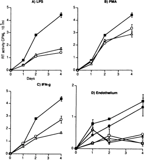
Effects ofcellular adherence on RT activity released from THP-1 cells. Washed, unstimulated HIV-infected THP-1 cellsdemonstratedincreasingsupernatantRT
activity
over4 days of culture. LPS stimulation of HIV-infected THP-1 cellscultured nonadherent in Teflon vialsresulted in reduced supernatant RTactivity.
Inaddition,
LPS stimulation of identical cells under adherent conditions further reduced VOL.67, 1993:;:--l
"I
on November 9, 2019 by guest
http://jvi.asm.org/
A)LPS
0
0-.52:
B) PMA C) IFN.y
Days
FIG. 3. Production of HIV fromstimulated THP-1 cells in nonadherent and adherent conditions. Tendaysafter infection withHIV-1RF, THP-1 cellswerewashedthree times in PBStoremoveall cell-freevirus, resuspended in freshmedium, culturedin either Teflon vialsor
tissue culture plastic, and stimulatedwith LPS(1,ug/ml;A),PMA(20 nM; B),orIFN-y (500U/ml;C). SupernatantswereanalyzedforRT
activityondays 1, 2, and 4 poststimulation. Symbols: El,notreatment,nonadherentconditions;0,treatment,nonadherentconditions;A, treatment,adherent conditions.Graphsillustrate the arithmeticmeanofaminimum of threeindependent experiments.
supernatant RT activity upon adhesion to tissue culture plastic (Fig. 3A). PMA stimulation of HIV-infected THP-1 cells cultured under nonadherent conditionsproduced little reduction in RT activity in comparison with unstimulated controls, in which activity was detected only at 4 days poststimulation. PMAtreatment of identical cells in adher-ent conditions removed all supernatant RT activity upon
cellular adherence to tissue culture plastic (Fig. 3B). In control experiments, incubation of viral supernatant of knownRTactivitywith conditionedsupernatant from
non-infected THP-1 cells treated with any of the stimuli in adherent conditions (data not shown) did not reduce RT activity.Thisfindingindicated that the detected reduced RT activities were not due to degradation of the enzyme in culture supernatants. Stimulation of
HIV-1RF-infected
THP-1 cells withIFN-y produced areduction in accumula-tion ofsupernatant RTactivity comparedwithunstimulated controls(Fig. 3C). Such reduction in RTactivitywassimilar in supematant from cells cultured in either tissue culture plasticorTeflonvials, asIFN-y didnotinduceadherence.
Effects of cellular adhesiononp24 release fromTHP-1 cells.
Theincrease insupernatantp24 antigenlevels fromwashed, unstimulated HIV-infected THP-1 cells over4 days of cul-tureparalleledthe increase in RTactivity. LPS stimulation ofHIV-infected THP-1 cellscultured innonadherent condi-tions in Teflon vials resulted in a similar pattern of p24 antigen release compared with released RT activity. In contrast tothe RTdata,cellular adherencetotissue culture plastic upon treatment with LPS resulted in little or no
reduction ofp24 levels (Table 1). PMAstimulation of cells
cultured in nonadherent conditions in Teflon vialsproduced
asmall reduction inp24levels. Similarly,incontrast tothe RTdata,tissue cultureplastic-adherentcells stimulated with PMA exhibited no reduction in p24 levels compared with controls. IFN-y stimulation of HIV-infected THP-1 cells cultured in tissue cultureplasticorTeflon vials reducedp24 levels.
Effects of adhesiontoendothelial cellsonviral release from HIV-infectedTHP-1cells. Adhesion ofwashed,unstimulated HIV-infected THP-1 cellstounstimulated confluent SGHEC 7 endothelial cells resulted in a reduction in released RT
activity comparedwith identical cells cultured in nonadher-entconditions in Teflon vials. Asignificantreduction in RT activitywasalsoseenin nonadherent cells cultured in Teflon
vials with conditioned medium from LPS-stimulated SGHEC 7 cells. Such reduction in released RTactivitywas
increased upon adherence of unstimulated THP-1 cells to LPS-prestimulated endothelial cells or adherence to endo-thelial cells in thepresenceofLPS(Fig. 4).
Effects of cellular activationonLTR-directedtranscription
inHIV-infectedTHP-1cells. To determine whether control of HIV LTRtranscriptionwas different inHIV-infected cells, THP-1 cells were transfected with HIV LTR-CAT
con-structs 10 days after HIV infection. Cells were stimulated andharvestedasinprevious experiments. CAT expression
[image:4.612.136.471.75.205.2]inunstimulated HIV-1-infected THP-1cells transfected with HIV-1LTR-CATproducedveryhighlevels of chloramphen-icolacetylation. Fifteenmicrograms ofcytoplasmicextract proteinfromunstimulated HIV-infected cellsproduced 89% acetylationof[14C]chloramphenicolafter 2 h of incubationat
TABLE 1. Comparison of HIV LTR-CAT expression, supernatantRT activity, and supernatant p24 levels
Valueobtained in:
Typeofanalysis Sample time Nonadherent conditions Adherent conditions
point (h)__ __
LPS PMA IFN-y LPS PMA IFN-y
CATexpression (fold increase) 24 1.0 1.2 2.0 3.0 1.6 1.5
RTactivity (relativetocontrol) 24 0.61 0.93 0.67 0.63 All 0.45
48 0.57 0.93 0.54 0.43 <0.01 0.82
96 0.96 0.48 0.59 0.49 0.59
p24 release(relative to control) 24 0.36 0.73 0.92 1.1 1.1 0.92
48 0.77 0.48 1.12 1.1 1.2 1.12
96 0.77 0.77 0.61 0.73 0.96 0.61
on November 9, 2019 by guest
http://jvi.asm.org/
[image:4.612.58.551.613.725.2]REGULATION OF HIV RELEASE BY MACROPHAGES 3573
A)LPS B) PMA
Days
FIG. 4. Effects on viral release of adherence to endothelial cells. Ten days after infection with HIV-1RF, THP-1 cells were washed threetimesinPBSto removeallcell-free virus, resuspended in fresh medium, andcultured innonadherent conditions in tissue culture plastic (-),in six-well tissueculture plates withconfluent untreated SGHEC 7endothelial cells(0), with SGHEC 7cells treated with LPS at 1 1xg/ml for 4 h and washed four times in PBS (V), with SGHEC 7 cellsinthepresenceof LPS at 1 1Lg/ml (OI), or in Teflon vials with conditioned medium (collected fromendothelial cells 24 h after they had been stimulated with LPS for 4 h and washed four times in PBS) (0).Supernatants were analyzedfor RT activity on days 1, 2, and 4poststimulation. Graphs illustrate the arithmetic mean (± standard errorof the mean) of three independent experi-ments.
37°C. Incomparison,300 ,ug ofproteinfrom uninfected cells transfected with HIV-1 LTR-CAT produced only a 1%
acetylationof[14C]chloramphenicolafter 6 h of incubation at
37°C. This high level of CATexpression was not reduced uponcellularstimulation; LPS, PMA, and IFN-yproduced 98, 94, and 91% acetylation, respectively. HIV-infected THP-1 cells transfected with AHIV-1 LTR-CAT showed low-levelCATexpression,withunstimulatedandstimulated
cells producingacetylation values of below3%.
Effects of cellular activation on RT activity from MDM. MDMspontaneouslyadheretotissuecultureplasticwithout
stimulation.Therewas anegligible difference in RTactivity
betweenadherentandnonadherent controls. Stimulation of
washed MDMinfectedwithHIV-lBaLwithLPS,PMA, or IFN--y resulted inasimilar reduction in released RTactivity
from cells cultured under nonadherent and adherent condi-tions (Fig. 5Ato C).
Effectsof adhesiontoendothelial cellsonviral releasefrom
HIW-infectedMDMcells.Adhesion of washed MDM infected withHIV-lBa L tounstimulated orLPS-stimulated endothe-lial cells resulted in a large reduction in RT activity. A similarreduction insupernatantRT
activity
was seenupon adherenceof MDM eithertoendothelial cells cultured in the presence of LPS or toglutaraldehyde-fixed LPSprestimu-lated SGHEC 7 cells. Culture of HIV-infected MDM in conditionedmedium from LPS-treated endothelial cells re-sulted inaminimal reduction in RTactivity (Fig. 5D).
DISCUSSION
The data presented in this study have demonstrated that cellular adherence may enhance HIV-1 LTR-CAT
transcrip-tion instimulated THP-1 cells. A
significant
enhancementof basal HIV LTR-CATexpression
was observedfollowing
LPS stimulation of THP-1 cells in adherent
conditions,
in agreement with previous studies(20).
We have shown that suchenhancementwasdependent
uponcellularadhesion,
as LPS stimulationof identical cells in nonadherent conditions failed to induce enhanced HIV LTR-CATtranscription.
Basal HIV LTR-CAT
expression
in unstimulated THP-1Days C)IFN-g
0 1 2 3 4
FIG. 5. Production of HIV from nonadherent and adherent MDM. Seven days postinfection, MDM infected with HIV-1BaL were washed three times in PBS to remove all cell-free virus, resuspended in fresh medium, stimulated with LPS (1 ,ug/ml; A), PMA (20 nM; B), or
IFN--y
(500 U/ml; C), and incubated under either nonadherentconditions in Teflon vials or adherent conditions in tissue culture plastic. Symbols: M, no treatment, nonadherent conditions; 0, treatment, nonadherent conditions; A, treatment, adherent conditions. (D) Effects on viral release of adherence of HIV-infected MDM to endothelial cells. Ten days postinfection, MDMwere cultured in nonadherent conditions in tissue culture plastic(M),insix-well tissue culture plates withconfluent untreated SGHEC 7 endothelial cells(0),with SGHEC 7 cells treated with LPSat 1 Lg/ml for 4h andwashed four times in PBS (A), with SGHEC 7 cells in the presence of LPS at 1 FLg/ml (0), or with glutaraldehyde-fixed SGHEC 7 cells which had been prestimulated withLPSat 1 pLg/ml for4h andwashed four timesinPBS (O) or cultured in Teflon vials with conditioned medium(collected from endothelialcells 24hafterthey had been stimulated with LPS for4 handwashed four times inPBS)(0).Inallexperiments, superna-tants wereanalyzed forRTactivityondays 1, 2,and 4 poststimu-lation.Graphs illustratethearithmeticmean(± standarderrorof the mean) of three independentexperiments.cells was in part mediated by NF-KB, constitutively
ex-pressedin suchcells (13),asreducedLTR-CAT
expression
wasobserved in cells transfectedwith AHIV-1 LTR-CAT.
EnhancementofHIVLTR-CAT expression followingLPS
stimulation of THP-1 cells in adherent conditions was
de-pendent upon functional NF-KB
binding
sites in theLTR,
since no enhancement was seen following adherence of LPS-stimulated THP-1 cells transfected with AHIV LTR-CAT. However, a
requirement
forintact NF-KB sites does not preclude the involvement of additionaltranscription
factors in induction of LPS-enhanced CAT
expression
ob-served in adherentconditions.In contrast, adherence of THP-1 cells to tissue culture
plastic following PMA stimulation did not enhance basal HIV-1 LTR-CAT
expression.
Such lackof enhancement of LTR-CATexpression
following
PMAstimulation is in agree-VOL. 67,1993,E
on November 9, 2019 by guest
http://jvi.asm.org/
[image:5.612.133.249.75.184.2] [image:5.612.329.566.78.340.2]ment with our previous observations (13) but at variance
with another report (20). Such differences may be due to
variablebasalexpressionofNF-KB indifferent subclonesof
THP-1cells. Furthermore, LPSand PMAhavebeen shown
tohave very different timecourses forinduction ofNF-KB
and are thought to act via different mechanisms (20). The
factthatthe combinationofLPS stimulation and adherence
enhanced HIV LTR-CAT expression whereas PMA
stimu-lationandadherencedid notaddssupporttothishypothesis. Monocyte adhesion may enhance HIV transcription only upon costimulation of the specific synergistic intracellular pathways.The natureofsuchpathways andtheireffectson
HIVtranscription remain tobedetermined.
As adhesion to tissue culture plastic may have artificial activatingproperties (19),weexaminedtheeffects of
adher-ence ofmonocytes to nonstimulated and stimulated human
umbilical endothelial cells (SGHEC 7 cells). Such studies
demonstrated that enhancement ofHIV LTR-CAT
expres-sion was similar following adhesion of LPS-stimulated
THP-1 cells to either endothelial cells or tissue culture plastic. Enhanced CATexpression was also observed upon
adherenceofunstimulated THP-1cellstoendothelial
mono-layers.Thiseffectwasinpart mediatedbysolublemediators
released fromtheendothelialcells,ascultureofTHP-1cells
in conditioned mediumproduced similarresults.
Incontrast toHIVLTR-CATtranscription, stimulation of
HIV-infected THP-1 cells with LPS or PMA resulted in
reducedRTactivityinnonadherentconditions.More impor-tantly, suchreductionwas greatlypotentiated under
adher-entconditions, indicatinganadherence-dependent reduction
in viral release (Table 1). The measured reduction in RT activity was not due to degradation of virus particles in supernatant, since RT activity was unchanged in control experiments inwhich tissueculturesupernatant from
LPS-orPMA-stimulated adherentTHP-1cellswasincubated for
24hwithsupernatant containing high-level RTactivity. Exposure ofHIV-infected THP-1cellsto IFN--y reduced
release of RT activity in both nonadherent and adherent conditions byequivalent amounts, demonstrating an
adhe-sion-independent mechanismofreduction.Thereductionin
RT activity following IFN--y treatmentwas consistentwith previous reports (26). Stimulation of HIV-infected MDM
also reduced RT supernatant activity. A similar pattern of
reductioninsupernatantRTactivitywasobservedin
adher-ent andnonadherent conditions. Theobservedreduction in
RT activityreleased fromHIV-infected MDMin
nonadher-ent conditions may have been facilitated by signals
gener-atedthroughautoadherence,as ahighlevel ofMDM clump-ingwasobserved inTeflonvials whichwasneverobserved
withTHP-1 cells.
Aparallel reduction in supernatant RT activity was
ob-served upon adherence of LPS-stimulated HIV-infected
THP-1 cells to tissue culture plastic or endothelial cell monolayers. In addition, adhesion of unstimulated
HIV-infected THP-1 cells to LPS-stimulated endothelial cells
inducedasignificantreductioninrelease ofRT activity.This finding suggested that adhesion per se to stimulated
endo-thelial cells was sufficient to induce a reduction in viral
release. The reduction in RT activity upon adhesion of
HIV-infected THP-1 cells tounstimulated SGHEC7
endo-thelial cells was, at least in part, mediated by soluble
mediators released from endothelial cells, as similar data
were obtained with use of culture conditioned medium. Furthermore,adhesionofMDMtounstimulatedor
prestim-ulatedconfluentendothelialcellsgreatly reducedRT activity independent ofsoluble mediatorsreleased fromendothelial
cells. Thisfinding suggested that adhesion of MDM may be sufficient to reduce RTactivity. Endothelial cells appearto have apassive role in such reduced RT release from
MDM,
as similar results wereobtained uponadherenceof MDMto
glutaraldehyde-fixedLPS-stimulatedendothelial cells.
Thus,
these data suggest that in vivo adherence may
downregulate
viral releasefrom infected
macrophages.
The described reduction in RT activity occurred even in the presence of high levels of HIV transcription, as HIV LTR-CAT expression was active at a rate far higher than that seen in non-HIV-infected cells. Such high levels of LTR-CAT expression were not surprising and may reflect
theaction oftatexpressedin HIV-infected cells
(24).
Hence,
reduction in released RT activity may be mediated
by
posttranscriptional events. Such posttranscriptional
down-regulation is different from previously described
transcrip-tional downregulation of HIV replication following LPS
stimulation of MDM (2). However, in agreement with pre-vious studies (1), use of HIV constructs with mutated KB sites demonstrated that the
efficiency
of transactivation of HIV LTR-CAT in infected cells was dependent on the presence ofintact KBbinding sites.In contrast to data described above on RT activity, there was little difference in p24 release upon stimulation with
LPS, PMA,or IFN--y ofHIV-infected THP-1 cellsin
adher-entconditions. RT activity detected intissue culture super-natants of cells infected with HIV has been shown to be associated almost exclusively with virus particles, whereas
capsid and envelope proteins maybe shed as free
(soluble)
antigen (7). IFN--y has been shown to reduce released supernatant RT activity from the persistently HIV-infected promonocyticcell lineUl (3). Such reduction in released RT
activity occurred without suppression of HIV mRNAs or proteins and reflected a redistribution of virions to intracy-toplasmic vacuoles. In addition, IFN-a treatment of HIV-infected T cells and human mononuclear cells haspreviously been shown to reduce supernatant RT activity while little affecting p24 antigen levels (7, 10). Differences in RT andp24 release mayreflect disruption of viral assembly and release but nottranscription and translation (7, 10). Our observation thata reduction in RT activity occurred even in the presence ofhigh levels of HIV transcription and withoutsuppression
of p24 antigen release suggests that activation of adherent monocytic cells interferes with HIV assembly and release. It remains to be determined whether such a reduction in release of intact HIV reflects an accumulation of intracyto-plasmic virions or disrupted viral assembly.
The relative importance in vivo of the differential effects of adhesion and activation on HIV transcription and release remain to be determined. In latently infected immature cells, adherence may prime cells to respond to other stimuli which initiate transcription (14), while in mature cells, adhesion alone and in conjunction with other cellular activators may, as was observed, downregulate viral release. Thus, macro-phage activation during an immune response may reduce rather than increase the amount of HIV released from infected macrophages.
ACKNOWLEDGMENTS
RobinJ. Shattockissupportedby theMedical Research Council (London, United Kingdom).Jon Friedland is a Medical Research Council Training Fellow, andGeorge Griffin is supported by the WellcomeTrust.
on November 9, 2019 by guest
http://jvi.asm.org/
REGULATION OF HIV RELEASE BY MACROPHAGES 3575 REFERENCES
1. Berkhout, B., A. Gatignol, A. Rabson, and K.-T. Jeang. 1990. TAR-independent activation of the HIV-1 LTR: evidence that tat requires specific regions of the promoter. Cell 62:757-767. 2. Bernstein, M., S. Tong-Starksen, and R. Locksley. 1991.
Activa-tion of human monocyte-derived macrophages with lipopolysac-charide decreaseshumanimmunodeficiencyvirus replication in vitro atthe level ofgeneexpression.J.Clin. Invest. 88:540-545. 3. Biswas, P., G. Poli, A. Kinter, J. Justement, S. Stanley, W. Maury, P. Bressler, J.Orenstein, and A. Fauci. 1992.Interferon -y induces theexpression of human immunodeficiency virus in persistentlyinfectedpromonocytic cells(Ul) and redirects the production of virions to intracytoplasmic vacuoles in phorbol myristate acetatedifferentiatedUlcells. J. Exp. Med. 176:739-750.
4. Bradford, M. 1976. A rapid and sensitive method for the quantitation of microgram quantities of protein utilizing the principle of protein-dye binding. Anal. Biochem.72:248-254. 5. Carlos, T.,andJ.Harlan.1990. Membraneproteins involvedin
phagocytosisadherence toendothelium. Immunol. Rev. 114:5-28.
6. Eierman, D. F.,C.E.Johnson, and J. S. Haskill. 1989.Human monocyteinflammatory mediatorgeneexpression is selectively regulatedby adherencesubstrates. J. Immunol. 142:1970-1976. 7. Fernie, B., G. Poli, and A. Fauci. 1991. Alpha interferon suppresses virion but not soluble human immunodeficiency virus antigen productioninchronically infected T-lymphocytic cells. J.Virol. 65:3968-3971.
8. Ferreira, 0. J., P. A. Garcia, and C. Bianco. 1990. Specific binding of the human monocyticcell line U937 to the alterna-tively splicedconnectingsegment(IIICS)offibronectin.J.Exp. Med. 171:351-356.
9. Gartner, A., P.Markovits, D.Markovits,K. Kaplan,R.Gallo, and M.Popovic. 1986. The roleofmononuclearphagocytesin HTLV3/LAV infection.Science 233:215-218.
10. Gendelman, H., L. Baca, J. Turpin, C. Kalter, B. Hansen, J. Orenstein, R. Froedman, and M. Meltzer. 1990. Restriction of HIVreplicationin infected Tcellsand monocytesby interferon alpha. AIDS Res.Hum. Retroviruses6:1045-1049.
11. Gendelman, M., J.Orenstein,L.Baca,B.Weiser,H.Burger,D. Kalter, and M. Meltser. 1989. The macrophage in the
persis-tenceandpathogenesisof HIV infection. AIDS 3:475-495. 12. Gorman, C., L. Moffat, and B. Howard. 1982. Recombinant
genomes which express chloramphenicol acetyltransferase in mammalian cells. Mol.Cell.Biol. 2:1044-1051.
13. Griffin,G.,K.Leung,T.Folks,S.Kunkel,andG. Nabel. 1989. Activation of HIV geneexpressionduringmonocyte differenti-ationby inductionofNF-KB.Nature(London) 339:70-73. 14. Haskill,S.,C.Johnson,D.Eierman,S.Becker,and K. Warren.
1988. Adherence induces selective mRNA expression of mono-cyte mediators and proto-oncogenes. J. Immunol. 140:1690-1694.
15. Jones, B., J. Nicholson, R. Holman, and M. Hubbard. 1989. Comparisonof monocyte separation methods using flow cyto-metric analysis. J.Immunol. Methods125:41-47.
16. Kasahara, K., R. Strieter, S. Chensue, T. Standiford, and S. Kunkel. 1991. Mononuclear cell adherence induces neutrophil chemotacticfactor/interleukin-8geneexpression. J. Leukocyte Biol.50:287-295.
17. Kornbluth, R. S., 0. P. Munis, Jr., P. M. Cleveland, and D. D. Richman. 1989. Interferons and bacterial lipopolysaccharide protect macrophages from productive infection by human im-munodeficiencyvirus in vitro. J. Exp. Med. 169:1137-1151. 18. Nabel, G. 1991. Tampering with transcription. Nature (London)
350:658.
19. Newman, S., and M. Tucci. 1990. Regulationofhuman mono-cyte/macrophage functionbyextracellular matrix. J. Clin. In-vest.86:703-714.
20. Pomerantz, R., H. Feinberg, D. Trono, and D. Baltimore. 1990. Lipopolysaccharide is a potentmonocyte/macrophage-specific stimulator of humanimmunodeficiencyvirus type-1expression. J. Exp.Med. 172:253-261.
21. Potts, B. 1990. Mini reversetranscriptase (RT)assay, p. 103-106. In A.Aldovini and B. Walkers(ed.),Techniques in HIV research.StocktonPress, New York.
22. Rosen, C., J. Sodroski, K. Campbell, and W. Haseltine. 1986. Construction of recombinant murineretroviruses that express the human T-cell leukemia virus type II and human T-cell lymphotropic virus type III trans activator genes. J. Virol. 57:379-384.
23. Sporn, S. A., D. F. Eierman, C. E. Johnson, J. Morris, G. Martin, M.Ladner,andS. Haskill. 1990. Monocyte adherence results inselectiveinduction of novel genes sharing homology withmediators ofinflammation andtissuerepair. J. Immunol. 144:4434-4441.
24. Steffy, K., and F. Wong-Staal. 1991. Genetic regulation of humanimmunodeficiency virus. Microbiol. Rev.55:193-205. 25. Tsuchiya, S., M. Yamabe, Y. Yamaguchi, Y. Kobayashi, T.
Konno, and K. Tada. 1980. Establishmentandcharacterisation ofa human acutemonocytic cell line (THP-1). Int. J. Cancer 26:171-176.
26. Tsunetsugu-Yokota, Y.,and M. Honda.1990.Effect ofcytokines
onHIVrelease andIL-2 receptoralpha expressioninmonocytic celllines. J.AcquiredImmuneDefic.Syndr.3:511-516. 27. Vlach,J., and P. Pitha. 1992. Activation of human
immunode-ficiencyvirus type 1 provirus in T cells andmacrophages is associated with induction of inducer-specific NF-<B binding proteins. Virology 187:63-72.
VOL. 67, 1993