Copyright ©)1992,American Society for Microbiology
Equine Infectious Anemia
Virus Gene Expression: Characterization
of the RNA Splicing
Pattern
and the Protein Products Encoded by
Open Reading Frames Si
and
S2t
R. LOUIS SCHILTZ,1 DING S. SHIH,1* STYAMAK RASTY,2RONALD C. MONTELARO,2 ANDKEITH E. RUSHLOW2
Departmentof Biochemistry andLouisiana State UniversityAgriculturalCenter, Louisiana State University, Baton Rouge, Louisiana 70803,1 and Department of MolecularGenetics andBiochemistry, School of Medicine,
University of Pittsburgh, Pittsburgh, Pennsylvania 152612
Received 25 November 1991/Accepted 10 March 1992
The utilization ofpredicted splice donor and acceptor sites in generating equine infectious anemia virus (EIAV) transcripts in fetal donkey dermal cells (FDD) was examined. A single splice donor site identified
immediately upstream ofthegag coding region joins the viral leader sequence toall downstream exons of
spliced EIAV transcripts. Thepredominant 3.5-kb transcript synthesized in EIAV-infected FDD cellsappears
tobe generated byasingle splicingeventwhich links the leadersequencetothefirst oftwofunctionalsplice acceptorsitesnearthe 5' endoftheSi open reading frame (ORF). The translation products encoded by the 3.5-kb transcriptwereexamined by producing in vitro transcripts fromacDNA corresponding tothis RNA followed by in vitro translation in wheat germ extracts. These transcripts directed the synthesis ofthree proteins: the virus trans-activator protein (EIAV Tat) encoded by ORF S1, a protein of unknown function
encoded byORFS2, and the virus envelope glycoprotein. When transfected into FDD cells, this cDNA also directed expression of EIAV Tat. Amino-terminal sequence analysis of the in vitro-synthesized Si protein supportsthe suggestion thattranslationofEIAV Tat is initiatedataCUG codon within the virus leader region.
Both invitro-synthesized S2 proteinandsynthetic peptides correspondingtoS2areshownto reactpositively withseraobtained from EIAV-infectedhorses, providing the first direct evidence of expression of this protein
in infected animals.
Equine infectious anemia virus (EIAV) is a member of the lentivirus subfamily of retroviruses, a group that includes the
ungulate (visna-maedi, caprine arthritis-encephalitis, and
bovineimmunodeficiency-like viruses) and the primate (hu-man immunodeficiency virus types 1 and 2 [HIV-1 and HIV-2] and simian immunodeficiency virus) retroviruses
(18). The genetic organization of EIAV is relatively simple
comparedwith that of other lentiviruses (6)inthat the gene
contains only three short open reading frames (ORFs),
Si,
S2, and S3, in addition to the gag, pol, and env genes common to all replication-competent retroviruses. ORFSi
is located within thepol-env intergenic region, while ORF S2
begins between ORF S1 and env and overlaps the amino
terminus of theenv gene in adifferentreadingframe. ORF S3 ispositionednearthe 3' end of the genome and is encoded within an alternate reading frame overlapping the
gp45-coding sequence of the env gene. The
Si
gene product,which shares both structural and functional homology with the Tat protein ofHIV-1, has been identified as the EIAV trans-activator protein (8, 20, 30). Although ORF S1 con-tainsnoAUGcodon, it has been suggested thattranslation of EIAV Tat may begin at a CUG codon within the viral
leadersequence,whichisjoinedin-frame with
Si
as aresultofanRNA splicingevent (8, 30). Onthebasis of sequence homology with HIV-1 Rev and nonsense mutations within
S3, which seem to impart a Rev-defective phenotype to EIAV-infected canine cells (30), ORF S3 is believed to encode the EIAV Rev protein. The predicted protein
en-*Correspondingauthor.
tApproved bytheDirector of theLouisianaAgricultural
Exper-iment Station forpublicationasmanuscriptnumber 91-12-551.
codedby ORF S2 bears nosignificant homology to any of the lentiviral ancillary proteins described to date and has yet to
be assigned a function. Thus far, no transcript or protein
product that corresponds to expression of ORF S2 has been identified.
EIAV gene expression has been primarily studied in canine and felinefibroblastcultures whicharepermissive for
persistentEIAV infections. Analyses ofRNAtranscription
patterns in such cells yielded variable observations in the temporal appearance and abundance ofEIAV-specific
tran-scripts (8, 21, 30). The simplest and most consistent
tran-scriptionpatternswere observed in EIAV-infectedprimary
horse macrophage cultures and equine fibroblasts (fetal
equine kidney [FEK] andfetaldonkeydermal [FDD] cells),
whichpredominantly synthesized transcripts corresponding to the 8.2-kb genomic RNA and the presumably singly
spliced 3.5-kb envelope message (25, 30). Small multiply
spliced transcriptsof1 to 2 kbwere mainlyundetectable in
EIAV-infected equine cells, asurprising observation, since viraltrans-activation factorsareexpressedinEIAV-infected
equine fibroblasts (25). In the present work, we have
sys-tematicallyexamined thesplicingpatternsofEIAV in FDD
cellsby usingcDNA
cloning,
Northern(RNA)
blothybrid-ization, and nuclease S1 protection assays.
Previous studies in our laboratory showed that EIAV infection of cultured FDD cells resulted in a
cytopathic
infection,whereas FEKcells becamepersistentlyinfected in
vitro (25). Interestingly, in cytopathically infected FDD
cells, the proportion of 3.5-kb mRNA to
full-length
8.2-kbtranscript was substantially greater than in
persistently
in-fected FEK cells, which synthesized the two RNAs in
approximately equal abundance at all stages of infection.
3455
on November 9, 2019 by guest
http://jvi.asm.org/
Examination of the properties of the predominant 3.5-kb EIAV transcript of FDD cells might provide some insight into the mechanisms of cytopathicity observed in this cell type. To address this question as part of the studydescribed in this report, in vitro transcripts were generated from a cDNA derived from the 3.5-kb RNA and used to prime translation in wheat germ extracts. These transcripts were found to produce theSi, S2, andenvelopeproteinsof EIAV as confirmed by radioimmunoprecipitation assays and ami-no-terminal sequence analyses of Si and S2. Sequence analysis of the Si EIAV Tat protein was consistent with the suggestion of translational initiation at a CUG codon within the leader region. The S2 protein was specifically
immuno-precipitated by sera from EIAV-infected horses,
demon-strating its production during EIAV infection of host ani-mals. The 3.5-kb RNA-derived cDNA was also tested for encoding trans-activation factors in cultured FDD cells by
cotransfection of a simian virus 40 (SV40) replacement
vector containing the cDNA with a long terminal repeat
(LTR)-driven reporter plasmid. The results of these studies
are presented in this report.
MATERIALSAND METHODS
Virus strains and cell cultures. Primary cultures of FDD cells were prepared and maintained as previously described (24, 25). An FDD-adapted stock of prototype EIAV was utilized in these studies. This stock was generated by prop-agation of the Wyoming cell-adapted strain of EIAV (15) in primary cultures of FEK cells to produce prototype virus followed by serial passage in FDD cells. Confluent mono-layers of FDD cells were infected with FDD-adapted virus at amultiplicity of infection of 1. Cells were harvested for RNA isolation as previously described (25).
Isolation and purification of RNA. Total cellular RNA was isolated from EIAV-infected or uninfected FDD cells by a modifiedguanidinium thiocyanate extraction method (4, 16) aspreviously described (25). Poly(A)+ RNA was purified by two cycles of oligo(dT)-cellulose chromatography of total cellular RNA (16) or by direct oligo(dT)-cellulose chroma-tography of cellular lysates by the commercially available Fast Track mRNAIsolation kit (Invitrogen Corp.) according
to themanufacturer's specifications.
cDNAcloning.Double-stranded cDNA was produced from poly(A)+ RNA from EIAV-infected cells by using a com-mercially available cDNA synthesis kit (Bethesda Research
Laboratories). First-strand synthesis was primed with a
30-nucleotide (nt) primer complementary to a region of the
envelope gene immediately downstream of theSmaI site (nt 5693 to 5722). The double-stranded cDNA products were tailed with oligo(dC) by using terminal deoxynucleotidyl transferase and ligated to PstI-digested pUC9 tailed with oligo(dG). Therecombinant plasmids were transformed into
DH5a Eschenchia coli cells and plated on
5-bromo-4-chloro-3-indolyl-o-D-galactopyranoside
(X-Gal) selective media. Whitecolonies were selected and screened for the presenceofEIAV-specificcDNA inserts by colony hybridization (16),
using as a probe an (x-32P-labeled BamHI-TaqI fragment
corresponding to a portion of ORF S2 located immediately
upstream of the priming site for first-strand cDNA synthesis. A total of 16 positive clones were further analyzed by Southern blot hybridization. Minilysate DNA from these clones was digested with PstI to release the cDNA inserts
and fractionated on 1% agarose gels. Following transfer to
nitrocellulose, the membrane was probed with an
at_32p_
labeled 226-bpMluI-BamHI restriction fragment (nt 156 to
386) corresponding to a portion of theviral LTR and leader sequence. Two clones, designated
pSR-1
and pSR-2, con-tained cDNA inserts which hybridized with this probe.These were subcloned as 270-bp
SmaI-BamfHI
restriction fragments intoM13mpi8
orM13mpi9
for sequenceanalysisby the dideoxy chain termination method (27).
Northern blot analysis.
Poly(A)+
RNA (2.5jig)
isolated from EIAV-infected FDD cells was fractionated on 1.4%agarose-formaldehyde denaturing gels and transferred to nitrocellulose membranes. The membrane was probedwith one of three
32P-labeled
synthetic oligonucleotides designed to be complementary to regions of the EIAV leader se-quence or amino-terminal portions of the gag codingregion. These probes were designed to be specific for the three putative splice donors in this region (see Fig. 2A). Probe 1(SD-1) is a 28-mer complementary to nt 428 to 455 within the
EIAV leader immediately upstream of the Gag polyprotein AUG initiation codon. Probe 2 (SD-2) is a 28-mer comple-mentary to nt 468 to 495 covering the amino-terminal region of the
pl5gag
protein. Probe 3 (SD-3) is a 27-mer comple-mentary to nt 518 to 544 within the p15 coding sequence.NucleaseS1 protection assays. Total cellular RNA isolated from EIAV-infected or uninfected FDD cells was annealed to one of four different end-labeled probes (see Fig. 3), and the resulting complex was subjected to Si nuclease digestion to identify the splice sites of EIAV-specific mRNAs. Probe 1
(P1)
was generated by subcloning a 308-bp TaqI-PvuII EIAV proviral DNA restriction fragment encompassing the three putative splice donor sequences near the amino terminus of the gag coding region intoAccI-SmaI-digested
M13mpl8
and then digesting it with
Nar.
The resulting probe con-tained a 294-bpNarI-PvuII
EIAV-specific sequence (nt 326 to 620) followed by a 250-bp sequence derived from theSmaItoNarI restriction sites of
M13mpi8.
This probe was 3' end labeled with[L-32P]dCTP
and the Klenow fragment of E. coliDNA polymerase
I.
Probe 2 (P2) was constructed by sub-cloning a 450-bpNcoI-BamHI
EIAV proviral restriction fragment encompassing the 3' end of thepol
gene and extending through the 5' end of the env gene (nt 4889 to 5337) intoBamHI-SmaI-digested M13mpi9
following treatment of theNcoI
site with the Klenow fragment of E. coli DNA polymerase I. The probe DNA was excised by digestion with BamHI andBglII
to yield a611-bp
restriction fragment containing 450 bp of EIAV sequence and 161 bp ofM13mpi9
sequence. This restriction fragment was dephosphorylated with calf intestine alkaline phosphatase and 5' end labeled with T4 polynucleotide kinase and
[_y-32P]ATP.
Probe 3(P3)
corresponds to a 617-bpPvuII-HindIII
proviral restriction fragment extending from within ORF Si to the 5' end of the env gene (nt 5161 to 5778). This restriction fragment was 3' end labeled with[a-32P]ATP
and[a-32P]CTP
by replacement synthesis with T4 DNA polymerase. Probe 4 (P4) was generated by digested of proviral DNA withDraI
andScal
and isolation of a 385-bp restriction fragment corresponding to a region of the env gene, which also includes the 5' end of ORF S3 (nt 7026 to 7412). This restriction fragment was 5' end labeled with
[_y-32P]ATP
and T4 polynucleotide kinase following dephosphorylation with calf intestine alkaline phosphatase.The 32P-labeled probes
(106
cpm) were combined with total cellular RNA (40,ug)
isolated from EIAV-infected or uninfected FDD cells and ethanol precipitated. The mixture was suspended in 30,ul
of Si nuclease hybridization buffer{80%deionized formamide, 40 mM PIPES
[piperazine-N,N'-bis(2-ethanesulfonic acid); pH6.4],
400 mM NaCl, 1 mMEDTA} and placed into an
85°C
water bath for 15min
toon November 9, 2019 by guest
http://jvi.asm.org/
denature the probe. Hybridization of the denatured probes to the RNAs was accomplished by overnight incubation at 50°C for probes P1 and P3 or at 55°C for probes P2 and P4.
Si nuclease digestion was accomplished by addition of the
following: 150
RIl
of 2x S1 nuclease buffer (0.56 M NaCl, 0.1M sodium acetate [pH 4.5], 9 mM
ZnSO41,
3RI
of2-mg/mlsingle-stranded calf thymus DNA, 147 ,u of diethyl pyrocar-bonate (DEPC)-treated doubly distilled H20, 300 U of Si nuclease [Bethesda Research Laboratories]). The mixture was incubated at 30°C for 60 min, after which the reaction wasstopped by the addition of 80
RI
ofS1 stop buffer (4 M ammonium acetate, 20 mM EDTA [pH 8.0], 40 mg of yeast tRNAperml). The products were concentrated by ethanolprecipitationand fractionated by gel electrophoresis on a 6%
acrylamide-urea sequencing gel. MspI-digested pBR322
plasmid DNA labeled with [a-32P]dCTP by the Klenow
fragment of E. coli DNA polymerase I was utilized as a molecularweight marker.
In vitro transcription and
eucaryotic
expression plasmid constructs.Invitrotranscriptionandeucaryotictransfectionplasmids were constructed by subcloning restriction
frag-mentsof the cDNA clone pSR-1 into pSP65 (Promega Corp.) or into an SV40 replacement vector, pSV2Acat, derived frompSV2cat (11). The pSV2Acatvector was produced by
digestion ofpSV2catwith HindIII and HpaI to remove the
chloramphenicol acetyltransferase (CAT) gene followed by
insertion of a synthetic linker that regenerates each of these sites and provides a uniqueXhoI site. In the pSR-1 cDNA clone, ORF Si, ORF S2, and env are spliced to the viral leader region through a splice donor site (sdl) localized immediately upstream of the gag gene (nt 459). pSR-1 was
digestedwith either StuI (nt256)orSmaI (nt 303) and then
digested with MscI (nt 5655) to generate a 720-bp
(Stul-MscI) or 585-bp (SmaI-MscI) restriction fragment. These
blunt-end DNAs weresubcloned intoSmaI-digested pSP65, generating pSP720 and pSP585, respectively, or into
StuI-digested pSV2Acat, generatingpSV720 and pSV585,
respec-tively(Fig. 5A). The resulting recombinant plasmids were screenedfor proper orientation by using appropriate restric-tion enzymes.
The EIAV LTR CAT plasmid, pLTRcat, used in trans-activation assays has beenpreviously described (7, 25). The
pSV2catplasmid (11), whichcontains the CAT gene under
the control of the SV40immediate-earlypromoter,wasused
as apositive control in CAT assay transfections.
Transfec-tionplasmidswerepurified bytworounds of cesium chloride
density centrifugation aspreviously described (10).
Plasmid DNA transfection and CAT assays. CsCl density
gradient-purifiedDNA(11 ,ug)wasused totransfect 60-mm
petri dishes of FDD cells by the calcium phosphate
copre-cipitation method(10). pLTRcat (5 ,ug)and
pSV3-gal
(1 ,ug)were cotransfected with pSV720, pSV585, orpSV2Acat (5
p,g).
pSV3-gal
plasmid(Promega Biotec)wasaddedto each transfection to control for transfection efficiency. Cell ly-sates were prepared 48 h posttransfection as previouslydescribed(10), andthe
P-galactosidase
activities of the celllysates
from eachtransfection weremeasured according tothesupplier's recommendations (Promega Corp.). pSV2cat
(5 ,ug)wasusedas apositivecontrolDNAfor CATactivity
andwascotransfectedwithpSV2Acat(5 ,g)and
pSVP-gal
(1R,g).
Lysatevolumescontaining equivalent
levels ofP-galac-tosidase activitywere assayedfor CAT enzyme activity by
the kinetic diffusion method of Neumann et al. (19) with
[14C]butyryl
coenzyme A(New England Nuclear,Dupont)
andchloramphenicol as substrates for CAT.
Peptide synthesis and ELISAs. Synthetic peptides were
prepared either by using a SAM-II automated peptide
syn-thesizer (BioSearch) or by using manual methods and the
RaMPS system.(Dupont). Reaction conditions used for synthesis were according to the manufacturers' specifica-tions. Peptides were initially purified by gel filtration on Sephadex G-25 followed by reverse-phase high-pressure
liquidchromatography and furthercharacterized by plasma
desorption mass spectrometry to confirm their purity and
sequences. Each peptide was reacted against a panel of horse sera in an enzyme-linked immunosorbent assay (ELISA) optimized for use with synthetic peptides antigens
(3). NegativeELISA values were established by examining a
panel of eight samples of normal horse serum and averaging thereactivity values against each peptide.
In vitro transcription and translation. Capped mRNAs were synthesized from linearized DNA templates by using SP6 RNA polymerase (Promega) and the mCAP mRNA
Capping Kit (StratageneCloning Systems) accordingto the
manufacturers' recommendations. The capped mRNAs were translated in wheat germ extracts
(Promega)
in the presence of [3H]leucine, [3H]glycine, or [ H]arginine (Am-ershamCorp.).Thetranslation productswereeither directlyanalyzed on 20% low-molecular-weight polypeptide gels
(HoeferScientificInstruments) or first subjectedto
radioim-munoprecipitation with the indicated antisera.
Autoradio-graphic image enhancement was achieved by the use of
Autofluor(National Diagnostics) according tothe manufac-turer's recommendations. The dried gels were exposed to Kodak XAR film at -70°C overnight.
Antiserum preparation and radioimmunoprecipitation as-says. Syntheticpeptides correspondingtoportionsofORFs
S1 and S2were linked to keyhole limpet hemacyanin and injected into New Zealand White rabbits in Freund's com-plete adjuvant. The rabbits were boosted with the same
antigen in Freund's incomplete adjuvant after 3 weeks to
producehyperimmune sera,whichwere harvested2weeks
after therabbitswereboosted. TheimmunoglobulinG(IgG)
fractionwas partially purified from thesera by ammonium sulfate precipitation and DEAE-Sephadex column
chroma-tographyas previously described (5). The appropriate
anti-serum was reacted with radiolabeled in vitro translation
products for radioimmunoprecipitation as previously
de-scribed (2). The precipitated proteins were analyzed on discontinuous sodiumdodecylsulfate(SDS)-polyacrylamide gelsasdescribed above.
Automatedprotein sequencing. The EIAV ORFS1protein
was synthesized in wheat germ extracts programmed with
pSP720 RNA in the presence of
[3H]arginine,
as describedabove. The pSP720 transcript encodes both S1 and S2,
which are difficult to completely resolve on SDS gels.
Becauseof thesimilarity in the gelmobilities of the S1 and
S2proteins,the
S1 protein
wasimmunoprecipitated
from thetranslation mixturepriortogelelectrophoresis.The ORF S2
proteinwassynthesizedin thepresenceof[3H]glycinefrom
pSP585RNA, which lacks sequencesrequiredforS1
expres-sion. The S1 and S2 proteins were fractionated on 15%
discontinuous gels
(14),
which wereplaced
directly
intoAutoflour for30min. The driedgelswereexposedtoKodak XAR film and placed at -70°C overnight. With the
autora-diographasatemplate,the
appropriate protein
bandwascutfrom the dried gel and rehydrated in a minimal volume of water. The gel slice was crushed, and the labeled
proteins
wereeluted in2 to3ml of Tris saline buffer
(50
mMTris-HCl[pH
7.5],
100 mMNaCl)
at37°C
for 3 hwithconstantmixing.
Thegelfragmentswereremovedby
centrifugation,
andthesupernatant was
lyophilized
andresuspended
in 100 ,ul ofon November 9, 2019 by guest
http://jvi.asm.org/
1-mg/mi
bovine serum albumin to act as a carrier protein. Theprotein
mixture was desaltedby
passing
it through a G-10Sephadex
column. Radioactive fractionswere pooled,lyophilized,
andresuspended
in 50 to 100,ul
of doubly distilled deionized water. Thesesamples
were subjected toautomated amino-terminal
sequence
analysis
on anAppliedBiosystems
470A ProteinSequencer.
Radioactivity from eachcycle
of thesequencer
was determined by directlyadding
thecycle
eluate to 3 ml ofLiquiscint
scintillationcocktail
(National Diagnostics)
and counting in a BeckmanLS 6000IC liquid scintillation counter.
RESULTS
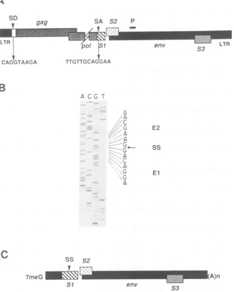
Nucleotide
sequence analysis
of cDNA clones generatedfrom the 3.5-kb
EIAV
RNA. Thesplice
site of the EIAV3.5-kb
RNA,
whichpredominates
incytolytically
infectedFDD
cells,
was identifiedby
cDNA cloning employing a30-nt
synthetic oligonucleotide primer
complementary to aportion
of theenvelope
RNAsequence
400 nt downstream of theputative envelope gene
initiator codon (Fig.1A).
Theposition
of thisprimer
downstream of all near-consensussplice
donor
sequences (17, 23)
should
preclude
synthesis ofcDNAs
corresponding
tomultiply
spliced
mRNAs and thusshould allow
synthesis only
ofcDNAs corresponding to the8.2-kb
full-length
genomic
RNA andthesinglyspliced3.5-kbenvelope
RNA. Two cDNAclones,
pSR-1
andpSR-2,whichhybridized
to both a114-bp
BamHI-TaqI
probe
(nt 5337 to5456)
derived from the envgene
and a 213-bpMluI-BamHI
probe
(nt
156 to
386)
derived
from LTR and leaderse-quences,
wereisolated,
indicating
that these cDNAs hadextended to the 5' terminus of the viral RNA. These two
clones,
found to be identicalby
dideoxy
nucleotidesequenc-ing
(Fig.
1B),
wereproduced
from aspliced
RNA, whichutilized a
splice
donor atnt459(sdl)
and asplice acceptor atnt 5135
(sal).
These cDNA clonescorrespond
to an RNAthat
contains a
majority
of
ORF
S1,
all of
ORF
S2, and theenv
gene
(Fig.
1C).
The
splicing
event removed
four codonsfrom the 5' end of
ORF
S1
as it wasoriginally
defined (26),that
is,
as the150
nt of EIAVsequence
from theterminationcodon
of the
pol
gene
to the next in-frame
terminationcodon.
However,
S1
is
extended an additional
38 codons in the amino-terminal directionthrough
the viral leader regionas a result of the
splicing.
These 38 codons combined withthe
46 codons
maintained from the
original
ORFS1
provide84 codons of
polypeptide coding
potential.
Interestingly, noAUG codon is found within
this 84-codon
sequence.Northern blot
analyses using splice
donor-specific
oligonu-cleotide
probes.
Although
we have
isolated
two identicalcDNA
clones
corresponding
to a
singly
spliced
subgenomicRNA,
the
possibility
that
other RNAs
with similar
mobilitiesin
denaturing
agarose gels
comigrate
with
this
identified3.5-kb
message
still
remains. We
were
particularly
interestedin
examining
whether two
additional
splice
donors,
sd2 (nt512)
and
sd3
(nt
546),
located near
the 5' end
of
thegag gene,were
functional in
FDD cells.
The
utilization
of sd2 couldresult in
the first
16
codons of the
gag
gene,
including
theAUG initiator
codon,
being
spliced
to
the
acceptors
at nt5135
(sal)
or nt
7243
(sa3)
to
provide
translational
initiationsignals
for
ORF
S1
and
ORF
S3,
respectively.
RNA
splicingfrom
sd3 to
these same
acceptors
would
result
in
a splicingeventthat
would
place
ORF
S1
or
ORF S3 out
offramewiththe
gag
coding sequence
and
would
supply
only
twoaddi-tional
codons,
neither
of
which is an
AUG, to
these ORFs.Northern
blot
analysis employing
splice
donor-specific
oli-gonucleotide
probes
and
nuclease
S1
protection
assayswereA
so a gag
I
CAGGTAAGA
B
SA S2 p
env S3 LTR
TTGTTGCAGG AA
ACG T
a
A
C
.DA
A
G
A
C .G
A
ES
C
SS S27meG
_MMS
Si
E2
sS
El
0A
)WM~~~
n A)nenv
1s
FIG. 1. Nucleotide sequence analysis of the splice junction of
the
predominant
3.5-kb RNA in EIAV-infected FDD cells. (A)Schematic
representation
of the EIAVproviral
genome, indicating theposition
ofpotential
splice
donor(SD)
and acceptor(SA) sites. Thesequences
surrounding
the 5' and 3'splice
sites of the 3.5-kb mRNA are shown. Theposition
of the 30-nt primer used for first-strand cDNAsynthesis
is shown with a heavy line marked P.(B)
Dideoxynucleotide
sequencing
gel
of the cDNA clone pSR-1, derived from thesingly
spliced
3.5-kb transcript. Nucleotidese-quences corresponding
to the twoexons are shown (El and E2), andthe
position
of thesplice
site(SS)
is indicated by an arrow. (C)Schematic
representation
of 3.5-kbsingly
spliced mRNA. ThismRNA
species
containsthe viral leaderregion
spliced to ORFS1,
which is followed
by
ORF S2 and the viral envelopegene. The cap structure is indicatedby
FmeG,
and thepoly(A)
tailis indicated by (A)n.performed
in order to determine the role of these putativesplicing signals
in EIAVgene
expression.By
employing
threesynthetic
oligonucleotide
probes(SD-1,
SD-2,
andSD-3)
that arecomplementary
to the RNAsequence immediately upstream
of each splice donor, wewere able
to
ascertain whether these two alternative
donorswithin the
gag gene
were utilized to
generate
splicedmRNAs
(Fig. 2A).
TheSD-1
oligonucleotide
shouldhybrid-ize to
spliced
messages utilizing
any
of
the
three putativesplice
donors. The SD-2
oligonucleotide
would
be unable tohybridize
to
spliced
mRNAs
utilizing
sdl
but
wouldhybrid-ize to those
utilizing
sd2 and
sd3. Similarly,
the SD-3oligonucleotide
could
hybridize
only
to spliced
mRNAsutilizing
sd3 and not to those
utilizing
sdl or
sd2. All threeoligonucleotide
probes
should
hybridize
to the
full-length8.2-kb
genomic
RNA. This fact wasexploited
as an internalcontrol for
hybridization
efficiency
of each of the
three splicedonor
probes.
The results of Northern
blot
analyses
of
poly(A)+
RNAI
aw6k.
==V\.l\\\lMon November 9, 2019 by guest
http://jvi.asm.org/
[image:4.612.328.562.84.376.2]__SD-Il_ 3.-CTC C ACAA G G ACCGGTCTTGT
QQQG.ACAGCACCAGC;;A AATrAACAC,AAC.TCTT!CTCCAGC.TGTTCCTGGCCAGAACA
__..
GTGTCCTCC-5 3-CCTCTGGC;CAAACT;TAC-D' _
CACAG GAG GAC AQGTAAGATo G GAGAC CC TTGAC A TG GAG CA AG G C G CTCA AGA AG
M G D P L T W S K A L K K
~pl5 S0-3
3 - CCA T CTTC CCAGA G TCUTA ATTGATG-5_
TTAG AG AAG!TGACGGTACAAGGGTCTCGAAATTAA'TACTGtTAACTGTAAT... L E K V T V C G S X K L T T G N C N
A
S2
P1 P2
P3
e
env WI-3"M LTR
S3 P4
B
P1
Kb
1 2 3 lP
8.2
3.5
1.5
'P/UP UP *
SP *,
130-,183-, 217-nt
P3
P *
SP *
114-.136-.144-. 273-nt
P2
B
N G
550-nt 611 -nt
H c
lP
*UP
450-nt SP
157-,206-nt
P4
D SA
[~
Fzl;-.1;,,s,11-* lPIUP
617-nt 385-nt * SP
178-nt
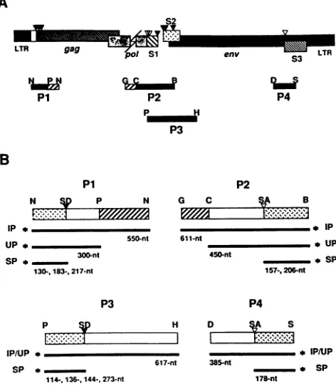
FIG. 2. Northernhybridization analysis of EIAV-infected FDD
cells using site-specific splice donor oligonucleotide probes. (A) Nucleotidesequenceof the EIAV leader RNA coding strand from
theSmaI site through the 5' end of thegag gene, illustrating the
positionsof the threepotential splice donors (arrows). Nucleotides ineach of the potential donor sites which conformtotheconsensus
areunderlined. The sequencesof all of thesynthetic splice donor-specific oligonucleotide probes (SD-1, SD-2, and SD-3) are dis-playedinboxes above theircomplementary sequences.The amino
acid sequence ofthep15 coding region of thegaggeneis shown
below the nucleotide codons. (B) Northern blot hybridization of poly(A)+ mRNA isolated from EIAV-infected FDD cells with each
of the threesplice donor probes. Lanes1to3 correspondtoRNA probed with radiolabeled oligonucleotides SD-1, SD-2, and SD-3, respectively.
isolated from EIAV-infected FDD cellsprobedwith eachof the three 32P-labeled splicedonor probes demonstrate that the 3.5-kb mRNAspeciescanbedetectedonly bytheSD-1 probe (Fig. 2B, lane 1), whichsuggests that sdl is the only splicedonor utilized in this cell type to generate the singly spliced envelopemessage.The 8.2-kbgenomicRNA hybrid-izes nearly equally well to each probe, suggesting consis-tencyin thetechniques employedand efficientlabelingofthe probes. The extremely low abundance ofmultiply spliced transcriptsin this celltype(25)made it difficulttodetermine thepotentialusageof these alternativespliceddonorsinthe generation of this class of RNAs. In an attempt to detect hybridizationof the variousprobestolow-molecular-weight transcripts, the autoradiograph shown in Fig. 2 has been intentionally overexposed. This didnot result in the detec-tion of any clear signal corresponding to EIAV-specific transcriptsof the 1.5-kb size class but rather resultedin the
appearanceofadiffusenonspecificsmearin this sizerange.
In order to more clearly define the role of these three putative splice donors,weused the more-sensitivetechnique of nuclease Si protection.
FIG. 3. Schematic representation of probes used in nuclease Si mapping of transcripts. (A) Schematic diagram of the EIAV proviral
genome, demonstrating the locations of the four DNA restriction endonucleasefragments (P1, P2, P3, and P4) usedasprobes forS1
nuclease mapping of potential splice donor (solid triangles) and
acceptor (open triangles)sites. Solid barsin eachproberepresent
EIAVsequences,while hatched barsindicate M13sequencesused
as tags to distinguish input probe from probe that annealed to
unspliced genomicEIAVRNA. Geneconstructsandpreparation of the probes are detailed in Materials and Methods. Restriction endonuclease abbreviations: N,Narl;P,PvuII; G, BglII; C, NcoI; B, BamHI; H, HindIll; D, DraI;S, ScaI. (B) Schematic
represen-tations ofS1 nuclease mapping restriction endonuclease fragment probes P1 to P4. The locations ofputative splice donor (SD) and
acceptor(SA)sitesareindicated. Thelength of the input probe (IP)
and those portionsof theprobe that areexpected to hybridize to
unspliced (UP)andspliced (SP)EIAVtranscriptsareindicated. The
asteriskrepresents the radiolabeled end of eachinput probe.
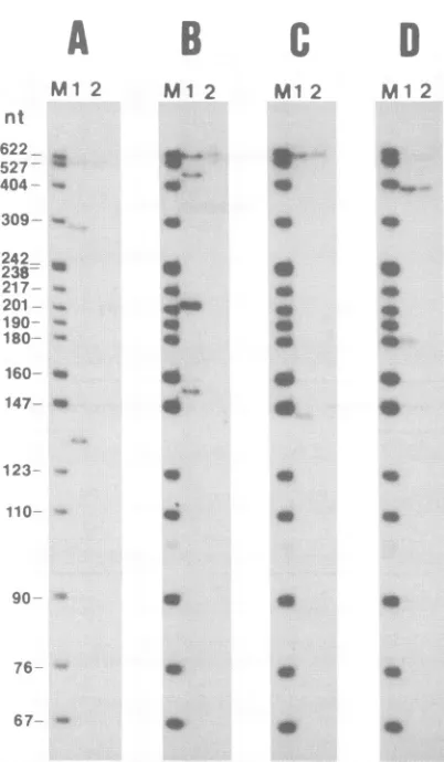
Nuclease Si protection analysis ofspliced EIAV mRNAs. The utilization of the various near-consensus splice donor
and acceptor sites (17, 23) identified within the EIAV
ge-nomic RNAbynucleotide sequenceanalysiswasexamined by nuclease Si protection assays of total cellular RNA
isolated from EIAV-infected FDD cells. 32P-labeled
restric-tionfragmentsof EIAVproviralDNAwere usedasprobes
in these assays (Fig. 3).
Analysis of thesplice donors at the5' end of thegenome
was accomplished by using a 5'-end-labeled 308-bp
TaqI-PvuII proviralrestriction fragment (nt 309 to 620) with an
M13 vectortag sequence at its 3' end (Fig. 3B, P1). When convenient, the probes were constructed with extraneous vector sequences to allow for distinction between input probe and probe annealed to full-length genomic RNA. Hybridization of P1 tospliced mRNAspecies utilizingsdl, sd2,orsd3 wouldyield protected fragmentsof130, 183, or
217nt,respectively. The resultsindicate thatonlya300- and a 130-nt probe fragment were protected from Si nuclease A
B
Nv
pon November 9, 2019 by guest
http://jvi.asm.org/
[image:5.612.48.297.75.359.2] [image:5.612.316.552.81.352.2]A
B
C
M1
2 Ml 2M1
2D
Ml 2
46 4W0.
40
0
0 40
0
0 0
* 0
*0 0
* 40
a *
* 0
*
a.
40 4W
[image:6.612.84.285.71.416.2]67- -x
FIG. 4. Analysis ofdigestion products from nuclease Si
protec-tion studies. Gels A to D correspond to results of Si nuclease protectionassaysoftotal RNAisolated fromEIAV-infected (lanes 1)oruninfected (lanes 2) FDD cells with probes P1toP4,
respec-tively. Molecular weight markers (lanes M) are 32P-labeled DNA restrictionendonuclease fragments fromanMspIdigest of pBR322.
digestion (Fig. 4A, lane 1). These fragments correspond to
probe annealed to full-length EIAV genomic RNA and spliced RNAutilizing sdl, respectively. No protected frag-mentscorrespondingtosd2 and sd3weredetected,
suggest-ingthatthesesequences arenotutilizedassplice donor sites
inEIAV-infectedFDDcells. Thisresult isinagreementwith that ofthe Northern blot experiment employing the differ-ential splicedonoroligonucleotide probes described above. Thatis, inFDD cells, apparently only the first splicedonor in theleader region is utilizedto generatesinglyormultiply
spliced mRNAs.
The splice acceptor sites in thepol-env intergenic region
were mapped with a 3'-end-labeled 450-bp NcoI-BamHI EIAV restriction fragment (nt 4889 to5337) with 161 bp of M13mpl9 sequence tagged toits 5' end (Fig. 3B, P2). This probe wasdesignedto investigate the utilizationof the two near-consensusspliceacceptorsiteslocatednearthe 5' ends
ofORFS1,sal(nt5135),and sa2 (nt 5183).Wehavealready shownbycDNAcloning andnucleotidesequenceanalysisof cDNAclonespSR-1 and pSR-2 thatsal is functional. Probe P2 will allow us to determine whether the second splice acceptor is utilized and what the relative distribution of spliced transcripts employing either acceptor is. Protection
of P2 from Si nuclease digestion by hybridization to full-length genomic DNA is expected to protect a450-ntportion
of the input probe, while hybridization to spliced mRNAs utilizing sal or sa2 would be expected to yield protected fragments of 206 and 157 nt, respectively(Fig. 3B, P2).
The experimental results clearly demonstrate protected
fragmentsrepresentative of all three classes of mRNAs, that is, unspliced genomic and spliced RNAs employing either sal or sa2 (Fig. 4B, lane 1).Althoughequivalent amounts of total cellular RNA were used in each experiment, differences in the autoradiograph signals of the protected fragments for different probes may vary depending on the efficiency of probe labeling and hybridization and on autoradiograph exposure time. However, within a given experiment, it is possible to draw quantitative conclusions aboutthe relative abundance of transcripts employing the individual splice sites. The intensity of the probe fragment protected by mRNAs utilizing sal is much greaterthan it is with mRNAs employing sa2, suggesting that mRNAs spliced at sa2 repre-sent a minor population in EIAV-infected FDD cells. Fur-thermore, the sal-protected probe fragment is considerably more intense than that protected by the full-length genomic RNA, which is in agreement with our previously reported result that the 3.5-kb mRNA species is the predominant viral RNA in EIAV-infected FDD cells (25).
The third probe (Fig. 3B, P3) was designed todetermine the role of potential splice donor sites within the pol-env intergenic region in the formation of multiply spliced mRNA transcripts. This probe was generated from a 617-bp 5'-end-labeledPvuII-HindHII EIAV proviral restriction fragment (nt 5162 to 5775). There are four near-consensus splice donors localized from the 3' end of ORFS1to the 3' end of ORF S2 at nt 5276, 5298, 5306, and 5435 (Fig. 3A). RNA species utilizing these donor sites would be expected to yield pro-tected fragments of 114, 136, 144, and 273 nt, respectively. In fact, only the 144-nt protected probe fragment could be detected (Fig. 4C, lane 1), suggesting that only the splice donorsequence localized to nt 5306 is utilized in FDD cells for thegeneration of multiply spliced mRNAs. This finding was somewhat surprising, in that multiply spliced ORF
Sl-ORF S3 RNAs isolated from canine fibroblasts were
shown to involve splicing from the donor site immediately downstream of ORF S1 at nt 5276 (30). We were unable to detect a protectedfragment 114 nt in length corresponding to asimilar splicing event in FDD cells. It should be noted that the design of probe P3 from thePvuII site prevents detection ofmultiply spliced mRNA species, which are produced by splicing events involving sa2, since the labeled portion of the probe would be unable to hybridize to such messages and would therefore be digested by nuclease S1. Therefore, we cannot conclude that the other splice donor sequences within this region are not utilized to generate multiply spliced messages. Further study isrequired to determine the role of sa2 in both singly andmultiply spliced RNAs.
ThefinalS1protection probe (P4) was designed to inves-tigate the potential usage of a splice acceptor site at the 5' end of ORF S3 (sa3, nt 7234), which may result in the provision of an AUG translational initiation codon for the protein product encoded by this ORF. Probe P4corresponds to a 3'-end-labeled-385 bp DraI-ScaI proviral restriction fragment (nt 7026 to 7412). This probe is expected to yield a 385-ntfragment when annealed to full-length genomic RNA and a 178-nt fragment when hybridized to spliced mRNA utilizing sa3. TheSi mapping results show the presence of the 178-nt protected fragment, indicative of usage of sa3.
Thenuclease S1 protection experiments described herein nt
622
527-404
309-242 .U
238-217 -201
-9o-0 180
160- us
147-- u
123
110
90
76
on November 9, 2019 by guest
http://jvi.asm.org/
demonstrate the utilization ofa
single
splicedonorsitethatprovides
the EIAV leader sequence to all spliced RNAs. Bothsplice
acceptorsites within the 5' end of ORFSi
arefunctional; however,
there isa strongbias towards splicingat the first site in FDD cells. Of the four splice donors
located in the
pol-env
intergenic region, only the third (nt5306)
wasused in FDD cells inthese experiments. Finally,the
splice
acceptorsitenearthe 5' border of ORFS3 is used toproduce presumptive multiply
spliced RNAs.Assays of trans-activation activity. As previously stated,
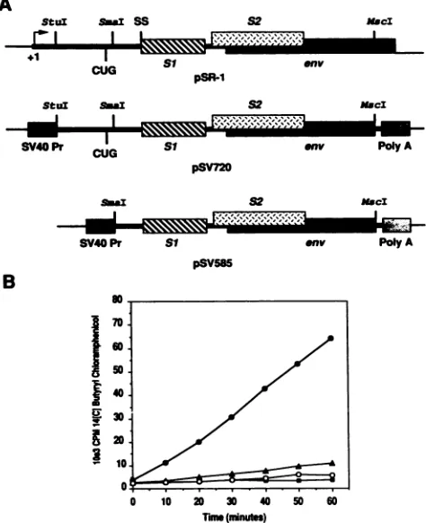
the
pSR-1
cDNAclonewehave characterized,whichcorre-sponds
to thesingly spliced
3.5-kb EIAVmRNA, contains the ORFSi,
ORFS2,
andenvgenes(Fig. 1C).Since ORFSiencodes theviraltransactivator EIAV Tat(8, 20),we were interestedtoascertain whether thepSR-1cDNA clone could direct the
synthesis
ofthe Tatproteinin cultured FDD cells. Towards thisend,restrictionfragments
ofthepSR-1cDNA sequenceweresubcloned into aeucaryotic expressionplas-mid,
pSV2Acat.
Twosubclones,
pSV720andpSV585,which differ in thatpSV585
lacks cDNA sequences between theStul
(nt 256)
restrictionsitelocated intheterminal redundant(R) region
and theSmaI(nt 393)
restrictionsitelocated in the leaderregion,wereconstructed(Fig.SA).
TheCUGcodon,which has been
proposed
toinitiate EIAV Tatsynthesis,lies betweenthesetworestriction endonucleasecleavagesitesat nt373(8, 30).
ThepSV720
andpSV585 expression
clonesare 3'coterminal, extending
tothe MscI(nt 5655)
restriction site within theenvgene. IfindeedEIAVTatsynthesisis initiated atthesuggested
CUGcodon,
thenpSV720
wouldcontain all the necessarygeneticinformation for EIAV Tatexpression,while
pSV585
wouldlack the translation initiation codon for thisprotein.
FDD cellmonolayers
were cotransfected with eitherpSV720
orpSV585
andaplasmid containing
theCAT reporter geneunder the controlof the EIAVLTR, pLTRcat(25).
Cotransfection of thepSV720 expression plasmid
withpLTRcat
clearly
results in trans activation of the EIAVLTR,
asevidencedby
anincrease in CAT enzymeactivity
of more than 60-foldcompared
with thatresulting
from trans-fection with thepLTRcat
plasmid
alone(Fig. SB).
Cotrans-fection ofpSV585
resulted innosignificant
trans activation of theLTR,
suggesting
that sequencesresiding
between theStuIsite within the EIAV R
region
of the LTR and theSmaIsite within the
leader,
which include the suggested CUG translational initiationcodon,
are absolutely required forexpression
of EIAV Tat from this cDNA clone in cultured FDD cells.Invitro
expression
ofORFSi,
ORFS2,
andenvgenes fromthe3.5-kbEIAV mRNA. Thetranslational
coding potential
ofthe
pSR-1
cDNA clone derived from the EIAV 3.5-kbtranscript
wasinvestigated by employing
invitrotranscrip-tion and translatranscrip-tion
techniques.
The presenceof three ORFs inthis cDNAsuggested
thepossibility
that the parent3.5-kb mRNA is tricistronic in nature,i.e., capable
ofencoding
EIAV
Tat,
the ORF S2protein,
and thegpi35
envelope
glycoprotein.
The SP6-basedinvitrotranscription plasmids
pSP720
andpSP585
areanalogous
totheSV40-based eucary-oticexpression
plasmids
pSV720
andpSV585
used in the trans-activation assays described above. Thatis,
these in vitrotranscription
plasmids
differonly
in thatpSP720
con-tains
pSR-1
cDNAsequencesstarting
from the StuI(nt 356)
restriction site in the R
region
of theLTR,
whilepSP585
contains cDNA sequences from theSmaI
(nt 393)
restriction site in the viral leaderregion.
The cDNA sequences of bothplasmids
are 3'coterminal,
ending
at the MscI(nt
5655)
restriction site located within the
gp9O
coding region
of theenvgene.
A
Stul Sa SS S2 maci
+1 I L S1J
CG Si
enlv
pSR-1
Stul s;au S2 m*ci
SV40 Pr CUG Si env Poly A
pSV720
smau
I
=
SV40Pr Si
pSV585
52
MaI
env PolyA
B
I
aU
I
0 10 20 0 40 50
Tin(minnute)
60
FIG. 5. Analysis of trans-activatedEIAV LTR-driven CAT en-zyme activity in transfected FDD cells. (A) Schematic representa-tionof cDNA from pSR-1corresponding to the singly spliced 3.5-kb EIAV mRNA. The positions ofrelevant restriction endonuclease sites forsubcloningand thepresumptive CUG translational initia-tioncodon of theSi EIAVTatprotein areindicated. Eucaryotic expression plasmidspSV720 and pSV585 were generated by sub-cloning either a 720-bp StuI-MscI or a 585-bp SmaI-MscI DNA restriction fragmentintoStuI-digestedpSV2Acat,respectively. This expression vector provides the SV40 immediate-early promoter (SV40 Pr) and polyadenylation signal (Poly A) for expression of cloned inserts in eucaryotic cells. (B) Graphical representation of theconversion of
[14Clbutyryl
coenzyme A to [14C]butyryl chlor-amphenicol by incubation with transfected FDD cell lysates as a function oftime. A controlplasmid,pSVO-gal,
was included in all transfectionssothatcelllysates could be normalized to 3-galactosi-dase activity to control for transfection efficiency. Solidsquares, pLTRcat alone; solid triangles, pSV2cat; solid circles, pLTRcat cotransfected with pSV720; open circles, pLTRcat cotransfected with pSV585.ThepSP720 and pSP585plasmidDNAs wereemployedas transcription templatestoproduce synthetic capped RNAs, whichweretranslated in wheat germextractsinthe presence
of[3H]leucine. Invitro translation ofpSP720RNAresulted
in the synthesis of three major polypeptide products with
relative molecularsizes of15, 8.5,and 7 kDa(Fig. 6A,lanes
1and8). These products correspond closelytothepredicted sizes of MscI-truncated gp9O envelope protein, the EIAV
Tatprotein, and theputative ORF S2protein,respectively.
ThepSP585RNAsupportedtranslation ofonlytwoofthese
products, the 15- and 7-kDa polypeptides (Fig. 6A, lane5).
The 15-kDa product from both pSP585 and pSP720 RNA-programmed translation reactions wasspecifically immuno-precipitated withamonoclonalantibodydirectedagainstan
epitope of the gp9O envelope protein mapping to a region
on November 9, 2019 by guest
http://jvi.asm.org/
[image:7.612.319.556.80.371.2]A
Kd
43
2 3 4 7 XI I s
29
18.4 14.3 t
(>-)
*w
s
*
^
w
6.2~ ~ ~ T gn.
3
4
S
B
K( NI I
43
29
18.4.
14.3
6.2
FIG. 6. In vitro translation andradioimmunoprecipitation
analy-sis ofpSP720andpSP585RNAs.(A) FluorographofanSDS-PAGE
analysisof wheatgerm extractsprogrammedwithpSP720(lanes1 and8)andpSP585 (lane 5) synthetic transcriptsin thepresenceof
[3H]leucine. Radioimmunoprecipitation of labeled translation
prod-uctswasdone withamonoclonalantibodydirectedagainstthegp9O envelope glycoprotein (lanes2 and6)and rabbit polyclonal
antise-rum specific to the Si (lane 3) or S2 (lanes 4, 7, and 9) protein products. (B) Immunoprecipitation of the S2 proteinwith
EIAV-infectedhorsesera.Invitrotranscripts producedfrompSP585were
translatedin wheatgermextractsinthepresenceof[35S]methionine (lane 1)andimmunoprecipitatedwith either 4or20 ,ug (lanes2and
3, respectively) of partially purified IgG from a pony naturally infected with EIAV. Molecular size markers (lanes M) are BRL
"4C-labeledlow-molecular-mass standards.
approximately 50 amino acids from its amino terminus (3) (Fig. 6A, lanes 2 and 6). The 8.5-kDa band specific for pSP720 RNAtranslation could beimmunoprecipitated by a
rabbit polyclonal antiserum produced against a synthetic peptide corresponding to a predicted immunoreactive
do-main of the EIAV Tatprotein (Fig. 6A, lane 3). The 7-kDa proteincommon toboth pSP720 andpSP585 RNA transla-tion reactransla-tionswasimmunoprecipitated byacombination of
low-titer rabbit polyclonal antisera produced against four differentoverlapping synthetic peptides that spanthe entire
ORFS2amino acidsequence (Fig. 6A, lanes 4and7). This combination of ORF S2 antisera cross-reacted with the EIAV Tat protein product (Fig. 6A, lane 4). However, a
high-titerORF S2antiserum obtainedmorerecently
demon-stratedno cross-reactivity withEIAVTat(Fig. 6A, lane 9).
This ORF S2 antiserum also
specifically precipitates
two minorproducts
with mobilities of 5.5 and 4kDa,
which arepresumed
to be eitherS2-related
peptides produced
by
translationalinitiation at the internal AUG codons of S2or
degradation
products
ofS2. These data indicate thatasingle
cDNA
species
corresponding
to the 3.5-kbsingly
spliced
mRNAof EIAV is
capable
ofdirecting
thesynthesis
of the EIAVTat,theS2protein,
and theenvelope protein
in vitro.Furthermore,sincenoEIAV Tat
protein product
is detectedupon translation of
pSP585 RNA,
sequences located be-tweentheStuI site in the Rregion
and theSmaIsite in theleader,which include the
predicted
CUG initiation codon ofEIAVTat, are
required
for in vitroexpression
of the EIAV Tatprotein.
Humoral immune responsetotheORF S2
protein
in EIAV-infected horses. Thepredicted
protein product
of ORF S2bearsno
significant
sequencehomology
toanyknown lenti-viralprotein,
andtodate,
noprotein product
hasbeen shown tobe encodedby
ORF S2 invivo. Inan effortto establishwhetheranORFS2-encoded
polypeptide
isproduced
during
productive EIAV
infection,
we have examined sera fromEIAV-infected horses for the presence ofantibodies tothe
putativeORF S2 gene
product.
We haveaccomplished
thisgoal by
examining
the abilities ofthesesera torecognize
thein vitro translated
product
in animmunoprecipitation
reac-tion andby
testing
thereactivity
ofapanel
of horseserato ORFS2-specific synthetic peptides
in ELISAs.The ORF S2
polypeptide
and aportion
of thegp9O
envelope
protein
weresynthesized
in wheat germ extract from asynthetic
RNA similar to thatproduced by
thepSP585
plasmid
described above. Thetranscription template
in this
experiment
differed frompSP585
in that itincluded sequences from the MscI(nt
5655)
to the first HindIII(nt
5775)
restriction endonucleasedigestion
sites in the envgene. This additional
envelope
sequenceresults in the syn-thesis ofatruncatedgp9O
protein
withanapparentmolecular size ofapproximately
20 kDaonSDS-polyacrylamide gels.
The 35S-labeled in vitro translationproducts
wereimmuno-precipitated
withseraobtained fromanEIAV-infected horse followedby
SDS-polyacrylamide
gel
electrophoresis
(PAGE) analysis.
Analysis
of the in vitro translationprod-ucts shows the
synthesis
of the 20-kDa truncatedenvelope
protein
andthe 7-kDa ORF S2polypeptide (Fig. 6B,
lane1).
These labeled
products
wereimmunoprecipitated
with 4or 20 ,ug ofpartially purified IgG
obtained from theserumofan EIAV-infected horse(Fig.
6B, lanes 2 and3,
respectively).
Asone would
anticipate,
the truncatedgp9O
envelope
pro-tein reactsstrongly
with theEIAV-infected horseIgG
frac-tion, particularly
atthehigher
concentration(Fig. 6B,
lane3).
The ORF S2protein
wasclearly
immunoprecipitated by
the EIAV-infected horseIgG
at thehigher
concentration(lane 3)
andfaintly precipitated
at the lower concentration(Fig.
6B,lane2).
Neither the truncatedgp9O
northe ORFS2polypeptide
demonstrated anyreactivity
with control sera from normal uninfected horses(data
notshown).
Further-more, ELISAs of a standardpanel
of horse immune sera with ORFS2-specific synthetic peptides
showed that 60to 75%of the horsesproduced
antibodies that reacted with thepeptides (data
notshown).
These dataindicate that the ORF S2protein
isexpressed during productive
EIAVinfection,
since EIAV-infected horses are
capable
ofmounting
hu-moral immune responses directedagainst
thisprotein.
Amino-terminal sequencing of the in
vitro-synthesized
EIAVTat andORF S2polypeptides. Inanattemptto
gain
a betterunderstanding
of theexpression
of the ORFSi,
ORFS2,
andenvelope
proteins
from thetricistronic 3.5-kbRNA,
on November 9, 2019 by guest
http://jvi.asm.org/
A
U
0.
.1%
C,)
ooo
B
300
c
0. oo
0
C 2
2'
!' 100
GLFGKGVTWSASHSMGGSOGESQ
S2 Amino Acid Residue
AD RR I PGTAEENFQKSSG
SI AminoAcidResidue
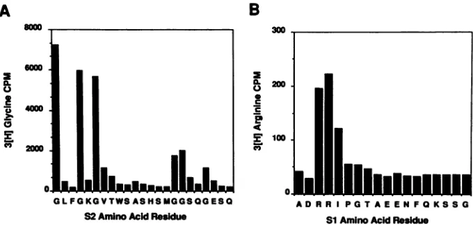
FIG. 7. Amino-terminalsequence analysis of the S2 and S1 proteins. In vitro-translated S2 protein produced in the presence of[3H]glycine in wheat germ extracts programmed with pSP585 RNA was purified from an SDS-polyacrylamide gel and subjected to amino-terminal sequencing. Amino acid fractions in 100% acetonitrile were directly placed in Liquiscint liquid scintillation solvent and assayed for radioactivity.The S1 protein was prepared in a similar manner by translation of pSP720 in vitro transcripts in the presence of[3H]arginine. The amino acid sequences of the predicted S2 and S1 polypeptides are aligned beneath the peaks of radioactivity assayed from the automated Sequenator.
wedetermined thepresumptive translation initiation sites of
the in vitro-synthesized ORF Si and ORF S2 proteins by amino-terminal sequencing of radiolabeled polypeptides. Theradioactive amino acidof choice forsequencing of the ORFS2 proteinwas [3H]glycine, sincethe first methionine codon inORF S2is followedimmediately byaglycine codon
and since three of the first seven codons correspond to glycine residues. The ORF Si protein was labeled with [3H]arginine, since translational initiation at the proposed CUG codon (8, 30) would place two contiguous arginine residuesneartheamino terminus.
TheORF S2productwassynthesizedfrompSP585 RNA,
and approximately 7 x 10⁴cpm ofgel-purified proteinwas subjected to amino-terminal sequence analysis. The
se-quencerfraction correspondingto thefirst amino acid resi-dueregisteredinexcessof7 x 103cpm,whilefurtherpeaks ofradioactivitywereobserved for aminoacidresidues 4 and 6 (Fig. 7A). Theanticipated aminoacid sequence of the S2 polypeptide initiating at the first methionine codon in this ORF is MG L F G K G.Assumingthatthe aminopeptidase activityof the wheatgermextract has cleaved the initiator methionine residue from this protein, thepeaks of radioac-tivity occurpreciselyasexpected, that is, in fractions 1, 4,
and 6. Additional peaks of radioactivity were noted in
fractions 16, 17, and 20, which are also predicted sites of glycine residues in theORFS2protein.These data confirm that the in vitro-synthesized polypeptide assigned to be encodedbyORFS2onthebasis of its abilitytospecifically react with ORF S2-specific peptide antisera is indeed the ORF S2 gene product and that its synthesis in vitro is apparently initiated at the first methionine residue in this ORF.
Thesequence of theORF Si protein, EIAVTat,was of particular interest, since it has been suggested that this proteinmayinitiate translationataCUG codon. Of thetwo CUG codons located withinthe leaderregion,theone more
downstream (nt 373) lies in the most favorable sequence
contextfor translation initiation(12). Initiationof EIAV Tat at this CUG codon would place two consecutive arginine codonsnearits amino terminus. The EIAV Tatproteinwas immunoprecipitated from pSP720 RNA-directed in vitro
translationproducts labeled with
[3H]arginine
andsubjectedto amino-terminal sequencing. Isolation by
immunoprecipi-tationwasnecessary topurifyEIAVTat from thesimilarly sized ORF S2protein as well as from a number of comigrat-ing wheat germ proteins, which become amino terminally
modifiedwith labeledarginine byanarginine-terminal
trans-ferase activity found in wheat germ extracts (9). Although wewereable to purifyrelatively large amountsof
[3H]argi-nine-labeled EIAV Tat protein, initial amino-terminal se-quenceanalysis attempts resulted in no distinctive sequence information, suggesting modification of the amino-terminal amino acid residue that prevented the Edman degradation
reaction. Toovercome thisproblem,wecarried out
transla-tion foronlyashort time(30 min)andimmediately boiled the translation reaction mixture after the incubation,
anticipat-ing that suchtreatmentcouldyieldafractionof the synthe-sized
Si
protein in an unmodified state. Amino-terminalsequencingof 5 x 10⁴ cpmofEIAVTatproteinpreparedin
this manner resulted in two clear consecutive peaks of radioactivity, each of approximately 2 x
102
cpm,corre-spondingto the third and fourth residues of thepolypeptide
(Fig. 7B). Thepositions of thesepeaks are consistent with
translational initiation at the previously proposed CUG codon,presumingaminopeptidaseprocessingof theinitiator methionine residue. On the basis of these findings, we proposethat the amino terminus of the invitro-synthesized
EIAV Tat protein is blocked by an unknown functional group and that limited translation time followed by heat treatment allowed us to capture a fraction of the newly
synthesizedTatprotein in anunblocked form.
DISCUSSION
EIAV RNA splicing in FDD cells. Although a complex
pattern
of RNA splicing has been observed in cultured canine fibroblastspersistentlyinfected with avirulent EIAV(21, 32), considerably less complex patterns have been
observedin cells ofequineorigin. In
primary
horse macro-phage cultures infected with the horse-virulentWyoming
strain of EIAV,
only
the 8.2-kbfull-length
genomic
and 3.5-kbsinglysplicedenvelopeRNAsweredetected(30).
WeI
1111
on November 9, 2019 by guest
http://jvi.asm.org/
[image:9.612.138.478.76.238.2]have previously reported similar results for primary equine fibroblasts (FDD) infected with avirulent cell culture-adapted EIAV (25). In this report, we have analyzed the EIAVsplicingpatterninFDDcellsand examined thecoding potential of the major 3.5-kb EIAV transcript produced in FDDcells.
The3.5-kb EIAV transcript(s) of FDD cells can be pro-duced by splicing of the 243-nt leaderregion from a unique splice donor (sdl, nt 459) to either oftwo functional splice acceptors(sal [r4 5135] and sa2[5184]) localized within ORF Si. The Si nuclease protection studies described here indi-catethat the first of thesetwo acceptorsites is preferentially utilized in FDD cells. Nucleotide sequence analysis of cDNA clones clearly demonstrate the involvement of this splicingeventintheproductionof the major 3.5-kb RNA of FDDcells. This splicingeventis identicaltothefirstof three splicing reactions in a previously defined ORF Si-ORF S3
RNA, whichwas showntoproduce functional trans-activa-tor protein encoded by ORF Si (22, 30). Since the major 3.5-kb singly spliced RNAin FDD cells contains the same
ORF Si sequences as this previously identified multiply spliced message, it follows that the 3.5-kb transcript may also be abletoproduce the EIAVTattrans-activator protein encoded by ORF Si.InadditiontoORF Si,this 3.5-kbRNA alsoincludes theoverlappingORF S2 andenv genes.
A second class of 3.5-kb transcripts canbe produced by
splicing the leader to the second acceptor located within ORF Si (sa2 [nt 5184]). RNAs of this class would not be expected to produce functional EIAV Tat protein, since
sequences shown to be critical for trans activation reside between the two alternative splice acceptor sites (8) and thereforewouldnotbepresentin RNAs splicedatsa2.Such RNAswould, however, contain the entire ORF S2 andenv
genes. It appears highly unlikely that any envelope RNA
which doesnotalso contain ORFS2 will be detected, since thereareonly 26ntseparating the presumptiveAUG initia-tion codons of S2 and the gpi35 envelopegeneproduct, and
no reasonable splice acceptor sequences lie in this region.
Indeed, we were unable to detect in FDD cells any
func-tional splice acceptor sites that map to this region by Si nuclease protection analysis. Therefore, singly spliced 3.5-kb RNAs involving sa2 may be bicistronic in nature,
encoding the S2 andgpi35 proteins in amanner similar to
that by which the HIV-1 Vpu and gpi60 proteins are
encoded (28).
Thepossibility remains thateither of the identified splice acceptorsites(sal andsa2) couldbeinvolved in the produc-tion of multiply spliced transcripts. Of the four putative splicedonorsequences in the pol-env intergenicregion,only
one,located between theORFS2 and theenvAUG initiation codons, was detected in these studies to be functional in FDDcells. The splice acceptor site (sa3)near the 5' end of
ORF S3 is also functional in these cells, suggesting the possibility that the ORF S2 AUG is splicedtoORF S3, thus providing atranslational initiation for S3 expression.
How-ever, directsplicing of the splice donor within ORF S2(nt 5306) totheORF S3 acceptorsite (nt 7235)wouldnotresult
inproperalignment of theS2 AUG with theS3 coding frame.
Only two additional codons, neither of which is an AUG,
would be placed in frame with S3. The third exon of the
triply spliced ORF Si-ORF S3 RNA identified in
EIAV-infected caninecells consists ofashortstretch ofsequence
fromtheenvgenewhich appears toprovide atranslational
initiation codon toORF S3 (30). Itremainspossible that an
exon within the env coding region provides a stretch of
amino acid-coding potential allowing for proper alignment of S3 with the S2 AUG.
Translational potential of the
major
3.5-kb transcript.The major 3.5-kb EIAV transcript of FDD cells is shown here to be tricistronic in vitro, encoding theprotein productsof the ORF Si, ORF S2, and env genesin the wheat germcell-free translation system. Amino-terminal sequence analysis of in vitro-synthesized EIAV Tat is consistent with initiation at a CUG codon (nt 373), which lies in an otherwise favorablesequence context for translational initiation, AAC CUG G, in which underlined nucleotides match the consensus se-quence for translation initiation (12). We have shown EIAV Tat synthesis from cDNA sequences corresponding to the 5' end of the 3.5-kb transcript in vivo, as evidenced by a 60-fold increase in CAT expression directed by the EIAV LTR. The relatively low levels of trans activation observed for EIAV compared with those reported for HIV may reflect differ-ences in the amount of trans-activator protein produced as a result of initiation at the suboptimal CUG codon.
Serum samples from EIAV-infected horses are immuno-reactive with both in vitro-synthesized S2 protein and syn-thetic peptides corresponding to portions of S2. Characteri-zation of viral envelope variants propagated from horse macrophage cultures infected with horse-virulent EIAV has shown that although a number of mutations arise throughout
thegp9O-coding sequence and in the major coding exon for
the S3 protein, no mutations occur in ORF S2 (1). Taken together, these data strongly indicate a role for the ORF S2 gene product during EIAV infection, although no function for this protein has yet been defined.
Although a number of singly spliced HIV-1 transcripts contain multiple ORFs (28, 29, 31), only the vpu-env (28) transcripts are known to act as multicistronic transcripts in vivo. TheHIV-1 transcript encoding the 72-amino-acid form of theTatprotein, Tat-1, is structurally similar to the major EIAV3.5-kbtranscript described here in that it contains the tat-i, vpu, and env genes in positions nearly identical to those of the EIAV tat, ORF S2, and env genes (28). This HIVtranscript has been shown to be monocistronic in vivo, producing only theTat-1 protein, presumably because of the relatively strong translation initiation codon of the Tat protein (GAA AUG G). Similarly, infrequent translation initiation of EIAV Tat from an inefficient non-AUG codon and ofthe S2 protein from an AUG codon in a relatively weaksequence context (UAU AUGG)may allow significant leakyribosomal scanning(13) to thehighly favorable gp135
AUG(AACAUG G) of the major 3.5-kb transcript(sdl-sal).
During thepreparation of this paper, a report which seems tosuggesttranslational initiation of EIAV Tat in transfected caninefibroblasts from an AUC codon (nt 388) upon deletion of sequences including the CUG (nt 373) initiation codon identified here was published (22). This result is somewhat surprising, since this AUC codon
(AGG
AUCC) lies in a less favorable context for translation initiation than does the CUGcodon (AACCUG G). It is possible that deletion of the morefavorable CUG could allow synthesis at the next most favorable codon, the AUC. Clearly, future studies involving site-specificmutagenesis of each codon and its surrounding sequence are required to determine which is the actual translation initiator in EIAV-infected cells.ACKNOWLEDGMENTS
Wethank C. Issel for providing equinefibroblast cells and virus. We thank J. Ball and M. Miller for peptide synthesis, peptide ELISAs, and preparation of some of the antisera used in these studies.
on November 9, 2019 by guest
http://jvi.asm.org/
This research was supported in part by funds provided by the LouisianaAgricultural Experiment Station and Public Health Ser-vicegrantCA49296.
REFERENCES
1. Alexandersen, S., and S. Carpenter. 1991. Characterization of variable regions in the envelope and S3 open reading frame of equine infectious anemia virus. J. Virol. 65:4255-4262. 2. Anderson, D. J., and G. Blobel. 1983. Immunoprecipitation of
proteins from cell-free translations. Methods Enzymol. 96:111-120.
3. Ball, J. M. 1990. Ph.D. thesis. Louisiana State University, Baton Rouge.
4. Chirgwin, J. M., A. E. Przybyl, R. J. MacDonald, and W. J. Rutter. 1979. Isolation of biologically active ribonucleic acid from sources enriched inribonuclease. Biochemistry 18:5249-5299.
5. Chua, N.-H., S. G. Bartlett, and M. Weiss. 1982. Preparation and characterization of antibodies to chloroplast proteins, p. 1063-1080. In M. Edelman, R. B. Hallick, and N.-H. Chua (ed.), Methods in chloroplast molecular biology. Elsevier Bio-medicalPress, Amsterdam.
6. Cullen, B. R. 1991. Humanimmunodeficiency virus as a proto-typic complex retrovirus. J. Virol. 65:1053-1056.
7. Derse, D., P. L. Dorn, L. Levy, R. M.Stephens,N. R. Rice, and J. W. Casey. 1987. Characterization of the equine infectious anemia viruslong terminalrepeat.J.Virol. 61:743-747. 8. Dorn,P.,L.DaSilva,L.Martrano, and D. Derse.1990.Equine
infectious anemia virustat:insights into thestructure,function, and evolution of lentivirus trans-activator proteins. J. Virol. 64:1616-1624.
9. Elias,S., and A.Ciechanover. 1990. Post-translational addition of an arginine moiety to acidic NH2 termini of proteins is required for their recognitionbyubiquitin-protein ligase.J. Biol. Chem. 265:15511-15517.
10. Fordis, M., and B. H. Howard. 1987. UseoftheCAT reporter genefor theoptimizationof gene transfer intoeucaryoticcells. MethodsEnzymol.151:382-397.
11. Gorman, C. M., L. F. Moffat, and B. H. Howard. 1982. Recombinant genomes which express chloramphenicol acetyl transferaseinmammaliancells. Mol. Cell.Biol.2:1044-1051. 12. Kozak, M. 1986. Pointmutations defineasequenceflankingthe
AUG initiator codon that modulates translation by eukaryotic ribosomes. Cell 44:283-292.
13. Kozak, M. 1989.Thescanning model for translation: anupdate. J. CellBiol. 108:229-241.
14. Laemmli,U. K.1970.Cleavage ofstructuralproteinsduringthe assembly ofthe head ofbacteriophage T4. Nature (London) 227:680-685.
15. Malmquist, W. A., D. Barnett, and C. S. Becvar. 1973. Produc-tion ofequine infectious anemia antigen in a persistently in-fected cell line. Arch.Virol. 42:361-370.
16. Maniatis, T., E. F.Fritsch,andJ.Sambrook. 1982. Molecular cloning: alaboratorymanual. ColdSpringHarborLaboratory, ColdSpring Harbor, N.Y.
17. Mount, S. M. 1982. A catalogueof splice junction sequences. Nucleic AcidsRes. 10:459-472.
18. Narayan, O., and J. E. Clements. 1990. Lentiviruses, p. 1571-1589. In B. N. Fields and D. M. Knipe (ed.), Virology, 2nd ed. Raven Press, New York.
19. Neumann, J. R., C. A. Morency, and K. 0. Russian. 1987. A novel rapid assay for chloramphenicol acetyltransferase gene expression. BioTechniques 5:444-447.
20. Noiman, S., A. Gazit, 0. Tori, L. Sherman, T. Miki, S. R. Tronick, and A. Yaniv.1990.Identification of sequences encod-ing the equine infectious anemia virus tat gene. Virology 176: 280-288.
21. Noiman, S., A. Yaniv, L. Sherman, S. R. Tronick, and A. Gazit. 1990. Pattern of transcription of the genome of equine infectious anemiavirus. J.Virol.64:1839-1843.
22. Noiman, S., A. Yaniv, T. Tsach, T. Miki, S. Tronick, and A. Gazit.1991.TheTatproteinofequine infectious anemiavirus is encodedbyatleast three types oftranscripts. Virology 184:521-530.
23. Ohshima, Y., and Y. Gotoh. 1987.Signals fortheselection of a splicesite inpre-mRNA. Computer analysisofsplice junction sequencesand like sequences. J. Mol. Biol. 195:247-259. 24. Orrego, A., C. J. Issel, R. C. Montelaro, and W. V. Adams, Jr.
1982. Virulence and in vitro growth of acell adapted strain of equine infectious anemiavirus after serial passage in ponies. Am. J. Vet. Res. 43:1556-1560.
25. Rasty,S., B. R. Dhruva, R. L. Schiltz, D. S. Shih, C. J. Issel, and R. C. Montelaro.1990.ProviralDNAintegrationand transcrip-tionalpatternsofequine infectious anemiavirusduring
persist-entandlytic infection.J. Virol.64:86-95.
26. Rushlow, K. E., K. Olsen, G. Stiegler, S. L. Payne, R. C. Montelaro, and C. J. Issel. 1986.Lentivirus genomic organiza-tion: thecomplete nucleotidesequenceof theenvgeneregionof equineinfectious anemia virus. Virology155:309-321. 27. Sanger, F., S. Nicklen, and A. R. Coulson. 1977. DNA
sequenc-ing with chain-terminating inhibitors. Proc. Natl. Acad. Sci. USA 74:5463-5467.
28. Schwartz, S.,B. K.Felber, E. Fenyo, and G. N.Pavlakis.1990. Env and Vpu proteins ofhuman immunodeficiencyvirus are
producedfrommultiplebicistronic mRNAs. J. Virol. 64:5448-5456.
29. Schwartz, S., B. K. Felber, and G. N.Pavlakis.1991.Expression of humanimmunodeficiencyvirus type 1 vif and vpr mRNAs is rev-dependentandregulated by splicing. Virology183:677-686. 30. Stephens,R.M.,D.Derse,and N. R. Rice. 1990. Cloningand characterization of cDNAsencoding equine infectious anemia virusTatandputative Revproteins.J. Virol.64:3716-3725. 31. Vaishnav, Y. N., and F.Wong-Staal. 1991. Thebiochemistryof
AIDS. Annu. Rev.Biochem.60:577-630.
32. Yaniv, A., L. Sherman, S. Noiman, 0. Tori, H. Lichtman-Pleban, T. Miki, S. R. Tronick, and A. Gazit. 1989. Studieson
theregulation and patterns ofexpression of the equine infec-tious anemia virus genome, p. 59-73. In D. Gaudy and W. Hennessen (ed.), Developments in biological standardization: progressin animalretroviruses,vol. 72. S.KargerAG,Basel.