Analysis of Efficiently Packaged Defective Interfering RNAs of
Murine
Coronavirus: Localization of
a
Possible
RNA-Packaging Signal
SHINJI
MAKINO,1.2*
KYOKO YOKOMORI,' AND MICHAEL M. C. LAI'Howard Hughes Medical Institute and Department of Microbiology, University of Southern California Schoolof Medicine, Los Angeles, California 90033,1 and Department ofMicrobiology,
The University of TexasatAustin, Austin, Texas 787122* Received 18 May 1990/Accepted 14September 1990
We have previously shownthatmostof the defective interfering (DI) RNAofmousehepatitis virus (MHV) arenotpackaged into virions. Wehave nowidentified, after 21 serialundilutedpassagesofMHV,asmall DI RNA, DIssF, which is efficiently packaged into virions. The DIssFRNAreplicatedata highefficiencyonits transfection into thehelpervirus-infectedcells. The virusreleasedfromthetransfected cells interferedstrongly with mRNA synthesis and growth of helper virus. cDNA cloning andsequenceanalysis of DIssFRNArevealed
that it is 3.6 kb and consists ofsequences derived from five discontinuous regions of the genome of the
nondefective virus. The first four regions (domains ItoIV) from the 5' end arederived fromgene 1, which presumably encodes the RNA polymerase of the nondefective virus. Theentiredomain I (859nucleotides) and the first750 nucleotides of domainIIarealso present inapreviously characterized DIRNA,DIssE, which is notefficiently packaged into virions. Furthermore, the junction between thesetwodomains isidentical between the two DI RNAs. The remaining 77 nucleotides at the 3' end of domain II and all of domains III (655
nucleotides) and IV (770 nucleotides)arenotpresentinDIssE RNA.Thesefour domainsarederived fromgene
1. In contrast,the3'-mostdomain (domain V,447nucleotides) is derived from the 3' end of thegenomicRNA
and is also present in DIssE. Thecomparisonofprimarysequencesandpackaging propertiesbetween DIssE and DIssF RNAssuggestedthat domains IIIandIVand part of the 3' end of domain II contain thepackaging signalfor MHV RNA. Thisconclusionwasconfirmed by inserting theseDIssF-uniquesequencesintoaDIssE cDNA construct;the invitro-transcribedRNAfromthishybridconstructwasefficiently packagedintovirion
particles.DIssF RNAalso containsanopenreadingframe,whichbeginsfrom domain Iand ends at the5'-end 20bases of domain III. In vitro translation of DIssFRNA and metaboliclabeling of the virus-infected cells showed thatthisopenreading frameisindeed translatedintoa74-kDaprotein.Thestructuresof both DIssE and DIssF RNAs suggestthata protein-encoding capabilityisacommoncharacteristic of MHV DI RNA.
Mouse hepatitis virus (MHV), a coronavirus, contains a
single-stranded, positive-sensed RNA genome of approxi-mately 31 kb (13, 23; H.-J. Lee, C.-K. Shieh, A. E. Gorbalenya, E. V. Koonin, N. La Monica, J. Tuler, A. Bagdzhyadzhyan, and M. M. C. Lai, Virology, in press). The virion iscomposedof four structural proteins,three of which are integral envelope proteins, including the pep-lomer-forming S(spike),M(membrane),and HE (hemagglu-tinin-esterase) proteins. The HE protein of MHV has es-teraseactivity (31) butnohemagglutinin activity (31) and is
present only in some, but not all, MHV strains (31). The fourth structuralprotein, N, isaninternalcomponentof the virus. It is a phosphoprotein of50 kDa (29) and binds to
virion RNA (30), forming the helical nucleocapsid of the virion. Each viral protein is synthesized from one of the seven oreight speciesofvirus-specific mRNA,which havea
3'-coterminal, nested-set structure (10, 14) and contain an
identical 5'-end leader sequence of 72to 77 nucleotides (9, 12, 28). The remaining mRNAs presumably encode other virus-specific proteins,whicharenotdetected inthe virion. These mRNAs rangefrom 1.8 kb tothegenomic size.
When MHV was serially passaged in tissue culture at a
high multiplicityofinfection, avarietyof defective interfer-ing (DI) RNAs of different sizes was detected (15, 19).
* Correspondingauthor.
Different DIRNAscanbegroupedintotwo types,whichare
produced atdifferentpassagelevels(15). One is DI RNA of nearly genomicsize, which is efficiently packaged intovirus particles. This type of DI RNA (termed DIssA) contains multiplesmalldeletionsinalmosteveryviralgeneexceptthe 5'-most gene (gene 1), which encodes the putative RNA polymerase, and the 3'-most 1.7 kb, which encodes N protein (15). Thistype ofDIRNAapparently encodes all of the proteins essential for itsown RNA replication andcan replicateitselfevenin the absenceofahelpervirus(17).This DI RNA hasaweak, homologous interference activity (17).
The second type of DI RNA is detectable in DI particle-infected cells inlarge quantity, butonlyatrace amountof it ispackagedinto virions(17).ThepackagedDIRNA, though
presentinonlyasmallamount,serves asthetemplatefor its
ownreplication,whichoccursataveryfastrate(17).One of this typeof DI RNA, DIssE,which is the smallest DI RNA detected so far, consists of three noncontiguous genomic
regions, representing the first 0.86 kb from the 5' end, an
internal 0.75 kb ofgene 1, and 0.6 kb from the 3' end of the parental MHVgenome (18). Thissequencecontains alarge open reading frame (ORF), from which two proteins were
translated(18);however,the function of theseproteinsisnot
known.
It has not been clear why the small DI RNAs are not
packaged efficiently into MHV virions. The failure ofthese
DI RNAs tobe packagedis reminiscent ofthepropertiesof 6045
0022-538X/90/126045-09$02.00/0
CopyrightC)1990, American Society for Microbiology
on November 10, 2019 by guest
http://jvi.asm.org/
6046 MAKINO ET AL.
MHVsubgenomicmRNAs. Oneexplanation isthatbothDI
and subgenomic mRNAs are too small to be packaged.
Alternatively, they may lack a specific RNA-packaging
signal. Curiously, both the 5'- and 3'-end sequences of the
DI RNA and subgenomic mRNAs are identical to those of
thegenomic RNA, which isefficiently packaged. Thus, the
possibleRNA-packaging signalmaybelocalizedsomewhere
within the 5'-most gene (gene 1) but not in the leader
sequence, since the latter is present in all of the MHV
mRNAs.
Inthispaper, wereport thedetection andcharacterization of a small DI RNA species of 3.6 kb, which is efficiently
packaged into MHV virions. By comparing this RNA with
DIssE RNA, which is not packaged efficiently, we have
localizedthepackaging signal ofMHV RNA tobewithinthe
internal 1.5-kbregions of gene 1.
MATERIALS ANDMETHODS
Viruses and cells.The JHM strain ofMHV (MHV-JHM)
wasoriginally obtained from the Microbiological Associates,
Bethesda, Md.It wasplaque cloned three times inDBTcells
(19)beforebeing usedasvirus stocks. The virus stock used
inthis study had been passaged 21 times undiluted inDBT
cells, aspreviously described (15, 19). This virus stockwas
further passaged nine times in DBT cells for a total of 30
passages. The plaque-cloned A59 strain of MHV (MHV-A59) was used as ahelper virus. Mouse L2 cells (10) were
usedforRNAtransfection. Propagation of viruseswasdone
inDBTcells.
Purification of viruses. Viruseswerelabeled with
32Pi
andharvestedat 14 hpostinfection, accordingto the published
procedures (17). Afterclarification by low-speed
centrifuga-tion, the supernatants were then placed on discontinuous
sucrosegradientsconsisting of10and50%(wt/wt)sucrosein
NTE buffer (0.1 M
NaCl,
0.01 M Tris hydrochloride [pH7.2], and0.001 MEDTA).Aftercentrifugationat26,000rpm
for3 h at4°C inaBeckman SW 28.1rotor,the virus bandat the
interphase
between 10 and 50% sucrose wascollected,
diluted with NTE buffer, and centrifuged in a 10 to 60%
continuous sucrose gradient at26,000rpm for 19 h at 4°C.
After
centrifugation,
fractionswere collected from thebot-tomof thecentrifuge tubes and examined for radioactivity.
Eachfractionwasthen diluted withNTEbufferandpelleted
bycentrifugationat40,000rpmfor 1h at4°Cin a Beckman
SW41rotor.
Preparation of virion RNA and intracellular RNA. Virion
RNA was extracted from the purified viruses by the
pub-lished procedure (19). For extraction of the intracellular
virus-specific RNA, a monolayer culture of infected DBT
cellswaslabeled with
32p;
from6 to9 hpostinfection inthepresence of2.5 ,ug of dactinomycin per ml. Cytoplasmic
extracts were prepared by
lysing
the cells in NTE buffercontaining
0.5%
Nonidet P-40. After removal of the nucleiby centrifugation at 15,000 rpm for 20 s, the cytoplasmic
extract wasincubatedwith 100
Rg
of proteinaseKper ml inthepresenceof1% sodiumdodecyl sulfate (SDS) for 30 min
at 37°C. RNA was then extracted with phenol-chloroform
andprecipitated with ethanol. For cDNA cloning of DIssF
RNA, poly(A)-containing RNAwas collected by
oligo(dT)-cellulose column chromatography prior to electrophoresis
(20).
Agarose gel electrophoresis. RNA was separated by
elec-trophoresis on
1%
agarose gels after denaturation with 1 Mglyoxal (22). Forpreparative gel electrophoresis, RNA was
separated on 1% low-melting-point agarose gels without
priortreatment with glyoxal (16). The RNA was extracted
from the gel slice according to published procedures(16).
cDNAcloning of DIssF RNA. cDNA cloning followed the previously described procedure with slight modifications(4,
18). First-strand cDNA synthesis was performed by using
oligo(dT)1218
as aprimer and gel-purified DIssF RNA as a template. The second-strandDNA was synthesized by pro-cedures described previously (4, 18). After completion of second-strandcDNA synthesis, the double-stranded DNAs were ligated to BamHI linkers and inserted into BamHI site of plasmid pTZ18U (United States Biochemical Corp.). The recombinant DNAs were transformed into Escherichia coli DH5a competent cells (Stratagene) and screened bycolonyhybridization (26) usingacompletecDNAclone of DIssE as
a probe (16). Colonies yielding a strong signal were further analyzedby Southern hybridization (21).
PCR. To obtain cDNA clones corresponding to the 5' region of DIssF, polymerase chain reaction (PCR) was utilized. Thegel-purified DIssF RNA was mixed with oligo-nucleotide 101(5'-CGCTCTTAACTAGTTTGTCC-3'),which is complementary to the sequence of the DIssF RNA at approximately 1.5 kb from the 5' end. After reverse tran-scription (18), one-fifth of the total reaction mixture was mixed with oligonucleotide 52 (5'-AAGCTTAATACGACT
CACTATAGTATAAGAGTGA1TGGCGTCCGTAC-3') (16),
which containsthe T7 promoter sequence followed by the 5' end sequence of MHVgenomicRNA,and incubatedat93°C for 60s,37°C for 40 s, and72°Cfor 100 s in PCR buffer(0.05 M KCl, 0.01 M Tris hydrochloride [pH 8.3], 0.0025 M
MgCl2, 0.01% gelatin, 0.17 mM (each) dNTPs, and 5 U of
Taq polymerase [Perkin Elmer-Cetus]). After 20 cycles of
reaction, the PCR product of1.5 kb was purified from the
agarose gel and used for cloning.
DNA and RNA sequencing. DNA sequencingwas carried outby thedideoxyribonucleotide chain terminationmethod,
as described previously (25). The dideoxyribonucleotide chain termination method, after adaptation for RNA
se-quencing (32), wasused forRNAsequencing.
Plasmid construction. Theproceduresfor the construction of the DIssE-DIssFhybrid plasmidsarediagrammedinFig. 1. The1.9-kbPpuMI-SpeIfragmentofcloneBA 114,
repre-sentingthe 3'portion of DIssF RNA,was inserted into the
PpuMI-SpeI site of the complete DIssE cDNA clone of
DE5-w4 (16). This construct was designated DF1-2. The DF1-2 DNA was digested withPvuI, treatedwith T4 DNA
polymerase, and thendigested with SphI. The 2.0-kb
PvuI-SphI cDNA fragmentwas ligated with the SphI-ScaI
frag-ment (4.0 kb) of DE5-w4, yielding plasmid MP44, which
contains a 1.6-kb internalfragmentof DIssF.
RNA transcription and transfection. PlasmidDNAs were
linearizedby XbaI digestion and transcribed withT7 RNA
polymerase aspreviouslydescribed(16). RNA transfection
wasperformed by the DEAE-dextran method(16, 17).
Invitro translation. AnmRNA-dependent rabbit
reticulo-cytelysate (Promega)wasused aspreviouslydescribed(27).
Labeling of intracellular proteins, imunoprecipitation, and SDS-PAGE. Labeling of intracellular
proteins,
immuno-precipitation, and SDS-polyacrylamide gel electrophoresis
(PAGE) wereperformedaspreviously described(18).
RESULTS
Efficient packaging ofDIssF RNA into MHV virion. We
have previously studied various intracellular DI RNA
spe-ciesdetectedduringthefirst20passages of MHV-JHM (15, 19). Among these DI RNAs, only the largest one, DIssA, J. VIROL.
on November 10, 2019 by guest
http://jvi.asm.org/
PACKAGING OF CORONAVIRUS DI RNA 6047
Pvu I PpuM I
CI
SphI SpeI Scal PpuMI
DE5-w4
PpuM I/ Spe ILargeFragment
S
aI
;ca
SphI SpeI Pvu I Ppi
\* I
A
IuM I
_
',::::::-,:'::-:---::,
-:.,-'...
_ ::::::--::::::S::::::. ::S::::
:--:~~~~~
:'SS.:S-:.-.:.:::.:.:.:...
:,_ ::- ::-::- :::. :--::- :R :-
~~~.:*:*
~
:-:::S.-..:-.:.S:R..
-::::::R:..-'.
IaDF 1-2
Pvu I/ Blunt End
SphI cDNAFragment
Pvu I ScaI
ScaIPartialDigestion
t
SphI Large Fragment
SphI ScaI PpuMI
ScaI
SphI Spel PpuMI
_
:
~~~~~~~~~~~~~~~~~.
{....>.>.. ..{~~~~~~~
,-<
~~~~~~~~~~~~..._..
M 44ScaI
FIG. 1. Construction of the clonesDF1-2and MP44 ofMHVDIcDNAs. The details of each step aredescribed in Materials and Methods. The open boxes andshaded boxes represent sequences derived fromDIssEandDIssF,respectively. The black boxes indicateT7promoter.
was efficiently packaged into MHV virions, while other DI
RNA species, e.g., DIssBl, DIssB2, DIssC, DIssD, and DIssE, were not packaged efficiently (15, 19). At passage level 21, a new DI RNA species, DIssF, became the pre-dominant DI RNA(15). To examine whether this DI RNA
was packaged into MHV virions, 32P-labeled MHV-JHM at passagelevel30waspurified. Carewastakentoensurethat virusparticles of lower density, whichmaycontain DI RNA, were included. A single peak ofradioactivitywasdetected,
which had the same buoyant density as the nondefective
virus(Fig. 2A). Viral RNAwasextracted from each fraction
of the sucrosegradientandanalyzed byagarosegel electro-phoresis. In the peak fractions containing virions, both the virion genomic RNA and DIssF RNA were present (Fig. 2B).Thedistribution of DIssFRNAwasidenticaltothatof the genomic RNA. Furthermore, the DIssF RNA from the purified virions comigrated with the DIssF RNA from in-fectedcells (Fig. 2C). Theamountof DIssF RNA in virions
wasmuchhigher than that of thecorresponding DIssE RNA
invirions foundinprevious studies (compare Fig. 2B, lane 9, of thisstudy withFig. 1, laneC, of reference 17). Densito-metryanalysis of variousRNAspecies in thevirion demon-strated that the molar ratio between DIssF RNA and the
genomic-sized RNA in the virion was approximately 1.
Thus, we conclude that DIssF RNA was as efficiently
packagedas thegenomicRNA into MHV virions.
Homologous interference by DIssF RNA. We have previ-ously demonstrated thatthesmallest MHV DIRNA, DIssE, didnotappreciably inhibit mRNA synthesisorvirus produc-tion ofhelpervirus (16, 17). This low level of interference activity isprobably due to thepoorpackaging efficiencyof DIssE RNA; thus, only a small proportion of cells was
infected with DIssE-containing DI particles. Since DIssF RNA was efficiently packaged into MHV virion, we
pre-dicted thatDIssF-containingDIparticles would havegreater
homologous interference activity toward helper virus. To
testthisprediction, 0.5 p.gof thegel-purified, nondenatured
DIssF RNA wastransfected intomonolayers ofmouse L2 cells which had been infected with MHV-A59 1 h prior to
transfection (17). The virus harvested from the transfected L2 cells was then passaged on DBT cells. The infectious
virus titer ateach passage levelwas assayed. Itwas found
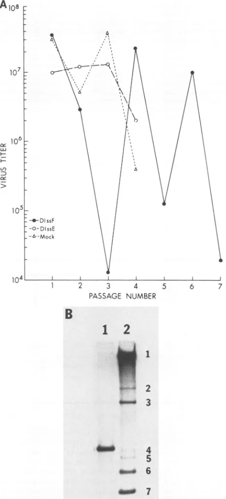
that at the third passage, the virus infectivity decreased drastically(Fig. 3A). Subsequently, thevirus titerfluctuated with each additional virus passage. Such a fluctuation of
infectious virus titer was consistently observed in three
SpeI
PniiMT/.Iqne T m;mnilFornment
I/
K
VOL.64,1990
'1'-FUI'I
N,
I
I
on November 10, 2019 by guest
http://jvi.asm.org/
6048 MAKINO ET AL.
5
10
15
20
NUMBER
C
12
pt* -1
F1
_
-DIssF
-6
-7
FIG. 2. Sucrose gradientsedimentation of DIssF
RNA-contain-ing MHV-JHM. (A) 32P-labeled virus released from DBT cells
infected with MHV-JHM (passage level 30)was purified by 10 to
60% sucrose gradient sedimentation at 26,000 rpm for 19 h in a
Beckman SW28.1rotor.Fractions (600,ul)werecollected.Portions
ofeach fraction were analyzed for radioactivity. (B) Agarose gel electrophoresis ofglyoxal-denatured virion RNAs extracted from eachfraction shown in panel A. Numbers atthetop of each lane representfraction numbers. Thegenomic RNA and DIssF RNAare
indicatedby arrowheads. (C) Agarose gelelectrophoresis of
glyoxal-denatured 32P-labeled virion RNA (lane 1) andintracellular RNAs
(lane 2) fromDBTcells infected with MHV-JHM(passage level30). Thepositions ofvirus-specific mRNA species (1, 3, 6, and 7) and DIssF RNAare indicated.
independent experiments. Thispatternofvirus fluctuation is consistent with the classical DI virus interference pattern
(24). The virus samples obtained from mock- or
DIssE-transfected cells were also passaged in parallel. In both cases, there was no significant decrease of virus infectivity
during the first three passages, even though DIssE RNA
replicated to ahigh level at each passage (datanot shown). Beginningatthe fourthpassage, however, the virus titers of
both mock- and DIssE-transfected cells decreased,
corre-spondingtotheappearanceofa newDI RNAspecies which
comigrated with the DIssA RNA species (data not shown).
This phenomenon was consistently observed in three
inde-pendentexperiments. The natureof this newDI RNAwas
not further studied. Toconfirm that DIssF RNA has ahigh
level ofhomologous interference activity, DBT cells were
coinfected with MHV-A59 and the DIssF-containing DI
particles whichhad undergonetwo passagesfollowing RNA
transfection;thegrowthofMHV-A59incoinfected cellswas
then compared with that from cells infected only with MHV-A59. The growth of MHV-A59was strongly (99.6%) inhibited by DIssF-containing DI particles. By contrast, DIssE-containing DI particles did not interfere with the
multiplicationof MHV-A59 (datanotshown). These results
demonstrated that DIssF RNA has a high level of
homolo-gous interference activity.
The interference activity of DIssF RNAwas also exam-ined atthe level of intracellular mRNA synthesis (Fig. 3B). Atpassage 3, DIssF RNAwasthe only RNAdetected in the cells coinfected with DIssF-containing virus and MHV-A59; the synthesis of mRNAs of helper virus was almost com-pletelyinhibited. No other DI RNA species was generated, evenafter additional DIssF passages (datanotshown). This result suggests that DIssF RNA is a highly interfering DI RNA. In contrast, DIssE RNA did not inhibit the synthesis of helper virus mRNAs, probably because it was poorly packaged into virions (17, 18).
The primary structure of DIssF RNA. We nextstudied the primary structure of DIssF RNA. Sequence analysis was performed with DIssF cDNA clones and PCR products. The 5'-most end of DIssF was also examined by direct RNA sequencing by using a specific primer (5'-CGCCGAATGGA CACGTC-3') complementary to nucleotides 172 to 188 from the 5' end of the genome of MHV-JHM (27). Sequence analysis revealed that DIssF RNA consists of five noncon-tiguous regions (Fig. 4). The first four regions (domains I through IV) are derived from MHV gene 1. Thefirst domain represents 859 nucleotides from the 5' end of the genomic RNA. The second domain, 827 nucleotides, corresponds to the region at 3.1 to 3.9 kb from the 5' end of the genomic RNA. Domain I and mostof thedomain IIof DIssF are also present in DIssE, and the junction between these two domains is identical to that of DIssE (18); however, DIssF contains 77 more nucleotidesthan DIssE at the 3' region of domain II. Domains III (655 nucleotides) and IV (770 nucle-otides) correspond to the regions at 7.0 to 7.7 kb and 19.7to 20.5 kb from the 5' end of the genomic RNA, respectively (23; H.-J. Lee et al., in press). These two domains are not present inthe DIssE RNA. The3'-most domain (domain V) represents 447nucleotides fromthe 3'endof the MHV-JHM genome. DomainVofDIssF is shorterby 155 nucleotidesat the 5' end than the corresponding region of DIssE (18).
Theentire sequence of DIssF isalmost identical tothat of the corresponding regions of MHV genomic RNA, with some exceptions. There are five nucleotide substitutions in domainIIandsixsubstitutions within the leaderregion (Fig. 5) (1). Three of the six nucleotide substitutions (nucleotides 12, 31, and 32) in the leader sequence werealso detected in theDIssE RNA (18). Inaddition, nine nucleotides (UUUAU AAAC) were deleted in DIssF at thejunction between the leader RNA and the remaininggenomic sequences.
Identification of the packaging signal. Sequence
compari-son between DIssE and DIssF RNAs showed that DIssF contained sequences in domains III and IV and partofthe3' end of domain II that are not present in DIssE. These
A30
-
20
0a
U
Bottom
FRACTION
B
7 8 9 10 11 12 13 14 15 16 17
V:
-!.
J. VIROL.
on November 10, 2019 by guest
http://jvi.asm.org/
[image:4.612.69.304.67.503.2]A
108107
c 106
Ln
---DissF
-0-DissE
-&--hAock
1 2 3
PASSAC
B
1
a-0
FIG. 3. Virus production and taining MHV-A59. (A) Infectiousv
wereinfected with MHV-A59(ata
then mocktransfected, transfecte RNA, or transfected with 0.5 ,ug presence ofDEAE-dextran (18). 1
was harvested at 14.5 h postinfec dilutionon DBT cells. The virus
infectivityof virus whichwasgrow
gelelectrophoresis ofintracellular RNAfromDBTcellsinfectedwit} fromDIssF-transfectedcells (lane 2)wasdenatured with glyoxal and
gel.
sequences could containRNA-packaging signals. To exam-ine this possibility, two cDNA clones, MP44 and
DF1-2,
which contain most of the DIssF-unique sequences and DIssE RNA sequences, were constructed (Fig. 1). The
possible packaging ofthese DI RNAs was then examined.
The MP44 clone was derived from a DIssE-specific
DE5-w4 clone butcontainsthe DIssF sequencefrom
nucle-otide 1489 to3089,which includesallof domain
III,
aportionfrom the 3' end ofdomainII, and almost all of domain IV (exceptits3'-most20nucleotides). Ithas deleted the 5'-most X | \ / \ 12 nucleotides of the third domain of DIssE (Fig. 4). The other clone, DF1-2, was derived by inserting the DIssF sequence from nucleotide 1489 to 3437 into DIssE cDNA clone DE5-w4. DF1-2 is very similar to DIssF, except for several nucleotide changes in the leader sequence and do-main II, since dodo-mains I and II of DIssF are closely related to those of DIssE. Theplasmids werelinearized by XbaI and transcribed by T7 RNA polymerase in the presence of a cap analog [m7G(5')ppp(5')G]. The RNA was transfected into monolayers of mouse L2 cells which had been infected with MHV-A59 1 h prior totransfection. Thevirus was harvested from the transfected L2 cells 14 h postinfection and passaged one time on DBT cells. The virus released from this DBT cell culture was then used to infect fresh DBT cells and was labeled with
32pi.
32P-labeled RNA was extracted from purified virus particles and analyzed by agarose gel electro-phoresis. The DI RNAs derived from bothDF1-2 and MP44 constructs wereefficiently packaged into MHV virions (Fig. 6). These data clearlydemonstrated that these threeDIssF-____________I_________
iunique
domains contain thepackaging
signal.
4 5 6 7 Translation of DIssF RNA in vitro and in vivo.
Sequence
:E NUMBER analysis of DIssF RNA reveals thepresenceofasingle, large
ORF. This ORF consists of domains I and II and the 5' portion of domain III. The first 215 amino acids correspond to the N terminus of the MHV gene 1 product, part of which
2
wascleaved to become a p28 protein (1, 3, 27). The ensuing 276amino acids were derived from the region at 3.1 to 3.9 kb from the 5' end of the genome. Since domain IIofDIssF
has 77 nucleotides more than the corresponding region of DIssE, the DIssF RNA encodes an extra 25 amino acids in domain II. There were five nucleotide substitutions in this domain;2 as aresult, two amino acids were converted from Arg to Lys 3 at position 248 and from Val to Ala at position 273 (Fig. 5). However, the sequence substitutions at nucleotides 1193 and 1194 did not alter the amino acids encoded. The last six amino acids at the C terminus of this ORF are encoded from domain III and utilize a reading frame which is different from 4 that utilized by the MHV genomic RNA. As a result, the 5 ORF inDIssFis terminated 21 nucleotides downstream from thejunction site between domains II and III. The predicted
_* 6 molecularweight of this ORF product is 54,670.
To examine whether the ORF ofDIssF RNA was
func-~
7 tional, the synthesis of DIssF-specific protein in DIparticle-mRNA synthesis of DlssF-con- infected cellswasexamined. DBTcells were mock infected
virus
titers(PFU/0.2
ml). L2cells(Fig. 7A, lane 1),
infected with MHV-A59 (Fig. 7A, lane 2),
multiplicity of infection of 5) and or infected with DIssF-containing MHV-A59 (passage 2) d with 0.5 ,ug ofpurified DIssF (Fig. 7A,lane 3). Aspecific, 74-kDaproteinwasdetected in of purified DlssE RNA in the the DI particle-infected cells. Since this protein was larger rhe culture fluid of infected cells thanthe predicted protein, the DIssF-specific nature of this -tion and then passaged without protein was further confirmed by in vitro translation. The titer at passage 1 represents the DIssF RNA was purified from DI particle-infected cells and enonce in DBTcells.(B)Agarose then translated in rabbit reticulocyte lysates. A 74-kDa
rRNA. 12P-labeledvirus-specific
protein
wasobtained
which
wasidentical
in size
to the hMHV-AS9(passage 2)obtained1)ormock-transfectedcells(lane
protein
foundin
the infected cells (Fig. 7A, lane 4). This electrophoresedon a1% agarose protein was immunoprecipitated with anti-p28 antibody (1) (Fig. 7B). Four minorprotein species were alsoon November 10, 2019 by guest
http://jvi.asm.org/
[image:5.612.65.298.83.598.2]6050 MAKINO ET AL.
A
(kb) 0 5
MHV Genes
GenomcIxc
RNA -L
\\ \'",1\ '~
Domalns\
[image:6.612.68.548.77.522.2]DIssF F
FIG. 4. Schematic diagram ofthe structureofDIssFRNAand 10 15 20 25 30 the strategy for sequencingDIssF cDNA clones. (A) Diagram of the
structureof
DIssF
RNA. ThestructureofDIssF
RNA iscompared
l 2 3 456 7 with that of the standard MHV-JHM genomic RNA and DIssEE 2
:1
|146,
71 RNA. Genes 1through
7,previously
called genes Athrough
G,__1__1__1__1__1_________ represent thesevengenesofMHV.Localizations of thelargeORFs
N \ / / inDIssEand
DIssF
areshown.Five domains ofDIssF
(domainsIN N N \ /X/ through V)areindicated abovethediagramofDIssF.(B)Structures
NN N : / / of sequenced DIssF-specific cDNAclones. PCR-1 representsthe XN N u/ / clone derived\
by
PCR. Theremaining
twoclones wereobtainedby
NN
"
NN / direct cDNAcloning. (C)Strategyforsequencingof DIssF. ArrowsN IV /V, starting withopencircles indicateDNAsequencing by the dideoxy chain termination method. Arrows
starting
with solid circlesindi-cate RNA sequencing of gel-purified
DIssF
RNA by using reverseORF/ / transcriptase (31).
/ /
/ /
/ /
DissE
3
(kb) 0 2 3 4
B
cDNAclonesPCR-1 BA1 14
BA49
c
Sequencingstrategy
_~~N . >
-0 g-0o-0
Do--o <
-0---.
4-*
-Scale
(kb) 0.5
-0
1.0 1.5 2.0
cipitated by the anti-p28 antibody; the origin of these
pro-teins isnotknown. From thesedata,weconcludethatDIssF
is afunctional mRNA and translates a74-kDa protein. The
sizedeviation of theDI-specific proteinsfrom theirpredicted molecularmassescouldnotbeexplainedatthis time and has also been observed withDIssE-specific protein (18).
DISCUSSION
Thepresent study identifieda newclassof MHV DI RNA which is subgenomic and yet packaged efficiently into viri-ons. Efficient packaging ofDIssF RNAwas unambiguously
demonstrated by several lines of evidence. First, a large
amount of DIssF RNA was present in the purified MHV virion. Second, DI particles containingDIssF RNAshowed
astronginterferenceactivity with thereplication andmRNA synthesis of helper viruses. Thisstronginterference activity
can be explained if the majority of the virus-infected cells
were also infected with the DIssF-containing DI particles.
Third, virus samples from the DIssF-transfected cells
showedafluctuatingpatternof virusinfectivity duringserial undilutedpassages. Thiscyclic natureof virusinfectivity is consistent with the classical pattern of DI viruses (24). Furthermore, in DI particle-infected cells only DIssF, but
not other DI RNAs, was present, thus establishing that DIssF RNA was responsible for the interference activity.
Since the molar ratio of thegenomicRNAtoDIssF RNA in thevirionswasapproximately 1, thepackaging efficiencyof DIssF RNA should be almost the same as that of the genomicRNA.
It is interesting that the DIssF RNA-containing virus particles had the same buoyant density as the infectious virus, despite the fact that DIssFRNAis 3.6 kb whereasthe MHVgenomic RNA is 31kb. Thislarge difference in RNA size could have altered the buoyant density of the virus particles, as demonstrated for the DI particles of vesicular
stomatitis virus (7). One possible explanation is that the DIssF RNAwascopackaged with MHV genomic RNA into
the same virus particle, whereas anotherpossibility is that
2.5 3.0 3.6
J.VIROL.
on November 10, 2019 by guest
http://jvi.asm.org/
5' -TATAAGAGGGAATGGCGTCCGTCCGTACCTAATCTACTCTAAAACTCTTGTAGTTTAAATCTAATCTAATCTAAACGGCACTTCCTGC 88
* * * *t*
89 GTGTCCATGCCCGTGGGCCTGGTCTTGTCATAGTGCTGACATTTGTGGTTCCTTGACTTTCGTTCTCTGCCAGTGACGTGTCCATTCGGC 178
M A K M G K Y G L G F K W A P E F P W M L
179 GCCAGCAGCCCACCCATAGGTTGCATAATGGCAAAGATGGGCAAATACGGTCTCGGCTTCAAATGGGCCCCAGAATTTCCATGGATGCTT 268
P N A S E K L G N P E R S E E D G F C P S A A Q E P K V K G
269 CCGAACGCATCGGAGAAGTTGGGTAACCCTGAGAGGTCAGAGGAGGATGGGTTTTGCCCCTCTGCTGCGCAAGAACCGAAAGTTAAAGGA 358
K T L V N H V R V D C S R L P A L E C C V Q S A I I R D I F
359 AAAACTTTGGTTAATCACGTGAGGGTGGATTGTAGCCGGCTTCCAGCCTTGGAGTGCTGTGTTCAGTCCGCCATAATCCGTGATATTTTT 448
V D E D P Q K V E A S T M M A L Q F G S A V L V K P S K R L 449 GTTGACGAGGATCCCCAGAAGGTGGAGGCCTCGACTATGATGGCATTGCAGTTCGGTAGTGCTGTCTTGGTCAAGCCATCCAAGCGCTTG 538
S V Q A W A K L G V L P K T P A M G L F K R F C L C N T R E
539 TCTGTTCAGGCATGGGCTAAGTTGGGTGTGCTGCCTAAAACTCCGGCCATGGGGTTGTTCAAGCGCTTCTGCCTGTGTAACACCACGGAG 628
C V C D A H V A F Q L F T V Q P D G V C L G N G R F I G W F
629 TGCGTTTGTGACGCCCACGTGGCCTTTCAACTTTTTACGGTCCAGCCCGATGGTGTATGCCTGGGTAACGGCCGTTTTATAGGCTGGTTC 718
V P V T A I P E Y A K Q W L Q P W S I L L R K G G N K G _S V 719 GTTCCAGTCACAGCCATACCGGAGTATGCGAAGCAGTGGTTGCAACCCTGGTCCATCCTTCTTCGTAAGGGTGGTAACAAAGGGTCTGTG 808
T S G H F R R A V T M P V Y D F N A T D V V Y A D E N Q D D
809 ACATCCGGCCATTCCCGCCGCGCTGTTACCATGCCTGTGTATGACTTTAATGCAACAGATGTTGTATATGCAGATGAAAACCAAGATGAT 898
A
D A D D P V V L V A D T O E E D G V A K E Q V D S A D S E I
899 GATGCTGACGATCCTGTAGTCCTTGTCGCCGATACCCAAGAAGAGGACGGCGTTGCCAAGGAGCAGGTTGATTCGGCTGATTCGGAAATT 988
C V A H T G GC E M T E P D A V G S Q T P I A S A E E T E V
989 TGTGTTGCGCACACTGTTGGTCAAGAAATGACTGAGCCTGATGCTGTCGGATCTCAAACTCCCATCGCCTCTGCTGAGGAAACCGAAGTC 1078
G E A C D R E G I A E V K A T V C A D A L D A C P D O V E A 1079 GGTGAGGCATGCGACAGGGAAGGGATTGCTGAGGTCAAGGCAACTGTGTGTGCTGATGCTTTAGATGCCTGCCCCGATCAAGTGGAGGCA 1168
F D I E K V E D S I L S E L Q T E L N A P A D K T Y E D V L
1169 TTTGATATTGAAAAGGTTGAAGACTCTATCTTAAGTGAGCTTCAAACCGAACTTAATGCGCCCGCGGACAAGACCTATGAGGATGTCTTG 1258
**
A F D A I' Y S E T L S A F Y A V P S D E T H F K V C G F Y S
1259 GCATTCGATGCCATATACTCAGAGACGTTGTCTGCATTCTATGCTGTGCCGAGTGATGAGACGCACTTTAAAGTGTGTGGATTCTATTCG 1348
? A I E R T N C W L R S T L I V M Q S L P L E F K D L C M Q
1349 CCAGCTATAGAGCGTACTAATTGTTGGCTGCGTTCTACTTTGATAGTAATGCAGAGTTTACCTTTGGAATTTAAAGACTTGGGGATGCAA 1438
K L W L S Y K A G Y D Q C F V D K L V K S A P K S I I L P Q
14 3 9 .A'AGCTCTGGTTGTCTTACAAGGCTGGCTATGATCAATGCTTTGTGGACAAACTAGTTAAGAGCGCGCCCAAGTCTATTATTCTTCCACAA 1528
C G Y V A D F A Y F F L S Q C S F K V H A N W R C L K C G M
1529 I^-TGGCTATGTGGCAGATTTTGCCTATTTTTTCCTAAGCCAGTGTAGCTTCAAAGTTCATGCTAACTGGCGTTGTCTAAAGTGTGGCATG 1618
- L K L. Q G L D A V F F Y G D V V S H M C K C S F K A Y F Z
1619 CAGTT.AAAGCTTCAAGGCTTGGACGCCGTGTTTTTCTATGGAGACGTTGTGTCTCATATGTGTAAGTGTAGCTTCAAAGCTTACT.TTTAA 1708
1709 G:TATGCTGTTTGGCCTTTAAGAATGCCTTACAGACGTTTAATTGGAGCGTTGTGTCTAGGGGTTTCTTCCTAGTGGCAACGTCTTTTTA 1798
17993--ATGGTTTAATTTTTTGTATGCCAATGTTATTTTGAGTGACTTTTATTTGCCTAATATTGGACCTCTCCCTATGTTTGTGGGGCAGATT 1888
1889 G-TGCCTTGGGTTAAGACTACATTTGGTGTTTTAACCATCTGCGATTTTTACCAGGTGACAGATTTAGGCTATAGGAGTTCGTTTTGTAAT 1978 1979 G0AAGTATGGTCTGTGAACTATGCTTCTCTGGTTTTGATATGCTGGACAACTATGATGCCATAAATGTTGTTCAACATGTTGTAGATAGG 2068 2069 CGGTGTCTCTTTTGACTACATTAGCCTATTTAAATTAGTAGTCGAACTTGTTATCGGCTACTCTCTATATACTGTGTGCTTCTACCCACTG 2158
2159 T TGTCCTTGTTGGAATGCAGTTGTTGACCACATGGTTGCCTGAATTCTTTATGCTGGGGACTATGCATTGGAGCGCTCGTTTGTTTGTT 22 4 8
2249 TTTGTTGCTAACATGCTCCCAGCTTTTACGTTACTGCGATTTTATATCGTGGTGACAGCTATGTATAAGGTCTATTGTCTTTGTAGACAT 2338
2339 GTTAAGACTTTTGATTTTTACAACCTTTGGAATACTTTTACTAGGCTCCAAAGTTTAGAAAATGTAGTGTACAATTTGGTTAATGCTGGA 24 28
A
2429 CACTTTGATGGCCGGGCGGGTGAACTGCCTTGTGCTGTTATAGGTGAGAAAGTCATTGCCAAGATTCAAAATGAGGATGTCGTGGTCTTT 2518
2519 AAAAATAACACGCCATTCCCTACCAATGTGGCTGTCGAATTATTTGCTAAGCGCAGTATTCGGCCCCACCCAGAGCTTAAGCTCTTTAGA 2608
2609 AATTTAAATATTGACGTGTGCTGGAATCACGTCCTTTGGGATTATGCTAAGGATAGTGTGTTTTGCAGTTCGACGTATAAGGTCTGCAAA 2698
2699 TACACAGATTTACAGTGCATTGAAAGCTTGAATGTACTTTTTGATGGTCGTGATAATGGTGCTCTTGAAGCTTTTAAGAAGTGCCGGAAT 2788
2789 GGCGTCTACATTAACACGACGAAAATTAAAAGTCTGTCGATGATTAAAGGCCCACAACGTGCCGACTTGAATGGCGTAGTTGTGGAGAAA 2878 2879 GTTGGAGATTCTGATGTGGAATTTTGGTTTGCTATGCGTAGAGACGGTGACGATGTTATCTTCAGCCGTACAGGGAGCCTTGAACCGAGC 2968 2969 CATTACCGGAGCCCACAAGGTAATCCGGGTGGTAATCGCGTGGGTGATCTCAGCGGTAATGAAGCTCTAGCACGTGGCACTATCTTTACT 3058 3059 CAAAGCAGATTTTTGTCCTCTTTCTCACCTCGATCGGAGATGGAGAAAGATTTTLGATAATGTAAGCGTTGCAAAGCCCAAAAGCTCTGT 314 8
3149 GCAGCGAAATGTAAGTAGAGAATTAACCCCTGAGGATCGCAGCCTTCTGGCTCAGATCCTAGATGATGGCGTAGTGCCAGATGGGTTAGA 3238
3239 AGATGACTCTAATGTGTAAAGAGAATGAATCCTATGTCGGCACTCGGTGGTAACCCCTCGCGAGAAAGTCGGGATAGGACACTCTCTATC 3328
3329 AGAATGGATGTCTTGCTGTCATAACAGATAGAGAAGGTTGTGGCAGACCCTGTATCAATTAGTTGAAAGAGATTGCAAAATAGAGAATGT 3418
3419 GTGAGAGAAGTTAGCAAGGTCCTACGTCTAACCATAAGAACGGCGATAGGCGCCCCCTGGGAAGAGCTCACATCAGGGTACTATTCCTGC 3508 3509 AATGCCCTAGTAAATGAATGAAGTTGATCATGGCCAATTCGAAGAATCAC-poly(A)-3' 3558
FIG. 5. DNA sequence and deduced amino acid sequence of the DIssF cDNA clones. A translation of the main ORF is shown as
single-letteramino acids. Solidtriangles indicate the sites where fusion of discontiguoussequences occurred. Thebasesubstitutions inDIssF compared with theparental MHV-JHMRNA areindicated by asterisks.
on November 10, 2019 by guest
http://jvi.asm.org/
[image:7.612.135.485.81.688.2]6052 MAKINO ET AL.
A
A B C
ABC
"I,w
M 1 2 3 4 5 200
97.4
-68*
43 ::
29 do
18.4Ak~.
-_M
FIG. 6. Agarose gel electrophoresis of glyoxal-denatured 32p_ labeled virion RNA from DI particle-infected cells. Viruses at
passagelevel 1 from mock-transfected (A), DF1-2 DI RNA-trans-fected (B), and MP44DI RNA-transfected(C) cells wereusedfor infection. DI RNAsareindicatedby arrows.
several molecules ofDIssF RNA were packaged together
intoavirion.Ourpreliminary studyexamining the effects of
serial virusdilutiononthesynthesis ofDIssF RNA in the DI
particle-infected cells has ruled out the first
possibility
(unpublished observation).
Of the two naturally occurring subgenomic DI RNA
species characterized sofar, DIssF is efficiently packaged
whereas DIssE is not. Thus, theextra sequence present in
DIssF RNA may contain the packaging signal for MHV
RNA. The existence of such a
packaging
signal wassug-gested from the finding thatother DI RNAs or mRNAs of
varioussizes, which areeither largerorsmaller thanor are
equivalent to DIssF RNA, were not packaged. From the
common structureofthese DI RNAs andmRNAs, itcanbe
deduced that the RNA-packaging signal must be present
withingene1,whichencompasses22kb (23; H.-J. Leeetal.,
inpress), butis outside the leadersequence.Itis evidentthat
thenucleotide substitutions indomain IIofDIssFwere not
responsible for the high level of packaging efficiency of
DIssFRNA, because DF1-2 DI RNA, in whichthe
substi-tuted nucleotides were converted to sequences similar to
those ofDIssE RNA, was efficiently packaged (Fig. 6). In
addition, the nine-nucleotide deletion (UUUAUAAAC) at
thejunction between the leader and the remaining DI
se-quenceinDIssF RNA(11, 18, 26)was alsonot
likely
toberesponsible
for the high level ofpackaging efficiency
ofDIssFRNA, since DIssE hasthis deletion but is not
pack-aged, whereas the nondefectivegenomic RNA doesnothave
thedeletionand yetispackaged. Rather,theextrasequence
presentinDIssF,which includes the 3'-end 77nucleotides of
domainII anddomainsIII andIV, ismostlikely responsible
for the specific RNA packaging. Analysis of the MP44 DI RNA, which contains most of these regions, conclusively demonstrated that the packaging signal resides within these three separate regions in gene 1 of no more than 1,480
nucleotides in total. This is the first identification of the
packaging signal in coronavirus RNA. Thispackaging signal
mayinteractwiththeN proteinduring the first step of viral
morphogenesis. Experiments using deletion mutants of
DIssF RNAareneeded to furtherdefinethis signal.
BothDIssEandDIssFRNAscontainanORF, whichcan
B
MA 1 2 3 4 S 200
97.4
68 * '4u-- .
43
fa
29 _ -P28
4.,
18.4 -l
FIG. 7. Translation of theDIssF-specificprotein. (A) Polyacryl-amidegelelectrophoresis of proteins fromDIparticle-infectedcells (lanes1 to3)and in vitro translationproducts ofgel-purifiedDIssF
RNAinarabbitreticulocytelysate(lanes4and5). DBTcellswere
mockinfected (lane 1)orinfectedwith MHV-A59(lane 2)orwith
MHV-A59(passage2) obtained fromDIssF-transfected cells(lane 3).At8hpostinfection, cultureswerelabeled with[35S]methionine for 20min, and thecytoplasmiclysateswerepreparedandanalyzed by SDS-PAGE. Lanes 4 and 5 are "S-labeled in vitro translation products of the gel-purified DIssF and no RNA, respectively. MHV-A59-specific structural proteinsareindicatedasS,N, andM. TheDlssF-specific protein is indicated byanarrowhead. LaneM,
14C-labeled marker proteins. (B) Immunoprecipitation of the
pro-teinsbyanti-p28serum."S-labeledsamplesshowninpanelA were
immunoprecipitated with anti-p28 serum and analyzed by SDS-PAGE. The order ofsamples is thesame asthat inpanelA. Lanes 1and2 wereexposed muchlonger than otherlanesin ordertodetect p28.
be translated into proteins in vitro and in vivo.
Puzzlingly,
both of these proteins had lower
electrophoretic
mobilitiesthanexpected fromthe
predicted protein
sizes. Whether thisanomalyofelectrophoreticmobilitywas dueto
posttransla-tional modifications orunusualamino acid compositions of
the DI-specific proteins is notclear. The function of these
proteins is currently unknown. It isinterestingto note that
poliovirusDI RNAsalsocontain functional ORFs(2,5, 8).It
is possiblethat proteinsynthesis is necessaryforthe
initia-tion of DI RNA replication or for interaction with other
host-derivedorhelpervirus-derivedproteins. Alternatively,
these proteins may interact with DI RNA itself in cis to
regulate DI RNA replication. Analyses of additional DI
J. VIROL.
on November 10, 2019 by guest
http://jvi.asm.org/
[image:8.612.386.494.70.416.2] [image:8.612.152.216.76.244.2]RNAs will likely shed further lighton thefunctional signif-icance of DI-specific proteins.
ACKNOWLEDGMENTS
We thank Chien-Kou Shieh for performing some of the prelimi-nary work and for valuable suggestions during the course of this study. We alsothank Susan Baker and Jennifer Fosmire for helpful
comments onthemanuscript.
M.M.C.L. is an investigator at the Howard Hughes Medical Institute. This work was supported in part by Public Health Service grants Al 19244, Al 29984, and NS 18146 from the National Institutes of Health.
LITERATURE CITED
1. Baker, S. C., C.-K. Shieh, L. H. Soe, M.-F. Chang, D. M. Vannier, and M. M. C. Lai. 1989. Identification of a domain required for autoproteolytic cleavage of murine coronavirus geneApolyprotein. J. Virol. 63:3693-3699.
2. Cole, C. N., and D. Baltimore. 1973. Defective interfering particles of poliovirus. II. Nature of the defect. J. Mol. Biol. 76:325-343.
3. Denison,M.R., and S. Perlman.1986. Translation and process-ing of mouse hepatitis virus virion RNA in a cell-free system. J. Virol. 60:12-18.
4. Gubler, U.,and B.J.Hoffman. 1983. Asimple and very efficient method forgeneratingcDNAlibraries. Gene 25:263-269.
5. Hagino-Yamagishi, K., and A. Nomoto. 1989. In vitro construc-tion of poliovirus defective interfering particle. J. Virol. 63: 5386-5392.
6. Hirano, N., K. Fujiwara, S. Hino, and M. Matsumoto. 1974.
Replication and plaque formation of mouse hepatitis virus (MHV-2) in mouse cell line DBT culture. Arch. Gesamte Virusforch. 44:298-302.
7. Holland, J. J.,S.I. T. Kennedy,B. L.Semler, C. L. Jones,L.
Roux, and F. A. Grabau. 1980. Defective interfering RNA
viruses and the host-cell response, p. 137-192. In H. Frankel-Conrat andR. R. Wagner(ed.), Comprehensive virology, vol. 16.
8. Kaplan, C. T., and V. R. Racaniello. 1988. Construction and characterization ofpoliovirus subgenomic replicons. J. Virol. 62:1687-1696.
9. Lai,M.M. C.,R.S.Baric,P.R.Brayton,and S. A. Stohlman.
1984. Characterization of leaderRNA sequences on thevirion and mRNAsofmousehepatitis virus,acytoplasmicRNAvirus.
Proc. Natl. Acad. Sci. USA81:3626-3630.
10. Lai,M. M. C.,P. R.Brayton, R.C.Armen, C. D.Patton, C.
Pugh, and S. A. Stohlman. 1981. Mouse hepatitis virus A59: mRNAstructureandgenetic localization of the sequence diver-gencefromhepatotropic strain MHV-3. J. Virol. 39:823-834.
11. Lai,M.M. C., S. Makino,L. H.Soe, C.-K. Shieh, J. G. Keck,
and J. 0. Fleming. 1987. Coronavirus: ajumping RNA
tran-scription.ColdSpringHarborSymp. Quant. Biol. 52:359-365.
12. Lai,M. M. C., C.D.Patton, R.S.Baric, andS. A. Stohlman. 1983. Presence of leader sequences in the mRNA of mouse
hepatitis virus. J. Virol. 46:1027-1033.
13. Lai, M. M. C., and S. A. Stohlman. 1978. RNA of mouse
hepatitis virus.J. Virol.26:236-242.
14. Leibowitz, J. L.,K. C.Wilhelmsen,and C. W. Bond.1981.The
virus-specific intracellular RNA species oftwo murine corona-viruses: MHV-A59 and MHV-JHM. Virology 114:39-51.
15. Makino, S.,N.Fujioka,and K.Fujiwara. 1985.Structure of the intracellulardefective viral RNAs of defective interfering parti-cles ofmousehepatitis virus. J. Virol. 54:329-336.
16. Makino, S., and M. M. C. Lai. 1989. High-frequency leader sequence switching during coronavirus defective interfering
RNAreplication. J. Virol. 63:5285-5292.
17. Makino, S., C.-K.Shieh, J. G. Keck, andM. M.C.Lai. 1988. Defective-interfering particles of murine coronaviruses: mech-anism ofsynthesis of defective viral RNAs. Virology 163:104-111.
18. Makino, S., C.-K. Shieh,L.H.Soe, S. C. Baker, and M. M.C. Lai. 1988. Primary structure and translation of a defective interferingRNAof murine coronavirus. Virology 166:550-560.
19. Makino, S., F. Taguchi, and K. Fujiwara. 1984. Defective interfering particles of mouse hepatitis virus. Virology 133:9-17. 20. Makino, S., F. Taguchi, N. Hirano, and K. Fujiwara. 1984.
Analysis of genomic and intracellular viral RNAs of small plaquemutantsofmousehepatitis virus, JHM strain. Virology 139:138-151.
21. Maniatis, T.,E. F.Fritsch,andJ. Sambrook. 1982. Molecular cloning:alaboratory manual. ColdSpring Harbor Laboratory, ColdSpring Harbor,N.Y.
22. McMaster, G. K., and G. G. Carmichael. 1977. Analysis of single- and double-stranded nucleic acids on polyacrylamide and agarose gels byusing glyoxal and acridine orange. Proc.
Natl. Acad.Sci. USA 74:4835-4838.
23. Pachuk,C.J.,P.J. Bredenbeek,P. W.Zoltick,W.J.M.Spaan,
andS.R. Weiss. 1989. Molecularcloning of the gene encoding the putative polymerse ofmouse hepatitis virus, strain A59.
Virology 171:141-148.
24. Palma, E. L., and A. S. Huang. 1974. Cyclic production of vesicular stomatitis virus caused bydefective interfering parti-cle. J. Infect. Dis.129:402-410.
25. Sanger, F.,S.Nicklen,andA. R.Coulson.1977. DNA sequenc-ing with chain-terminating inhibitors. Proc. Natl. Acad. Sci. USA 74:5463-5467.
26. Shieh, C.-K., L. H. Soe, S. Makino, M.-F. Chang, S. A.
Stohlman,and M. M.C.Lai. 1987.The5'-end sequence of the murine coronavirus genome: implications for multiple fusion sites inleader-primed transcription.Virology 156:321-330. 27. Soe, L. H., C.-K. Shieh, S. C. Baker, M.-F. Chang, and
M. M.C. Lai. 1987. Sequence and translation of the murine coronavirus 5'-endgenomicRNAreveals theN-terminal struc-tureoftheputative RNApolymerase.J. Virol.61:3968-3976.
28. Spaan, W.,H.Delius,M.Skinner, J. Armstrong,P. Rottier, S. Smeekens, B. A. M. van der ZeUst, and S. G. Siddell. 1983. Coronavirus mRNA synthesis involves fusion of
non-contigu-oussequences. EMBOJ.2:1939-1944.
29. Stohlman, S. A.,and M. M. C.Lai. 1979. Phosphoproteins of murinehepatitis virus. J. Virol. 32:672-675.
30. Sturman,L.S.,K. V.Holmes,andJ.Behnke.1980.Isolation of coronavirus envelope glycoproteins and interaction with the viral nucleocapsid. J. Virol. 33:449-462.
31. Yokomori, K., N. La Monica, S. Makino, C.-K. Shieh, and M. M. C. Lai. 1989. Biosynthesis, structure, and biological activities of envelope protein gp65 of murine coronavirus. Virology173:683-691.
32. Zimmern, D., and P. Kaesburg. 1978. 3'-Terminal nucleotide sequence ofencephalomyocarditis virus RNA determined by
reverse transcriptase and chain terminating inhibitors. Proc.
Natl. Acad. Sci. USA 75:4257-4261.