STUDY OF CORRELATION BETWEEN P WAVE VERTICAL
AXIS AND COPD SEVERITY BY PULMONARY FUNCTION
TEST
DISSERTATION SUBMITTED FOR M.D GENERAL MEDICINE
BRANCH –I
APRIL 2015
THE TAMILNADU
Certificate from the DEAN
This is to certify that this dissertation entitled “STUDY OF
CORRELATION BETWEEN P WAVE VERTICAL AXIS AND COPD
SEVERITY BY PULMONARY FUNCTION TEST” is the bonafide work of
Dr S.S.SRINIVASAN.., in partial fulfillment of the university regulations of
the Tamil Nadu Dr. M.G.R. Medical University, Chennai, for M.D General
Medicine Branch I examination to be held in April 2015.
Captain Dr.B.SANTHAKUMAR,M.Sc(F.Sc.) ,
M.D(F.M)., PGDMLE., Dip.N.B (F.M) .,
THE DEAN
Madurai Medical College and Government Rajaji Hospital,
Certificate from the HOD
This is to certify that this dissertation entitled “STUDY OF
CORRELATION BETWEEN P WAVE VERTICAL AXIS AND COPD
SEVERITY BY PULMONARY FUNCTION TEST” is the bonafide work of
Dr S.S.SRINIVASAN., in partial fulfillment of the university regulations of the
Tamil Nadu Dr. M.G.R. Medical University, Chennai, for M.D General
Medicine Branch I examination to be held in April 2015.
Dr. S. VadivelMurugan, M.D.
Professor and HOD,
Department Of General Medicine,
Government Rajaji Hospital,
Madurai Medical College,
Certificate from the GUIDE
This is to certify that this dissertation entitled “STUDY OF
CORRELATION BETWEEN P WAVE VERTICAL AXIS AND COPD
SEVERITY BY PULMONARY FUNCTION TEST” is the bonafide work of
Dr S.S.SRINIVASAN., in partial fulfillment of the university regulations of the
Tamil Nadu Dr. M.G.R. Medical University, Chennai, for M.D General
Medicine Branch I examination to be held in April 2015.
Dr. M.NATARAJAN, M.D
Professor of Medicine,
Department Of General Medicine,
Government Rajaji Hospital,
Madurai Medical College,
DECLARATION
I , DR S.S.SRINIVASAN , solemnly declare that this dissertation titled
“STUDY OF CORRELATION BETWEEN P WAVE VERTICAL AXIS
AND COPD SEVERITY BY PULMONARY FUNCTION TEST” is a
bonafide record of work done by at the Department Of General Medicine ,
Government Rajaji Hospital , Madurai , under the guidance of
Dr. M.NATARAJAN ,M.D, Professor , Department of General Medicine ,
Madurai Medical college , Madurai.
This dissertation is submitted to The Tamil Nadu Dr. M.G.R Medical
University, Chennai in partial fulfillment of the rules and regulations for the
award of M.D Degree General Medicine Branch- I; examination to be held in
April 2015.
Place: Madurai
Date:
ACKNOWLEDGEMENT
I would like to thank Captain Dr.B. SANTHAKUMAR , M.Sc(F.Sc) ,
M.D (F.M)., PGDMLE., Dip.N.B (F.M) ., Dean Madurai Medical College and
Government Rajaji Hospital, for permitting me to utilize the facilities of
Madurai Medical College and Government Rajaji Hospital facilities for this
dissertation.
I wish to express my respect and sincere gratitude to my beloved teacher
and Head of The Department, Prof. Dr. S.VADIVELMURUGAN,
M.D.,Professor of Medicine for his valuable guidance and encouragement
during the study and also throughout my course period.
I would like to express my deep sense of gratitude, respect and thanks to my
beloved Unit Chief and Professor Of Medicine,Prof. Dr.M.NATARAJAN,
M.D., for his valuable suggestions , guidance and support throughout the study
and also throughout my course period .
I am greatly indebted to my beloved Professors , Dr. V.T.PREMKUMAR ,
M.D., Dr. R.BALAJINATHAN, M.D., Dr. J.SANGUMANI, M.D.,
Dr. G.BAGYALAKSHMI , M.D., Dr. DHARMARAJ, M.D., and
Dr. R.PRABAKARAN ,M.D., for their valuable suggestions throughout the
I am extremely thankful to Assistant Professor of Medicine of my Unit,
DR. K.MURALIDHARAN M.D.AND DR.B.PALANIKUMAR M.D
.,
fortheir valid comments and suggestions.
I sincerely thank the Assistant Professor of Thoracic Medicine,
Dr. VIVEKANANTHAN, M.D. (chest) for his guidance and suggestions in
my dissertation work.
I sincerely thank all the staffs of Department Of Medicine and Department Of
Thoracic Medicine for their timely help rendered to me, whenever needed.
I express my thanks to Dr.VAIRAKANI., DR.J.ARUN KUMAR.,
DR.LENIN SANKR., DR.B.KARTHI., DR.G.CHINNAMARIYAPAN., for
their help and support in my dissertation work .
I extend my thanks to all my friends, batch mates any senior and junior
colleagues who have stood by me and supported me throughout my study and
course period
Finally, I thank all my patients, who form the backbone of my study, for their
patience and co-operation .I pray to god for their well-being and their speedy
CONTENTS
S.NO CONTENTS PAGE.NO.
1. HISTORICAL PERSPECTIVE 1
2.
INTRODUCTION
2
3.
AIM OF STUDY
3
4.
REVIEW OF LITERATURE
4
5.
MATERIALS AND METHODS
73
6.
RESULTS AND INTERPRETATION
76 7. DISCUSSION 96 8. CONCLUSION 101 ANNEXURES BIBLIOGRAPHY PRO FORMA ABBREVIATIONS MASTER CHART
ETHICAL COMMITTEE APPROVAL LETTER
INTRODUCTION
In India, COPD is the second most common lung disorder after
pulmonary tuberculosis. The disease frequently encountered in middle aged
patients and also COPD increasing public importance around the world.
Estimates suggest that COPD will rise from the 6th to the 3rd most common
cause of death worldwide by 2020.
AIMS AND OBJECTIVES
To estimate P wave vertical axis and FEV1 in patients with features of COPD.
To study P wave vertical axis inversely correlating with FEV1.
TO analyse whether P wave vertical axis can be substitute for FEV1 in
assessing the severity of COPD.
METHODS AND MATERIALS
The study is to be conducted on 100 patients of Government Rajaji
Hospital, who are all attending internal medicine and thoracic medicine op with
clinical features and ECG changes of COPD .inclusion criteria: Age >45 year,
Normal sinus rhythm with ECG change , past medical history of
COPD,Imaging studies with COPD changes, Pulmonary function tests with
COPD changes. Exclusion criteria: Congenital heart disease ,valvular heart
STUDY PROTOCOL
The design for study is Observational study done in the
period of July 2014 to September 2014 with simple Statistical analysis
and participants are Patients attending in medicine and thoracic medicine
op with clinical features and ECG changes of COPD
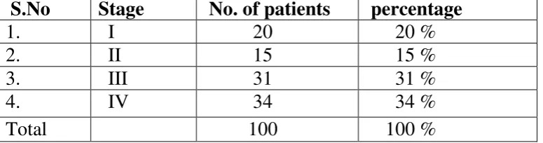
RESULTS
Among 100 patients, 17 patients had P wave vertical axis of
65-75 degree,19 patients had p wave vertical axis of 71-65-75 degree,20 patients had P
wave vertical axis 76-80 degree,44 had P wave vertical axis of >80 degree. P
wave vertical axis>80 degree is superior to P wave vertical axis 75 -80 degree is
correlating with COPD severity and statically significant as p value is < 0.005.
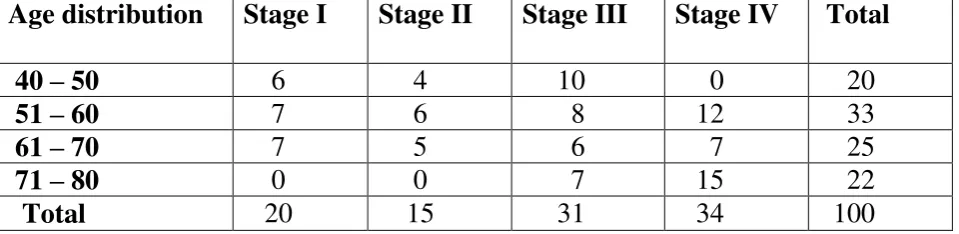
CONCLUSION
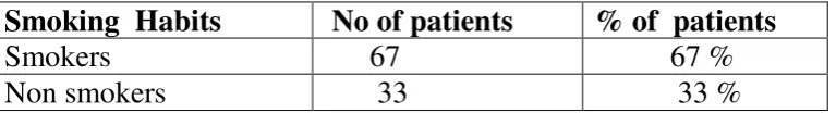
From our study we concluded that , increasing age is
associated with severity of the disease .Males are more commonly
affected then females because of smoking.occupational exposure to risk
factors confounding the risk of developing the disease when associated
with smoking. Incidence of pulmonary hypertension and right ventricular
dilatation increase with increase in severity of the diseases. p wave
vertical axis is directly proportional to severity of COPD and inversely
KEY WORDS
Vertical P-wave axis, the electrocardiographic synonym for pulmonary
emphysema, The electrocardiogram in pulmonar emphysema,electrocardiogram
in chronic cor pulmonale,Pulmonary emphysema: classical, quasi-diagnostic
HISTORICAL PERSPECTIVE
The beginning of modern chest medicine can be traced to the classic
volume by Laennec, “A treatise on diseases of the chest” which appeared in 1821 laid the corner stone of modern chest medicine.
In his treatise, Laennec, devoted one chapter to “Pulmonary Catarrh or
Bronchitis” and emphysema. The chapter on bronchitis distinguishes between
acute and chronic form and sub divides chronic bronchitis into two types – the humid (Copious Expectoration) and dries (Scarcely any Expectoration). He
identified “Dilatation of air cells” as the essential feature of emphysema.
Recognition of chronic bronchitis as a potentially grave illness rather than
as a trivial but not disabling disease had to wait the “London Fog” of 1953, which was brought about by bad weather and air pollutants, carried with it a
surge in morbidity and mortality due to chronic lung disease .
After World War II, clinical investigations of pulmonary disease were provided
with a new diagnostic armamentarium; Pulmonary Function tests were extended
beyond simple spirometry and innovative techniques were developed for
assessing the distribution of gases within the lungs which greatly improved our
INTRODUCTION
In India, COPD is the second most common lung disorder after
pulmonary tuberculosis. The disease frequently encountered in middle aged
patients and also COPD increasing public importance around the world.
Estimates suggest that COPD will rise from the 6th to the 3rd most common
cause of death worldwide by 2020.
Emphysema of any pathogenesis nearly always due to chronic obstructive
pulmonary disease and rarely due to alpha 1 antitrypsin deficiency produce a
state of abnormal lung hyperinflation and has been shown to carry on
association with a vertical frontal p wave axis.
Patients with COPD show that vertical p wave axis in ECG and forced
expiratory volume in pulmonary function test were inversely correlating, so
ECG can be used to assess the severity of COPD in place of pulmonary
function test and also vertical P wave axis (>60) during a sinus rhythm can be
easily detected by a simple glance at the electrocardiogram.
The present study is to compares the P wave vertical axis, which is
reproducible, patient friendly, less procedure related complication, minimal
time consuming against the more cumbersome method of FEV1 measurement in
AIMS AND OBJECTIVES
1. To estimate P wave vertical axis and FEV1 in patients with features of
COPD.
2. To study P wave vertical axis inversely correlating with FEV1.
3. TO analyse whether P wave vertical axis can be substitute for FEV1 in
REVIEW OF LITERATURE
DEFINITION
Chronic obstructive pulmonary disease is Characterised by
persistent airflow limitation that is not fully Reversible, usually progressive and
associated with a chronic Inflammatory response in the airway and the lung to
noxious .
Particles or gases.
1. EMPHYSEMA
Emphysema is defined as destruction and enlargement in The lung
alveoli.
2. CHRONIC BRONCHITIS
This condition associated with excessive mucous Production sufficient to
cause cough with expectoration At least 3 months a year for more than 3
consecutive years
EMPHYSEMA
Centriacinar emphysema: proximal portion of respiratory unit and central part
are destructed and enlarged. Apices and upper lobes are predominantly
involved. This predominantly seen in male patients with chronic bronchitis
Panacinar emphysema: these type of emphysema seen in alpha 1 antitrypsin
deficiency individual.it is particularly involve lower lobes because lower lobes
are rich in blood supply .protease rich enzymes are present in neutrophils that
destruct the alveoli which is not able to inhibit by anti-protease enzymes in this
individuals due to deficiency.
Paraseptal emphysema: this type of emphysema can lead to pneumothorax
because its mainly found near to pleura. Distal acinus is predominantly
involved.
Irregular emphysema: this could be seen in any type of emphysema.
SPECIAL VARITIES OF EMPHYSEMA
Compensatory emphysema: in response to pathology to same lung or opposite
lung, normal lung become hyper inflate as a compensatory mechanism. Here the
alveoli septea are not destroyed .clinically there is no signs of emphysema.
Mediastinal emphysema: this type of emphysema occurs due to rupture of over
distended alveoli .this may occur in following condition
Rupture of emphysematous bulle
Rupture of oesophagus
Subcutaneous emphysema: this type of emphysema occur due to escaped air
tracks up into the subcutaneous tissues of neck. If the COPD patient present
with neck swelling, we have to think in terms of subcutaneous emphysema
possibility.
CHRONIC BRONCHITIS
It is subdivided into
a.simple chronic bronchitis
b.chronic mucopurulent bronchitis
c.chronic bronchitis with obstruction
REID INDEX
Reid index is expressed as ratio of sub mucosal glands to that of bronchial wall.
In normal individuals, its 0.44±0.09
In chronic bronchitis, its 0.52±0.08
When the sub mucosal layer thickness >50% of bronchial wall thickness it is
DIFFERENTIATING FEATURE BETWEEN EMPHYSEMA AND
CHRONIC BRONCHITIS
FEATURES PREDOMINANT
EMPHYSEMA (PINK PUFFER) PREDOMINANT BRONCHITIS (BLUE BLOATER)
Age of onset 6th decade 5th decade
Cough After dyspnoea Before dyspnoea
Dyspnoea Severe Mild
Sputum Scanty,mucoid Copious, purulent
Infections Less common Common
Respiratory
insufficiency
Often terminal Repeated attacks
Chest x ray Hyperinflation ±
bullous changes;
small heart
Increased
bronchovascular
markings; large
heart
Paco2 (mm Hg) 35 – 40 50 – 60 Pao2 (mm Hg) 65 -70 45-60
Pulmonary
hypertension
Cor pulmonale Preterminal stage Common
Diffusing capacity Decreased Normal to slight
reduction
Lung compliance Increased Normal
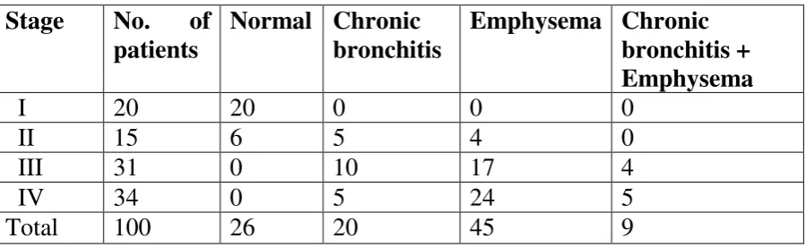
GOLD CRITERIA FOR COPD SEVERITY
GOLD stage Severity Spirometry
I Mild FEV1/FVC < 0.70
FEV1≥80% predicted
II Moderate FEV1/FVC < 0.70
50% ≤ FEV1 < 80%
predicted
III Severe FEV1/FVC < 0.70
30% ≤ FEV1 < 50%
predicted
IV Very severe FEV1/FVC < 0.70
FEV1 < 30% predicted
or FEV1 < 50%
predicted plus chronic
EPIDEMIOLOGY
COPD is expected to be the third most common cause of mortality
and the fifth for loss of DALY worldwide according to Global Burden of
disease study.
PREVALENCE IN INDIA
The exact prevalence in our country could not be
ascertained because of misdiagnosis, underassessment, lag of extensive
studies, poor statistical information. The prevalence rate varies from 2 to 22
per cent in males and 1.2 to 20 percentage in females. Recently ICMR has
done the INSEARCH study in four cities and reported prevalence of 5% in
males and 3.2% in females more than 35 years of age. The total population
affected by the disease has increased to 14.84 million in 2011 from 6.45
million in 1971. In India the sex ratio and the smoker to non smoker ratio are
not as high when compared to western statistics. The reason for disparity is
biomas fuel combustion which is important risk factor in women more so in
villages. Data on mortality statistics are limited; 7% mortality has been
RISK
FACTORS
INFLUENCING
ONSET
AND
PROGRESSION
SMOKING
Tobacco smoking is the most important and the well-studied risk factor.
About 85% of COPD is related to smoking. The remaining 15% is attributed
to smoke from burning biomass fuels like woods, from occupational exposure
to dust and smoke and cow dung for cooking.
The effect of smoking on decline lung function has been proved in many
studies. cigarette smokers show high annual rate of decline in FEV1 of about
50 ml, this is nearly double the average value of 30 ml annually present in
non-smokers .even though ,there is considerable variation in FEV1,with some
smokers showing very rapid rate of decline. The decline in FEV1 might be
faster in natural history of disease before COPD is established.
But still not all smoker developed the disease and even among those who
smoke there are variations in response to the duration of smoking which
suggest that other factors particularly environment modulate, the effect of
smoking, content of smoke such as tar, nicotine ,other constituents and genetic
in these patients.
Mortality due to COPD is twofold higher in smokers who smoke ≥
PASSIVE SMOKING
There is statistically significant correlation showed in relation with lung
cancer in passive smokers .these is shown mainly in patients who are
chronically exposed since childhood with significant lower of FEV1 in
adulthood.
AIRWAY RESPONSIVENESS
Though the concept of airway hyper responsiveness is proved beyond doubt in
bronchial asthma, in COPD there are conflicting reports even though there are
recent studies supporting the airway hyper responsiveness in COPD patients.
RESPIRATORY INFECTIONS
Infections associated with risk
1. Childhood respiratory infections
2. Previous history of tuberculosis even when adequately treated with ATT.
3. Inadequately treated bronchial asthma
INDOOR AIR POLLUTIONS
Though in developed countries, smoking is the major risk factor, in
to half of all cases. Among them chronic exposure to carbon monoxide,
HCHO and nitric oxide and sulphur dioxide and others released from biomass
fuel combustion is the important risk factor particularly in female patients.
WHO identified that, if the sulphur dioxide level ≥150 microgram /m3 shows
increased morbidity in terms of symptoms and hospital admissions in adult
COPD.
Those who have poor cardiopulmonary reserve and elderly people show
increased mortality if the sulphur dioxide level or black smoke
≥500microgram/m3
GENES
The difference in expression of disease among smokers could possibly be
explained by the genetic factors. Alpha 1 antitrypsin deficiency due to alteration
in SERPINA 1 locus encoding the enzyme is the only proven genetic factor
related to the COPD.
Allele Alpha 1 Anti trypsin
M Normal
S Slightly reduced
Z Markedly reduced
The above picture shows that pattern of PiM,PiZ and Pimz alpha 1 AT on
isoelectric focus. Because of its multiheterogenisity ,it appear as multiple
bands.PiM and PiZ shows different band patterns, while PiMZ shows
One has to suspect alpha 1 antitrypsin deficiency as the cause in patients with
1. Age of onset <40 years of age
2. Insignificant history of smoking
3. Predominant lower lobe involvement.
4. Necrotizing panniculitis(weber Christian disesase)
5. C ANCA positive vasculitis (wegeners granulomatosis)
6. Early onset of emphysema in family members or non-smoking related
emphysema.
7. Bronchiectasis without other aetiology
PiZZ is the most common form of severe alpha 1 antitrypsin deficiency
showing greatly accelerated decline in FEV1 .
PiM is the commonest allele in all population and the most common
genotype is PiMM.
Treatment for this subset of individuals is weekly intravenous administration
of alpha 1 antitrypsin.
Increasing age physiologically decreases the function of lung which may
also contribute.
In vertical axis FEV1 plotted and horizontal axis age of the person is plotted.
This shows that ,in non-smoker or not susceptible to smoking will lose 25% of
his /her lung function throughout his life. Those who smoke or are susceptible
to smoking will have decline in lung function rapidly as shown in this picture.
Even though abnormal lung function is detected in these patients ,symptoms
The above picture represents natural history of COPD with hypothetical case
scenario who continues smoking upto the age of 45 years then he discontinues
the smoking.here once the patient stops smoking before development of
symptoms the lung fuction goes to normal range. Once the patient who is
detected to have abnormal lung function before the development of symptoms
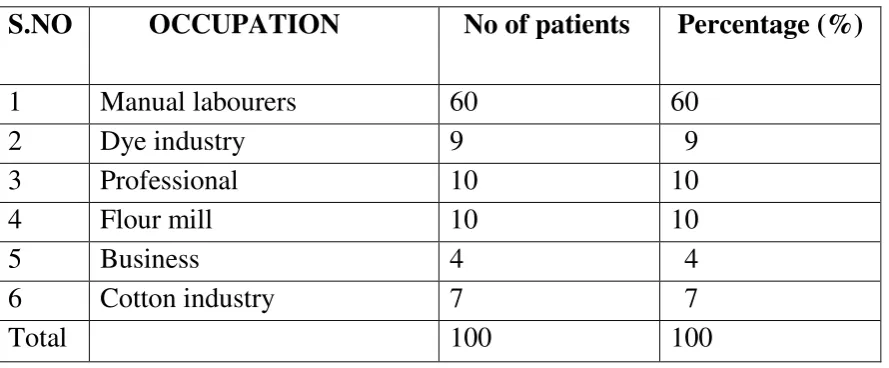
OCCUPATION
There is causal correlation between occupational dust exposure and
development of mucus hyper secretion. Coal miner’s shows development of
small excess decline in FEV1 and increased mortality.
Those who are exposed to welding fumes are also showing development of
COPD and workers who are exposed to cadmium also develop COPD in
later days.
GENDER
In the past , the prevalence and mortality were more among men. As on date
it is almost equal among men and women. When compared to men, women’s
susceptibility to the ill effects of tobacco smoking is significantly higher.
LUNG GROWTH AND DEVELOPMENT
The factors that affect the lung growth and development During the
intrauterine life, and childhood increases an individual’s susceptibility to
develop COPD in the later years of life.
LOW SOCIOECONOMIC STATUS
The risk of developing COPD is inversely proportional to socioeconomic
PATHOPHYSIOLOGY
Pathological changes in the lungs lead to corresponding physiological
changes characteristic of the disease which occur in the following order:
Mucus hyper secretion
Ciliary dysfunction
Airflow limitation
Pulmonary hyperinflation
Gas exchange abnormalities
Cor pulmonale
AIRFLOW LIMITATION
Airflow limitation due to decreased airway calibre and increased airway
resistance, impaired elastic lung recoil during expiration manifest in
spirometry as a reduction in the ratio of FEV1/FVC < 0.7 (FEV1- forced
expiratory volume in 1 second, FVC- forced vital capacity) and reduced
forced bronchodilator FEV1 predicted value is a cardinal feature for the
disease diagnosis and dividing into stages.
FVC at 6 seconds (FEV6) in patient with COPD is adequate and equivalent
to FVC in individuals with severe obstruction.
During forced expiration there is airway instability and narrowing ,this the
cause for discrepancies between inspiratory and expiratory flow.
From the flow pressure curve we can quantify the diminished elastic recoil
and increased airway resistense by reduced maximal expiratory air flow.
Decreased elastic recoil shows –normal slope with premature termination.
MEFV –maximum expiratory flow volume
MIFV-maximum inspiratory flow volume
LEFT-shows normal subject
MIDDLE-mild airway obstruction due to COPD
RIGHT-shows severe obstruction due to COPD
HYPERINFLATION
In COPD patients there is hyperinflation as shown by increased RV and
increased ratio of RV/TLC and later increase in TLC. ( RV- residual volume,
Mechanism
The primary mechanism during expiration from small airways and alveoli is
driven from the inward elastic recoil pressure of the lung which is also
contributed by the counteracting thoracic wall outward pressure. At the end
of tidal expiration both these balance each other resulting in particular
amount of air remaining in the lungs named as FRC(Functional residual
capacity). In COPD patients, due to destruction of lung parenchyma the
inward pressures are low and it comes into equilibrium with the outward
pressure at increased volumes of FRC resulting in hyperinflation
Hyperinflation may be an advantage as it favours conservation of
maximum expiratory airflow by the following ways
1. Increase lung volume
2. Increase elastic recoil pressure
3. Enlarge airway lumen
4. Decrease airway resistance
However hyperinflation has untoward effects on mechanics of the thorax.
1. increase work of breathing
2. induce dyspnea
But hyperinflation of the lung keeps the diaphragm at a mechanical
disadvantage i.e flattening of diaphragm resulting in functional diaphragmatic
paralysis.this flatting of diaphragm leads to number of ill effects.those are
1. Because of loss of apposition zone between diaphragm and
abdominal wall, during inspiration positive abdominal pressure not transmitted
as virtually to chest. This leads to hampering rib cage movement and impairing
2. Because of flatting of diaphragm muscle fibres are shorter than those
of more normally curved diaphragm, this ultimately lead to less effective
generation of inspiratory pressure.
Concurrent with hyperinflation, in patient with COPD inspiratory capacity also
commonly reduced. This finding has prognostic significance that is independent
of FEV1.
GAS EXCHANGE
The partial pressure of oxygen will be maintained until FEV1 s less
than 50% of the predicted value and co2 retention will be observed only when
the FEV1 is less than 25% of the predicted value. Gas exchange impairment in
COPD is mainly due to the mismatch between the ventilation and perfusion.
Large bullae will complicate this issue further because they are non-functioning
airspaces with both poor perfusion and ventilation and moreover they occupy a
significant volume of lung and hence compress and compromise the adjacent
functional parenchyma.
V/Q MISMATCH
The characteristic feature of COPD is ventilation perfusion mismatch, this
heterogeneous process affect not only the lung parenchyma but also affect the
Ventilation – perfusion mismatching is the primary cause for the reduced Po2 that occur in COPD.From this view, modest increase in inhaled oxygen are
effective in treatment of COPD.
DYSPNEA
Most of the COPD patient seek medical care only when they develop
dyspnea.dyspnea impairs their day to day activites and affect the qualities of
life.patient present with complaints of dypnea only when the FEV1 fall to
The mechanism for breathlessness in COPD is +currently not well known.
Neural signals pertaining to abnormalities of chest wall and airway mechanics
seems to be important.
1.An increased feeling of expenditure relating to the pressure required from
respiratory muscles relating to their maximum pressure generating capacity.
2.Impuse of “length-tension inappropriateness “from the respiratory muscles because of hyperinflation
3.Also,signals from airways leading abnormal dynamic compression
during exhalation have been described.
4.except in acute scenario hypercarbia and hypoxia play little role in
dyspnoea.
PATHOGENESIS
ACTIVATION OF MACROPHAGES
Macrophages are the cells found in the normal lower airways. Cigarette
smoke contains oxidants which inactivates the histone deacetylase II with
a resulting increase in acetylated chromatin thereby promoting increased
transcription of pro inflammatory cytokines and matrix metalloproteinase.
This pro inflammatory cytokines recruits neutrophils and stimulates
neutrophil elastase. Macrophage secreted matrix metalloproteinase and
LOSS OF CILIA
Cigarette smoke causes loss of cilia in the airways and predispose to
bacterial infection with neutrophilia.
ANTIELASTASE HYPOTHESIS
Alpha 1 anti trypsin deficiency
Neutrophils in the alveoli are stimulated by variable triggers which get
activated and recruited and they release the granules such as proteinases,
catapsins, elastase and matrix metalloproteinase and also reactive oxygen
species which inhibit alpha 1 anti trypsin. Because of deficiency of alpha
1 anti trypsin , counteraction with granules released by neutrophis is
decreased and this results in destruction of air spaces
SMOKING
In smokers both neutrophils and macrophages are increased in alveoli.
Neutrophils upon activation release the above said proteases and activate
macrophages release a variety of cytokines, ROS, chemokines and a
variety of matrix mataloproteinases which are not inhibited by alpha 1
antitrypsin rather these enzymatically degrade alpha 1 antitrypsin and
increase apoptosis of alveoli and CD4 cells triggers autoimmunity against
native lung tissue.
OXIDANT STRESS
The normal lung contains high amount of anti oxidants such as
glutathione to handle the oxidant stress. Smoking activates inflammatory
cells by the above said mechanism and releases free radicals which shift
causing lung destruction in patients even with normal alpha 1 AT.
Oxidant stress also result in loss of surfactant, reduction in elastin
synthesis, Extra cellular matrix apoptosis.
CLINICAL PRESENTATION
HISTORY
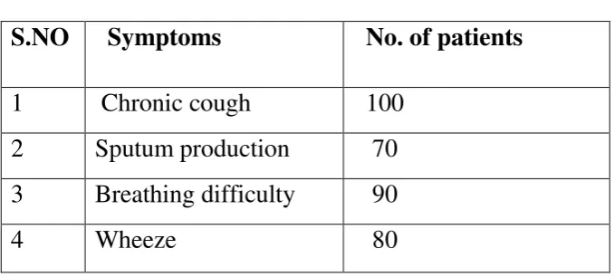
SYMPTOMS
1. Chronic cough
2. Expectoration
3. Dyspnea on exertion, insidious in onset and gradually progressive
over time. Sudden worsening of dyspnea could be either on due to
exacerbation or pneumothorax or other complications.
4. Wheeze
5. Loss of weight – in severe disease ( other causes should be ruled out before attributing it to the disease per se i.e. bronchogenic carcinoma)
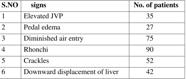
SIGNS:
The physical examination might be completely normal in patients in the
beginning stages till there is significant deterioration in pulmonary
function.
GENERAL EXAMINATION
1. Stigmata of smoking – nicotine stains, etc. 2. Cyanosis may be present.
3. Clubbing is not a sign of disease per se and if clubbing is present one
4. Pedal edema, ascites, elevated JVP due to pulmonary hypertension
and cor pulmonale.
5. Patient may have hypoxemia.
RESPIRATORY SYSTEM EXAMINATION
a. Patient with severe disease may have acting accessory muscles of
respiration with the patient assuming “tripod” position to enhance the
synergistic action of these muscles.
b. Hoover’s sign – the movement of costal margins towards the midline during inspiration.
c. Signs of hyperinflation – barrel shaped chest, hyper resonance on percussion
d. On auscultation patient may have prolonged expiratory phase,
diminished intensity of breath sounds, expiratory wheeze.
e. Signs of pulmonary hypertension and cor pulmonale – loud P2 may not be present due to hyperinflation, tricuspid regurgitation murmur
DIFFERENTIAL DIAGNOSIS
1. Bronchial asthma – distinguished by near total post bronchodilator FEV1 reversibility.
2. Bronchiectasis – differentiated by symptoms like hemoptysis, presence of clubbing and radiography.
3. Cystic fibrosis – history since childhood, recurrent infections, presence of other features such as cirrhosis.
4. Broncho pulmonary mycosis
5. Central airway obstruction such as developmental anomalies,
tumours, stenosis - differentiated by history and PFT using
flow-volume loops.
COMPLICATIONS
1. Recurrent lung/airway infection
2. Bronchogenic carcinoma
3. Cor pulmonale and pulmonary hypertension
4. PTE
5. Pneumothorax especially in those with emphysema
6. Cardiovascular effects – CAD, arrhythmias.
RADIOGRAPHY
1. CHRONIC BRONCHITIS – increased bronchovascular markings
2. EMPHYSEMA – hyper inflated lung fields Diaphragm flattening
Decreased peripheral vascular markings
3. Pulmonary hypertension – enlarged main and right descending pulmonary artery.
4. To look for co morbidities and complications like malignancy,
pneumothorax etc.
COMPUTED TOMOGRAPHY
1. Not routinely done for diagnostic purposes
2. To assess patients fitness for surgical management.
LABORATORY TESTS
PULMONARY FUNCTION TEST:
1. FEV1/FVC < 0.70
2. TLC, FRC, RV may increase often more than normal with disease progression.
Apart from diagnostic utility, FEV1 is also used to monitor treatment response
and worsening FEV1 is a poor prognostic factor.
ABG :
1. Normal in early stages.
2. To determine the degree of hypoxemia and the acid base status and to
differentiate between acute exacerbations and chronic respiratory failure.
TREATMENT
STABLE PATIENTS
Survival improving strategies
1. Smoking cessation
2. Oxygen
3. Lung volume reduction surgery
Other management strategies are to the relief of symptoms, improving the
quality of life and to reduce exacerbations.
DRUGS
BRONCHODILATORS
These drugs are the mainstay of management in that they provide symptom
relief and enhance the well-being of patient but do not prevent the decline in
FEV1.
INHALED BETA2 AGONISTS :
1. Short acting beta 2 agonist : used in all stages of the disease ‘on demand’ basis to provide immediate relief from dyspnoea and not to be used on a routine basis.
Drugs used are salbutamol and levo salbutamol.
2. Long acting : used on a scheduled basis as maintenance therapy to provide
sustained relief.
Side effects; tachycardia, tremor, decrease in serum
potassium.
INHALED ANTI-CHOLINERGICS:
1. Short acting : for symptom relief similar to short acting beta 2 agonists but are
devoid of their sympathetic side effects and also has prolonged action compared
to them.
Drug used is ipratropium.
2. Long acting : for maintenance therapy
Side effects : dryness of mouth, primary angle closure glaucoma is an absolute
contra indication, symptoms of BPH may aggravate.
COMBINATION THERAPY
As the disease worsens, combination therapy with long acting
sympathetic and anticholinergic provides maximum symptom benefit by
synergistic mechanisms rather than single drug alone.
CORTICOSTEROIDS
INHALED :
ICS are not to be used as a single agent in management of these patients but
used in combination with other inhaled bronchodilators.
1. Stage 3 disease
2. ≥2 exacerbations / year
Side effects :
1. Oral candidiasis – prevented by mouth gargling after each use or by spacer.
2. Hoarseness of voice.
SYSTEMIC STEROIDS
Oral steroids are not indicated in the regular treatment of stable patients because
the adverse effects overweigh the benefits but they are important part of acute
exacerbation treatment.
METHYLXANTHINES
Sustained release theophylline is commonly used in patients who do not have
adequate symptom relief with inhalational therapy because of its effects on
diaphragm function, reducing airway resistance and anti inflammatory action.
Side effects:
These drugs have a narrow therapeutic window and hence supervision is
essential.
CNS: tremor, decreases seizure threshold, insomnia.
GIT: gastritis, nausea.
ANTIBIOTICS – no role in stable patients.
NON PHARMACOLOGICAL MEASURES
OXYGEN
Oxygen therapy improves the morbidity of the patient and reduces the mortality.
Indications
1. Arterial partial pressure of oxygen≤ 55 mm of hg or oxygen saturation by pulse
oximetry≤ 88 %
2. Arterial partial pressure of oxygen < 60 mm hg and oxygen saturation < 90% in
the presence of pulmonary hypertension, Hct > 55% or cor pulmonale.
VACCINATIONS
For all stages of the disease yearly influenza vaccine and pneumococcal vaccine
(zero dose and a booster after 5 years)
PULMONARY REHABILITATION
It is a multi system modality including respiratory exercises, psychological
breathlessness and quality of life. Indicated in patients failing optimal medical
therapy and also for severe disease.
SURGICAL MANAGEMENT
1. LUNG TRANSPLANTATION
A. Done either as one lung or sequential double lung transplant.
B. Requirements include severe disease inspite of optimal medical management,
poor quality of life, absence of other organ system dysfunction.
C. PHT and the region of lung involved are not contraindications.
2. LUNG VOLUME REDUCTION SURGERY
3. Patients with upper lobe emphysema and severely compromised exercise
tolerance are model candidates. Presence of PHT/ cor pulmonale are contra
This picture shows that selection of patient for lung volume reduction
surgery
EXACERBATION OF COPD
PRECIPITATIING CAUSES
Infections
1. Bacterial infections
a. Pneumococcus
b. Haemophilus influenza
c. Moraxella catarrhalis
d. Pseudomonas in special risk groups.
2. Viral infections
Environmental
1. Pollution
2. Exposure to toxic gases
3. Climate changes
SYMPTOMS
1. Increase in cough
2. Worsening dyspnoea
3. Increased sputum with purulence
To assess the severity of exacerbation complete physical examination is
mandatory.
Investigations include
1. X ray chest – to detect pneumothorax, consolidation
2. ECG – to detect rhythm disturbance, most common being MAT 3. ABG – to detect respiratory failure.
Based on the severity patient general status, supportive care, presence of
other co morbidities like CAD, diabetes, ABG measurement and the need for
assist ventilation, the decision is taken whether the patient has to be treated
at home or hospital or ICU.
Detecting the COPD mortality in exacerbation of COPD, BODE index is
BODE INDEX
DRUGS
BRONCHODILATORS
Short acting beta agonists are used in primary treatment for exacerbation of
COPD, either alone or in addition with an anticholinergic delivered through
nebulisation or inhalation if patient can perform. Those patients who are already
on methyl xanthine, it should not be stopped as it causes deterioration but
monitoring of drug level is mandatory. During an exacerbation theophylline
should not be prescribed.
CORTICOSTEROIDS
For short course systemic corticosteroids either oral or parenteral is a important
cornerstone in management of acute exacerbation because the decrease hospital
Variable 0 1 2 3
FEV1 (% of predicted) ≥65 50-64 36-49 ≤35
Distance walked in 6 min (meters) ≥350 250-349 150-249 ≤149
MMRC dyspnea scale 0-1 2 3 4
stay improves lung function decreases the subsequent hospital admissions due
to exacerbation.
ORAL CORTICOSTEROIDS
Prednisolone at a dose of 30 to 40 mg or its equivalent has to be given for 10 to
14 days.
The proven advantages are early recovery, lung function improvement,
improvement in arterial hypoxemia, decrease in length of hospital stay.
INHALED CORTICOSTEROIDS
Nebulised budesonide can be used as an alternative for oral corticosteroids.
ANTIBIOTICS
Pro calcitonin III – specific for bacterial infection could be used to guide antibiotic therapy.
INDICATIONS FOR ANTIBIOTICS IN ACUTE EXACERBATION
1. Increase in dyspnea
2. Increase in sputum volume
3. Purulent sputum
4. Patient requiring mechanical ventilation.
OXYGEN THERAPY
Spo2 should be maintained ≥ 90%. Oxygen must be given and should not
be with held for fear of respiratory depression due to removal of
hypercarbic stimulus because it is very important to prevent tissue
hypoxemia but care should be exercised in monitoring the patient.
After 6 minutes of oxygen therapy ABG should be performed to ensure
adequate oxygenation without acidosis.
Ventury masks are preferred because of it’s controlled delivery and its accuracy.
NON INVASIVE POSITIVE PRESSURE VENTILATION
Currently it is preferred for patients with COPD requiring mechanical
ventilator support for reason like
1. Discomfort to the patient is very less.
2. Less risk of infections.
3. It counter balances the disadvantage of PSV and CPAP when used
alone.
INDICATIONS FOR NIPPV INCLUDES
1. Severe dyspnoea with respiratory muscle fatigue
2. Respiratory acidosis
INDICATION FOR INVASIVE MECHANICAL VENTILATION
1. Nov-invasive positive pressure ventilation failure
2. Severe bradycardia
3. Cardiac and or respiratory arrest
4. Severe tachypnea
5. Uncontrolled respiratory secretions
6. Hemodynamic instability
7. Aspiration
8. Altered consciousness
SYSTEMIC MANIFESTATION OF COPD
It has been identified from the experiences in the past decades that
inflammation is no longer confined to lungs in COPD and that GOLD
definition of the disease at present includes ‘some significant extra pulmonary effects that may contribute to the severity in individual
patients’. Therefore the disease could be renamed as chronic systemic inflammatory syndrome.
MARKERS OF SYSTEMIC INFLAMMATION IN COPD
1. white blood cell count
2. ferritin
4. hsCRP
5. TNF alpha receptor polymorphism
6. TGF beta 1
7. Interleukins
8. ROS
These markers are present in mild markers of disease and their levels
increase with increasing disease severity.
HYPOTHESIS
1. Probable inflammation spreading over to CVS, CNS etc.
2. A pro inflammatory phenotype is happening independent of lung
component in systemic inflammation.
TRIGGERS
1. Smoking an important risk factor for the disease per se by way of
endothelial function dearangement and oxidative stress to the tissues.
2. Hypoxia of severe disease enhances HIF – 1 expression, upregulates genes invlolved in new vessel formation, inflammation, vascular
remodelling. TNF alpha levels have been shown to be in correlation
with hypoxemia severity and that the enhanced survival of patients on
LTOT might be due to fact that oxygen decreases inflammation.
4. Autoimmune process
5. Oxidative stress of the disease shorten the telomere and causing
ageing of lungs and other systems.
CONSEQUENCES
The consequence of the systemic inflammatory process are non
pulmonary morbidities and mortalities shown to have immediate cause
effect relationship across studies consistently and also have been
identified that their association is not just by chance.
CARDIOVASCULAR SYSTEM
1. CORONARY ARTERY DISEASE
These patients have three fold increased risk of developing CAD and
the mortality rates in these patients due to CAD are correlate to those
due to worsening of disease per se. cardiac injury biomarkers are
increased in studies during acute exacerbations when the level of
systemic inflammation is high.
2. ARRHYTHMIAS
The common rhythm disturbance include
a. multifocal atrial tachycardia
b. ventricular tachycardia
3. CARDIAC FAILURE
Cardiac failure is precipitated by end diastolic pressure changes
induced by pressure variation caused by hyperinflation which
interferes with ventricular remodelling.
SKELETAL MUSCLE WASTING
1. More common in patients with severe COPD
2. Proposed mechanisms include reduced IGF-1, decreased IL-6, decreased
testosterone, decreased intake, hypoxemia.
3. Fat mass will be relatively preserved when compared to skeletal muscle
mass.
4. It will have ill impact on exercise capacity and respiratory function.
It is one of the negative predictor for outcome in COPD patients.
OSTEOPOROSIS
1. Seen in COPD patients who have never been on steroids
2. It is related to tumour necrosis factor alpha and IL-1.
3. It helps the macrophages to convert to osteoclasts.
4. Other factors that may worsen the problem are increasing age,
smoking, malnutrition, poor physical activity.
Undoubtedly, increased prevalence of diabetes in COPD patients are
seen due to systemic inflammation which is strengthened by finding of
elevated levels of inflammatory markers such as TNF alpha, IL-6 and
CRP which further compounds the risk for developing CVD.
DEPRESSION
Increased prevalence due to
1. systemic inflammation
2. dependence due to increased disability
3. impaired quality of life.
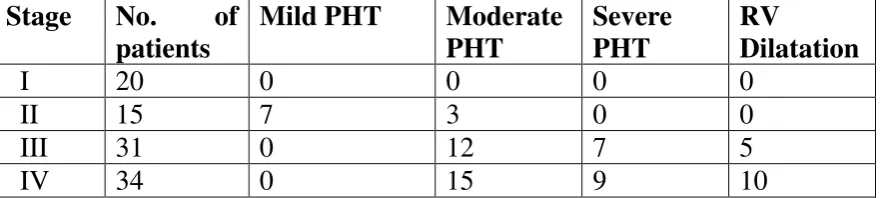
PULMONARY HYPERTENSION IN COPD
Mild PHT to some extent occur in most of the patients with advanced
disease and rarely severe pulmonary hypertension in few.
CAUSES
1. Pulmonary vasoconstriction due to alveolar hypoxia acidosis,
hypercapnia.
2. There is decrease in small vessels in areas of destroyed lung tissue
due to terminal air space destruction seen in patients with
emphysema.
3. Increased lung volume leading to compression of pulmonary
4. For every acute exacerbation there is approximately 20 mm of hg
rise in pulmonary arterial pressure. This ultimately end up in
pulmonary hypertension in repeated exacerbation.
MECHANISM OF PULMONARY HYPERTENSION BY
HYPOXIA
1. Vascular intimal thickening
2. Distal vessels vascularisation
3. Hypertrophy of the vascular media of the proximal vessels.
TREATMENT
Pulmonary vasodilators result in worsening of V/Q mismatch because
it is not the level of pulmonary hypertension but the degree of hypoxia
which produce the clinical symptoms and that these drugs further
worsen the situation.
The only proven treatment is oxygen therapy which decreases both
morbidity and mortality.
To keep the haemoglobin in upper limit of normal because low
haemoglobin is not tolerated in these patients due to hypoxemia.
Nocturnal or ambulatory SaO2 can assist in optimal O2 concentration.
it can happen either acutely during exacerbations or chronically due to
disease progression and worsening gas exchange and produces
irreversible vascular remodelling.
DIAGNOSIS
1. Echocardiogram
To identify the presence of right ventricular enlargement but the
study is difficult due to hyperinflated lungs and rotation of the
heart. It can be supplemented by ABG-showing PaO2 < 50 mm Hg
and Pco2 >50 mm hg.
2. Electrocardiogram
3. Cardiac catheterization – the gold standard investigation. MANAGEMENT
1. Acute cor pulmonale
a. Treatment of the precipitating cause
b. Supplemental o2 to maintain adequate oxygenation.
2. Chronic cor pulmonale
Should be very careful while using diuretics and digoxin in
patients with cor pulmonale because it sensitizes the heart to
digoxin and precipitates life threatening rhythm disturbances in
the presence of hypoxia and acidosis.
SPIROMETRY IN COPD
The test to confirm or refute a diagnosis of the disease is
spirometric evaluation as suggested by GOLD. As of them a post
bronchodilator Forced expiratory volume 1/ Forced vital capacity <
70% is used for diagnosis and the previous concept of absence of
post bronchodilator reversibility has been abandoned. Spirometry
has its own limitations and therefore a combination of clinical
symptomatology and spirometry improves the diagnostic accuracy.
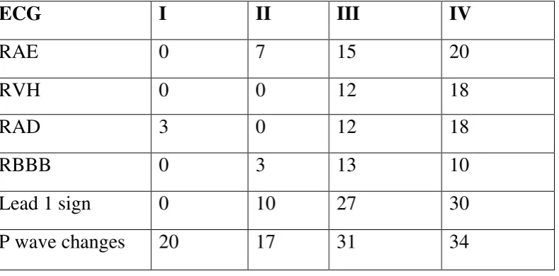
ELECTOCARDIOGRAM IN COPD
Chronic obstructive airways disease influence the electrical events of the
heart in the following basic respects:
1. the voluminous lungs have an insulating effect and thereby diminish the
transmission of electrical potential to the registering electrodes
2. the heart descends to a lower position within the thorax due to a
lowering of the diaphragm. This will alter the position of the heart
relative to conventional precordial electrode postions.
3. the right ventricle and right atrium become compromised due to a
reduction of the pulmonary vascular bed. This will result in right
ventricular hypertrophy as well as right atrial enlargement.this right atrial
enlargement lead to change in P wave vertical axis.
The voluminous lungs impair electrical transmission. The QRS and T
deflexions are therefore markedly diminished in magnitude.
Lead I sign
In patients with COPD the frontal plane P, QRS and T wave axes are not
infrequently all directed at around + 90 degree which are either precisely or
almost perpendicular to the standard lead I axis. As a result of this Lead I
reflects absent or very low amplitude P, QRS and T wave complexes giving the
appearance of minimally disturbed base line. This ECG phenomenon is known
as the Lead I sign.
Right atrial enlargement
Depolarization of atrium represented by the P wave. Normal frontal plane
P wave axis is directed to the right of +60 degree. In lead I and lead II and left
precordial leads, the P wave is upright. Lead III biphasic P wave may be seen
.Initial positive portion of the P wave indicate, activity of right atrium. Terminal
negative portion indicate, activity of left atrium.
The cause for P-axis verticalisation in lung hyperinflation is that the right
atrium is sturdy attached to the diaphragm by means of dense pericardial
ligament around the inferior vena cava. Due to progressive flattening of the
diaphragm, the right atrium is distorted/displaced inferiorly causing a significant
rightward deviation (verticalisation) of the P-wave axis.
In contrast, in pure restrictive (fibrotic) lung disease, correlating with the degree
The vertical P wave axis degree inversely correlating with FEV1, directly
proportional to disease severity
P pulmonale
It is reflected by P waves which are tall and peaked in standard leads II,
III, aVF, and is the expression of right atrial enlargement.
The combination of right axis deviation and tall peaked P waves is
Called P pulmonale. A comparison of interstitial pulmonary fibrosis and COPD
showed that a deviation of the frontal plane P wave axis to the right of +70
degree only occurs with COPD.
Abnormalities of the QRS complex
Right QRS axis deviation
The frontal plane QRS axis is deviated to the right and commonly
directed to +90 degree. When it is deviated further, the frontal plane leads will
usually reflect an S1Q3R3 pattern. In very severe cases it may be directed to
the “northwest” region.
Left QRS axis deviation
This occurs in about 10% of cases. The mechanism is still speculative.
S1, SII, SIII syndrome
I, II and III giving rise to the SI, S II, S III syndrome. This indirectly reflects
posterior displacement of the apex.
Posterior displacement of the mean QRS axis
The mean QRS axis may also be displaced somewhat posteriorly so that
it tends to be more obliquely oriented to the horizontal plane.
13
Abnormalities of the precordial QRS form
There is diminution of QRS magnitude in all the leads. It is not
uncommon for all the precordial leads to reflect rS complexes. In very severe
cases the R : S ratio is usually less than 1 in leads V4 to V6, and R wave
amplitude in lead V6 may be less than 5 mm. The transition zone is frequently
displaced to V6 or even further to the left.
Right bundle branch block
There may occasionally be transient complete or incomplete RBBB with
an exacerbation of the emphysema and increase in oxygen desaturation.
Right ventricle hypertrophy is mainly reflected by:
a. right axis deviation
b. prominent terminal S waves in the left precordial leads
The single most characteristic ECG feature of diagnosis in COPD is said
to be P wave axis of +70 to +90 degrees.
Abnormalities of T waves
It is usually similar in direction to that of QRS axis. The T wave is
diminished in amplitude in all leads. The T wave may be inverted in the right
precordial leads especially when pulmonary hypertension is marked.
Exacerbation of the disease with an increase in oxygen desaturation may
be associated with elevation of ST segment in leads II, III and AVF. This
manifestation is reversible.
USES
1. Diagnosis
2. Grading of severity In COPD patients the rate of decrease in
forced expiratory volume 1 per year is about 75-100 ml in sharp
contrast to normal persons’ 30 ml/ year.
3. Lung “age” assessment
By correlating patient’s FEV1 to the predicted FEV1 of his age
matched control. It may be used to encourage smokers to quit
smoking.
5. Pre-operative evaluation
These patients have a higher chance of developing
post-operative pulmonary complications.
1. Thoracic surgery
Predicted post operative FEV1 is calculated by applying
“rule of 5” in that 1/5th function is contributed by each lobe and the predictive post operative value is calculated by lobe
to be removed. A PPO FEV1 < 40% predicted is a contra
indication to surgery.
2. Non thoracic surgery
Factors predicting post-operative complications are
a. FEV1/FVC < 50%
b. Maximum voluntary ventilation< 50%
c. FEV1 or diffusing capacity <20% predicted
d. High partial pressure of Co2
e. Short distance of surgical site from the chest
SMOKING CESSATION
Smoking cessation is most important intervention in stopping the disease
process.
A. Counselling
B. Pharmacological assistance.
COUNSELLING
Counselling either by the clinician or non clinician increases cessation rate
many times than self initiated attempts.
5A intervention approach
1. ASK – information about tobacco use at every visit.
2. ADVICE – should be strong and individualized depending on patient’s health status and other considerations.
3. ASSESS – patient’s willingness. If patient is not willing he should be insisted to quit.
4. ASSIST – by speaking with the patient a give up plan; setting a date to stop smoking in the next 2 weeks, to reduce alcohol use, educate
households and providing psycho social support.
5. ARRANGE – follow-up.
PHARMACOTHERAPY
First line agents
Nicotine replacement therapy
Gum
Dose : 2 mg – 1-24 cigarettes/d; 4mg ->25 cigarettes/d
Duration : 12 weeks
Adverse effects : dyspepsia
Lozenge
Dose : 2mg – first smoke > 30 min after waking up from bed; 4mg – first smoke < 30 min after waking up from bed.
Maximum of 20 mg/d
Duration : 12 weeks
Side effects : nausea, insomnia.
Inhaler
Dose : 6-16 cartridges of 4 mg each/d
Duration : 6 months with dose reduction in last 3 months
Side effects : rhinitis, local irritation
Spray
Dose : 0.5 mg in Each nostril 1-2 doses/he
Duration : 3-6 months
Side effects : local irritation
Patch
Dose: 16 or 24 hr patch for total duration of 8 weeks with dose reduction
Side effects : local skin reaction, insomnia
Advantage : good compliance, requires less skill.
Contra indication to NRT :
1. CAD – MI or unstable angina 2. CVA
3. Untreated acid peptic disease
Bupropion sustained release
Mechanism of action
nor-epinephrine and dopamine reuptake inhibitor
dose : 150 mg once daily x 3 days; 150 mg bd x 7-12 weeks
duration: 6 to 12 months
side effects : insomnia, dry mouth
particularly preferred in patients with concomitant depression
varnecline
mechanism of action
partial agonist of nicotinic ach receptor
dose : 0.5 mg od x 1- 3 days; 0.5 mg bd x 4-7 days; 1mg bd from then up to
8 weeks.
Side effects : CNS – suicidal intention which has resulted in FDA warning.
Second line agents
1. Clonidine :
Mechanism of action : post synaptic alpha2 agonist
Dose : 0.1 – 0.4 mg/d x 2-6 weeks 2. Anxiolytics
Diazepam, buspirone, beta blockers
3. Sensory replacement
Citric acid inhaler, denicotinised tobacco, black pepper extract.
4. Acupuncture : by releasing endorphins
ANTAGONISTS
Mecamylamine
1. Naltrexone
Aversion causing drugs
Silver acetate – poor compliance SMOKING PREVENTION
Smoking behaviour in around 90% smokers were initiated during
adolescence. So prevention is more important and effective than
cessation after the behaviour has begun.
Measures :
1. Health education – target population : adolescents, young adults 2. Public health programs
3. Smoke free public places.
EMERGING THERAPIES
The imbalance between pulmonary oxidant load and anti-oxidants
leads to oxidative stress and causes inflammation and airway
modelling. This functions as an important pathogenetic mechanism
and probably the reason for steroid resistance and so has been
targeted for newer therapies.
So newer drugs are developed with the aim to inhibit this one of
the several steps involved in the inflammatory pathogenesis of
COPD with the concern of developing effective but also safe drugs.
Smoking cessation is considered to be an important and effective
intervention to halt the decline of lung function beyond any doubts.
And now recent research has developed anti-free nicotine
antibodies.
These anti free nicotine antibodies bind with free nicotine denying
their access across the blood brain barrier. And hence nicotine from
cigarettes cannot stimulate nicotine receptors causing smoking an
unpleasant experience
NEWER BRONCHODILATORS
Bronchodilators provide immediate symptom relief even though
they do not arrest the disease process and thereby improve
wellbeing of the patient. Hence this serves as an important
component of the treatment plan.
So for this purpose, newer long and ultra long acting
bronchodialtors have been developed with the aim of meeting the
short comings of short acting ones which is poor compliance due to
multiple dosing. These ultra long acting bronchodilators are ultra
long acting muscarinic antagonists and ultra long acting beta2
agonists.
ULTRA LONG ACTING MUSCARINIC ANTAGONISTS
These agents have been shown to decrease the number of
so they have the ability to attenuate the disease process partially
and hence provide mortality benefit.
DRUGS UNDER RESEARCH :
1. Dexpironium
2. Daratropium bromide
3. Aclidinium
4. TD-4208
5. GSK-573719
6. Glycopyronium bromide
ULTRA-LONG ACTING BETA2 AGONISTS
This includes
1. Vilanterol – safe and efficacious
2. Indacaterol – approved in Europe and USA
3. Carmoterol – using modulate technology, the amount of inhaled drug reaching the target has been increased.
4. GSK-642444
5. MILVETEROL – PROVED EFFICACIOUS IN ASTHMA, NOT YET RESEARCHED ON copd
6. BI-1744-CL
7. UK-503590
NOVEL COMBINATIONS
Rationale of combining long acting drugs
1. Synergism between two agents
2. Patients are analysed whether beta or muscarinic receptor
predominance in their airway respond to their respective drugs
more than the other. So that their combination can overcome the
problem of receptor variation.
Though several combination of these agents along with steroids to
decrease exacerbations are under development, there is a potential
limitation of using these combination. The limitation is that
delivery of these agents at different site which decrease their
synergism. But still a solution which could overcome these
limitation has been witnessed. It is by combining them into a dimer
which serves to deliver the agents at the same site – dual acting muscarinic antagonist beta 2 agonist.
Even other combinations under development are combining a
LABA with an ICS or combining LABA with inhaled steroid and
LAMA.
Over the decades there has been search for an effective and safe
anti inflammatory drug with the potential of reducing lung
inflammation. But still it has turned out to be in vain.
But one of the recent break through finding is that oxidative stress
causing decrease in HDAC in these patients has been the reason for
corticosteroid unresponsiveness in them. And that theophylline
increases this enzyme at cellular levels. Hence theophylline has
made a comeback in the management of the disease with the aim of
additive anti inflammatory effect when given with steroids and
studies are underway.
PDE-4 INHIBITORS
Phosphodiesterase-4 is an enzyme found specifically in most
inflammatory cells. This has been targeted to reduce the
inflammatory process of the disease. So a selective PDE-4 inhibitor
has been approved for use. This has shown to increase FEV1
comparatively much better than tiotropium. And hence these drugs
could be the best adjuvant therapy with bronchodilators. It is under
study to introduce an inhalational form of this class. The key
problem is that the side effects of this drug – gastro intestinal and upper respiratory tract effects. And this has led to search for