QUANTIFICATION OF PLASTIC REMNANTS ON
TITANIUM SURFACE AFTER INSTRUMENTATION
AND EVALUATION OF EFFICACY OF ITS REMOVAL
AFTER IRRIGATION BY USING A CONFOCAL
MICROSCOPE
Dissertation submitted to
THE TAMILNADU Dr. M.G.R. MEDICAL UNIVERSITY
In partial fulfilment for the Degree of
MASTER OF DENTAL SURGERY
BRANCH II
CERTIFICATE
This is to certify that this dissertation titled, “QUANTIFICATION OF PLASTIC
REMNANTS ON TITANIUM SURFACE AFTER INSTRUMENTATION AND
EVALUATION OF EFFICACY OF ITS REMOVAL AFTER IRRIGATION BY
USING A CONFOCAL MICROSCOPE” is a bonafide record of work done by
Dr. NIMISHA GOPAL under our guidance and to our satisfaction, during her
postgraduate study period of 2016-2019.
This dissertation is submitted to THE TAMILNADU Dr. M.G.R. MEDICAL
UNIVERSITY in partial fulfillment for the award of the degree of MASTER OF
DENTAL SURGERY - PERIODONTICS,BRANCH II. It has not been submitted
(partial or full) for the award of any other degree or diploma.
Dr. Koshy Chithresan, MDS, Dr. J. Srihari, MDS, Professor & Head of the Department, Professor and Guide,
Department of Periodontics, Department of Periodontics, Sri Ramakrishna Dental College & Sri Ramakrishna Dental
Hospital. College & Hospital.
Dr. V. Prabhakar, MDS, Principal,
Sri Ramakrishna Dental College & Hospital.
Date:
DECLARATION
TITLE OF DISSERTATION
QUANTIFICATION OF PLASTIC REMNANTS ON TITANIUM SURFACE AFTER
INSTRUMENTATION AND
EVALUATION OF EFFICACY OF ITS REMOVAL AFTER IRRIGATION BY USING A CONFOCAL MICROSCOPE
PLACE OF STUDY
SRI RAMAKRISHNA DENTAL COLLEGE AND HOSPITAL, COIMBATORE-641006.
DURATION OF THE COURSE 3 YEARS
HEAD OF THE DEPARTMENT Dr. KOSHY CHITHRESAN
NAME OF THE GUIDE Dr. J. SRIHARI
I hereby declare that no part of the dissertation will be utilized for gaining
financial assistance or any promotion without obtaining prior permission of the
Principal, Sri Ramakrishna Dental College and Hospital, Coimbatore. In addition, I
declare that no part of this work will be published either in print or in electronic media
without the permission of the guide who has been actively involved in this dissertation.
The author has the right to reserve for publish of work solely with the prior permission
of the Principal, Sri Ramakrishna Dental College and Hospital, Coimbatore.
Head of the Department PG Guide Signature of the Candidate
Urkund Analysis Result
Analysed Document: THESIS.docx (D46462454)
Submitted: 1/6/2019 6:59:00 PM
Submitted By: [email protected]
Significance: 0 %
Sources included in the report:
Instances where selected sources appear:
0
CERTIFICATE – II
This is to certify that this dissertation work titled
“QUANTIFICATION OF PLASTIC REMNANTS ON TITANIUM SURFACE
AFTER INSTRUMENTATION AND EVALUATION OF EFFICACY OF ITS REMOVAL AFTER IRRIGATION BY USING A CONFOCAL MICROSCOPE”
of the candidate Dr. NIMISHA GOPAL with registration number 241613352 for the
award of MASTER OF DENTAL SURGERY in the branch of PERIODONTICS
(BRANCH II). I personally verified the urkund website for the purpose of plagiarism
check. I found that the uploaded thesis file contains all pages from introduction to
conclusion and the result shows 0% plagiarism in the dissertation.
Head of the Department PG Guide
ACKNOWLEDGEMENTS
Foremost, I thank God Almighty for all the blessings bestowed upon me.
I thank my guide Dr. J.Srihari, MDS, Professor, Department of Periodontics,
Sri Ramakrishna Dental College and Hospital, for his able guidance throughout and
instilling in me that learning is a continuous and everlasting process. His perfectionist
ways have helped me grow in many ways.
I would like to express my whole hearted gratitude to Dr. Koshy Chithresan,
MDS, Head of the Department, Department of Periodontics, Sri Ramakrishna Dental
College and Hospital for taking out the time and being patient with me throughout my
period of dissertation and for helping me understand the subject better.
I thank Dr. Arun Maradi, MDS, Reader, Dr. Praveen Krishna, MDS, Reader,
and Dr. Rajesh, MDS, Senior Lecturer, Department of Periodontics, Sri Ramakrishna
Dental College and Hospital for the constant support and guidance.
I also thank Dr. Surya, Dr. Vimala and Dr. Cynthia for their support.
I thank Dr. V. Prabhakar, MDS, Principal, Sri Ramakrishna Dental College and
Hospital, for providing me with the necessary facilities.
I take this moment to thank my seniors; Dr. Subash, Dr. Fazal Ilahi, Dr. Megha
Madhusoodhanan, Dr. K.K.N Saranya and Dr. Asha for their steady support and
guidance throughout this period. I thank my juniors; Dr. Sheetal and Dr. M. Indrajit for
being supportive throughout. My sincere thanks to Dr. M. Junaid for his expertise in
statistics.
Special thanks to Dr. Ritika Chhalani and Ms.Sneha Venkataramana for keeping
I express my sincere thanks to Dr. M.R.Ganesh, PhD, Assistant Professor and
Ms.Saranya Sekar, MPhil, National Facility for Clinical Trial, Interdisciplinary
Institute of Indian System of Medicine (IIISM) at SRM University, Kattankalathur for
their time and effort.
I sincerely acknowledge Dr. P.Radhakrishnan, PhD, Professor and Dean of PSG
Institute of Advanced Studies and Mr.Sakthi Vignesh, Assistant Professor, Department
of Robotics and Automation Engineering, PSG College of Technology for their time
and effort.
None of this would have been possible without the support of my family. I thank
my parents Mr. K. Gopalan and Mrs.Pushpa Gopal for always standing by me, believing
in me and for being my strength. I thank my sister Ms.Nithu Mahendhiran and family
for always being my support system in all ways.
CONTENTS
S.NO. INDEX PAGE NO.
1. INTRODUCTION 1
2. AIMS & OBJECTIVES 2
3. REVIEW OF LITERATURE 3
4. MATERIALS & METHODS 26
5. RESULTS 44
6. DISCUSSION 67
7. SUMMARY & CONCLUSION 73
8. BIBLIOGRAPHY 75
LIST OF FIGURES
Figure
No. Title Page No.
Flowchart Methodology 27
1 Group I – Plastic Curette (Hu-Friedy) 31
2 Group II – Carbon Composite Tip (Satelec) 31
3 Group III – Peek Tip (EMS) 31
4 Surface modified titanium discs before instrumentation 32
5 Instrumentation using Plastic curette (Hu-Friedy) 33
6 Instrumentation using Carbon composite tip (Satelec) 33
7 Instrumentation using PEEK tip (EMS) 34
8 Irrigation using 0.2% chlorhexidine 35
9 Irrigation using air-water spray 35
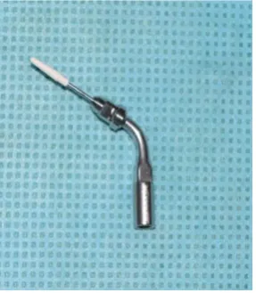
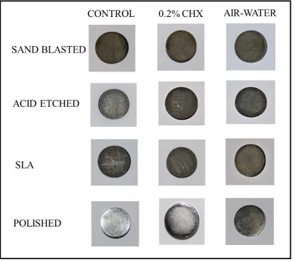
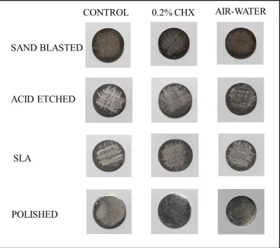
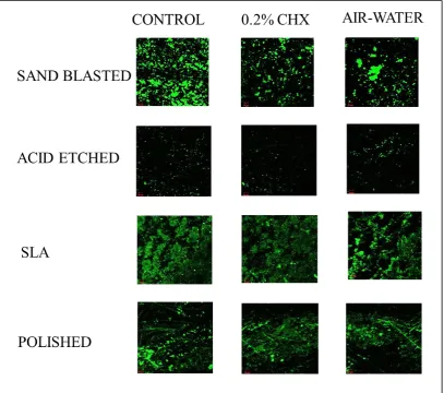
10 Surface modified titanium discs following instrumentation
and irrigation with Group I – Plastic curette 36
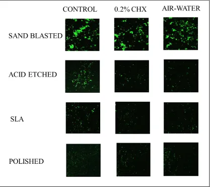
11 Surface modified titanium discs following instrumentation
and irrigation with Group II – Carbon composite tip 37
12
Surface modified titanium discs following instrumentation
and irrigation with Group III- PEEK tip 38
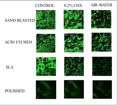
14 Titanium disc in focus under confocal laser scanning microscope 39
15 Surface modified titanium discs before instrumentation
under confocal laser scanning microscope 40
16
Surface modified titanium discs following instrumentation and irrigation with Group I – Plastic curette (Hu-Friedy)
under confocal laser scanning microscope 41
17
Surface modified titanium discs following instrumentation and irrigation with Group II – Carbon composite tip
(Satelec) under confocal laser scanning microscope 42
18
Surface modified titanium discs following instrumentation and irrigation with Group III- PEEK tip (EMS) under confocal laser scanning microscope
LIST OF TABLES
Table Title Page No.
1 Mean area covered by plastic remnants after instrumentation
with 3 Groups 50
2 Comparison among the groups 50
3 Mean area covered by plastic remnants after instrumentation
and irrigation in Group I 51
4 Mean area covered by plastic remnants after instrumentation and irrigation in Group II 51
5 Mean area covered by plastic remnants after instrumentation
and irrigation in Group III 51
6 Mean area covered by plastic remnants after instrumentation on various modified surfaces followed by irrigation in Group I 52
7 Mean area covered by plastic remnants after instrumentation on various modified surfaces followed by irrigation in Group II
53
8 Mean area covered by plastic remnants after instrumentation on various modified surfaces followed by irrigation in Group III
54
9 Intergroup comparison among various modified surfaces after
irrigation with 0.2% CHX 55
10 Intergroup comparison among various modified surfaces after
irrigation with air-water spray 55
11 Mean area covered by plastic remnants in 3 groups after
instrumentation and irrigation with 0.2% CHX 56
12 Mean area covered by plastic remnants in 3 groups after
[image:11.595.112.524.89.775.2]LIST OF GRAPHS
Graph Title Page No.
1 Mean area covered by plastic remnants after instrumentation
in 3 Groups 57
2 Mean area covered by plastic remnants after instrumentation
and irrigation in Group I 58
3 Mean area covered by plastic remnants after instrumentation and irrigation in Group II 59
4 Mean area covered by plastic remnants after instrumentation
and irrigation in Group III 59
5 Mean area covered by plastic remnants after instrumentation
and irrigation in Group I sand blasted titanium discs 60
6 Mean area covered by plastic remnants after instrumentation
and irrigation in Group I acid etched titanium discs 60
7 Mean area covered by plastic remnants after instrumentation
and irrigation in Group I SLA titanium discs 61
8 Mean area covered by plastic remnants after instrumentation
and irrigation in Group I polished titanium discs 61
9 Mean area covered by plastic remnants after instrumentation
and irrigation in Group II sand blasted titanium discs 62
10 Mean area covered by plastic remnants after instrumentation
and irrigation in Group II acid etched titanium discs 62
11 Mean area covered by plastic remnants after instrumentation
12 Mean area covered by plastic remnants after instrumentation
and irrigation in Group II polished titanium discs 63
13
Mean area covered by plastic remnants after instrumentation
and irrigations in Group III sand blasted titanium discs 64
14
Mean area covered by plastic remnants after instrumentation
and irrigation in Group III acid etched titanium discs 64
15
Mean area covered by plastic remnants after instrumentation
and irrigation in Group III SLA titanium discs 65
16
Mean area covered by plastic remnants after instrumentation
and irrigation in Group III polished titanium discs 65
17
Mean area covered by plastic remnants in 3 Groups after
irrigation with air-water spray 66
18
Mean area covered by plastic remnants in 3 Groups after
GLOSSARY
Abbreviation Expansion
ASTM American Society for Testing and Materials
AFM Atomic Force Microscopy
BI Bleeding Index
BOP Bleeding On Probing
CA Citric Acid
CAL Clinical Attachment Level
CFU Colony Forming Unit
CHX Chlorhexidine
CI Calculus Index
CLSM Confocal Laser Scanning Microscope
DNA Deoxyribo Nucleic Acid
EDS Energy Dispersive X-Ray Spectroscopy
EDTA Ethylene diamine tetra acetic acid
ERL Erbium-doped Yttrium Aluminium Garnet Laser
GI Gingival Index
H2O2 Hydrogen Peroxide
HA Hydroxy apatite
HCL Hydrochloric acid
HeNe Helium Neon
ICPMS Inductively Coupled Plasma Mass Spectrometry
Lr Waviness
MGI Modified Gingival Index
MP Machine Polished
NaCl Sodium Chloride
PBS Phosphate Buffered Saline
PD Probing Depth
PEEK Polyetheretherketone
PI Plaque Index
Ra Average Surface Roughness
RBM Resorbable Blast Material
RSR Relative Specular Reflectance
Rz Maximum Surface Roughness
SEM Scanning Electron Microscope
SI Stain Index
SLA Sand Blasted and acid etched
Ti Titanium
TIE Trans mucosal Implant Extensions
TPS Titanium Plasma Spray
INTRODUCTION
1
Complete debridement of plaque and all other decontaminants from the implant
surface is of paramount importance for long term success and survival of dental
implants. Various techniques have been used which includes, non-surgical and surgical
decontamination with mechanical instruments, antimicrobial therapies and/or lasers1.
Among all these techniques, surface debridement using plastic and titanium
curettes appears to be the treatment protocol for implant mucositis and
peri-implantitis. It has been proposed that treating the titanium surface with plastic or non-
metal tip prevents damage to the implant surface as compared to the use of metal
instruments as the use of metal instruments leads to increased roughness of implant
surface and increased biofilm formation2. But, it has been known that instrumenting
with any material softer than titanium may leave remnants on the titanium surface, 3,4
which can influence bacterial attachment, cell attachment and effectiveness of oral
hygiene measures, thereby impairing the biocompatibility of the implant surface5.
To investigate the nature of these effects, it is necessary to ascertain the amount
of plastic debris left on various modified titanium surfaces after treatment. Plastic
materials have been known to show significant auto fluorescence when visualised under
laser irradiation6. Based on this background, the current in-vitro study was conducted
under non-simulated conditions to quantify the plastic remnants on surface modified
titanium after instrumentation with plastic instruments by using a Confocal laser
scanning microscope and also investigate the efficacy of removal of these remnants by
AIMS & OBJECTIVES
2
AIM: The aim of this in-vitro study was to quantify the surface area covered by plastic
remnants on different surface modified titanium discs after instrumentation with
various plastic instruments and to evaluate the efficacy of the removal of these remnants
by using a confocal laser scanning microscope under non-simulated conditions.
OBJECTIVES:
1. To quantify the plastic remnants on titanium discs that are sand blasted, acid
etched, SLA treated and polished after instrumentation with various plastic
instruments such as PEEK ultrasonic tip (EMS Piezon Systems), carbon
composite ultrasonic tip (PH1, Satelec, Suprason) and plastic curette (Columbia
4R/4L, Hu Friedy) by confocal microscopy.
2. To evaluate the efficacy of the removal of these remnants after irrigation with
water spray and 0.2% chlorhexidine for 10 seconds by confocal microscopy.
3. To compare the amount of plastic remnants remaining on various surface
3
REVIEW OF LITERATURE
INSTRUMENTATION:
An in-vitro study was conducted by Fox et al in 1990 to evaluate the effects of
scaling titanium implant surface (IMZ titanium implant system) using titanium alloy
curette, a stainless steel curette and a plastic instrument specifically designed for
instrumentation of dental implants. Alterations of the surfaces due to instrumentation
were evaluated by a Helium Neon (HeNe) laser and was reported as relative specular
fluorescence (RSR). It was concluded that plastic instruments produced an insignificant
alteration of the titanium implant surface following instrumentation, while metal
instruments such as titanium and stainless steel curettes significantly altered the
titanium surface7.
A study was conducted by Dmytryk et al in 1990 to evaluate the effects of
scaling titanium implant surfaces with Plastic (Branemark), titanium-alloy (Norton) and
stainless steel curette (Hu-Friedy) on fibroblast cell attachment. Counts of attached cells
were made at 24 and 72 hours; the implants were then processed for scanning electron
microscopy (SEM). It was found that the cell attachment to stainless steel curette
instrumented titanium surfaces was significantly reduced compared to untreated
control, titanium alloy curette or plastic instrumented surfaces. SEM observations
showed that fibroblast on stainless steel instrumented surfaces tended to show a
somewhat rounded morphology and a relatively reduced degree of spreading; while
fibroblasts on untreated control, plastic, or titanium-alloy instrumented surfaces showed
a well-spread, polygonal morphology, which were more typical of fibroblasts in
favourable culture conditions. Hence it was concluded that such observations of cell
4
are indicative of in-vivo biocompatibility and might have clinical implications for the
proper maintenance of titanium dental implants8.
The cleaning effectiveness of different treatment methods on titanium
abutments was evaluated using scanning electron microscope (SEM) by Speelman JA
et al in 1992. Titanium abutments were installed on beagle dogs. After 16 weeks of
plaque accumulation, the abutments were instrumented using 1) Metal Gracey curettes
(LM Dental); 2) Plastic scalers (Nobelpharma); 3) Ultrasonic scalers (Cavitron,
Dentsply); 4) Air-polishing (Stainbuster with Prophy-Jet cleaning powder, Dentsply);
5) Rubber cup polishing with pumice and 6) Brushing with a multitufted brush (Butler).
It was found that regular rubber cup polishing and regular brushing resulted in highest
surface cleanliness while the air polishing showed the lowest cleanliness score.
Treatment with metal, plastic and ultrasonic instruments clinically resulted in clean
surfaces. The finding that daily brushing resulted in clean surfaces stresses the
importance of daily oral hygiene for implant patients. Finally, taking into consideration
the cleanliness, the surface roughness and the possible adverse effects on the
biocompatibility, it was concluded that plastic scalers may be the instruments of choice
for professional debridement of titanium implant surface9.
In an in-vitro study by Alan Homiak et al in 1992, the surface of titanium
implant abutments using light and scanning electron microscope and also the effects of
various forms of hygiene prophylaxis instrumentation on the abutments were evaluated.
The instruments used were stainless steel scaler (American Dental Mfg,Co) using
moderate finger pressure, plastic scaler (Nobelpharma), rubber cup polishing (Densco
Prophy Cup) and air powder abrasive unit (Cavi-Jet, Dentsply). The metal scaler system
was found to roughen the titanium surface. All other modalities tested appeared to
5
sharp machined grooves present on the untreated abutment surface. These findings
suggested that the use of plastic scaler, rubbercup polishing and air powder abrasive
system did not harm the titanium surface and could be thus used for debridement10.
The surface texture of titanium implant abutments after instrumentation with
plastic scaler, air-powder abrasive and rubber cup polishing was evaluated in an in-vitro
study by Mc Collum et al in 1992. Plaque accumulation was also compared following
instrumentation. The untreated control abutments revealed prominent milling marks
and slight pits, the plastic scaler was found to slightly smoothen the milling mark and
it caused micro scratches. The air-powder abrasive largely obliterated the milling marks
and caused surface pitting whereas the rubber cup with polishing removed the milling
marks and created a smooth swirl pattern. It was found that all abutments collected
plaque and there was no statistically significant difference between the groups. Thus, it
was concluded that, for maintenance and prophylaxis, any of these methods may be
used without damaging the abutment surface or enhancing plaque accumulation11.
Brookshire et al in 1993 in an in-vitro study compared the surface quality of
both commercially pure titanium and titanium alloy abutments, subjected to various
hygiene methods and instruments such as the 1) Implarette scaler - instrument tip was
fabricated from a gold palladium alloy with a gold coating; 2) Plasteel scaler
(Implacarea, Hu-Friedy) fabricated from a high grade resin called Plasteel; 3) Universal
scaler (Steri-Oss Inc.) which was fabricated from graphite fiber; 4) A slow speed
handpiece and prophy angle with screw-type rubber cup and tin oxide slurry; 5) The
Prophy-Jet air-powder abrasive system (Dentsply). The surfaces were then analysed
under a scanning electron microscope (SEM). Results showed that, no significant
surface alterations were produced by the air abrasive system. Implarette scaler,
6
after instrumentation. It was also found that surfaces treated with the Implarette scaler
exhibited the most damage among all the hygiene instruments. Hence, from the findings
it was concluded that the air abrasive system seemed to produce the least surface
alterations as compared to the Implarette scaler, Plasteel scaler and Universal scaler12.
The effect of modified scaler tips on variously structured titanium surfaces using
stereomicroscopy, scanning electron microscopy and laser profilometry was
investigated by Ruhling et al in 1994. Instrumentation was done on different implant
surfaces such as smooth machined titanium, etched and sandblasted surfaces,
titanium-plasma-sprayed (TPS) and hydroxyapatite coated surfaces (HA) using Cavimed-200
ultrasonic scaler with Teflon coated tip, Sonicflex-2000 sonic scaler with Teflon coated
tip, Light curette, Implacare curette and metal instruments such as Cavimed-200
ultrasonic scaler with stainless steel tip, Sonicflex-2000 sonic scaler with stainless steel
tip, Implarette gold plated curette and stainless steel Gracey curette (Hu-Friedy). The
results revealed that no discernible damage was caused by Teflon coated sonic and
ultrasonic scalers or implant curettes made of plastic on smooth titanium surfaces.
Surface roughness increased with the use of metal instruments on smooth titanium
surfaces. Instrument material residues were found on rough implant surfaces. Thus, it
was concluded that coating of sonic and ultrasonic scaler tips with Teflon could be used
for supragingival and subgingival cleaning of titanium implant surfaces3.
Kuempel et al in 1995 conducted a study to examine the epithelial growth on
titanium surfaces after instrumentation with plastic scaler, stainless steel scaler and gold
coated curettes. The discs were then seeded with a microdot of rat gingival cells. At the
end of 5 days, the surface area covered by the epithelial cells were then measured.
Results showed that gold coated curette exposed surfaces had less epithelial growth
7
coverage did not vary significantly among groups. But, the specific characteristics of
the cellular morphology were found to be different among the groups. Thus, it was
concluded that the reduced epithelial growth in gold coated curette instrumented discs
might be due to the surface contaminants originating from the gold curette13.
Meschenmoser et al in 1996 assessed quantitatively and qualitatively effects of
various instruments such as stainless steel curette (Schweickhardt); plastic curette
(Nobelpharma); a prototype of pure titanium curette, an air abrasive polishing system
(Airflow II, EMS) and an ultrasonic system (Cavitron) on titanium abutments. The
surface structures were compared with scanning electron microscope (SEM),
profilometry and Confocal laser scanning microscope. Evaluation revealed surface
alterations for all instruments and systems except the plastic curette which did not
roughen the surface. The steel curette and the ultrasonic system proved to be totally
unsuitable for cleaning titanium abutments. Even though plastic curette did not roughen
the surface, the effectiveness of plastic curette for removing hard calculus and the
resultant plastic debris on the implant surface were not evaluated in this study14.
Surface alterations on titanium implant necks following different prophylaxis
procedures such as ultrasonic scaler, Plastic tip ultrasonic scaler, Stainless steel curette,
Titanium curette, Teflon curette, Air powered system, Abrasive rubber cups, polishing
rubber cup and brush was evaluated by Matarasso et al in 1996. SEM and laser
prophylometer analysis was done to measure the roughness in terms of average surface
roughness (Ra) and maximum surface roughness (Rz). Results showed that the use of
ultrasonic scaler, stainless steel curette, titanium curette and air jet polishing increased
the implant surface roughness as compared to controls whereas abrasive rubber cups
increased the implant surface smoothness. Use of rubber cup polishing, brush polishing,
8
Hallmon et al in 1996compared the effects of metallic, non-metallic and sonic
instrumentation on titanium abutment surface in-vitro, using scanning electron
microscopic (SEM) examination. The instruments used were Stainless steel Gracey
curette (Hu-Friedy), Implacare plastic curette (Hu-Friedy), Plastic curette (Steri-Oss),
Plastic curette (Implant Support), Sonic scaler with metal tip (Titan – S) and Sonic
scaler with plastic tip (Dynatip). The highest surface alteration was seen with the
Implarette scaler followed by sonic scaler, Gracey curette, Dynatip and Steri-Oss. The
Implacare and Implant Support non-metallic scaler had the least surface alteration. It
was concluded that the Implacare and Implant support non-metallic (plastic) scalers
appear to be the instruments of choice for debridement of titanium abutment surfaces if
preservation of surface integrity is the primary objective16.
Mengel et al in 1998 examined the work traces left by various instruments such
as Titanium curette (Deppeler SA), Gracey curette (Hu-Friedy), Plastic curette (Nobel
Biocare), Rubbercup with Zircate prophypaste (Dentsply), Cavitron Jet ultrasonic
scaler with universal insert (Dentsply), Cavitron Jet air polishing nozzle with
Prophy-Jet cleaning powder (Dentsply), Densonic sonic scaler with SofTip disposable prophy
tip (Dentsply) and Densonic sonic scaler with universal tip (Dentsply) on implants and
abutments by scanning electron microscope and determined the quantity of substance
removal by optical laser profilometry. It was found that the Gracey curette, the Cavitron
Jet ultrasonic scaler and the Densonic scaler with universal tip left moderate to
pronounced work traces and caused increased substance removal followed by the
titanium curette and the Densonic sonic scaler with SofTip disposable prophy tip which
left slight working traces The rubber cup, the plastic curette and the Cavitron Jet air
polishing system caused no visible change to the implant surfaces and caused the least
9
Augthun et al in 1998 examined the effect of specific cleaning procedures such
as Plastic curette (DIA 238), Metal curette (Hu-Friedy), Diamond polishing device
(Perioset/blue), Ultrasonic scaler (Satelec), Air powder spray with sodium
hydrocarbonate solution (Plaque Sweep) and 0.1% CHX solution rinse on the surface
of 3 implant types with different coatings and shapes (plasma sprayed, hydroxyapatite
coated implants and smooth titanium surface screws) using SEM. The air powder
abrasive system, CHX rinse and curettage with the plastic instrument caused little or no
surface damage in all but hydroxyapatite coated fixtures. The growth of vital cells on
contaminated implants was also observed after treatment. It was found that implants
sprayed with the air- abrasive system had the most vital cells. Hence it was concluded
that, the use of plastic scalers and air abrasive system had the least damaging effect on
plasma coated and smooth titanium implant surfaces18.
The effects of Er: YAG laser (ERL) and the Vector ultrasonic system on the
biocompatibility of titanium implants with four different surfaces (sand-blasted and
acid-etched (SLA), titanium plasma-sprayed (TPS), machine-polished (MP) and
hydroxyapatite-coated (HA)) in cultures of human osteoblast-like cells was investigated
by Schwarz et al in 2002. Cells were counted using a reflected light microscope and the
cell density per mm2 was calculated. Additionally, cell morphology and surface
alterations of the titanium discs after treatment were investigated using SEM. It was
found that the highest number of cells per mm2 were seen on SLA surfaces, followed
by the TPS and MP surfaces. The HA- coated surfaces showed the least cell density per
mm2. In the laser-treated groups, no thermal side effects such as melting or loss of
porosity were observed. However, all surfaces treated with the Vector system showed
10
that Er:YAG laser did not damage titanium surfaces and subsequently did not influence
the attachment rate of SAOS-2 cells4.
Sato et al in 2004 compared the effects of a new ultrasonic scaler with carbon
tip (Vector), a conventional ultrasonic scaler with plastic tip (Satelec) and a plastic
scaler on titanium surfaces. The roughness was measured with a Profilometer and
observed by SEM. It was found that the rate of debris removal by the Vector scaler and
the conventional ultrasonic scaler were higher than the plastic scaler. There were no
significant differences in surface roughness among the 3 instruments. Hence, it was
concluded that the new ultrasonic scaler and conventional ultrasonic scaler were useful
for removing artificial debris and produced no significant damage to titanium surfaces
compared to plastic scalers19.
Karring et al in 2004 compared the effectiveness of treatment of peri-implantitis
with a Vector system and carbon composite curette. Instrumentation was done at
baseline and at the end of 3 months. Plaque, BOP and PPD were recorded on all implant
surfaces at baseline, and after 3 and 6 months. At the end of 6 months, it was found that
four of the Vector treated sites and one site treated with carbon curettes had stopped to
bleed. Thus, it was concluded that there was greater reduction in the number of sites
with BOP following treatment with the Vector system than following instrumentation
with carbon fiber curettes, but the difference was not found to be statistically
significant20.
In an in-vitro study by Ramaglia et al in 2006, the effects of different
instrumentations used in the treatment of peri-implantitis on implant surfaces coated
with hydroxyapatite or titanium plasma spray (TPS) was investigated. The implant
11
(Implant scaler, Premier), ultrasonic scaler tip (Satelec) and air-powder-water spray
(Airflow). Profilometry and SEM were used to examine the instrumented surfaces. It
was found that the plastic curette and air-powder-water spray induced less implant
surface alterations, though these instrumentations left deposits on the surface that may
affect, in-vivo, the tissue healing process21.
Kawashima et al in 2007 evaluated the treatment of titanium implants with
ultrasonic scalers with Carbon tip (Vector), Plastic tip (Satelec) or Metallic tip (Enac).
The abutment surface characteristics were examined after instrumentation using SEM
and laser profilometer. The amounts of remaining plaque and calculus were estimated
using the modified remaining plaque and calculus score developed by Speelman. The
surface alterations were evaluated using the modified roughness score developed by
Hallmon. The abutments treated with the Vector scaler and plastic scaler had essentially
clean and smooth surfaces. No calculus was observed, although some small particles of
amorphous material were seen. The abutments treated with the metallic tip scaler had
irregularities and defects but had clean surfaces with no calculus. Thus it was concluded
that piezoelectric scalers with non-metal tips were suitable for use in dental implant
maintenance22.
Mann et al in 2011 conducted a study to assess the effect of plastic covered
ultrasonic scalers on titanium implant surface. The inserts used included a TFI 10
metallic tip and a plastic coated ultrasonic implant insert (SofTip, Dentsply) driven by
a Cavitron SPS 30 kHz ultrasound generator. The plastic cover of the modified insert
probe was screwed into place on an adapted metallic scaler. The implant surfaces were
then scanned using laser profilometer and SEM. It was found that the metal scalers
12
minimal damage to implant surface. It was also found that the plastic coated scalers had
a polishing action and left plastic deposits behind on the implant surface23.
A study to evaluate the safety and efficiency of novel ultrasonic scaler tips,
conventional steel tips and plastic tips on titanium surface. Mechanical instrumentation
was carried out using conventional scalers with a novel metallic implant tip (Cetatech),
a plastic headed tip (EMS), Plastic tip (Satelec) and a conventional stainless steel tip
(EMS) was conducted by Baek et al in 2011. The instrumented surface samples were
viewed with a SEM and surface profile was investigated using an atomic force
microscope (AFM). SEM images on surfaces scaled by the novel metallic implant tip
and the EMS plastic tip showed no marked differences in surface morphology. Surfaces
instrumented using the conventional stainless steel tip showed higher surface
roughness24.
Sahm et al in 2011 conducted a study to evaluate the effectiveness of air abrasive
device and carbon curette with antiseptic therapy and CHX for non-surgical treatment
of peri-implantitis. At the end of 6 months it was found that both the treatment
procedures resulted in comparable but limited CAL gains and air abrasive device was
found to be associated with significantly higher BOP reductions than carbon curette
with antiseptic therapy and CHX25.
Schmage et al in 2012 evaluated the effects of variety of implant cleaning
instruments on different implant surfaces, especially surface roughness and cleaning
efficiency. Biofilm layers of Streptococcus mutans were cultivated on titanium discs
with four different surface modifications (polished, grit blasted, acid etched, and acid
etched/grit blasted). The instruments used were 1)Plastic curette (Hu-Friedy), 2)Carbon
13
Neos), 5)Sonic driven PEEK plastic tip (Sonic- Flex Clean), 6)Ultrasonic driven PEEK
tip (Piezon Master), 7)Ultrasonic driven carbon composite tip (Satelec), 8)Vector
system, 9)Air polishing (ProphyJet) and 10) Er:YAG laser. Results showed that the
surface roughness for the acid etched surfaces, polished and the grit blasted surfaces
showed no significant differences between the different cleaning instruments compared
to control groups. Significantly lower surface roughness was seen on grit blasted/acid
etched implant surfaces following use of prophylaxis brush and plastic curette, followed
by sonic driven PEEK tip, Vector system, ultrasonic driven PEEK tip, rubber cup,
Er:YAG, air polishing and carbon curette26.
Park et al in 2012 evaluated the effects of oral hygiene instruments including
various types of ultrasonic tips such as 1) ultrasonic scaler with metal tip (EMS Piezon
Systems), 2) ultrasonic scaler with plastic tip (EMS Piezon Systems), 3) ultrasonic
scaler with metal tip (Suprason; Satelec), 4) ultrasonic scaler with plastic tip (PH1;
Satelec), and 5) brush (Implant care brush) (Implant Care; TePe) in simulated clinical
settings and brushing with dentifrice on machined and SLA titanium surface with
confocal microscopy. It was concluded that metal or plastic ultrasonic scaler tips may
be applied as usual to treat the SLA surface without increasing the irregularities on the
titanium surfaces. However, in case of machined surfaces, ultrasonic metal tips cannot
be recommended because the surface becomes rougher after treatment27.
Fakhravar et al in 2012 investigated the surface roughness on the apical collar
of implant abutments caused by probing and scaling instruments. The instruments used
were 1) UNC -15 metal probe, 2) Periowise plastic probe, 3) Mc Call SM 17/18 metal
scaler and 4) Universal plastic scaler (Hu-Friedy). Surface roughness was assessed with
a contact profilometer. The plastic probe and plastic scaler did not significantly affect
14
the surface of the abutment both through mechanical attachment to the machining
grooves on the abutment and through electrostatic forces based on charge differences
between the plastic particles and the metal surface. This debris then creates large
“positive” artifacts on the surface, thus contributing significantly to surface roughness.
On the other hand, the metal probe seems to have had limited or no effects on the
abutment surface. Thus, it was concluded that probing around implant abutments with
a metal probe seemed to have no effect on the surface but, instrumentation with scalers
(plastic and metal) and plastic probe may cause surface roughness28.
Unursaikhan et al in 2012 characterized changes in the roughness of titanium
surfaces treated by various scaling instruments such as piezoelectric ultrasonic scaler
with a newly developed metallic tip (B & L Biotech), a piezoelectric ultrasonic scaler
with a conventional tip (EMS), a piezoelectric root planer ultrasonic scaler with a
conventional tip (EMS), and a plastic hand curette (Hu-Friedy). The treated titanium
surfaces were observed by SEM and a profilometer. Most of the procedures increased
Rz, the exception was treatment with the plastic hand curette. Hence it was concluded
that, the roughness values (Ra and Rz) of the titanium surfaces increased in all, except
plastic hand curette and the newly developed metallic tip groups, which showed
decreased roughness relative to the untreated control group29.
Park et al in 2013 compared the effects of different instruments on surface
roughness and removal of bacteria from Resorbable blast material (RBM) titanium
implant discs. The instruments used were 1) ultrasonic scaler with metal tip (EMS), 2)
ultrasonic scaler with plastic tip (EMS), 3)Ultrasonic scaler with metal tip (Satelec), 4)
ultrasonic tip with carbon tip (Satelec) and 5) Toothbrush (Implant care). The changes
in surface roughness were measured using confocal microscopy. A statistically
15
treatment with an ultrasonic scaler with a metal tip. The discs were incubated with
bacteria and instruments were used to remove the bacteria. The amount of remaining
bacteria was evaluated using a crystal violet assay. It was found that the metal tip and
brushing was more efficient in removing bacteria from the contaminated titanium
surface according to the crystal violet assay30.
Park et al in 2013 conducted a study to evaluate the removal of Porphyromonas
gingivalis from SLA titanium discs after the discs were instrumented by various
ultrasonic scaler tips such as 1) ultrasonic scaler with metal tip (EMS), 2) ultrasonic
scaler with plastic tip (EMS), 3)Ultrasonic scaler with metal tip (Satelec) 4) ultrasonic
tip with carbon tip (Satelec) 5) Toothbrush (Implant care) using crystal violet assay and
SEM and also to assess the change in surface roughness after the treated discs. The
smoothest surfaces were produced by EMS metal curette tip and toothbrush followed
by EMS plastic tip, Satelec plastic tip and Satelec metal tip. Quantification of remaining
bacteria was also assessed. Lowest number of adhering bacteria was noted with metal
tip groups. Highest adherence of bacteria was seen in the brushing group even though
brushing with dentifrice seemed to produce the surface with lowest roughness31.
Blasi et al in 2014 conducted a study to compare the efficacy of different
instruments on biofilm removal from implant supported restorations. Patients with
peri-implant mucositis was treated with ultrasonic scaler with plastic tip, titanium curette,
airflow with glycine powder and rubber cup with polishing paste.Results showed that
there was no significant difference between the four groups in inflammatory status
reduction of peri-implant mucosa. Thus it was concluded that non-surgical therapy was
effective in reducing peri-implant mucositis. Although a higher efficacy was seen with
ultrasonic scaler with plastic tip and rubber cup with polishing paste when compared to
16
Schmage et al in 2014 evaluated the effects of implant prophylaxis instruments
on polished and acid etched implant surfaces. Biofilm layers of Streptococcus mutans
were grown on the titanium discs. They were instrumented using Plastic curette
(Hu-Friedy), Carbon curette (Hawe Neos), Prophylaxis brush (Sonic-Flex Clean -KaVo),
Rubber cup (Hawe Cleanic, Hawe Neos), Sonic driven PEEK plastic tip (Sonic- Flex
clean – Kavo), Ultrasonic driven PEEK plastic tip (Piezon Master 400 with Pi-
instrument EMS) and Air polishing (Dentsply). After cleaning, the surfaces with
remaining bacteria were assessed by light microscopy. The best cleaning effectiveness
with less than 4% residual biofilm was observed with sonic and ultrasonic oscillating
PEEK tips and air polishing followed by prophylaxis brush and rubber cup. The worst
cleaning effectiveness was obtained with the manual plastic and carbon curette, with
up to 18 % residual biofilm33.
Smith et al in 2015 evaluated in-vitro topographical and composition changes
after instrumentation using ultrasonic scaler with metal tip and plastic coated PEEK tip
(EMS) on machined and moderately roughened titanium surfaces. Surface topography
analysis was performed using SEM and confocal laser scanning microscopy (CLSM).
Surface element composition and rinsing solutions were evaluated using energy-
dispersive spectroscopy (EDS) and trace elemental analysis using inductively coupled
plasma mass spectrometry (ICPMS). Results demonstrated severe surface
topographical alterations with metallic tips and mild to moderate changes for plastic tip
instrumented sites. ICPMS analysis of rinsing solutions identified titanium and other
metal traces with the use of metallic tips and mainly titanium and carbon when plastic
tips were used. Thus, it was concluded that the use of metallic tips produces more
17
Bertoldi et al in 2015 evaluated changes to titanium implants smooth surfaces
after instrumentation using low vacuum scanning electron microscope (LV-SEM) and
white light confocal (WLC) profilometry. The surfaces were instrumented using 1)
Stainless steel Gracey curette (Hu-Friedy), 2) Titanium Langer curette, 3) ultrasonic
device with probe covered with plastic tip (Cavitron Softip). It was found that the
surfaces were significantly roughened after use of stainless steel curette compared to
titanium curette and plastic tip.Moreover, an accumulation of titanium after treatment
with stainless steel curette and plastic debris, after plastic tip ultrasonic device
treatment, inside the implant-abutment gap was recorded. Thus, it was concluded that
careful use of titanium curette produced only a slight smooth surface alteration even
over prolonged treatments, without debris production that could endanger implant
preservation unlike the plastic curette35.
Ronay et al in 2015 assessed the cleaning potential of commonly used implant
debridement methods, stimulating non-surgical peri-implantitis therapy in-vitro. Ink
stained implants were instrumented using 1) A Gracey steel curette (Hu-Friedy), 2) An
ultrasonic device with a steel tip (PiezoLED Scaler Tip 201, KaVo), 3) An air powder
abrasive device (AIRFLOW Master, EMS) with glycine powder and a nozzle for
subgingival use. Micro-morphologic surface changes were analysed using SEM. SEM
evaluation displayed considerable surface alterations after instrumentation with Gracey
curettes and ultrasonic devices, whereas glycine powder did not result in any surface
alterations. Among all the treatments, the air powder abrasive device showed a superior
cleaning potential36.
Al-Hashedi et al in 2016 evaluated the effect of four commonly used
decontamination methods such as 1) Metal curettes (Hu-Friedy), 2) plastic curettes
18
and bacterial load of biofilm-contained Ti implants. Evaluation was done using SEM
and X-ray photoelectron spectroscopy. The presence and viability of bacteria were
evaluated with live-dead assays. The organic layer tightly adhered to Ti surfaces could
not be completely removed with any of the methods assessed. Ti brushes achieved
greater elimination of organic contaminants and bacteria than curettes and Er:YAG
laser; however, none of them were able to restore the original surface chemistry. Thus
it was concluded that Ti brushes were more effective than curettes (metal or plastic)
and Er:YAG laser in decontaminating Ti implant surfaces and Er:YAG laser was more
effective than curettes and Ti brushes in killing the biofilm bacteria37.
Schmidt et al in 2016evaluated surface characteristics of implants after using
different instruments and biofilm formation following instrumentation under SEM. The
implants were instrumented using 1) stainless steel curette (Hu-Friedy) 2) titanium
curettes; air-polisher using glycine-based 3) perio (PP) or 4) soft (SP) powders or 5)
erythritol powder (EP); and an ultrasonic device using 6) stainless steel (PS) or 7)
plastic-coated instruments (PI). Implants were then rinsed and subjected twice to
bacterial colonisation with Streptococcus gordonii (2 hours) and a mixed culture (S.
gordonii, Actinomyces naeslundii, Fusobacterium nucleatum, Porphyromonas
gingivalis and Tannerella forsythia; 24 hours). Quantitative scoring of the photographs
revealed that Stainless steel curette caused a significantly rougher surface followed by
air polishing with Perio powder, soft powder and erythritol powder, titanium curette,
ultrasonic device with metal tip and the least was with ultrasonic device with plastic
tip. No significant differences in the surface characteristics (except for stainless steel
curette) or bacterial colonization based on one-time instrumentation was concluded38.
19
IRRIGANTS USED ON IMPLANT SURFACES:
Zablotsky et al in 1992 conducted a study to determine the nature of residual
hydroxyapatite (HA) coated implant surface after treatment with various
chemotherapeutic agents such as citric acid, CHX, hydrogen peroxide, tetracycline
HCl, stannous fluoride, polymyxin B and a prototype plastic Cavitron tip. Implant
surfaces after treatment were evaluated SEM and spectrometrically using Energy
dispersive spectrometry (EDS) and X ray diffraction. All treatments left either
microscopic residues or loss of surface roughness when viewed on SEM. Results
suggested that both citric acid and the plastic cavitron tip had the least residual
lipopolysaccharide (LPS) counts. On the other hand, CHX and stannous fluoride left
significantly greater amounts of LPS on surfaces than controls. Thus, it was concluded
that treating the infected HA-coated implant surface with a 30- 60 seconds application
of citric acid was more beneficial in detoxifying the HA coating prior to regenerative
procedures as compared to CHX39.
Dennison et al in 1994 in an in-vitro study investigated the relationship between
implant surfaces and decontamination treatments to determine which treatment was the
most effective for treating a particular implant surface. The implants used in the study
were press fit cylindrical titanium units with machined, plasma sprayed and
hydroxyapatite-coated surfaces. Implants were coated with 125I-LPS and treated by
burnishing with a cotton pellet soaked in water, citric acid solution (CA), or 0.12%
CHX; or treated with an air-powder abrasive (AIR). It was found that the air abrasives
were equal to or better than the other treatments on all implant surfaces treated. Air
abrasive treatment was the most effective of the four treatments on plasma-sprayed
implants, was equally as effective as citric acid on hydroxyapatite-coated implants, and
20
have a poor ability to remove the endotoxin from the hydroxyapatite surface. This may
be related to the soponifying effect of the detergents found within CHX. Thus, CHX
was found to better distribute the endotoxin on the implant surface, rather than
removing the endotoxin from the surface. Thus it was concluded that CHX tended to
function poorly when used to detoxify contaminated implant surfaces40.
Felo et al in 1997 conducted a study to evaluate the effect of irrigation with
0.06% CHX using a powered oral irrigator (Water Pik) with a special subgingival
irrigating tip (Pik Pocket Subgingival Tip) compared to rinsing with 0.12% CHX once
daily in peri-implant maintenance. Modified Gingival Index (MGI), Plaque Index (PI),
Bleeding Index (BI), Calculus Index (CI) and Stain Index (SI) was measured at 3
months. Intergroup comparisons showed that CHX irrigation produced statistically
significantly greater reduction than CHX rinsing in the PI, MGI, and SI. The irrigation
group also showed a greater reduction in BI and CI than the rinsing group but these
differences were not statistically significant41.
Porras et al in 2002 conducted a study to determine the clinical effects of CHX
on peri-implant mucositis at 1 and 3 months as determined by the MPI, mSBI, CAL
and PD. The effect of CHX on the microbial flora of mucositic lesions was also
evaluated using DNA probes. Test group included mechanical cleansing with rubber
cups and polishing paste, plastic scalers for removing calculus and oral hygiene
instructions, supplemented by local irrigation with 0.12% CHX using a plastic syringe
and the topical application of CHX gel. Control group received only mechanical
cleansing and oral hygiene instructions. It was concluded that both modalities of
treatment were effective in reducing peri-implant mucositis and probing depths and
21
Trejo et al in 2006 performed an experiment to evaluate clinically and
histologically the effect of mechanical therapy with or without antiseptic therapy on
peri-implant mucositis lesions in nine cynomolgus monkeys. Peri-implant lesions were
induced by placing silk ligatures and allowing plaque to accumulate for 6 weeks. The
monkeys were randomly assigned to three treatment groups: group A, mechanical
cleansing only using rubber cups and polishing paste; group B, mechanical cleansing
and local irrigation with 0.12% CHX and application of 0.2% CHX gel; and group C,
control, no treatment. It was concluded that for pockets of 3-4mm, (1) mechanical
therapy alone or combined with CHX resulted in the clinical resolution of peri-implant
mucositis lesions, (2) histologically, both treatments resulted in minimal inflammation
compatible with health, and (3) the mechanical effect alone was sufficient to achieve
clinical and histologic resolution of mucositis lesions43.
Sennhenn- Kirchner et al in 2009 conducted a study to evaluate the efficacy of
four common antimicrobial agents in the reduction of aerobic bacteria grown in
biofilms on rough titanium samples. The solutions investigated contained CHX,
essential oil, octenidine, or citric acid. Results showed significant differences in
antimicrobial efficacy for the different regimens depending on bacterial species or even
the subtype as compared to untreated controls. The reduction rates achieved varied from
30% after 2 minutes of rinsing with CHX to 99.8% after 8 minutes of rinsing with
octenidine. Thus it was concluded that the irrigation regimens reduced bacterial
colonization in a mature biofilm grown intraorally on rough titanium surfaces. The
highest absolute reduction was achieved after 8 minutes, but only the 2-minute
reduction rates are significant for clinical practice. Taking this into consideration, the
distinct decontamination efficacy of octenidine and citric acid was found to be
22
Gosau et al in 2009 conducted a human in-vivo study to evaluate the efficacy of
six antimicrobial agents on the surface decontamination of an oral biofilm attached to
titanium implants. The specimens were treated with six antimicrobial agents such as 1)
sodium hypochlorite 2) 3% hydrogen peroxide 3) 0.2 % CHX 4) Plax 5) Listerine and
6) 40% citric acid for 1 minute. After which the total bacterial load was quantified and
analysed with fluorescence microscopy. Results suggested a significantly lower ratio
between dead and total adhering bacteria (bactericidal effect) after incubation with
control phosphate-buffered saline (PBS), Plax mouth rinse and citric acid than after
incubation in sodium hypochlorite, hydrogen peroxide, CHX and Listerine45.
Muhling et al in 2010 conducted a study to investigate whether an additional
full mouth disinfection would result in a greater clinical and microbiological
improvement compared to sole mechanical debridement within one session in patients
with peri-implant mucositis and treated chronic periodontitis. After randomized
assignment to a test and a control group, patients received a one-stage full-mouth
scaling with or without CHX. Clinical and microbiological examination was performed
at baseline, after 1, 2, 4 and 8 months. Additional microbial samples were taken 24 h
after treatment. Microbiological analysis was performed by real-time PCR. Results
showed that both treatment modalities led to an improvement of the clinical parameters
and a temporary reduction of the microflora at implants with mucositis, but without
significant inter-group differences after 8 months46.
Ntrouka et al in 2010 conducted a study to assess the effectiveness of different
chemotherapeutic agents on biofilm-contaminated titanium surfaces. In experiment 1,
Streptococcus mutans biofilms grown on titanium discs were treated with (1) EDTA,
(2) citric acid (CA), (3) cetylpyridium chloride, (4) Ardox-X, (5) H2O2, (6) CHX and
23
effectiveness in killing polymicrobial biofilms grown on titanium discs was tested in
experiment 2. The biofilms were treated for 5 minutes either with one of the
monotherapies, (1) CA, (2) Ardox-X or (3) H2O2, or with combined therapies of (4)
Ardox-X (2.5min), followed by CA (2.5min) or (5) H2O2 (2.5min), followed by CA
(2.5min). Results showed that H2O2, Ardox-X and CA killed significantly more S.
mutans compared to the other treatments. H2O2 and CA removed significantly more
protein than water. CA and the combination treatments were significantly more
effective against the polymicrobial biofilms than CHX, H2O2 and Ardox-X. Thus it was
concluded that among the chemicals tested, CA demonstrated the greatest
decontamination capacity with respect to both the killing and the removal of biofilm
cells47.
Burgers et al in 2012 conducted a study to evaluate the antibacterial efficacy of
six different topical antiseptics on three test microorganisms attached to titanium
implant specimens. Machined pure titanium specimens were used in the study. The
titanium discs were incubated either in Candida albicans, Streptococcus sanguinis, or
Staphylococcus epidermidis for 2 hours. The specimens were then treated with different
topical antiseptics for 60 s (1% sodium hypochlorite, 3% H2O2, 0.2% CHX, 40% citric
acid, Plax, or Listerine) and with sterile saline as control. Remaining vital fungi were
quantified by means of a bioluminometric assay and the bacterial load and the viability
of adhering S. epidermidis and S. sanguinis by live or dead cell labelling in combination
with fluorescence microscopy. It was found that sodium hypochlorite was effective
against all three species, whereas hydrogen peroxide was solely effective against C.
albicans. CHX and Listerine showed antimicrobial activity against S. sanguinis and C.
24
Charalampakis et al in 2014 conducted a study to investigate the combined
effect of mechanical and chemical cleansing on a 4 day biofilm grown intra orally on
titanium discs with different surface characteristics. Four titanium discs with four
different surface characteristics(OsseoSpeedTM, TiOblastTM, experimental and turned
surface) were used. After 4 days of biofilm growth,titanium discs from the right side
of the splint were cleaned for 5 seconds each, using three strokes with a cotton pellet
soaked in saline while the discs from the left side were cleaned in the same manner but
using cotton pellets soaked in CHX. The titanium discs were then processed for SEM
analysis. It was found that the combination of mechanical and chemical cleansing was
ineffective in complete biofilm removal from all four titanium discs. It was found that
Listerine had the largest effect against anaerobes and smallest effect on aerobes
(streptococci). Whereas, CHX had better antimicrobial efficacy on streptococci
aerobes49.
Yang et al in 2015 conducted a study to quantify the surface area covered by
plastic remnants after instrumentation with various plastic instruments and also to
evaluate the efficacy of removal of these remnants after irrigation. The discs were
instrumented with 1) Plastic curette (Hu-Friedy), 2) Carbon tip (Satelec) and 3) PEEK
(Polyetherether ketone tip (EMS)). The discs were then cleaned with 0.2% CHX soaked
cotton pellets and air water spray for 10 seconds. It was found that 10-20% of the
surface was covered with plastic remnants irrespective of the instrument used. These
remnants could not be completely removed with the air water spray or CHX soaked
pellet. Thus, it was concluded that plastic remnants remained after instrumentation,
regardless of the irrigation used5.
Lee et al in 2018 conducted a study to investigate the factors that interfere with
25
Grade 4 titanium discs were randomly divided into 5 groups and each group was divided
into 2 subgroups, with one contaminated with A. actinomycetemcomitans, and the other
contaminated with P. gingivalis. Group 1 did not receive bacterial inoculation or surface
debridement and served as a control. Group 2 received A. actinomycetemcomitans or P.
gingivalis inoculation, separately. Group 3 received bacterial inoculation and titanium
curette debridement, followed by normal saline irrigation. Group 4 received bacterial
inoculation, curette debridement, normal saline irrigation and ultrasonication. Group 5
received bacterial inoculation, curette debridement, normal saline irrigation and
placement in 0.12% CHX. Results showed that after treatment, A.
actinomycetemcomitans and P. gingivalis biofilms noticeably reduced surface
hydrophilicity. Groups 3-5 showed decreased hydrophilicity and fewer adhered
osteoblast cells compared with the control group. Although ultrasonication was more
effective in removing LPS than curette debridement and CHX, cell adhesion was not as
high as with clean titanium discs. Thus it was concluded that the non-surgical treatment
used in this study was not effective in removing LPS from titanium surfaces and
26
MATERIALS & METHODS
This in-vitro study was conducted at the Department of Periodontics, Sri
Ramakrishna Dental College and Hospital, Coimbatore.
ARMAMENTARIUM:
1. Sandblasted titanium discs
2. Acid etched titanium discs
3. SLA treated titanium discs
4. Polished titanium discs
5. PEEK ultrasonic tip (EMS Piezon Systems)
6. Carbon composite ultrasonic tip (PH1 Satelec, Suprason)
7. Plastic curette (Columbia 4R/4L, Hu- Friedy)
8. Forceps
9. Air-water spray (3 way syringe)
10. 0.2% chlorhexidine mouthrinse
11. 2 ml disposable syringe
12. Cover slip
27
TITANIUM DISCS (72)
10mm x 2 mm
GROUP I PLASTIC CURETTE
(HU-FRIEDY) - 24
SAND BLASTED (6)
● CONTROL (2) ● 0.2% CHX (2) - 10 secs ● AIR-WATER
SPRAY(2)-10 secs
ACID ETCHED (6)
●CONTROL (2) ● 0.2% CHX (2)-10 secs ● AIR-WATER
SPRAY(2)-10 secs
SLA (6)
●CONTROL (2) ● 0.2% CHX (2)-10 secs ● AIR-WATER
SPRAY(2)-10secs
POLISHED (6)
●CONTROL (2) ● 0.2% CHX (2)-10 secs ● AIR-WATER
SPRAY(2)-10secs
GROUP II CARBON COMPOSITE
TIPS (ACTEON) - 24
SAND BLASTED (6)
●CONTROL (2) ● 0.2% CHX (2)-10 secs ● AIR-WATER
SPRAY(2)-10 secs
ACID ETCHED (6)
●CONTROL (2) ● 0.2% CHX (2)-10 secs ● AIR-WATER
SPRAY(2)-10secs
SLA (6)
● CONTROL (2) ● 0.2% CHX (2)-10 secs ● AIR-WATER
SPRAY(2)-10secs
POLISHED (6)
●CONTROL (2) ● 0.2% CHX (2)-10 secs ● AIR-WATER
SPRAY(2)-10secs
GROUP III PEEK TIPS
(EMS) - 24
SAND BLASTED (6)
●CONTROL (2) ● 0.2% CHX (2)-10 secs ● AIR-WATER
SPRAY(2)-10secs
ACID ETCHED (6)
●CONTROL (2) ● 0.2% CHX (2)-10 secs ● AIR-WATER
SPRAY(2)-10secs
SLA(6)
●CONTROL (2) ● 0.2% CHX (2)-10 secs ● AIR-WATER
SPRAY(2)-10secs
POLISHED (6)
●CONTROL (2) ● 0.2% CHX (2)-10 secs ● AIR-WATER
SPRAY(2)-10secs
28
TITANIUM DISCS:
72 Titanium discs made of commercially pure Ti (ASTM Grade 4) measuring
10 mm in diameter and 2 mm in thickness were used in the study. The titanium discs
were fabricated from Ti bars of 10 mm in diameter. The discs were fabricated at VR
Industries, Ekkaduthangal, Chennai.
The discs were divided into 3 Groups of 24 discs each. They were instrumented
using the following instruments:
Group I: A plastic curette (Columbia 4R/4L, Implacare, Hu- Friedy) which is made
of Plasteel – a high grade unfilled resin.
Group II: A carbon composite tip on ultrasonic scaler A (PH1, Satelec, Suprason).
These tips are made of fibre reinforced plastic containing carbon fibres.
Group III: A plastic tip on ultrasonic scaler B (Polyetheretherketone tip-PEEK, EMS
Piezon Systems). These tips are made of polyetheretherketone (PEEK) - a thermoplastic
polymer.
Ultrasonic scaler A was applied at power setting 3 at 25 to 32 kHz and ultrasonic
scaler B was applied at a power setting of 3 at 27 to 33 kHz according to the
manufacturer’s manual.
The discs were surface modified to mimic surface topography of commercially
available dental implants. The following surface modifications were done:
Sand blasting
Acid etching
Sand blasting and acid etching ( SLA )
29
Out of the 24 discs in each Group, 6 discs were sand blasted, 6 were acid etched, 6
were SLA treated and 6 were polished.
All the discs were polished using # 800 grit silicon carbide metallographic papers,
washed in distilled water, cleaned and dried at room temperature. These discs were then
subjected to the following surface modifications:
Sand blasting: Sand blasting was done on one side of the disc, with 250 µm alumina
particles at 20 psi for 1 minute with a fixed distance of 1 cm between the sample and
blasting tip.
Acid etching: Discs were acid etched by boiling in 5% sulphuric acid for 15 hours at
60°c.
SLA: Discs were sand blasted with 250 µm alumina particles followed by chemical
treatment in boiling 5% sulphuric acid for 15 hours at 60°c.
Polishing: Discs were manually polished using # 800 - # 2000 grit silicon carbide
metallographic papers.
INSTRUMENTATION OF DISCS:
The discs were instrumented by vertical 40 strokes and 40 horizontal strokes.
The scaler tips and the plastic curette were angulated tangentially, and care was
taken to place minimal lateral pressure on the titanium.
EFFICACY OF REMOVAL OF PLASTIC REMNANTS:
From each instrument group of 6 discs, 2 discs each were treated with:
1. Air-water spray from a three way syringe for 10 seconds
2. Irrigated with 0.2 % chlorhexidine mouthwash using a 2 ml syringe for 10
seconds.
30
The discs were then dried in open air for 1 hour.
EVALUATION OF THE SURFACE AREA OF PLASTIC REMNANTS AFTER
INSTRUMENTATION:
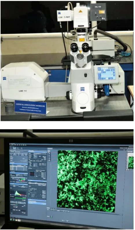
An image was taken of the centre of each disc with a Confocal Laser Scanning
Microscope (Carl Zeiss LSM 700) equipped with a 488nm argon laser using a X20 Plan
– Apochromat objective lens. Images sized 461.2µm X 461.2µm were captured and
digitized.
A wavelength of 488 nm was used to capture the images in green.MATLAB
(version R2009b, The MathWorks Inc., USA), a digital image analysis software was
used to quantify the area with auto fluorescence. The surface area was calculated in
percentage. The evaluation using Confocal Laser Scanning Microscope was done at the
National Facility for Clinical Trials, Interdisciplinary Institute of Indian System of
31
FIGURES
Figure 1: Group I - Plastic Curette (Hu-Friedy)
[image:46.595.352.496.340.505.2]
Figure 2: Group II – Carbon Composite Tip (Satelec)