IJOCR
ORIGINAL RESEARCH
Comparative Evaluation of Microleakage in Class I
Cavities Restored with Amalgam, Bulk-fill Composite
and Cention-N – An
In Vitro
Confocal Laser Scanning
Microscope Study
Shorya Sahu1, Naushad Ali2, Abhinav Misuriya3, Prashansa Vijaywargiya4, Suparna Ganguly Saha5, Anuj Bharadwaj6
ABSTRACT
Aim: The aim of the study was to evaluate and compare microleakage in Class I cavities restored with amalgam, bulk-fill composite, and cention-N using confocal laser scanning microscope (CLSM).
Materials and Methods: Class I cavities in 80 non-carious human mandibular premolars were prepared and the teeth were randomly divided into four experimental groups of 20 teeth each. The prepared cavities were then restored with amalgam (n = 20) (Dispersalloy, Dentsply, India); bulk-fill composite (n = 20); cention-N without adhesive (n = 20), and with adhesive (n = 20) as per manufacturer’s instruction. The teeth were stored in distilled water at room temperature in a thermocycler. The apices of the teeth were then sealed with acrylic and all tooth surfaces, except for a 1 mm wide zone around the margins of each restoration, were sealed with two coats of nail polish. The teeth were then immersed for 48 h in a 6% solution of rhodamine B dye. The teeth were rinsed and then sectioned longitudinally in a mesiodistal direction, coinci-dent with the center of the restoration, using slow speed water cooled diamond disc. The two sections of each tooth showing dye penetration were selected and observed under 10 × CLSM (Olympus FV 1200 E, Japan).
Statistical Analysis: Chi-square test, One-way ANOVA, and Post Hoc Tukey test were performed.
Results and Conclusion: Within the limitations of the present study, it was found that amalgam exhibited least microleak-age in comparison to composite resins. The recently used cention-N, showed lesser dye penetration thus promising lower microleakage when compared to composite resin especially when used in combination with an adhesive and can be a bet-ter albet-ternative in present scenario for leakage free restoration. Keywords: Amalgam, Cention-N, Class I cavity, Composite, Confocal laser scanning microscope, Microleakage
1,2Postgraduate Students, 3,6Reader, 4Senior Lecturer, 5Professor and Head
1-6Department of Conservative Dentistry and Endodontics,
College of Dental Science and Hospital Rau, Indore, Madhya Pradesh, India
Corresponding Author: Dr. Shorya Sahu, Postgraduate Student, Department of Conservative Dentistry and Endodontics, College of Dental Science and Hospital Rau, Indore, Madhya Pradesh, India. e-mail: [email protected]
How to cite this article: Sahu S, Ali N, Misuriya A, Vijaywargiya P, Saha SG, Bharadwaj A. Comparative Evaluation of Microleakage in Class I Cavities Restored with Amalgam, Bulk-fill Composite and Cention-N – An In Vitro Confocal Laser Scanning Microscope Study. Int J Oral Care Res 2018;6(1):S81-85.
Source of support: Nil Conflicts of interest: None
INTRODUCTION
Marginal microleakage causes marginal staining and secondary caries around restorations and may lead to pulpal pathology.[1,2] Therefore, controlling microle-akage has always been an important goal in operative dentistry.[3] Research is being conducted to introduce materials which are able to fulfill all the prerequisites of ideal restorative material.
Dental amalgam has been used as a restorative mate-rial for more than a century due to its various advantages. Initial microleakage has been a major challenge in an amalgam restoration which, however, improves over time with the aging of the restoration, due to the accumulation of corrosion products at the tooth restoration interface.[4,5]
Over a period of time, direct composite restorations have gained preference over conventional amalgam restorations due to their superior esthetic properties, micromechanical retention, and no mercury content. However, polymerization shrinkage and the resulting microleakage remain major drawbacks of this esthetic restorative material.[6]
The recently introduced material cention-N which is proclaimed as an esthetic alternative to amalgam, is an “alkasite” restorative such as compomer or ormocer, and is essentially a subgroup of the composite resins with compressive strength comparable to amalgam.[7]
lars extracted and were then cleaned with tap water. Polishing was done with a rubber cup and pumice and teeth were stored in distilled water at room temperature until they were used for the study.
Occlusal surfaces of all teeth were ground with a coarse diamond bur, under profuse water cooling, to produce a flat surface perpendicular to the long axis of the tooth, without removing the whole of the occlusal enamel. Class I cavities of approximately 3 mm length, 2 mm width and 3 mm depth were prepared using straight fissure bur (FG 111 012, Horico, Germany), with a high-speed handpiece and copious amount of water as a coolant. No bevels were placed.
After every five cavity preparations, the bur was replaced. Dimensions of each cavity were measured using a William’s graduated periodontal probe to main-tain uniformity among the size of cavities.
The teeth were randomly divided into four experi-mental groups of 20 teeth each. The prepared cavities were then restored with amalgam (n = 20) (Dispersalloy, Dentsply, India); bulk-fill composite (n = 20) (Tetric N-Ceram Bulk Fill, Ivoclar Vivadent, India); cention-N without adhesive (n = 20) (Ivoclar Vivadent, India), and with adhesive (Tetric N Bond, Ivoclar Vivadent, India) (n = 20) as per manufacturer’s instruction.
All preparations, restorations, and finishing were car-ried out by a single operator simulating clinical conditions. The teeth were then stored in distilled water at room temperature for 15 days and then were subjected to 1000 thermal cycles between 5°C and 15°C water baths with a dwell time of 1 min and a transit time of 5 s between baths.
wide zone around the margins of each restoration, were sealed with two coats of nail polish. The teeth were then immersed for 48 h in a 6% solution of rhodamine B dye.
The teeth were rinsed and then sectioned longitu-dinally in a mesiodistal direction, coincident with the center of the restoration, using slow speed water cooled diamond disc.
One of the two sections of each tooth showing bet-ter dye penetration were selected, observed under 10x confocal laser scanning microscope(Olympus FV 1200 E, Japan) and scored according to an ordinal ranking system [Figure 1].[8]
RESULTS
Figure 2 displays microleakage in different groups seen under 10x confocal laser scanning microscope (Olympus FV 1200 E, Japan) and an inter-group com-parison of frequency distribution of various samples showing different depth of penetration was done using Chi-square test [Table 1].
The four groups had significantly different number of samples units in different categories with respect to depth of penetration as seen in Frequency distribution graph [Graph 1] (P < 0.05).
Overall, maximum sample units (26.2%) showed 0.25 depth of penetration, most of which belonged to Group 4.
Maximum sample units showed 0.00 depth of pen-etration in Group 1, 1 depth of penpen-etration in Group 2, 0.5 depth of penetration in Group 3, and 0.25 depth of penetration in Group 4.
Graph 1: Frequency distribution of sample units based on depth dye penetration
Table 1: Comparison amongst four groups based on the frequency distribution of study sample units with different depth
of dye penetration
groups Number of samples (%
within group) Chi‑square value
λP value*
0 1 2 3 4 Total
Group 1 13 5 2 0 0 20 53.871 0.000* Group 2 0 1 6 6 7 20
Group 3 1 4 7 4 4 20 Group 4 2 9 6 2 1 20
*P<0.05 was considered statistically significant. λChi-square test, Df- 12.
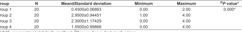
Table 2: Description of mean penetration scores of the specimen of four groups
Group N Mean±Standard deviation Minimum Maximum ΩP value*
Group 1 20 0.4500±0.06863 0.00 2.00 0.000*
Group 2 20 2.9500±0.94451 1.00 4.00
Group 3 20 2.3000±1.17429 0.00 4.00
Group 4 20 1.5500±0.99868 0.00 4.00
Microleakage in class I cavities IJOCR
The mean penetration score of Group 1 specimen was minimum (0.4500 ± 0.68633) and Group 2 specimen showed the maximum depth of penetration (2.3000 ± 0.94451). Statistically significant difference was observed between the four groups by applying One Way Anova (P < 0.05) [Table 2].
According to Post Hoc analysis [Table 3], the mean penetration score of Group 1 specimen was significantly less than that of Group 2, 3 and 4 specimen.
The mean penetration score of Group 2 specimen was significantly greater as compared to Group 4 specimen.
The mean penetration score of Group 2 and Group 3 specimen did not differ significantly. Similarly, the mean penetration score of Group 3 specimen did not differ significantly from Group 4 specimen.
DISCUSSION
Dye penetration test is used as an adjunct by which cli-nicians and researchers can predict the performance as well as longevity of restorative material. Evaluation of marginal microleakage for any restorative material is of utmost importance because it is directly related to the success or failure of the restorations.[8]
In the present in vitro study, Class I cavities were prepared and restored to replicate clinical situations which are associated with maximum polymerization shrinkage thus resulting in microleakage encountered with composites due to high C-factor.
Thermocycling of restored teeth was done to mimic intraoral temperature variations compatible with the oral cavity.[9]
Dye penetration method, despite its limitations, was used in this study as microleakage studies are still the most popular test methods employed to obtain a pre-liminary idea about the sealing ability of the restorative materials. They also have an advantage of low cost and simplicity of technique.[10] Rhodamine dye was selected for this study because it has a low molecular size which allows for the detection of minutest leakage where even bacteria cannot penetrate.[11]
According to the present study, amalgam restoration showed least microleakage, followed by cention-N with adhesive, cention-N without adhesive, and composite demonstrating maximum microleakge.
When an amalgam restoration is initially placed, a micro space exists between it and the restoration and the tooth structure. The gradual obliteration of this space may be attributed to the self-sealing of the tooth restoration margins by deposition of corrosion products formed by the interaction of the metallic ions from amalgam with chlorine and oxygen in the oral environment.[12]
Bulk-fill composite used in this study presented with maximum microleakage among all the four groups. High polymerization shrinkage of composites due to their high C-factor may be responsible for the microgap formation at the tooth restoration interface leading to marginal staining and recurrent caries, thus affecting the longevity of the restoration.[4]Results of the present study are in accordance with the studies conducted by Baghdadi, and in which Class I amalgam restorations consistently showed lower microleakage when com-pared to bonded composite restorations.[13]
Figure 1: Scoring criteria for the depth of penetration of dye at the tooth restoration interface[24]
Table 3: Post hoc analysis
Groups P value
Group 1 versus Group 2 0.000* Group 1 versus Group 3 0.000* Group 1 versus Group 4 0.003* Group 2 versus Group 3 0.154 Group 2 versus Group 4 0.000* Group 3 versus Group 4 0.076
Figure 2: Microleakage of amalgam (a), composite (b) cention-N without adhesive(c) and cention-N with adhesive (d) as seen under the confocal microscopic image (×10)
d c
ison to its usage without adhesive, a reason which could be attributed to the formation of an acid-resistant res-in-dentin inter-diffusion zone formed by the adhesive layer.[7]Results of the present study are in accordance with the study conducted by John Burgess who also observed minimum dye penetration in cention-N when used with adhesive as compared to without adhesive.[14]
The relatively lesser degree of microleakage observed with cention-N, both with and without adhesive as compared to bulk-fill composite may be assigned to the introduction of an Ivocerin based patented isofiller tech-nology (partially functionalized by silanes). This acts as a stress reliever which keeps the shrinkage stress to a minimum, thus improving the mechanical properties of the restoration by reinforcing its structural integrity in load bearing areas where amalgam is usually the mate-rial of choice.[15]
Amalgam has been an age-old successful material used in restorative dentistry. However, in the present times due to the increased esthetic demand and with the renewed worldwide concern regarding its poten-tial toxicity this material is gradually being phased out by the more esthetic composite restoration. The major drawback associated with composite resin restorations is shrinkage during polymerization which affects the longevity of the restoration.[16]
Cention N, is a self curing material which resembles amalgam in compressive strength (300 MPa) as well as in terms of bulk placement.
Cention-N uses hydrogen peroxide initiator and has a setting time of 4 min. Similar to that of composites it has a light curing option with the presence of photoini-tiator Ivocerin and an acyl phosphine oxide iniphotoini-tiator.[17]
Furthermore, similar to the bulk-fill composites like Tetric N-Ceram, it contains a shrinkage stress reliever as filler which is responsible for its low modulus of elastic-ity (10 GPa) allowing it to act as a spring in contrast to standard glass fillers used in composites which have a higher modulus of elasticity (71 GPa).[18]
Cention-N releases fluoride like glass ionomer cement (GIC) with flexural strength and compressive strength superior to it. Cention-N is a more esthetic material as compared to GIC due to to its higher parency of 11% in contrast to GIC which presents trans-parency of 3–4%.
Numerous studies have been conducted to evalu-ate the microleakage of amalgam and composite.[19-22] However, this is one of the pioneer studies that evaluated and compared the microleakage of a new innovative dental restorative material, Cention-N with amalgam and composite.
optical sections out of thick specimens, by either reflec-tion or fluorescence. It can view specimens in planes running parallel to the line of sight; scan images deep into light scattering samples and can produce impres-sive 3-dimensional views at very high resolution.[23]
CONCLUSION
Within the limitations of the present study, it may be concluded that the age-old amalgam still serves to be one of the most efficient materials as far as microleakage is concerned.
In comparison, composite resins though esthetic, demonstrate higher microleakage, thus increasing the incidences of secondary caries.
Cention-N, which is a relatively recently introduced tooth-colored material in dentistry and has been shown to present with a lesser degree of dye penetration, thus promising lower microleakage when compared to com-posite resin restorations especially when used in combi-nation with an adhesive.
However, very little literature is yet available to substantiate the results obtained in the present study. Further research is, therefore, required to establish this material as an effective alternative to amalgam and composite.
REFERENCES
1. Kidd EA. Microleakage: A review. J Dent 1976;4:199-205. 2. Brännström M, Vojinovic O. Response of dental pulp to
invasion of bacteria round three filling materials. ASDC J Dent Child 1976;43:83-9.
3. Eakle WS, Ito RK. Effect of insertion technique on microle-akage in mesio-occluso distal composite resin restorations. Quintessence Int 1990;21:369-74.
4. Ben-Amar A, Cardash HS, Judes H. The sealing of the tooth/ amalgam interface by corrosion products. J Oral Rehabil 1995;22:101-4.
5. Johansson BI, Mjör IA. Marginal degradation and corro-sion of a dispersed high copper amalgam. Scand J Dent Res 1988;96:75-82.
6. Alptekin T, Ozer F, Unlu N, Cobanoglu N, Blatz MB. In vivo
and in vitro evaluations of microleakage around class I
amal-gam and composite restorations. Oper Dent 2010;35:641-8. 7. Samanta S, Das U K, Mitra A. Comparison of microleakage
in class V cavity restored with flowable composite resin, glass ionomer cement and cention N. Imp J Interdiscip Res 2017;3:180-3.
8. Santhosh L, Bashetty K, Nadig G. The influence of different composite placement techniques on microleakage in prepa-rations with high C-factor: An in vitro study. J Conserv Dent 2008;11:112-6.
Microleakage in class I cavities IJOCR
10. Tolidis K, Boutsiouki C, Gerasimou P. Microleakage in com-bined amalgam/Composite resin restorations in MOD cavi-ties. Braz J Oral Sci 2013;12:100-4.
11. Alahbdan AA. Review of microleakage evaluation tools. J Int Oral Health 2017;9:141-5.
12. Vogt BF, Xavier CB, Demarco FF, Padilha MS. Dentin pene-trability evaluation of three different dyes in root-end cavi-ties filled with mineral trioxide aggregate (MTA). Braz Oral Res 2006;20:132-6.
13. Mahler DB. Research on dental amalgam: 1982-1986. Adv Dent Res 1988;2:71-82.
14. Baghdadi ZD. Microleakage of a single-bottle adhesive sys-tem with 3 restorative materials: In vitro study and clinical considerations. Compend Contin Educ Dent 2003;24:755-8. 15. Microleakage of Cention N Compared to Dental Amalgam.
Final Report. John Burgess, Assistant Dean, School of Dentistry. Birmingham, USA: University of Alabama; 2015. 16. Burtscher P, Rheinberger V. Germanium based
photoini-tiator as an alternative to camphorquinone/amine. IADR Abstr 2008;1:1611.
17. Moszner N, Fischer UK, Ganster B, Liska R, Rheinberger V. Benzoyl germanium derivatives as novel visible light pho-toinitiators for dental materials. Dent Mater 2008;24:901-7. 18. Ensaff H, O’Doherty DM, Jacobsen PH. Polymerization
shrinkage of dental composite resins. Proc Inst Mech Eng H 2001;215:367-75.
19. Cention N. Scientific Documentation. Schaan, Liechtenstein: Ivoclar-Vivadent Press; 2016.
20. Jang JH, Park SH, Hwang IN. Polymerization shrinkage and depth of cure of bulk-fill resin composites and highly filled flowable resin. Oper Dent 2015;40:172-80.
21. Kasraei S, Azarsina M, Majidi S. In vitro comparison of microleakage of posterior resin composites with and with-out liner using two-step etch-and-rinse and self-etch dentin adhesive systems. Oper Dent 2011;36:213-21.
22. Helvatjoglou-Antoniades M, Theodoridou-Pahini S, Papadogiannis Y, Karezis A. Microleakage of bonded amalgam restorations: Effect of thermal cycling. Oper Dent 2000;25:316-23.
23. Redwan H, Bardwell DN, Ali A, Finkelman M, Khayat S, Weber HP, et al. Composite replacement of amalgam res-toration versus freshly cut dentin: An in vitro microleakage comparison. Oper Dent 2016;41:E73-82.
![Figure 1: Scoring criteria for the depth of penetration of dye at the tooth restoration interface[24]](https://thumb-us.123doks.com/thumbv2/123dok_us/8858608.1805892/3.594.80.257.393.569/figure-scoring-criteria-depth-penetration-tooth-restoration-interface.webp)
