understanding
.
White Rose Research Online URL for this paper:
http://eprints.whiterose.ac.uk/90406/
Version: Published Version
Article:
Davey, James orcid.org/0000-0003-1336-1414, Rueschemeyer, Shirley-Ann
orcid.org/0000-0001-6432-0917, Costigan, Alison et al. (4 more authors) (2015) Shared
neural processes support semantic control and action understanding. Brain and Language.
pp. 24-35. ISSN 0093-934X
https://doi.org/10.1016/j.bandl.2015.01.002
[email protected]
https://eprints.whiterose.ac.uk/
Reuse
Items deposited in White Rose Research Online are protected by copyright, with all rights reserved unless
indicated otherwise. They may be downloaded and/or printed for private study, or other acts as permitted by
national copyright laws. The publisher or other rights holders may allow further reproduction and re-use of
the full text version. This is indicated by the licence information on the White Rose Research Online record
for the item.
Takedown
If you consider content in White Rose Research Online to be in breach of UK law, please notify us by
Shared neural processes support semantic control and action
understanding
James Davey, Shirley-Ann Rueschemeyer, Alison Costigan, Nik Murphy, Katya Krieger-Redwood,
Glyn Hallam, Elizabeth Jefferies
⇑Department of Psychology and York Neuroimaging Centre, University of York, UK
a r t i c l e
i n f o
Article history:
Accepted 3 January 2015 Available online 3 February 2015
Keywords:
Semantic Action Control Executive fMRI
a b s t r a c t
Executive–semantic control and action understanding appear to recruit overlapping brain regions but existing evidence from neuroimaging meta-analyses and neuropsychology lacks spatial precision; we therefore manipulated difficulty and feature type (visual vs. action) in a single fMRI study. Harder judge-ments recruited an executive–semantic network encompassing medial and inferior frontal regions (including LIFG) and posterior temporal cortex (including pMTG). These regions partially overlapped with brain areas involved in action but not visual judgements. In LIFG, the peak responses to action and diffi-culty were spatially identical across participants, while these responses were overlapping yet spatially distinct in posterior temporal cortex. We propose that the co-activation of LIFG and pMTG allows the flex-ible retrieval of semantic information, appropriate to the current context; this might be necessary both for semantic control and understanding actions. Feature selection in difficult trials also recruited ventral occipital–temporal areas, not implicated in action understanding.
Ó2015 The Authors. Published by Elsevier Inc. This is an open access article under the CC BY-NC-ND license (http://creativecommons.org/licenses/by-nc-nd/4.0/).
1. Introduction
Our conceptual knowledge encompasses a large body of infor-mation but only particular aspects of concepts will be useful in any given context or task: as a consequence, executive control pro-cesses are engaged to guide conceptual processing in a context-dependent manner (Badre, Poldrack, Paré-Blagoev, Insler, & Wagner, 2005; Jefferies, 2013; Noonan, Jefferies, Corbett, & Lambon Ralph, 2010). We can match objects on the basis of specific features, even when these are not prominent aspects of the items, and this is crucial for intelligent behaviour – for example, when trying to pitch a tent, we can understand that a shoe has properties that make it suitable for banging pegs into the ground, even though these properties are not directly related to its dominant associa-tions. Semantic control processes in left inferior frontal gyrus (LIFG) are thought to be critical for this selection of task-relevant attributes (Thompson-Schill, D’Esposito, Aguirre, & Farah, 1997) and the controlled retrieval of weak associations (Noonan, Jefferies, Visser, & Lambon Ralph, 2013; Wagner, Paré-Blagoev, Clark, & Poldrack, 2001). However, little is known abouthow con-trol processes are deployed to focus neural activity on specific,
task-relevant aspects of knowledge – and whether the same mech-anisms are recruited for different types of features (e.g., action vs. visual properties).
Contemporary theories of semantic cognition agree that modal-ity-specific sensory and motor areas, plus multi-modal regions capturing specific features, contribute to semantic representation (Meteyard, Rodriguez Cuadrado, Bahrami, & Vigliocco, 2012; Patterson, Nestor, & Rogers, 2007; Pobric, Jefferies, & Lambon Ralph, 2010; Pulvermüller, 2013). As a result, semantic judgements about manipulable objects are thought to draw on representations across the cortex, including inferior parietal, premotor and poster-ior middle temporal (pMTG) regions, which support motor and praxis features (Chouinard & Goodale, 2012; Liljeström et al., 2008; Pobric et al., 2010; Rueschemeyer, van Rooij, Lindemann, Willems, & Bekkering, 2010; Vitali et al., 2005; Watson, Cardillo, Ianni, & Chatterjee, 2013; Yee, Drucker, & Thompson-Schill, 2010; Zannino et al., 2010). Although some research suggests that sen-sory and motor regions are recruited rapidly and automatically fol-lowing word presentation (Hauk & Pulvermüller, 2004; Shtyrov, Butorina, Nikolaeva, & Stroganova, 2014), recent neuroimaging studies have examined how activity within modality-specific areas might be modulated on the basis of task demands (Hoenig, Sim, Bochev, Herrnberger, & Kiefer, 2008; Rüeschemeyer, Brass, & Friederici, 2007; Tomasino & Rumiati, 2013). Action words (e.g., kick) and their semantic associates do not necessarily activate
http://dx.doi.org/10.1016/j.bandl.2015.01.002
0093-934X/Ó2015 The Authors. Published by Elsevier Inc.
This is an open access article under the CC BY-NC-ND license (http://creativecommons.org/licenses/by-nc-nd/4.0/).
⇑Corresponding author at: Department of Psychology, University of York, YO10 5DD, UK. Fax: +44 (0)1904 323181.
E-mail address:[email protected](E. Jefferies).
Contents lists available atScienceDirect
Brain & Language
motor regions when presented in isolation; this response is seen more strongly for literal sentences (‘kick the ball’) in which the action properties are relevant to the task (Raposo, Moss, Stamatakis, & Tyler, 2009; Schuil, Smits, & Zwaan, 2013; van Dam, van Dijk, Bekkering, & Rueschemeyer, 2012). Such findings challenge the assumptions of strong ‘embodied’ accounts of semantic cognition, in which neural connections between distrib-uted sensory and motor features are sufficient for conceptual rep-resentation. Furthermore, they raise questions about how semantic representations are applied in a controlled way, to suit the partic-ular task or context.
In addition to the role of distributed visual and motor/praxis representations in object knowledge, some theories suggest these disparate features are drawn together in an amodal semantic ‘hub’ in the anterior temporal lobes (ATL;Patterson et al., 2007). This proposal remains controversial (Simmons & Martin, 2009) because although data from multiple methods – including patients with semantic dementia (Bozeat, Lambon Ralph, Patterson, Garrard, & Hodges, 2000), TMS (Ishibashi, Lambon Ralph, Saito, & Pobric, 2011; Pobric et al., 2010) and PET (Devlin et al., 2002) – reveal a contribution of ATL to conceptual knowledge across modalities, fMRI is relatively insensitive to signals from ATL due to magnetic susceptibility artefacts that produce signal loss and distortion in this brain region (Visser, Jefferies, Embleton, & Lambon Ralph, 2012; Visser, Jefferies, & Lambon Ralph, 2010). Con-sequently the fMRI literature does not uniformly emphasise a role for ATL and instead focuses on the contribution of pMTG to multi-modal tool/action knowledge, with some recent studies suggesting pMTG is a semantic hub for tool and action understanding (Martin, 2007; Martin, Kyle Simmons, Beauchamp, & Gotts, 2014; van Elk, van Schie, & Bekkering, 2014).
An alternative view about the contribution of pMTG to semantic cognition is provided by work on semantic control (for reviews, see
Jefferies, 2013; Noonan et al., 2013). Although this research has lar-gely focussed on the role of LIFG in selection and controlled seman-tic retrieval (Badre et al., 2005; Hoffman, Jefferies, & Lambon Ralph, 2010; Thompson-Schill, D’Esposito, Aguirre, & Farah, 1997; Wagner et al., 2001), a recent meta-analysis revealed that manip-ulations of the executive demands of semantic tasks activate a dis-tributed cortical network, including left and right inferior frontal gyrus (LIFG; RIFG), medial PFC (pre-SMA), dorsal angular gyrus (dAG) bordering intraparietal sulcus (IPS) and, most notably, pMTG (Noonan et al., 2013). These sites all show greater activation during difficult tasks that tap less prominent aspects of meaning, or require strongly related distracters to be suppressed (Rodd, Johnsrude, & Davis, 2010; Wagner et al., 2001; Whitney, Kirk, O’Sullivan, Lambon Ralph, & Jefferies, 2011). Moreover, inhibitory TMS to LIFG and pMTG produces equivalent disruption of semantic tasks tapping controlled retrieval, but has no effect on semantic judgements to highly-associated word pairs, which rely largely on automatic spreading activation (Whitney et al., 2011). This network for semantic control overlaps with the ‘‘fronto-parietal control network’’ involved in cognitive control across domains – which includes inferior frontal sulcus, intraparietal sulcus and occipital–temporal regions (Duncan, 2010; Woolgar, Hampshire, Thompson, & Duncan, 2011; Yeo et al., 2011), although some sites appear to make a relatively restricted contribution to control pro-cesses important for semantic cognition, particularly anterior parts of LIFG and pMTG (Devlin, Matthews, & Rushworth, 2003; Noonan et al., 2013; Whitney, Jefferies, & Kircher, 2011; Whitney et al., 2011, 2012).
In summary, separate literatures on executive–semantic pro-cessing and action understanding have linked similar left hemi-sphere networks – encompassing IFG/premotor cortex, IPL and pMTG – with diverse aspects of semantic cognition (Noonan et al., 2010; Watson et al., 2013). Since these regions are associated
with understanding actions, tools, verbs and events, it has been suggested they might represent motion, action, or praxis features (Chouinard & Goodale, 2010; Liljeström et al., 2008; Spunt & Lieberman, 2012; Watson et al., 2013). However, left IFG, pMTG, and dorsal IPL are also activated during semantic tasks with high executive demands, suggesting they might support controlled retrieval/selection processes that shape semantic processing to suit the current context (Noonan et al., 2013). Damage to this network in semantic aphasia (SA) produces difficulty controlling conceptual retrieval to suit the task or context, both in verbal tasks like picture naming and non-verbal tasks like object use (Jefferies & Lambon Ralph, 2006; Noonan et al., 2010). These deficits can be overcome through the provision of cues that reduce the need for internally-generated control (i.e., phonological cues for picture naming; pho-tographs of the recipients of actions in object use;Corbett, Jefferies, Ehsan, & Lambon Ralph, 2009; Corbett, Jefferies, & Lambon Ralph, 2011), suggesting that damage to this network does not produce a loss of semantic information about words or actions, but instead poor control over conceptual retrieval. However, both neuropsy-chological studies and neuroimaging meta-analyses have poor spa-tial resolution, and thus it is not yet known whether semantic control and action understanding recruit adjacent (yet distinct) or overlapping regions in pMTG and LIFG.
We addressed this question in an fMRI study with a 22
design that (1) contrasted decisions about action and non-action (visual) features and (2) compared easy, low-control judgements, in which participants selected a globally semantically-related item with more difficult, high-control judgements, in which the target was only related via a specific feature. We predicted that the recruitment of sensory/motor regions would vary according to the feature, with more activity within visual areas for visual deci-sions (e.g., lateral occipital cortex, occipital pole), and within motor/praxis areas for action decisions (e.g., precentral gyrus; IPL; pMTG). Executive–semantic regions were expected to show stronger responses for more demanding judgements irrespective of the feature to be matched. Furthermore, we examined whether brain regions recruited during the retrieval of action knowledge would overlap with those implicated in semantic control in both group analyses and at the single-subject level.
2. Method
2.1. Participants
20 right-handed, native English speaking participants were recruited from the University of York, UK. All subjects had nor-mal/corrected to normal vision. Three participants had to be excluded from the final analysis due to head movement (>2 mm) and poor accuracy. A total of 17 participants were entered into the analysis (mean age = 22.7 years, 10 females).
2.2. Study design
A fully-factorial 22 within-subjects design was used. The
two factors were judgement type (action or visual form match-ing) andcontrol demands(contrasting easy decisions about glob-ally related items with difficult decisions based on specific features).
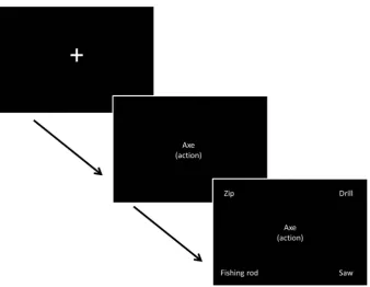
contrasted ‘easy’ trials in which the probe and target were taken from the same semantic category and shared either overlapping action or visual properties (i.e.,KETTLEandJUGshare action proper-ties and are both kitchen items) with ‘difficult’ trials in which the probe and target were not semantically related andonly shared an action or visual feature (e.g.,KETTLEwithHOURGLASS, which only share a tipping action). Moreover, in the difficult trials, there were globally-related distracters which shared category membership with the probe butnotthe relevant feature (e.g.,SCALESandTOASTER are categorically related toKETTLEbut are not targets because they do not share action features). In both types of trial, there were two response options that were globally-semantically related, and two that were not, but the trials varied as to whether these constituted the target or distracters. A complete list of probes and targets is provided in thesupplementary materials (Table S1). A four-alternative forced-choice paradigm was used; partici-pants matched centrally presented probe words to one of four potential items, based on the nature of the association for that block. A reminder of the association being probed was present on every trial, in parentheses underneath the probe word. The exper-iment was organised into sixteen blocks; eight blocks for each fea-ture type (action or vision) with control demands randomised within a block. An instruction slide stating the relevant feature to be matched (action/vision) appeared before each block for 1000 ms. Blocks contained seven or nine events. In blocks with seven events, there were six semantic decisions with one null event (screen was blank for 6000 ms). In blocks with nine events, there were seven trials with two null events. Probe words were presented for one second, and then the response options appeared and remained on the screen until the participant responded via a button press, with a maximum duration of 7.5 s. There was a jit-tered inter-trial interval of 4000–6000 ms between all events (including null events) with 10–12 s of rest between each block. Null events were combined with the rest between blocks to pro-vide a baseline measure for analysis. Before participants took part in the fMRI experiment they were given a practice session, equiv-alent to one fMRI run (seeFig. 1).
2.3. Stimuli
Each condition had 25 targets (100 in total; seeTable S1). In the easy condition, 25 semantically related items were used as distract-ers, combined with 50 unrelated distracter items. In the hard condi-tions, 50 semantically related items were used, with the remaining 25 distracters consisting of semantically unrelated items. All of the words were concrete nouns denoting manipulable objects. Individ-ual words were used a maximum of four times throughout the experiment. Target words were matched across conditions for fre-quency (CELEX database, Max Planck Institute for Psycholinguistics, 2001), number of letters, and imageability, with no significant dif-ferences between conditions. Frequency and letter length were obtained using the program N-watch (Davis, 2005). Details of imageability ratings, descriptive statistics and ANOVA results can be found in thesupplementary materials (Tables S2 and S3).
2.4. Data acquisition
Brain images were acquired using a 3T GE HDx Excite MRI scan-ner, utilising an 8 channel head coil. We obtained high-resolution structural images for every participant (3D FSPGR MRI). Functional data were recorded from the whole brain using gradient-echo EPI (FOV: 192192, matrix: 6464, slice thickness: 4.5 mm, voxel
size; 334.5 mm, flip angle: 90°, TR: 2000 ms, TE: 30 ms) with
bottom-up sequential data acquisition. Each session was split into two 14 min runs, with a total of 420 volumes for each run. Co-reg-istration between structural and functional scans was improved using an intermediary scan (T1 FLAIR) with the same parameters as the functional scan. NBS Presentation version 14 (Neurobehav-ioral Systems Inc., 2012) was used to present stimuli and capture responses (reaction time and accuracy) during fMRI. Stimuli were projected using a Dukane 8942 ImagePro 4500 Lumens LCD projec-tor onto an in-bore screen with a 4530 visual degree angle.
[image:4.595.125.464.468.730.2]Responses were collected using two Lumitouch two button response boxes, in a custom built case allowing all four buttons to be operated using the left hand.
2.5. Data analysis
The analysis used an event-related design to examine the tran-sient responses to each trial separately. fMRI analysis was con-ducted using FSL 4.1.9 (Analysis Group, FMRIB, Oxford, UK;
Jenkinson, Beckmann, Behrens, Woolrich, & Smith, 2012; Smith et al., 2004; Woolrich et al., 2009). First and higher level analyses were conducted using FEAT (fMRI Expert Analysis Tool). Pre-pro-cessing of the data included McFLIRT motion correction (Jenkinson, Bannister, Brady, & Smith, 2002), skull-brain segmenta-tion (Smith, 2002), slice timing correction, spatial smoothing using a Gaussian kernel FWHM of 5 mm, and high-pass temporal filter-ing (100 s). Time-series data were modelled usfilter-ing a general linear model (FILM; FMRIB Improved Linear Model), correcting for local autocorrelation (Woolrich, Ripley, Brady, & Smith, 2001). Each experimental variable (EV) was entered as a boxcar function, con-volved with a hemodynamic response gamma function, using a variable epoch model (Grinband, Wager, Lindquist, Ferrera, & Hirsch, 2008): the start of each epoch was defined as the onset of the probe word, with epoch duration determined by the response time on each trial. The following EVs were used: correct responses from each of the four conditions, rest (null events and time between blocks, modelled independently since we were initially interested in potential differences between them, driven by the instructions, but these were not observed) and errors (a temporal derivative was added to all variables). Four contrasts were defined from the correct responses; individual conditions > rest (easy action, hard action, easy visual, hard visual).
2.5.1. Whole brain group analysis
A first analysis examined the effect offeature typeby comparing brain activity to action and visual decisions separately. Analysis of the complete behavioural data from the scanner revealed a small but significant difference in accuracy between the action and visual conditions. Therefore, the whole brain analysis was conducted on a subset of 84 trials (i.e., 21 trials per condition, using the same probe words across conditions). All of the trials related to four spe-cific probe words were removed across all conditions and partici-pants, and entered as a covariate of no interest. The trials in the analysis were matched for psycholinguistic properties, accuracy and RT (seeTables S1, S2 and S3). Contrasts of each of the condi-tions over rest were entered into a higher level contrast of action decisions (hard action > rest + easy action > rest) vs. visual deci-sions (hard visual > rest + easy visual > rest) and vice versa. To con-trol for multiple comparisons, cluster-based thresholding was applied to all analyses. Voxel inclusion was set atz= 2.3 with a cluster significance threshold at FWEp< .05. The minimum cluster size for significance atp= .05 was 615 contiguous voxels.
In a second analysis, the manipulation ofdifficultywas maxi-mised by selecting 60/100 trials with accurate responses which generated the fastest and slowest decisions for each participant. This was done in order to maximise the sensitivity of the study to the effects of this variable. These trials were divided evenly between the action/visual conditions (15 easy action; 15 easy visual; 15 hard action; 15 hard visual). The fastest trials were based on global semantic similarity while the slowest were based on a specific feature in the presence of globally-related distracters. The same contrasts described above were repeated using these 60 trials. Voxel inclusion was set atz= 2.3 with a cluster signifi-cance threshold atp< .05. The minimum cluster size for signifi-cance atp= .05 was 561 contiguous voxels.
2.5.2. Regions of interest (ROI) group analysis
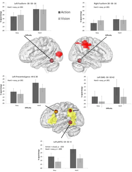
We examined 8 mm spherical ROIs placed at key coordinates taken from the literature. The coordinates used in this analysis are shown inFigs. 4 and 5. The FEATquery tool in FSL was used
to extract unthresholded percentage signal change for each ROI and each of the four conditions using the matched set of 84 items. The average change across all voxels within the ROI was computed and subjected to ANOVA to examine the effects of difficulty and task, and their interaction at each location.
(i) The first set of ROIs focussed on regions implicated in exec-utive–semantic control by a recent meta-analysis of neuro-imaging studies (Noonan et al., 2013). This highlighted a distributed network, involving left posterior and anterior IFG (corresponding to BA44, BA45, and BA47 respectively), right posterior IFG (RBA44), medial PFC (pre-SMA), pMTG and dAG/IPS.
(ii) In addition, we included peaks designed to localise addi-tional brain responses involved in understanding actions. These were taken from a study which contrasted responses to action and object pictures (Liljeström et al., 2008) and from a meta-analysis investigating action concepts in the brain (Watson et al., 2013). In the Liljeström et al. (2008)
study, the strongest action-selective peak was in left precen-tral gyrus, motivating the choice of this site as an ROI. We also examined the strongest peak in left IPL identified from the same contrast. Finally, we examined a pMTG site for actions identified in a recent meta-analysis (Watson et al. (2013).
(iii) We examined a left fusiform peak implicated in the retrieval of visual features (Thompson-Schill, Aguirre, D’Esposito, & Farah, 1999). This was transformed to the right hemisphere to investigate bilateral fusiform contributions to visual and action judgements.
2.5.3. Individual analysis
3. Results
3.1. Behavioural results
Descriptive statistics are provided in thesupplementary mate-rials (Table S4). A repeated-measures ANOVA on the set of 84 trials revealed significant main effects of difficulty for both reaction time and accuracy (RT:F(1, 16) = 61.70,p< .001, eta2= 0.79; accuracy: F(1, 16) = 35.86,p< .001, eta2= 0.69). Participants took longer and were less accurate in the hard conditions compared to the easy conditions, irrespective of feature type. There were no significant main effects of feature type (RT: F(1, 16) = 1.26, p= .28, eta2= 0.07; accuracy:
F(1, 16) = 1.21, p= .29, eta2= 0.07) and no interactions (RT: F(1, 16) = 0.41, p= .53, eta2= 0.03; accuracy: F(1, 16) = 0.10,p= .76, eta2= 0.01).
3.2. Whole brain analysis: Action vs. visual decisions
To examine differences between action and visual feature judgements, direct contrasts of these two tasks were performed.
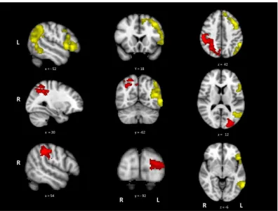
Fig. 2shows the activation maps for the contrasts of action > visual judgements and visual > action judgements. Cluster maxima and sub-peaks are in thesupplementary materials (Table S5). A con-trast of actions > rest and visual > rest can be found in the supple-mentary materials S3 and S4. The action > visual contrast revealed large clusters in left hemisphere areas previously implicated in action processing and semantic cognition, including LIFG, premo-tor cortex, IPL and pMTG. The opposite contrast of visual over action judgements revealed bilateral areas involved in visual pro-cessing, including right supramarginal gyrus, left lateral occipital cortex (LO) and left occipital pole.
3.3. Whole brain analysis: The effects of task difficulty
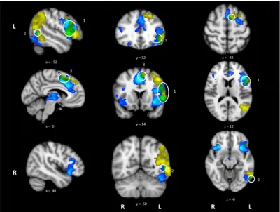
The activation map for the contrast of hard > easy decisions is shown in Fig. 3. Coordinates for cluster maxima and sub-peaks can be found in thesupplementary materials (Table S6). Consistent with our predictions, the manipulation of difficulty for the seman-tic judgements produced activation in a distributed network asso-ciated with executive control of semantic decisions. The most extensive and strongest activity was in LIFG, but the network was bilateral, extending to RIFG, medial PFC/anterior cingulate/ paracingulate and posterior temporal areas in left posterior ITG/ MTG/fusiform gyrus.
Activation revealed by the hard > easy contrast partially over-lapped with several regions also activated by action > visual judge-ments (see Fig. 3). These areas of overlap were found in LIFG, extending into left precentral gyrus and superior frontal gyrus (site 1), pMTG (site 2) and left paracingulate gyrus/medial PFC (site 3). In contrast, there was no overlap between the difficulty and visua-l > action contrasts. These findings suggest common brain regions are involved in action understanding and in dealing with the exec-utive demands of semantic tasks.
We also explored the possibility of an interaction between task (action vs. visual) and difficulty in the whole brain analysis; how-ever no such effects were found.
3.4. ROI analysis
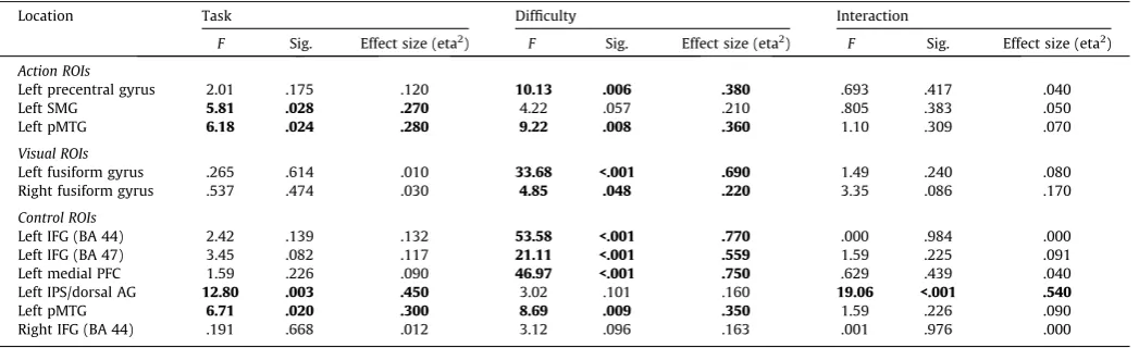
Within each ROI, we extracted the mean percentage signal change for the four conditions (easy action, hard action, easy visual, and hard visual) for each participant and submitted the data to a 22 repeated-measures ANOVA, examining the factors of
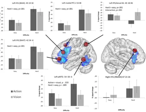
[image:6.595.93.490.65.366.2]task (action vs. visual judgements) and difficulty (easy vs. hard). ANOVA results are shown inTable 1, whileFigs. 4 and 5display
ROI locations on a rendered 3D brain, plus graphs displaying mean percentage signal change for each condition.
3.4.1. Executive–semantic control peaks
The ROI analysis revealed significantly greater signal change for difficult vs. easy trials for all left hemisphere PFC/IFG sites (there were no significant effects in right BA44). In addition, left BA 47 showed a near-significant effect of task, reflecting somewhat greater signal change for action than visual trials. No other effects of task were observed and there were no significant interactions.
Left dorsal AG/IPS showed a highly significant interaction between control demands and task. While difficult visual feature decisions involved increased recruitment of left dAG/IPS, this site showed deactivation for hard action decisions: there was a highly significant difference between hard action and visual trials (t(16) = 5.32, uncorrected p< .001) but no difference between easyaction and visual judgements.
Left pMTG displayed significant effects of task and difficulty, with a greater response for action trials compared to visual trials, and for difficult trials compared to easy trials, with no interaction.
3.4.2. Action peaks
Left precentral gyrus demonstrated a significant effect of con-trol, with a stronger response to hard judgements compared to easy judgements. There were no significant effects or interactions with task: therefore, although this site has been previously impli-cated in action understanding, it is also involved in executive– semantic control, even when the task involves visual feature matching.
Left SMG, a site implicated in hand praxis, showed a stronger response to action than visual trials. No significant main effects or interactions with difficulty were observed, indicating that this site is recruited by action judgements irrespective of difficulty.
The pMTG peak fromWatson et al. (2013)demonstrated signif-icant effects of both task and category, with no interaction. Greater signal change was observed for action trials relative to visual trials, and for harder trials relative to easy trials. The pattern of results mirrors those observed for the semantic control peak in pMTG from Noonan et al. (2013), and indeed, these two ROIs selected from different literatures were spatially similar and partly overlapping.
3.4.3. Visual peaks
The fusiform gyrus bilaterally demonstrated significant effects of control with stronger responses to hard than easy trials, irre-spective of task.
3.5. Individual overlap between contrasts examining task difficulty and action retrieval
3.5.1. LIFG
[image:7.595.102.503.66.368.2]88% of participants (N= 15) showed a response for both feature type (action > visual) and difficulty (hard > easy). For these partic-ipants, we counted the number of voxels within the mask respond-ing to (i) both contrasts, (ii) feature type only, (iii) difficulty only and (iv) neither contrast, in order to establish whether the number of voxels showing effects ofbothcontrasts was greater than would be expected by chance (supplementary materials; Table S7). Nine participants (60% of the sample) showed a significant conjunction between the two contrasts when each contrast was thresholded atp= 0.05 (z= 1.96) (i.e., number of voxels > 0; mean cluster size 47.6 voxels, s.d. = 60.6, mean MNI coordinates; 50 22 6, pars tri-angularis); however, the number of voxels showing a conjunction was highly variable across subjects. Loglinear analysis examined the frequencies of voxels responding to difficulty and feature type in the 15 participants who showed both effects, with participant
identity included as an additional predictor. The final model retained all three effects and their interaction terms (for k= 3,
v
2(14) = 369.7, p< .001), with a significant partial associationbetween voxels responding to difficulty and those responding to feature type (
v
2(1) = 55.9,p< .001). Follow-up chi-square analysesconfirmed that across subjects, more voxels responded toboth dif-ficulty and feature type than would be expected by chance (
v
2(1) = 9.19, p= .002 with continuity correction; see Table S7).In addition, within-subjects MANOVA was used to examine the coordinates of the peak responses for each contrast across partici-pants (N= 15). Descriptive statistics are provided inTable S8. There was no difference in the location of the peaks associated with dif-ficulty and action feature retrieval,F(3, 12) = 1.89,p= .19, suggest-ing overlappsuggest-ing responses.
3.5.2. Posterior temporal cortex
94% of participants (N= 16) showed a response to both feature type and difficulty. 8 individuals (50%) showed a significant con-junction between the two contrasts that reached p= 0.05 (mean cluster size 42.1 voxels, s.d. = 57.1, mean MNI coordinates; 50 60 0 pMTG); however, as for LIFG, the number of voxels showing a conjunction was highly variable across subjects. Loglin-ear analysis was conducted using the model described above for LIFG. The final model retained all three effects and their interaction terms (fork= 3,
v
2(15) = 1011.6,p< .001), with a significant partialassociation between voxels responding to difficulty and those responding to feature type (
v
2(1) = 41.6,p< .001). Follow-upchi-square analyses confirmed that across subjects, significantly more voxels responded tobothdifficulty and feature type than would be expected by chance (
v
2(1) = 209.6,p= .002 with continuitycorrec-tion; seeTable S7). Within-subjects MANOVA was used to examine the coordinates of the peak responses for each contrast across par-ticipants (N= 16). Descriptive statistics are provided inTable S8. This analysis revealed that difficulty and action retrieval elicited overlapping yet spatially distinct peaks,F(3, 13) = 4.75,p= .02, with significant differences in thex(F(1, 15) = 8.83,p= .01) and thez dimension (F(1, 15) = 6.98, p= .02). The peak for difficulty was more ventral and medial than the peak for action retrieval.
In conclusion, overlapping voxels responded to difficulty and feature type (action > visual) in both LIFG and pMTG. The location of the peak responses for these two contrasts across individual par-ticipants did not differ within LIFG yet was spatially distinct in pos-terior MTG/ITG.
4. Discussion
[image:8.595.55.535.63.430.2]Neuropsychological studies (Corbett, Jefferies, & Lambon Ralph, 2009; Corbett et al., 2011) and neuroimaging meta-analyses have identified apparently overlapping left-hemisphere sites which
respond to both action knowledge (Watson et al., 2013) and semantic tasks with high executive demands (Noonan et al., 2013), yet both of these methods lack spatial resolution. In the
[image:9.595.77.532.64.656.2]current study, group-level and single-subject analyses examined the extent to which the brain regions implicated in difficult semantic judgements also responded to the requirement to
retrieve action as opposed to visual features. We established that there is significant overlap between these contrasts in both LIFG and posterior temporal cortex (with peaks in pars triangularis and pMTG respectively). However, while the response to these contrasts in LIFG was spatially identical, there were overlapping yet distinct responses to difficulty and action retrieval in posterior temporal cortex. These findings suggest that there is a common distributed functional system for executive control over semantic processing and action understanding, involving both prefrontal and posterior temporal components; however, the data also point to differences in the roles and organisation of these regions.
First, the study revealed differential activation in modality-spe-cific areas during action and visual feature judgements, which was flexibly driven by the task instructions. The retrieval of action fea-tures over visual feafea-tures revealed an exclusively left-hemisphere network, including left inferior frontal and precentral cortex, infe-rior parietal lobule (IPL) and pMTG – regions linked to action pro-cessing and representation (Ghio & Tettamanti, 2010; Liljeström et al., 2008; Sasaki, Kochiyama, Sugiura, Tanabe, & Sadato, 2012; Watson et al., 2013; Yoon, Humphreys, Kumar, & Rotshtein, 2012). Left IPL has been implicated in the planning of tool use (Johnson-Frey, Newman-Norlund, & Grafton, 2005) and in tool-action observation and naming (Liljeström et al., 2008; Peeters et al., 2009), while pMTG is thought to be important for action, tool and event knowledge and responds across a variety of modalities (Chao, Haxby, & Martin, 1999; Liljeström et al., 2008; Noppeney, Price, Penny, & Friston, 2006). In contrast, visual > action decisions yielded bilateral activation in lateral occipital (LO) cortex impli-cated in object perception (Grill-Spector, Kourtzi, & Kanwisher, 2001; Grill-Spector et al., 1999), while right IPL and occipital pole were recruited during visual judgements (Liljeström et al., 2008). These findings confirm that participants were able to selectively focus their semantic processing for tools ontask-relevantsensory and motor areas: activation was enhanced in sensory/motor areas relevant to the decision being performed (Schuil et al., 2013; van Dam, Rueschemeyer, Lindemann, & Bekkering, 2010).
An interesting question to emerge from these findings ishow participants are able to focus attention on specific semantic fea-tures in a flexible way, depending on the task. There was little evi-dence that sites specifically implicated in processing visual and action features showed a selective response to task difficulty for those features. In fact, posterior fusiform cortex, associated in pre-vious studies with visual-semantic processing (Thompson-Schill, Aguirre, D’Esposito, & Farah, 1999), showed an increased response when hard trials were contrasted with easy trials for both visual and action features; possibly reflecting increased use of visual
imagery in both visual and action trials, and/or an increased response linked to word reading when the decision was hard. Instead, the goal-driven retrieval of both visual and action features in difficult trials recruited a network of regions implicated in con-trolled semantic processing (and, in many cases, other aspects of cognitive control), including LIFG, RIFG, medial PFC, pMTG and ventral temporal-occipital cortex. Trials in the ‘easy’ condition were relatively undemanding of executive–semantic processes, because the target items that shared the relevant action or visual feature were globally semanticallyrelated to the probe word. In contrast, for more difficult decisions, participants had to identify a target word on the basis of the task-relevant features and inhibit globally-related distracters that shared task-irrelevant features. This required the application of a varying ‘goal set’ to control the allocation of attention and to bias selection processes in a task-appropriate way.
Posterior MTG has been implicated in executive–semantic con-trol, along with LIFG, by convergent lines of evidence: first, patients with semantic aphasia show deregulated semantic cognition in the absence of degraded semantic knowledge following either left pre-frontal or left temporoparietal lesions (Jefferies & Lambon Ralph, 2006; Noonan et al., 2010); secondly, TMS to both LIFG and pMTG specifically disrupts semantic decisions that maximise controlled retrieval/selection but not automatic aspects of semantic retrieval (Whitney et al., 2011); (iii) a recent meta-analysis of fMRI studies found that left IFG and pMTG were reliably activated across differ-ent manipulations of executive–semantic demands (Noonan et al., 2013). However, the proposal that pMTG helps to support execu-tively-demanding semantic decisions remains controversial, because differing theoretical perspectives ascribe alternative roles to pMTG, including the view that pMTG captures aspects of seman-tic representation linked to action/event/tool knowledge (Kellenbach, Brett, & Patterson, 2003; Kilner, 2011; Martin, 2007; Peelen, Romagno, & Caramazza, 2012; Romagno, Rota, Ricciardi, & Pietrini, 2012). Moreover, prior to this investigation, the role of pMTG in action/event knowledge and in semantic control has always been examined in separate studies.
[image:10.595.33.557.86.246.2]Given this controversy, perhaps the most significant finding to emerge from the current study was the overlap between the regions implicated in executive–semantic judgements and retriev-ing actions (as opposed to visual features). In the whole-brain anal-ysis, areas of overlap were observed in left IFG/precentral gyrus, medial PFC (pre-SMA) and pMTG. Significant overlap was also con-firmed for individual participants in LIFG and left posterior tempo-ral lobe. In contrast, there were no areas of overlap between executive–semantic processing and the retrieval of visual features.
Table 1
ANOVA results for the ROI analysis.
Location Task Difficulty Interaction
F Sig. Effect size (eta2) F Sig. Effect size (eta2) F Sig. Effect size (eta2)
Action ROIs
Left precentral gyrus 2.01 .175 .120 10.13 .006 .380 .693 .417 .040
Left SMG 5.81 .028 .270 4.22 .057 .210 .805 .383 .050
Left pMTG 6.18 .024 .280 9.22 .008 .360 1.10 .309 .070
Visual ROIs
Left fusiform gyrus .265 .614 .010 33.68 <.001 .690 1.49 .240 .080 Right fusiform gyrus .537 .474 .030 4.85 .048 .220 3.35 .086 .170
Control ROIs
Left IFG (BA 44) 2.42 .139 .132 53.58 <.001 .770 .000 .984 .000 Left IFG (BA 47) 3.45 .082 .117 21.11 <.001 .559 1.59 .225 .091 Left medial PFC 1.59 .226 .090 46.97 <.001 .750 .629 .439 .040 Left IPS/dorsal AG 12.80 .003 .450 3.02 .101 .160 19.06 <.001 .540
Left pMTG 6.71 .020 .300 8.69 .009 .350 1.59 .226 .090
Right IFG (BA 44) .191 .668 .012 3.12 .096 .163 .001 .976 .000
Table reports results for 22 repeated measures ANOVAs examining the effects of task (visual vs. action feature selection) and difficulty (easy vs. hard) plus their interaction.
To explain these findings, we tentatively suggest that action retrie-val and executively-demanding semantic tasks may share some cognitive processes that are supported by the network revealed here. Representations of actions and events must be flexibly con-trolled to suit the context or task – for example, we can retrieve very different actions for the object ‘shoe’ if the task is to bang in tent pegs rather than fasten our laces. The action decisions in this experiment required participants to establish contexts in which the probe and target objects could be used in a similar way, and in many trials this would have involved linking actions to their recipients (e.g., easy action trials involved recognising that both a highlighter and a felt tip are drawn across a sheet of paper; hard action trials involved recognising that a similar action is made when drawing a match across the box). Arguably, the matching of visual features for tools in the easy condition did not involve retrieval of a spatiotemporal context to the same degree – e.g., when thinking about the shape of a ‘‘TV remote’’, it is perhaps not necessary to think about the object interacting with other objects within its environment to see the shape similarity with ‘‘mobile phone’’. However, for more difficult trials loading seman-tic control, even those involving visual decisions, there was a requirement to match items on a specific feature and disregard a globally-related distracter (e.g., ‘‘TV remote’’ with ‘‘soap bar’’ not ‘‘radio’’): thus, activation within the semantic system had to be tai-lored to suit the context specified by the instructions within each block. Manipulations of semantic control demands generally have this quality: they require participants to retrieve specific associa-tions and features which may be non-dominant but which are required for that trial or task (e.g., associations such as ‘‘slippery’’ and ‘‘mud’’ must be retrieved for the word ‘‘bank’’, in the context of ‘‘river’’). This might explain why action retrieval (in both easy and hard trials) and specific feature matching on harder trials (irre-spective of feature type) recruited an LIFG-pMTG network. We pro-pose that this network shows activation when semantic cognition is tailored in a flexible way to suit the context in which retrieval occurs. These sites may be involved in the creation and mainte-nance of a task set or semantic ‘context’ which facilitates the con-trolled and flexible retrieval of stored multimodal semantic information such that it is appropriate to ongoing goals. This pro-posal is compatible withTurken and Dronkers’s (2011)suggestion that interactions between ventral PFC and pMTG allow selected aspects of meaning to be sustained in short-term memory such that they can be integrated into the overall context.
Although we propose that the sites within this functional net-work are recruited together, and that controlled aspects of seman-tic cognition emerge from their interaction, it is also likely that they each make a unique contribution to our flexible retrieval of concepts. Indeed, there were some differences in their responses in the current study. ROIs in posterior LIFG and medial PFC demon-strated strong effects of control demands irrespective of the semantic feature to be retrieved. This pattern was observed not only for LIFG (within ROIs determined by the semantic control lit-erature) but also in left premotor cortex (within an ROI associated with action understanding). Moreover, individual participants’ peak responses to contrasts examining difficulty and action retrie-val were not spatially distinct in LIFG, suggesting that the same voxels were recruited in both action understanding and difficult feature selection. In contrast, in our pMTG ROI, there was a main effect of both difficulty and feature type – i.e., pMTG showed greater activity for hard relative to easy trials, and for action deci-sions compared with visual decideci-sions. Individual participants’ peak responses to these contrasts were overlapping in pMTG yet spa-tially distinct within posterior temporal cortex, suggesting that LIFG co-activates with somewhat different neuronal populations during action retrieval and difficult feature selection. One possibil-ity is that while LIFG and pMTG both contribute to the shaping of
semantic retrieval in line with a semantic context (driving their engagement in both action understanding and difficult trials across feature types, according to the arguments above), posterior ITG is additionally recruited during difficult feature selection: resting-state functional connectivity analyses show coupling of this region with networks implicated in semantic control (Spreng, Stevens, Chamberlain, Gilmore, & Schacter, 2010; Yeo et al., 2011), and there is common recruitment of this site across executively-demanding tasks involving visual inputs (Duncan & Owen, 2000). This could potentially pull the peak for the difficulty contrast in a ventral and medial direction, relative to the peak for the action contrast in single subject analyses, in line with our observations. Interestingly, in this way, our data hints at the possibility that there might be more than one response in posterior temporal cor-tex associated with semantic control: a region in pMTG withinYeo et al.’s (2011)‘frontoparietal control system’ which might support the retrieval of contextually-appropriate but non-dominant semantic information, and an adjacent region in ITG within the ‘dorsal attention network’, which might be recruited to resolve competition during feature selection more widely (Hindy, Altmann, Kalenik, & Thompson-Schill, 2012; Hindy, Solomon, Altmann, & Thompson-Schill, 2013), and which is also recruited when non-semantic tasks are executively demanding (e.g.,
Duncan, 2013).
Sites within left inferior parietal cortex are also variably impli-cated in knowledge of events and semantic associations, praxis for tools, and semantic control (Binder, Desai, Graves, & Conant, 2009; Humphries, Binder, Medler, & Liebenthal, 2007; Kim, 2011; Kim, Karunanayaka, Privitera, Holland, & Szaflarski, 2011; Noonan et al., 2013; Pobric et al., 2010; Wirth et al., 2011). However, a com-mon area of activation across contrasts examining action retrieval and semantic control was not observed in this study, presumably because there are multiple regions within left IPL with different response profiles (Noonan et al., 2013; Seghier, Fagan, & Price, 2010). Anterior SMG/IPS is associated with action observation and tool praxis (Caspers, Zilles, Laird, & Eickhoff, 2010; Watson et al., 2013), while dorsal AG/IPS emerged as part of the semantic control network in the meta-analysis ofNoonan et al. (2013). In contrast to both of these sites, more ventral/posterior aspects of AG show a stronger response to semantic than non-semantic tasks, particularly for concrete concepts (Binder, Westbury, McKiernan, Possing, & Medler, 2005; Wang, Conder, Blitzer, & Shinkareva, 2010), yet no effect of control demands (Noonan et al., 2013).
Dorsal AG/IPS, unlike other regions showing a response to semantic control demands, showed an increased response with dif-ficulty for visual features, but task-related deactivation for hard action trials. This interaction between difficulty and task is a novel finding which speaks to the role of dorsal AG within and beyond semantic cognition. Broadly speaking, IPL has been proposed to play a crucial role in reflexive visual attention (Corbetta & Shulman, 2002; Konen, Kleiser, Wittsack, Bremmer, & Seitz, 2004; Nobre, Coull, Walsh, & Frith, 2003). Left IPL may therefore show deactivation when participants perform more demanding tasks which would be disrupted by allocating attention to changing visual inputs. In line with this proposal, we found that dorsal AG/ IPS showed above baseline activation when attention to visual fea-tures was necessary to perform the task, particularly for harder judgements. In contrast, it showed deactivation when attention was directed towards non-visual features (e.g., actions), again, par-ticularly when these decisions were hard. In short, this site might play an important role in allocating attention towards different types of features according to the task requirements, even when these features are internally represented and not present in the input.
overlapping responses to semantic control demands and action knowledge in left IFG/precentral gyrus, medial PFC (pre-SMA) and pMTG at both the group and single-subject level. We also iden-tified a distinct response to semantic selection but not action retrieval in pITG.
Acknowledgments
We would like to thank Pavel Gogolev and Yannan Liu for their contribution to experiment design, data acquisition and pre-pro-cessing, Silvia Gennari for her advice regarding data analysis and Hannah Thompson for her useful comments on a draft of this paper. The research was partially supported by BBSRC grant BB/ J006963/1. Jefferies was supported by a grant from the European Research Council (283530 – SEMBIND).
Appendix A. Supplementary material
Supplementary data associated with this article can be found, in the online version, at http://dx.doi.org/10.1016/j.bandl.2015.01. 002.
References
Badre, D., Poldrack, R. A., Paré-Blagoev, E. J., Insler, R. Z., & Wagner, A. D. (2005). Dissociable controlled retrieval and generalized selection mechanisms in ventrolateral prefrontal cortex.Neuron, 47, 907–918.
Binder, J. R., Desai, R. H., Graves, W. W., & Conant, L. L. (2009). Where is the semantic system? A critical review and meta-analysis of 120 functional neuroimaging studies.Cerebral Cortex (New York, NY: 1991), 19(12), 2767–2796.
Binder, J. R., Westbury, C. F., McKiernan, K. A., Possing, E. T., & Medler, D. A. (2005). Distinct brain systems for processing concrete and abstract concepts.Journal of Cognitive Neuroscience, 17(6), 905–917. http://dx.doi.org/10.1162/ 0898929054021102.
Bozeat, S., Lambon Ralph, M. A., Patterson, K., Garrard, P., & Hodges, J. R. (2000). Non-verbal semantic impairment in semantic dementia.Neuropsychologia, 38, 1207–1215.
Caspers, S., Zilles, K., Laird, A. R., & Eickhoff, S. B. (2010). ALE meta-analysis of action observation and imitation in the human brain.NeuroImage, 50, 1148–1167. Chao Haxby & Martin (1999). Attribute-based neural substrates in temporal cortex
for perceiving and knowing about objects.Nature Neuroscience, 2, 913–919. Chouinard, P. A., & Goodale, M. A. (2010). Category-specific neural processing for
naming pictures of animals and naming pictures of tools: An ALE meta-analysis.
Neuropsychologia, 48, 409–418.
Chouinard, P. A., & Goodale, M. A. (2012). FMRI-adaptation to highly-rendered color photographs of animals and manipulable artifacts during a classification task.
NeuroImage, 59(3), 2941–2951.
Corbett, F., Jefferies, E., Ehsan, S., & Lambon Ralph, M. A. (2009). Different impairments of semantic cognition in semantic dementia and semantic aphasia: Evidence from the non-verbal domain.Brain, 132, 2593–2608. Corbett, F., Jefferies, E., & Lambon Ralph, M. A. (2009). Exploring multimodal
semantic control impairments in semantic aphasia: Evidence from naturalistic object use.Neuropsychologia, 47, 2721–2731.
Corbett, F., Jefferies, E., & Lambon Ralph, M. A. (2011). Deregulated semantic cognition follows prefrontal and temporo-parietal damage: Evidence from the impact of task constraint on nonverbal object use. Journal of Cognitive Neuroscience, 23(5), 1125–1135.
Corbetta, M., & Shulman, G. L. (2002). Control of goal-directed and stimulus-driven attention in the brain.Nature Reviews Neuroscience, 3(3), 201–215.
Davis, C. J. (2005). N-watch: A program for deriving neighborhood size and other psycholinguistic statistics.Behavior Research Methods, 37(1), 65–70.
Desikan, R. S., Segonne, F., Fischl, B., Quinn, B. T., Dickerson, B. C., Blacker, D., et al. (2006). An automated labeling system for subdividing the human cerebral cortex on MRI scans into gyral based regions of interest.Neuroimage, 31(3), 968–980.
Devlin, J. T., Matthews, P. M., & Rushworth, M. F. S. (2003). Semantic processing in the left inferior prefrontal cortex: A combined functional magnetic resonance imaging and transcranial magnetic stimulation study. Journal of Cognitive Neuroscience, 15, 71–84.
Devlin, J. T., Russell, R. P., Davis, M. H., Price, C. J., Moss, H. E., Fadili, M. J., et al. (2002). Is there an anatomical basis for category-specificity? Semantic memory studies in PET and fMRI.Neuropsychologia, 40, 54–75.
Duncan, J. (2010). The multiple-demand (MD) system of the primate brain: Mental programs for intelligent behaviour.Trends in Cognitive Sciences, 14(4), 172–179. Duncan, J. (2013). The Structure of Cognition: Attentional episodes in mind and
brain.Neuron, 80, 35–50.
Duncan, J., & Owen, A. M. (2000). Common regions of the human frontal lobe recruited by diverse cognitive demands. Trends in Neurosciences, 23(10), 475–483.
Fedorenko, E., Duncan, J., & Kanwisher, N. (2013). Broad domain generality in focal regions of frontal and parietal cortex.Proceedings of the National Academy of Sciences of the United States of America, 110(41), 16616–16621.
Frazier, J. A., Chiu, S., Breeze, J. L., Makris, N., Lange, N., Kennedy, D. N., et al. (2005). Structural brain magnetic resonance imaging of limbic and thalamic volumes in pediatric bipolar disorder.American Journal of Psychiatry, 162(7), 1256–1265. Ghio, M., & Tettamanti, M. (2010). Semantic domain-specific functional integration
for action-related vs. abstract concepts.Brain and Language, 112(3), 223–232. Goldstein, J. M., Seidman, L. J., Makris, N., Ahern, T., O’Brien, L. M., Caviness, V. S. Jr.,
et al. (2007). Hypothalamic abnormalities in schizophrenia: Sex effects and genetic vulnerability.Biological Psychiatry, 61(8), 935–945.
Gouws, A., Woods, W., Millman, R., Morland, A., & Green, G. (2009). DataViewer3D: An open-source, cross-platform multi-modal neuroimaging data visualization tool.Frontiers in Neuroinformatics, 3, 9.
Grill-Spector, K., Kourtzi, Z., & Kanwisher, N. (2001). The lateral occipital complex and its role in object recognition.Vision Research, 41, 1409–1422.
Grill-Spector, K., Kushnir, T., Edelman, S., Avidan, G., Itzchak, Y., & Malach, R. (1999). Differential processing of objects under various viewing conditions in the human lateral occipital complex.Neuron, 24, 187–203.
Grinband, J., Wager, T. D., Lindquist, M., Ferrera, V. P., & Hirsch, J. (2008). Detection of time-varying signals in event-related fMRI designs. NeuroImage, 43(3), 509–520.
Hauk, O., & Pulvermüller, F. (2004). Neurophysiological distinction of action words in the fronto-central cortex.Human Brain Mapping, 21(3), 191–201.
Hindy, N. C., Altmann, G. T. M., Kalenik, E., & Thompson-Schill, S. L. (2012). The effect of object state-changes on event processing: Do objects compete with themselves?The Journal of Neuroscience. The Official Journal of the Society for Neuroscience, 32(17), 5795–5803.
Hindy, N. C., Solomon, S. H., Altmann, G. T. M., & Thompson-Schill, S. L. (2013). A cortical network for the encoding of object change.Cerebral Cortex (New York, NY: 1991).
Hoenig, K., Sim, E.-J., Bochev, V., Herrnberger, B., & Kiefer, M. (2008). Conceptual flexibility in the human brain: Dynamic recruitment of semantic maps from visual, motor, and motion-related areas. Journal of Cognitive Neuroscience, 20(10), 1799–1814.
Hoffman, P., Jefferies, E., & Lambon Ralph, M. A. (2010). Ventrolateral prefrontal cortex plays an executive regulation role in comprehension of abstract words: Convergent neuropsychological and repetitive TMS evidence. The Journal of Neuroscience. The Official Journal of the Society for Neuroscience, 30(46), 15450–15456.
Humphries, C., Binder, J. R., Medler, D., & Liebenthal, E. (2007). Time course of semantic processes during sentence comprehension: An fMRI study.
NeuroImage, 36, 924–932.
Ishibashi, R., Lambon Ralph, M. A., Saito, S., & Pobric, G. (2011). Different roles of lateral anterior temporal lobe and inferior parietal lobule in coding function and manipulation tool knowledge: Evidence from an rTMS study.Neuropsychologia, 49(5), 1128–1135.
Jefferies, E. (2013). The neural basis of semantic cognition: Converging evidence from neuropsychology, neuroimaging and TMS.Cortex; A Journal Devoted to the Study of the Nervous System and Behavior, 49(3), 611–625.
Jefferies, E., & Lambon Ralph, M. A. (2006). Semantic impairment in stroke aphasia versus semantic dementia: A case-series comparison. Brain: A Journal of Neurology, 129(8), 2132–2147.
Jenkinson, M., Bannister, P., Brady, M., & Smith, S. (2002). Improved optimization for the robust and accurate linear registration and motion correction of brain images.NeuroImage, 17(2), 825–841.
Jenkinson, M., Beckmann, C. F., Behrens, T. E. J., Woolrich, M. W., & Smith, S. M. (2012). FSL.NeuroImage, 62(2), 782–790.
Johnson-Frey, S. H., Newman-Norlund, R., & Grafton, S. T. (2005). A distributed left hemisphere network active during planning of everyday tool use skills.Cerebral Cortex (New York, NY: 1991), 15(6), 681–695.
Kellenbach, M. L., Brett, M., & Patterson, K. (2003). Actions speak louder than functions: The importance of manipulability and action in tool representation.
Journal of Cognitive Neuroscience, 15, 30–46.
Kilner, J. M. (2011). More than one pathway to action understanding.Trends in Cognitive Sciences, 15(8), 352–357.
Kim, H. (2011). Differential neural activity in the recognition of old versus new events: An activation likelihood estimation meta-analysis. Human Brain Mapping, 34(4).
Kim, K. K., Karunanayaka, P., Privitera, M. D., Holland, S. K., & Szaflarski, J. P. (2011). Semantic association investigated with functional MRI and independent component analysis.Epilepsy & Behavior: E&B, 20(4), 613–622.
Konen, C. S., Kleiser, R., Wittsack, H.-J., Bremmer, F., & Seitz, R. J. (2004). The encoding of saccadic eye movements within human posterior parietal cortex.
NeuroImage, 22(1), 304–314.
Liljeström, M., Tarkiainen, A., Parviainen, T., Kujala, J., Numminen, J., Hiltunen, J., et al. (2008). Perceiving and naming actions and objects.NeuroImage, 41(3), 1132–1141.
Makris, N., Goldstein, J. M., Kennedy, D., Hodge, S. M., Caviness, V. S., Faraone, S. V., et al. (2006). Decreased volume of left and total anterior insular lobule in schizophrenia.Schizophrenia Research, 83(2), 155–171.
Martin, A., Kyle Simmons, W., Beauchamp, M. S., & Gotts, S. J. (2014). Is a single ‘hub’, with lots of spokes, an accurate description of the neural architecture of action semantics? Comment on ‘‘Action semantics: A unifying conceptual framework for the selective use of multimodal and modality-specific object knowledge’’ by van Elk, Van Schie and Bekkering.Physics of Life Reviews, 11(2), 261–262.
Meteyard, L., Rodriguez Cuadrado, S., Bahrami, B., & Vigliocco, G. (2012). Coming of age: A review of embodiment and the neuroscience of semantics.Cortex; A Journal Devoted to the Study of the Nervous System and Behavior, 48(7), 788–804. Nobre, C., Coull, J. T., Walsh, V., & Frith, C. D. (2003). Brain activations during visual search: Contributions of search efficiency versus feature binding.NeuroImage, 18, 91–103.
Noonan, K. A., Jefferies, E., Corbett, F., & Lambon Ralph, M. A. (2010). Elucidating the nature of deregulated semantic cognition in semantic aphasia: Evidence for the roles of prefrontal and temporo-parietal cortices. Journal of Cognitive Neuroscience, 22(7), 1597–1613.
Noonan, K., Jefferies, E., Visser, M., & Lambon Ralph, M. A. (2013). Going beyond inferior prefrontal involvement in semantic control: Evidence for the additional contribution of dorsal angular gyrus and posterior middle temporal cortex.
Journal of Cognitive Neuroscience, 25(11), 1824–1850.
Noppeney, U., Price, C. J., Penny, W. D., & Friston, K. J. (2006). Two distinct neural mechanisms for category-selective responses.Cerebral Cortex (New York, NY: 1991), 16(3), 437–445.
Patterson, K., Nestor, P. J., & Rogers, T. T. (2007). Where do you know what you know? The representation of semantic knowledge in the human brain.Nature Reviews Neuroscience, 8(12), 976–987.
Peelen, M. V., Romagno, D., & Caramazza, A. (2012). Independent representations of verbs and actions in left lateral temporal cortex. Journal of Cognitive Neuroscience, 24(10), 2096–2107.
Peeters, R., Simone, L., Nelissen, K., Fabbri-Destro, M., Vanduffel, W., Rizzolatti, G., et al. (2009). The representation of tool use in humans and monkeys: Common and uniquely human features.The Journal of Neuroscience. The Official Journal of the Society for Neuroscience, 29(37), 11523–11539.
Pobric, G., Jefferies, E., & Lambon Ralph, M. A. (2010). Category-specific versus category-general semantic impairment induced by transcranial magnetic stimulation.Current Biology, 20(10), 964–968.
Pulvermüller, F. (2013). How neurons make meaning: Brain mechanisms for embodied and abstract-symbolic semantics.Trends in Cognitive Sciences, 17(9). Raposo, A., Moss, H. E., Stamatakis, E. A., & Tyler, L. K. (2009). Modulation of motor and premotor cortices by actions, action words and action sentences.
Neuropsychologia, 47(2), 388–396.
Rodd, J. M., Johnsrude, I. S., & Davis, M. H. (2010). The role of domain-general frontal systems in language comprehension: Evidence from dual-task interference and semantic ambiguity.Brain and Language, 115(3), 182–188.
Romagno, D., Rota, G., Ricciardi, E., & Pietrini, P. (2012). Where the brain appreciates the final state of an event: The neural correlates of telicity.Brain and Language, 123(1), 68–74.
Rüeschemeyer, S.-A., Brass, M., & Friederici, A. D. (2007). Comprehending prehending: Neural correlates of processing verbs with motor stems.Journal of Cognitive Neuroscience, 19, 855–865.
Rueschemeyer, S.-A., van Rooij, D., Lindemann, O., Willems, R. M., & Bekkering, H. (2010). The function of words: Distinct neural correlates for words denoting differently manipulable objects. Journal of Cognitive Neuroscience, 22(8), 1844–1851.
Sasaki, A. T., Kochiyama, T., Sugiura, M., Tanabe, H. C., & Sadato, N. (2012). Neural networks for action representation: A functional magnetic-resonance imaging and dynamic causal modeling study.Frontiers in Human Neuroscience, 6, 236. Schuil, K. D. I., Smits, M., & Zwaan, R. A. (2013). Sentential context modulates the
involvement of the motor cortex in action language processing: An fMRI study.
Frontiers in Human Neuroscience, 7, 1–13.
Seghier, M. L., Fagan, E., & Price, C. J. (2010). Functional subdivisions in the left angular gyrus where the semantic system meets and diverges from the default network. The Journal of Neuroscience. The Official Journal of the Society for Neuroscience, 30(50), 16809–16817.
Shtyrov, Y., Butorina, A., Nikolaeva, A., & Stroganova, T. (2014). Automatic ultrarapid activation and inhibition of cortical motor systems in spoken word comprehension.Proceedings of the National Academy of Sciences of the United States of America, 111(18), E1918–E1923.
Simmons, W. K., & Martin, A. (2009). The anterior temporal lobes and the functional architecture of semantic memory.Journal of the International Neuropsychological Society: JINS, 15(5), 645–649.
Smith, S. M. (2002). Fast robust automated brain extraction.Human Brain Mapping, 17(3), 143–155.
Smith, S. M., Jenkinson, M., Woolrich, M. W., Beckmann, C. F., Behrens, T. E. J., Johansen-Berg, H., et al. (2004). Advances in functional and structural MR image analysis and implementation as FSL.NeuroImage, 23, S208–S219.
Spreng, R. N., Stevens, W. D., Chamberlain, J. P., Gilmore, A. W., & Schacter, D. L. (2010). Default network activity, coupled with the frontoparietal control network, supports goal-directed cognition.NeuroImage, 53(1), 303–317. Spunt, R. P., & Lieberman, M. D. (2012). Dissociating modality-specific and
supramodal neural systems for action understanding. The Journal of Neuroscience. The Official Journal of the Society for Neuroscience, 32(10), 3575–3583.
Thompson-Schill, S. L., Aguirre, G. K., D’Esposito, M., & Farah, M. J. (1999). A neural basis for category and modality specificity of semantic knowledge.
Neuropsychologia, 37, 671–676.
Thompson-Schill, S. L., D’Esposito, M., Aguirre, G. K., & Farah, M. J. (1997). Role of left inferior prefrontal cortex in retrieval of semantic knowledge: A reevaluation.
Proceedings of the National Academy of Sciences of the United States of America, 94(26), 14792–14797.
Tomasino, B., & Rumiati, R. I. (2013). At the mercy of strategies: The role of motor representations in language understanding.Frontiers in Psychology, 4, 27. Turken, A. U., & Dronkers, N. F. (2011). The neural architecture of the language
comprehension network: Converging evidence from lesion and connectivity analyses.Frontiers in Systems Neuroscience, 5, 1–20.
van Dam, W. O., Rueschemeyer, S.-A., Lindemann, O., & Bekkering, H. (2010). Context effects in embodied lexical-semantic processing.Frontiers in Psychology, 1, 1–6.
van Dam, W. O., van Dijk, M., Bekkering, H., & Rueschemeyer, S.-A. (2012). Flexibility in embodied lexical-semantic representations.Human Brain Mapping, 33(10), 2322–2333.
van Elk, M., van Schie, H., & Bekkering, H. (2014). The scope and limits of action semantics: Reply to comments on ‘Action semantics: A unifying conceptual framework for the selective use of multimodal and modality-specific object knowledge’.Physics of Life Reviews, 11(2), 273–279.
Visser, M., Jefferies, E., Embleton, K. V., & Lambon Ralph, M. A. (2012). Both the middle temporal gyrus and the ventral anterior temporal area are crucial for multimodal semantic processing: Distortion-corrected fMRI evidence for a double gradient of information convergence in the temporal lobes.Journal of Cognitive Neuroscience, 24(8), 1766–1778.
Visser, M., Jefferies, E., & Lambon Ralph, M. A. (2010). Semantic processing in the anterior temporal lobes: A meta-analysis of the functional neuroimaging literature.Journal of Cognitive Neuroscience, 22(6), 1083–1094.
Vitali, P., Abutalebi, J., Tettamanti, M., Rowe, J., Scifo, P., Fazio, F., et al. (2005). Generating animal and tool names: An fMRI study of effective connectivity.
Brain and Language, 93(1), 32–45.
Wagner, A. D., Paré-Blagoev, E. J., Clark, J., & Poldrack, R. A. (2001). Recovering meaning: Left prefrontal cortex guides controlled semantic retrieval.Neuron, 31(2), 329–338.
Wang, J., Conder, J., Blitzer, D., & Shinkareva, S. (2010). Neural representation of abstract and concrete concepts: A meta-analysis of neuroimaging studies.
Human Brain Mapping, 31(10), 1459–1468.
Watson, C. E., Cardillo, E. R., Ianni, G. R., & Chatterjee, A. (2013). Action concepts in the brain: An activation likelihood estimation meta-analysis.Journal of Cognitive Neuroscience, 25(8), 1191–1205.
Whitney, C., Jefferies, E., & Kircher, T. (2011). Heterogeneity of the left temporal lobe in semantic representation and control: Priming multiple versus single meanings of ambiguous words.Cerebral Cortex (New York, NY: 1991), 21(4), 831–844.
Whitney, C., Kirk, M., O’Sullivan, J., Lambon Ralph, M. A., & Jefferies, E. (2011). The neural organization of semantic control: TMS evidence for a distributed network in left inferior frontal and posterior middle temporal gyrus.Cerebral Cortex (New York, NY: 1991), 21(5), 1066–1075.
Whitney, C., Kirk, M., O’Sullivan, J., Lambon Ralph, M. A., & Jefferies, E. (2012). Executive semantic processing is underpinned by a large-scale neural network: Revealing the contribution of left prefrontal, posterior temporal, and parietal cortex to controlled retrieval and selection using TMS. Journal of Cognitive Neuroscience, 24(1), 133–147.
Wirth, M., Jann, K., Dierks, T., Federspiel, A., Wiest, R., & Horn, H. (2011). Semantic memory involvement in the default mode network: A functional neuroimaging study using independent component analysis.NeuroImage, 54(4), 3057–3066. Woo, C.-W., Krishnan, A., & Wager, T. D. (2014). Cluster-extent based thresholding
in fMRI analyses: Pitfalls and recommendations.NeuroImage, 91, 412–419. Woolgar, A., Hampshire, A., Thompson, R., & Duncan, J. (2011). Adaptive coding of
task-relevant information in human frontoparietal cortex. The Journal of Neuroscience. The Official Journal of the Society for Neuroscience, 31(41), 14592–14599.
Woolrich, M. W., Jbabdi, S., Patenaude, B., Chappell, M., Makni, S., Behrens, T., et al. (2009). Bayesian analysis of neuroimaging data in FSL. NeuroImage, 45, S173–S186.
Woolrich, M. W., Ripley, B. D., Brady, M., & Smith, S. M. (2001). Temporal autocorrelation in univariate linear modeling of FMRI data.NeuroImage, 14(6), 1370–1386.
Yee, E., Drucker, D. M., & Thompson-Schill, S. L. (2010). FMRI-adaptation evidence of overlapping neural representations for objects related in function or manipulation.NeuroImage, 50(2), 753–763.
Yeo, B. T. T., Krienen, F. M., Sepulcre, J., Sabuncu, M. R., Lashkari, D., Hollinshead, M., et al. (2011). The organization of the human cerebral cortex estimated by intrinsic functional connectivity.Journal of Neurophysiology, 106(3), 1125–1165. Yoon, E. Y., Humphreys, G. W., Kumar, S., & Rotshtein, P. (2012). The neural selection and integration of actions and objects: An FMRI study.Journal of Cognitive Neuroscience, 24(11), 2268–2279.