0022-538X/95/$04.0010
Copyright 1995, American Society for Microbiology
Transgenic Mice Support Replication of Hepatitis Delta Virus
RNA in Multiple Tissues, Particularly in Skeletal Muscle
JOHN M. POLO,1,2† KING-SONG JENG,1,2BERNARD LIM,3SUGANTHA GOVINDARAJAN,3
FLORENCE HOFMAN,4FRANK SANGIORGI,5
ANDMICHAEL M. C. LAI1,2*
Howard Hughes Medical Institute,1Departments of Microbiology2and Pathology,4and Norris Cancer Center,5
University of Southern California School of Medicine, Los Angeles, California 90033, and Pathology and Clinical Laboratories, Rancho Los Amigos Medical Center, Downey, California 902423
Received 13 February 1995/Accepted 3 May 1995
Hepatitis delta virus (HDV) is hepatotropic and frequently causes fulminant hepatitis in both human and nonhuman primate hosts. To understand the molecular basis of HDV tissue tropism and the mechanism of pathogenesis, transgenic mice in which replication-competent HDV dimeric RNA is expressed under the control of either liver-specific or universal transcriptional promoters were developed. The expressed RNA replicated efficiently in the liver and several tissues of nonhepatic origin. Surprisingly, maximal replication of HDV RNA occurred in skeletal muscle and was almost 100-fold greater than in the liver. These findings suggest that the hepatotropism of HDV is most likely a receptor-mediated restriction and that muscle-specific factors may facilitate HDV RNA replication. No evidence of cytopathology was apparent in most of the tissues examined, including the liver, supporting the contention that hepatocellular disease is not mediated by direct cytopathological effects associated with HDV RNA replication and gene expression. However, mild muscle atrophy in some of the transgenic mice was noted. Delta antigen was detected in the nuclei of myocytes. Only the small form, not the large form, of delta antigen was detected, suggesting that the RNA editing event which causes the conversion of delta antigen did not occur in transgenic mice. Furthermore, the 0.8-kb antigenomic RNA species, which is postulated to be the mRNA for delta antigen, was not detected in mice. The preferential replication of HDV RNA in skeletal muscle suggests that HDV RNA replication can be facilitated by certain muscle-specific factors.
Hepatitis delta virus (HDV) is a satellite virus associated with hepatitis B virus and is the causative agent of acute and chronic human liver diseases, often leading to fulminant hep-atitis (17). HDV particles are composed of a single-stranded, circular RNA genome of 1.7 kb (29, 47) that is bound tightly by the only HDV-encoded protein, hepatitis delta antigen (HDAg), forming a ribonucleoprotein core complex (4, 22, 40). The ribonucleoprotein core is surrounded by an outer enve-lope of hepatitis B surface antigen (HBsAg), provided by its helper hepatitis B virus (38). Delta antigen is synthesized from the antigenomic RNA strand and usually exists as two related protein species, a 27-kDa large form (LHDAg) and a 24-kDa small form (SHDAg) (1, 48). LHDAg (214 amino acids) is identical to SHDAg (195 amino acids) except that it contains a 19-amino-acid carboxyl-terminal extension, and it is produced as a result of an RNA editing event by which an amber termi-nation codon is replaced and the length of the open reading frame is increased (23, 51). The two proteins have many similar biochemical and functional properties, yet they have very dif-ferent biological roles in the viral life cycle. SHDAg serves as a transactivator of HDV RNA replication (20), while LHDAg suppresses RNA synthesis (5) and initiates virion assembly (3, 39, 46).
HDV RNA replicates via a double rolling-circle mechanism similar to that of some plant virusoid RNAs (2). This
replica-tion mechanism requires autocatalytic cleavage and self-ligation activities of the RNA (21, 28, 42, 49, 50). Replication appears to be mediated by host cell RNA polymerase II (11, 27) and can be initiated in tissue culture cells by the transfec-tion of HDV cDNA multimers linked to various eukaryotic transcriptional promoters (20) or circularized monomer HDV cDNA without a foreign promoter (25, 44). Although a wide range of cultured cell lines can support HDV replication when this in vitro transfection approach is used, the virus itself has been demonstrated to replicate only in the liver in humans and experimental animals, such as chimpanzees and woodchucks (36). Furthermore, virus fails to infect any cultured cell lines except primary cultures of hepatocytes, which it infects at a very low efficiency (45). Thus, transfection of HDV cDNA multimers into cultured cell lines has been the primary means for obtaining information concerning HDV replication. How-ever, this approach does not allow one to assess the ability of HDV to replicate in extrahepatic tissues in vivo, a property which may contribute to viral pathogenesis.
The mechanism of hepatitis pathogenesis by HDV remains unclear. One possibility is that hepatocellular damage results from cytotoxicity associated with HDV RNA replication and/or delta antigen expression. An alternative possibility is that hepatitis is a consequence of immune-mediated host re-sponses to the infected hepatocyte (36). To date, all of the reported studies have addressed only the potential cytotoxicity of delta antigen. SHDAg, but not LHDAg, has been shown to be cytotoxic when expressed at high levels (9, 26). However, transgenic mice expressing either form of delta antigen at levels approximating that found with natural infection did not show any evidence of cytotoxicity or hepatitis (13).
To further examine the tissue specificity and cytopathogenic potential of HDV replication in vivo, transgenic mice that
* Corresponding author. Mailing address: Department of Microbi-ology, University of Southern California, School of Medicine, 2011 Zonal Ave., HMR-503, Los Angeles, CA 90033. Phone: (213) 342-1748. Fax: (213) 342-9555. Electronic mail address: [email protected]. edu.
† Present address: Department of Viral Therapeutics, Viagene, Inc., San Diego, CA 92121.
4880
on November 9, 2019 by guest
http://jvi.asm.org/
express replication-competent genomic dimers of HDV RNA from two different promoters were derived. The data described in this report show that expressed RNA transcripts replicated in the liver and several tissues of nonhepatic origin. Surpris-ingly, HDV RNA replicated most efficiently in skeletal muscle. Therefore, the liver specificity of HDV infection apparently is not a consequence of restriction of replication capability. Fur-thermore, HDV replication and gene expression did not pro-duce any gross deleterious effects on the liver, suggesting that the possible cytotoxicity caused by HDV RNA replication or delta antigen expression is not the primary mechanism in HDV-induced liver disease. The efficiency with which HDV RNA is able to replicate in skeletal muscle also provides an unexpected insight into the mechanism of HDV RNA replica-tion.
MATERIALS AND METHODS
Plasmid construction.All DNA manipulations were performed by standard techniques (41), unless otherwise noted. For both liver- and non-liver-specific expression of HDV genomic RNA, head-to-tail cDNA dimers of HDV RNA were linked to either a mouse albumin promoter (kindly provided by F. Chisari, Scripps Clinic, La Jolla, Calif.) (13, 33) or a humanb-actin promoter (kindly provided by L. Kedes, University of Southern California, Los Angeles) (14). The cDNA dimer used for both constructions was derived from a replication-com-petent HDV clone in the simian virus 40 promoter-containing pECE vector (28). In addition, a bovine growth hormone gene transcription termination-polyade-nylation sequence (BGHpA) was inserted immediately downstream of the HDV dimer in the unique KpnI site of the pECE polylinker (10). To make these constructs, a 247-bp BGHpA sequence was removed from plasmid pRC/CMV (Invitrogen, San Diego, Calif.) by digestion with SacI and PvuII, blunt ended with the Klenow fragment of DNA polymerase I, purified by agarose gel electro-phoresis, and ligated into pECE/dimer plasmid that had been linearized with
KpnI and blunt ended. The resulting plasmid, pECE/dimerBGHpA, contains an
HDV cDNA dimer and adjacent BGH poly(A) sequence that can be released by cutting at the flanking HindIII and XbaI sites in the pECE polylinker.
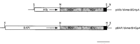
For the albumin promoter construct, plasmid 2335A-1 (13, 33) was modified by destroying a unique NotI site immediately upstream of the albumin promoter by using Klenow fragment to fill in NotI-digested DNA and then religating the blunt ends. A new NotI site was introduced immediately downstream of the promoter by ligation of NotI linkers into a unique BamHI site that had been digested and filled in with Klenow enzyme. Plasmid pECE/dimerBGHpA was digested with HindIII and XbaI, blunt ended with Klenow fragment, and flanked with NotI linkers, after which gel purification and ligation of the fragment into the NotI site of the modified 2335A-1 vector were performed. The resulting plasmid, pAlb/dimerBGHpA, contained flanking SacI sites which were used to release the 6.6-kb insert for use in the generation of transgenic mice (Fig. 1).
For theb-actin promoter construct, plasmid pHbAPr-1 (14) was modified by changing a unique EcoRI site immediately upstream, and a unique BamHI site immediately downstream, of the promoter to XbaI sites by digestion and linker insertion. Plasmid pECE/dimerBGHpA was digested with XbaI, blunt ended, and ligated to HindIII linkers, after which the original HindIII site in the polylinker region was digested with HindIII to release the HDV dimer-BGHpA sequence. The HindIII site-flanked sequences were gel purified and ligated into the unique HindIII site immediately downstream of theb-actin promoter and just upstream of the engineered XbaI site, which generated plasmid pBAP/ dimerBGHpA. This plasmid was digested with XbaI to release the 7.9-kbb-actin promoter–HDV sequence for use in the generation of transgenic mice.
37813 and 38796 to 38777, respectively (43), were used as controls for PCRs and DNA quality. PCR mixtures were first heated to 948C for 5 min and then subjected to 25 cycles of denaturation at 948C for 1 min, annealing at 558C for 1.5 min, and extension at 728C for 2 min. Products were analyzed by electrophoresis on a 1% agarose gel.
Characterization of integrated HDV sequences was performed by Southern blot hybridization, by standard procedures (41). Tail DNA (10ml, undiluted, approximately 10mg) was digested with appropriate restriction enzymes, elec-trophoresed through 0.7% agarose gels, and transferred to Hybond-C Extra nitrocellulose membrane (Amersham, Arlington Heights, Ill.). HDV-specific,
32P-labeled DNA probes were synthesized by using the Random Primed DNA
Labeling Kit (Boehringer Mannheim) with 1.7 kb of monomeric HDV cDNA as a template.
Northern (RNA) blot analysis.Unless otherwise noted, adult transgenic ani-mals were sacrificed at approximately 8 weeks of age. Total RNA was extracted from various tissues by using guanidinium isothiocyanate and acid phenol as described previously (8). RNA pellets were washed in 70% ethanol, resuspended in distilled H2O, and quantitated spectrophotometrically. Equal amounts of
RNA (10mg) from individual tissues were treated with formaldehyde, electro-phoresed through 1.2% agarose gels containing formaldehyde, and transferred to Hybond-C Extra nitrocellulose membrane. RNA blots were hybridized with
32P-labeled, strand-specific HDV riboprobes, synthesized by using T7 RNA
poly-merase and linearized plasmid templates as described previously (28). RNA extracted from H1d9 cells, which express and replicate HDV RNA from an integrated cDNA dimer (26), was used as a positive control in all Northern blots. Western blot (immunoblot) analysis.Protein lysates were obtained by extract-ing equivalent amounts of tissue with TRIzol reagent (GIBCO/BRL, Gaithers-burg, Md.) as described by the manufacturer. Proteins were denatured, separated in 10% polyacrylamide gels containing sodium dodecyl sulfate (SDS), and blot-ted onto Immobilon-NC (Millipore, Bedford, Mass.) nitrocellulose membrane. Filters were probed with a rabbit antibody against delta antigen (19) and then with125
I-labeled protein A (ICN, Costa Mesa, Calif.). Negative control lysates were obtained by the same procedure from normal mouse tissue, and positive controls for delta antigen were cell lysates from DBT cells infected with recom-binant vaccinia viruses expressing either large or small delta antigen (33a).
Immunohistochemistry and histopathological examination. Tissue blocks were fixed in 10% buffered formalin, embedded in paraffin, and cut into 4-m m-thick sections. For histological examinations, serial sections were stained with hematoxylin and eosin. For detection of HDAg by indirect immunoperoxidase staining, tissue sections were prepared as follows: endogenous peroxidase activity in deparaffinized sections was blocked by using 3% hydrogen peroxide, and rabbit serum protein was then used as a blocking protein to reduce background caused by nonspecific hydrophobic interactions. The sections were subsequently incubated with a 1:100 dilution of human antidelta serum for 30 min in a moist chamber at room temperature, washed in phosphate-buffered saline (PBS) three times for 10 min per washing, and treated with horseradish peroxidase-conju-gated rabbit anti-human immunoglobulin G (DAKO Corporation, Carpinteria, Calif.) at a 1:16 dilution for 30 min. Thereafter, sections were rinsed in PBS for 10 min and treated for 10 min with 3-30-diaminobenzidine hydrochloride and hydrogen peroxide, according to the method of Graham and Karnovsky (12). After being dehydrated, the sections were covered with a coverslip and examined by light microscopy. Positive and negative controls were run in parallel with the test samples.
RESULTS
Generation of HDV RNA-expressing transgenic mice. In
order to examine the properties of HDV RNA replication and its possible cytopathological effects in the liver, we generated transgenic mice which were expected to express a
on November 9, 2019 by guest
[image:2.612.61.296.67.142.2]sense HDV dimer RNA in the liver by using a liver-specific promoter. The dimer HDV RNA previously has been shown to be replication competent, and the albumin promoter used has been shown to direct the expression of HDAg specifically in the livers of transgenic mice (13). Figure 1 depicts schemati-cally the albumin promoter-HDV construct (pAlb/dimer BGHpA) used in this study.
Offspring of mice transplanted with microinjected eggs were screened for the presence of HDV sequence by PCR analysis. With tail DNA as the template, PCR was performed with two sets of primers: one set specific for HDV transgene sequences and a second set specific for mouseb-globin sequences, used as a control. Only the HDV-positive animals produced a 400-bp, HDV-specific band, while both positive and negative animals produced ab-globin-specific, 1,000-bp band (data not shown). Three of 20 mice obtained from pAlb/dimerBGHpA DNA-injected eggs were found to be positive for the HDV transgene and were designated founders 5, 11, and 14. Separate trans-genic lineages were established from these founder animals. To determine the nature of the integrated HDV cDNA se-quences, the founder mice and their progeny were character-ized by Southern blot analysis, by using the HDV-specific probe. The results showed that the three transgenic founder lines have different restriction endonuclease digestion pat-terns, suggesting that the HDV cDNA was integrated at dif-ferent chromosomal locations (Fig. 2). These data also showed that each transgenic mouse contained at least one full-length copy of the HDV transgene.
Replication of HDV RNA in transgenic mice.To examine
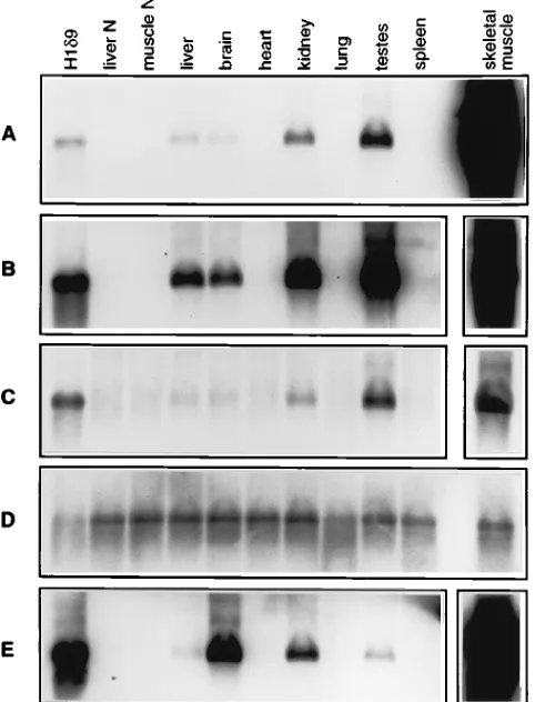
the synthesis of HDV RNA in pAlb/dimerBGHpA transgenic mice, total RNA was isolated from various tissues of an 8-week-old transgenic animal and subjected to Northern blot analysis with the HDV strand-specific probe. Figure 3A and B (panels show different exposure times) show that a 1.7-kb HDV genomic RNA was detected in the liver, as would be expected with the albumin promoter. Since the primary tran-script of this transgene is a dimeric HDV RNA, the monomeric RNA represents either the autocatalytically cleaved product of the primary dimer RNA transcript or the product resulting from HDV RNA replication and processing. Surprisingly, sev-eral other tissues, including brain, kidney, and testis tissue, and
particularly skeletal muscle, also expressed a similar or even larger amount of HDV genomic RNA. Quantitation of the HDV-specific bands by scanning densitometry showed that the level of HDV RNA in skeletal muscle was more than 100-fold higher than that in the liver or any other tissues. The detection of abundant HDV genomic RNA in nonhepatic tissues, par-ticularly skeletal muscle, was unexpected, since a liver-specific promoter was used to drive the transgene. This result suggests that the HDV RNA detected is replicated HDV RNA rather than primary transcripts from the integrated cDNA and that the levels of replicated HDV RNA, once expressed from the transgene, differed among tissues. To confirm this interpreta-tion, the expression of antigenomic HDV RNA in these tissues was examined. Figure 3C shows that all five tissues (liver, brain, kidney, testis, and skeletal muscle) also expressed 1.7-kb, an-tigenomic HDV RNA. The relative levels of anan-tigenomic RNA in different tissues were comparable to those of genomic RNA. Detection of antigenomic HDV monomer RNA is possible only after processing and replication of the primary genomic dimer transcript. These results indicate that the HDV RNAs FIG. 2. Southern blot characterization of albumin-HDV transgenic mice.
DNA from founder mice 5, 11, and 14 was digested with StuI (one site in HDV) or BglII (no sites in HDV), electrophoresed through a 0.7% agarose gel, and transferred to nitrocellulose membrane. 32
[image:3.612.316.557.74.390.2]P-labeled HDV-specific, random-primed DNA probes encompassing the entire HDV sequence were used as probes for hybridization. HindIII-digested lambda DNA markers are indicated to the left of the autoradiograph (numbers indicate kilobases).
FIG. 3. Northern blot analysis of RNAs from various tissues of pAlb/dimer BGHpA transgenic mice. RNA was separated by electrophoresis, transferred to membrane, and hybridized with a32P-labeled HDV riboprobe of either genomic
or antigenomic sense. The H1d9 sample was from a stably transformed cell line expressing HDV monomer RNA (26); liver N and muscle N samples were from normal mice. (A) Detection of genomic HDV RNA in transgenic lineage 14. The autoradiograph exposure time was 2 h. The line represents 1.7-kb HDV mono-mer RNA. (B) Same as for panel A, except that the exposure time was 30 h. The exposure time for skeletal muscle (inset) was 30 min. (C) Detection of anti-genomic HDV RNA in the tissues shown in panels A and B. The exposure times were identical to those for panel B. (D) Detection of 18S rRNA present in the samples shown in panel C. (E) Detection of genomic HDV RNA in transgenic lineage 5. Exposure times were as for panels B and C.
on November 9, 2019 by guest
http://jvi.asm.org/
different tissues showed slight variations. Again, the amount of HDV RNA in skeletal muscle was at least 100-fold greater than that in the liver or any other tissue. This elevated level of HDV RNA expression in skeletal muscle was also confirmed in the third transgenic mouse lineage (founder 11) (data not shown). These results established the unexpectedly high effi-ciency of HDV RNA replication in nonhepatic tissues, partic-ularly skeletal muscle. In contrast, the replication of HDV RNA in the liver was relatively poor.
HDV RNA replication from ab-actin promoter in
trans-genic mice.To further rule out the possibility that the albumin
promoter used may have other previously unrecognized prop-erties, additional transgenic mice which express HDV RNA from a cDNA dimer driven by a humanb-actin promoter were generated. This promoter was expected to be active in most tissues (14). Northern blot analysis with HDV-specific genomic and antigenomic riboprobes was performed with various tis-sues. Figure 4A shows that again the skeletal muscle was the tissue which contained the most abundant HDV genomic RNA; most of the other tissues also expressed some HDV genomic RNA. Surprisingly, the liver expressed the smallest amount of HDV RNA among all the tissues examined. The same pattern of expression was seen for the antigenomic RNA (Fig. 4B). Curiously, in this transgenic mouse, most of the antigenomic RNA was in dimeric form. The reason for this poor efficiency of processing is not clear; sequence analysis of the transgene detected no mutation in the autocatalytic cleav-age region (data not shown). A control blot using cho A se-quence (15) as a probe showed that the same amounts of RNA were loaded in all lanes (Fig. 4C). This transgenic mouse, in which a universal promoter (b-actin) was used, thus confirmed the findings obtained with the transgenic mice with a liver-specific promoter. Therefore, we concluded that HDV RNA replicates most efficiently in skeletal muscle.
Expression of delta antigen in transgenic mice. We next
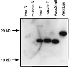
examined whether the delta antigen could be properly ex-pressed in HDV cDNA-transgenic animals. Because delta an-tigen is encoded by the anan-tigenomic strand of HDV RNA, proper processing and replication of the genomic primary tran-script must occur prior to the synthesis of delta antigen. Total protein extracts were prepared from liver and skeletal muscle tissues, separated by SDS-polyacrylamide gel electrophoresis (PAGE), and subjected to Western blot analysis with a rabbit polyclonal antibody specific for delta antigen. As shown in Fig. 5, the small form of delta antigen (p24), encoded by the HDV transgene, was detected in both liver and skeletal muscle tis-sues. Its amount in the muscle was at least 40-fold greater than that in the liver; thus, the levels of delta antigen present in these two tissues correlated roughly with the levels of RNA expression. Surprisingly, in each case, no large delta antigen
was detected, suggesting that the specific RNA editing event (23) required for the generation of the open reading frame for the large delta antigen did not occur.
Cellular localization of delta antigen and histopathology of
transgenic animals.The cell types in which delta antigen was
[image:4.612.314.551.75.331.2]expressed were determined by immunohistochemical analysis of tissue sections from the liver and muscle. By using human serum from HDV-infected individuals or rabbit polyclonal an-tibody against delta antigen in an indirect immunoperoxidase FIG. 4. Northern blot analysis of RNA from pBAP/dimerBGHpA transgenic mice. The method of detection was the same as used for Fig. 3. (A) Genomic (G) RNA. (B) Antigenomic (aG) RNA. (C) Control cho A RNA. Computer images were generated by using Adobe Photoshop software, version 2.5.1. M, muscle.
FIG. 5. Western blot detection of delta antigen in transgenic mice. Total proteins from liver and muscle tissue were separated by SDS-PAGE on a 10% polyacrylamide gel, transferred to nitrocellulose membrane, and probed with rabbit polyclonal antibody specific for HDAg (19). Liver N and muscle N samples were from normal mice. VaccSmdand VaccLgdsamples were from tissue culture cells infected with vaccinia virus recombinants expressing small and large delta antigens, respectively. Liver T and muscle T samples were from the transgenic mouse lineage 14 used in the experiment shown in Fig. 3. A 40-fold greater liver sample than muscle sample was used.
on November 9, 2019 by guest
[image:4.612.362.506.512.652.2]system (see Materials and Methods), delta antigen was dem-onstrated to be present in skeletal muscle and liver and brain tissue. In skeletal muscle, delta antigen was localized in the nuclei of myocytes (Fig. 6B and C). The same pattern of expression was seen in different sections of several different muscles. The specificity of the staining was demonstrated by the omission of the delta-antigen-specific antibody (Fig. 6A) and by the inclusion of a nontransgenic animal (data not shown). In the liver, the cells which expressed the most delta antigen, surprisingly, were epithelial cells of bile ducts (Fig. 7B and C). Only a few hepatocytes expressed a high level of delta antigen. In the brain, delta antigen appears to be localized in the glial cells, the precise type of which was not further deter-mined (data not shown).
Tissue sections also were stained with hematoxylin and eosin and examined for possible cytopathological effects resulting from HDV RNA replication and delta antigen expression. In either 10-week- or 9-month-old mice, no evidence of pathology was seen in either muscle, brain, or liver tissue (Fig. 7A and data not shown). There was no evidence of necrosis or inflam-mation. These data indicate that HDV RNA replication and delta antigen expression, at the level seen in these animals, did
not appear to cause cytopathology. However, in two pAlb/ dimerBGHpA-transgenic f1animals, some skeletal muscle re-vealed evidence of nuclear crowding and mild atrophy (Fig. 6D). We could not determine whether this was specifically related to the expression of delta antigen or RNA.
DISCUSSION
Studies of HDV replication to date have been limited pri-marily to tissue culture systems. Animal models, such as chim-panzee or woodchuck models (35, 37), have provided limited information on HDV replication in vivo. There is also a small-animal (mouse) model, in which HDV infects a small number of hepatocytes after intraperitoneal or intravenous injection of the virus (32). Unfortunately, in this model HDV replicates very inefficiently and causes neither hepatitis nor chronic in-fection (32). In all of these animal models, HDV RNA repli-cation is restricted to liver tissue, similar to the situation for natural HDV infection in humans. This exclusive hepatotro-pism is likely the result of trohepatotro-pism imposed by the surface antigen of HDV’s helper virus (hepatitis B virus). The poten-FIG. 6. Histopathology and immunocytochemistry of skeletal muscle from pAlb/dimerBGHpA transgenic mice. (A) Indirect immunoperoxidase staining of skeletal muscle in the absence of antibody against the delta antigen (magnification, 180X). (B) Indirect immunoperoxidase staining of skeletal muscle showing the staining of HDAg in the nuclei of myocytes (indicated by arrows) (magnification, 190X). (C) Same as panel B except for magnification (magnification, 380X). (D) Histopathology of skeletal muscle showing mild atrophy and nuclear crowding (indicated by an arrow) (hematoxylin and eosin staining; magnification, 70X).
on November 9, 2019 by guest
http://jvi.asm.org/
[image:5.612.65.554.73.451.2]tial of HDV to replicate in extrahepatic tissues of animals has not been explored.
In this report, we have described the generation of trans-genic mice in which HDV RNA is expressed and replicated under the control of two different eukaryotic transcriptional
scription of mRNA from a DNA transgene and is therefore controlled entirely by the tissue-specific expression pattern of the promoter. It is likely that a low basal level of transcription from the albumin promoter occurred in nonhepatic tissues and that self-replication and amplification of the primary transcript allowed these RNAs to become readily detectable. Thus, the amounts of RNA detected in these tissues would reflect the replication efficiencies of the RNA in the various tissues, rather than the strength and specificity of the promoter. There-fore, our detection of the largest amount of HDV RNA in skeletal muscle, despite the use of a liver-specific promoter, suggests that HDV RNA replicates most efficiently in skeletal muscle. This observation was confirmed with transgenic mice containing HDV cDNA transgene under control of theb-actin promoter. In these transgenic mice, more tissues expressed HDV RNA, reflecting the universal nature of this promoter. The studies of transgenic mice thus establish that HDV RNA can replicate in many tissues, particularly skeletal muscles, and that the hepatotropism of HDV is solely the result of tissue tropism imposed by the HBsAg on the viral envelope.
[image:6.612.64.293.74.605.2]How does one account for muscle displaying such a high propensity for HDV RNA replication? It has been demon-strated that replication of HDV RNA is dependent on cellular transcription machineries, including RNA polymerase II (11, 27) and cellular transcription factors (6). It is possible that muscle-specific transcription factors may interact efficiently with HDV RNA or delta antigen, thus serving as efficient factors for HDV RNA replication. In agreement with this finding, we have also shown that HDV cDNA dimers, under the control of simian virus 40 or cytomegalovirus immediate-early promoters, when injected as naked DNA directly into mouse muscle, achieved high levels of RNA replication (34), such that a humoral immune response to the expressed delta antigen was induced. Taken together, these data suggest that skeletal muscle supports very efficient HDV RNA replication. Whether this phenomenon is a fortuitous result of muscle factors interacting with HDV RNA or delta antigen or whether it represents an evolutionary origin of HDV RNA is not clear. If the latter is true, HDV may have been associated with muscle originally and have become hepatotropic only because of its association with HBsAg. Indeed, among all the tissues in the transgenic mice, the liver was a relatively poor site for HDV RNA replication. The high efficiency of HDV RNA replication in skeletal muscle also suggests that HDV RNA may potentially be used as a vehicle for efficient expression of foreign genes in skeletal muscles. Another tantalizing possibil-ity is that HDV-like agents may be associated with certain muscular diseases. So far, HDV is the only animal virus of its kind. Discovery of additional HDV-like agents will be of great interest.
FIG. 7. Histopathology and immunocytochemistry of liver tissue from pAlb/ dimerBGHpA transgenic mice. (A) Hematoxylin and eosin staining of liver tissue showing no pathology (magnification, ca. 50X). (B and C) Indirect immu-noperoxidase staining of portal area showing HDAg staining in the bile duct epithelial cells (arrows) (magnification, ca. 100X).
on November 9, 2019 by guest
This transgenic mouse model also revealed two additional interesting features of the mechanism of HDV replication. First of all, no 0.8-kb antigenomic RNA, which has been pos-tulated to be the mRNA for delta antigen (18), was detected. Because of the extremely large amount of HDV RNA gener-ated in this system, the presence of this mRNA species should have been detected readily. Yet no 0.8-kb RNA species was detected, even after prolonged exposures of the Northern blots (Fig. 3). The complete absence of this mRNA suggests that delta antigen is likely synthesized from the full-length mono-meric or multimono-meric antigenomic HDV RNA. Thus, the 0.8-kb RNA species is not absolutely required for the translation of delta antigen, in contrast to previous suggestions (18). There is likely an alternative mechanism for the translation of this pro-tein. Secondly, no large delta antigen was detected in either muscle or liver tissue of these transgenic mice, suggesting that the HDV-specific RNA editing event (23) did not occur during HDV RNA replication in transgenic mice. This cDNA clone has previously been demonstrated to undergo RNA editing in some tissue culture cell lines (24, 28). These data suggest that RNA editing is not the direct result of HDV RNA replication and that HDV RNA editing may be regulated by host factors. However, we could not with certainty rule out the possibility that minor differences in plasmid sequences interfered with RNA editing.
These studies further suggested that the hepatotropism ob-served in natural infection with HDV is due solely to the presence or absence of a functional receptor for the virus on various cell types. In humans, chimpanzees, and woodchucks, HDV RNA is detectable in hepatocytes only (31). Recent work on HDV infection of mice (32) also showed that HDV infected only the liver, at very low efficiency. The transgenic mice de-scribed in this report are the first experimental animals in which the virus entry process has been bypassed, allowing di-rect examination of HDV RNA replication in nonhepatic tis-sues in vivo. The data obtained from the transgenic mice also support the contention that HDV does not cause any direct cytopathological effects on the host. Previously, Guilhot et al. (13) reported that the expression of either large or small delta antigen in liver tissue of transgenic mice did not produce any cytopathological effects on the hepatocytes. In the present study, we further showed that HDV RNA replication itself also does not lead to cytopathic effects. Thus, the mechanism of HDV pathogenesis may not be direct cytopathicity. However, the possibility that interactions between HDV and its helper, HBV, within the same cell may exert a synergistic effect leading to cellular damage still remains. Curiously, several transgenic mice did develop mild muscular atrophy (Fig. 6D). Whether this was due to specific effects associated with HDV RNA or delta antigen expression or to nonspecific effects caused by high levels of viral RNA or proteins in the cells is not clear. This transgenic mouse model is expected to provide very in-teresting insights into the mechanism of regulation of HDV RNA replication and pathogenesis.
ACKNOWLEDGMENTS
We thank F. Chisari of Scripps Clinic for the albumin promoter plasmid 2335A-1, L. Kedes of the University of Southern California for theb-actin promoter plasmid pHbaPr-1, and S. Elmore of the Uni-versity of North Carolina, Chapel Hill, for the mouseb-actin-specific primers.
This work was partially supported by NIH research grant AI 26741 (to M.M.C.L.) and National Cancer Institute Cancer Center core grant CA5P3014089 to Norris Cancer Center, University of Southern Cali-fornia. J.M.P. and K.-S.J. are research associates and M.M.C.L. is an investigator at Howard Hughes Medical Institute.
REFERENCES
1. Bergmann, K. F., and J. L. Gerin. 1986. Antigens of hepatitis delta virus in the liver and serum of humans and animals. J. Infect. Dis. 154:702–706. 2. Branch, A. D., and H. D. Robertson. 1984. A replication cycle for viroids and
other small infectious RNAs. Science 223:450–455.
3. Chang, F.-L., P.-J. Chen, S.-J. Tu, C.-J. Wang, and D.-S. Chen. 1991. The large form of hepatitisdantigen is crucial for assembly of hepatitisdvirus. Proc. Natl. Acad. Sci. USA 88:8490–8494.
4. Chang, M.-F., S. C. Baker, L. H. Soe, T. Kamahora, J. G. Keck, S. Makino, S. Govindarajan, and M. M. C. Lai.1988. Human hepatitis delta antigen is a nuclear phosphoprotein with RNA-binding activity. J. Virol. 62:2403–2410. 5. Chao, M., S.-Y. Hsieh, and J. Taylor. 1990. Role of two forms of hepatitis delta virus antigen: evidence for a mechanism of self-limiting genome rep-lication. J. Virol. 64:5066–5069.
6. Chao, M., and M. M. C. Lai. Unpublished data.
7. Chisari, F. V., P. Filippi, J. Buras, A. McLachlan, H. Popper, C. A. Pinkert, R. D. Palmiter, and R. L. Brinster.1987. Structural and pathological effects of synthesis of hepatitis B virus large envelope polypeptide in transgenic mice. Proc. Natl. Acad. Sci. USA 84:6909–6913.
8. Chomczynski, P., and N. Sacchi. 1987. Single-step method of RNA isolation by acid guanidinium thiocyanate-phenol-chloroform extraction. Anal. Bio-chem. 162:156–159.
9. Cole, S. M., E. J. Gowans, T. B. Macnaughton, P. D. M. Hall, and C. J. Burrell.1991. Direct evidence for cytotoxicity associated with expression of hepatitis delta virus antigen. Hepatology 13:845–851.
10. Ellis, L., E. Clauser, D. O. Morgan, M. Edery, R. A. Roth, and W. J. Rutter. 1986. Replacement of insulin receptor tyrosine residues 1162 and 1163 compromises insulin-stimulated kinase activity and uptake of 2-deoxyglu-cose. Cell 45:721–732.
11. Fu, T.-B., and J. Taylor. 1993. The RNAs of hepatitis delta virus are copied by RNA polymerase II in nuclear homogenates. J. Virol. 67:6965–6972. 12. Graham, R. C., Jr., and M. J. Karnovsky. 1966. Glomerular permeability:
ultrastructural cytochemical studies using peroxidases as protein tracers. J. Exp. Med. 124:1123–1133.
13. Guilhot, S., S.-N. Huang, Y.-P. Xia, N. La Monica, M. M. C. Lai, and F. V. Chisari.1994. Expression of the hepatitis delta virus large and small antigens in transgenic mice. J. Virol. 68:1052–1058.
14. Gunning, P., J. Leavitt, G. Muscat, S.-Y. Ng, and L. Kedes. 1987. A human b-actin expression vector system directs high-level accumulation of antisense transcripts. Proc. Natl. Acad. Sci. USA 84:4831–4835.
15. Harpold, M. M., R. M. Evans, M. Salditt-Georgieff, and J. E. Darnell. 1979. Production of mRNA in Chinese hamster cells: relationship of the rate of synthesis to the cytoplasmic concentration of nine specific mRNA sequences. Cell 17:1025–1035.
16. Hogan, B., F. Constantini, and E. Lacy. 1986. Manipulating the mouse embryo: a laboratory manual. Cold Spring Harbor Laboratory Press, Cold Spring Harbor, N.Y.
17. Hoofnagle, J. H. 1989. Type D (delta) hepatitis. JAMA 261:1321–1325. 18. Hsieh, S.-Y., M. Chao, L. Coates, and J. Taylor. 1990. Hepatitis delta virus
genome replication: a polyadenylated mRNA for delta antigen. J. Virol. 64:3192–3198.
19. Hwang, S. B., and M. M. C. Lai. 1993. A unique conformation at the carboxyl terminus of the small hepatitis delta antigen revealed by a specific mono-clonal antibody. Virology 193:924–931.
20. Kuo, M. Y.-P., M. Chao, and J. Taylor. 1989. Initiation of replication of the human hepatitis delta virus genome from cloned DNA: role of delta antigen. J. Virol. 63:1945–1950.
21. Kuo, M. Y.-P., L. Sharmeen, G. Dinter-Gotlieb, and J. Taylor. 1988. Char-acterization of self-cleaving RNA sequences on the genome and antigenome of human hepatitis delta virus. J. Virol. 62:4439–4444.
22. Lin, J.-H., M.-F. Chang, S. C. Baker, S. Govindarajan, and M. M. C. Lai. 1990. Characterization of hepatitis delta antigen: specific binding to hepatitis delta virus RNA. J. Virol. 64:4051–4058.
23. Luo, G., M. Chao, S. Y. Hsieh, C. Sureau, K. Nishikura, and J. Taylor. 1990. A specific base transition occurs on replicating hepatitis delta virus RNA. J. Virol. 64:1021–1027.
24. Macnaughton, T. B. Unpublished data.
25. Macnaughton, T. B., M. R. Beard, M. Chao, E. J. Gowans, and M. M. C. Lai. 1993. Endogenous promoters can direct the transcription of hepatitis delta virus RNA from a recircularized cDNA template. Virology 196:629–636. 26. Macnaughton, T. B., E. J. Gowans, A. R. Jilbert, and C. J. Burrell. 1990.
Hepatitis delta virus RNA, protein synthesis and associated cytotoxicity in a stably transfected cell line. Virology 177:692–698.
27. Macnaughton, T. B., E. J. Gowans, S. P. McNamara, and C. J. Burrell. 1991. Hepatitisdantigen is necessary for access of hepatitisdvirus RNA to the cell transcriptional machinery but is not part of the transcriptional complex. Virology 184:387–390.
28. Macnaughton, T. B., Y.-J. Wang, and M. M. C. Lai. 1993. Replication of hepatitis delta virus RNA: effect of mutations of the autocatalytic cleavage sites. J. Virol. 67:2228–2234.
29. Makino, S., M. F. Chang, C. K. Shieh, T. Kamahora, D. M. Vannier, S. Govindarajan, and M. M. C. Lai.1987. Molecular cloning and sequencing of
on November 9, 2019 by guest
http://jvi.asm.org/
F. Bonino, R. H. Purcell, and J. L. Gerin.1984. Transmission of hepatitis B virus-associated delta agent to the eastern woodchuck. Proc. Natl. Acad. Sci. USA 81:2208–2212.
36. Purcell, R. H., and J. L. Gerin. 1990. Hepatitis delta virus, p. 2275–2287. In B. N. Fields and D. M. Knipe (ed.), Virology, 2nd ed. Raven Press, New York.
37. Rizzetto, M., M. G. Canese, J. L. Gerin, W. T. London, D. L. Sly, and R. H. Purcell.1980. Transmission of the hepatitis B virus-associated delta antigen to chimpanzees. J. Infect. Dis. 141:590–602.
38. Rizzetto, M., B. Hoyer, M. G. Canese, J. W.-K. Shih, R. H. Purcell, and J. L. Gerin.1980. Delta agent: association ofdantigen with hepatitis B surface antigen and RNA in serum ofd-infected chimpanzees. Proc. Natl. Acad. Sci. USA 77:6124–6128.
39. Ryu, W.-S., M. Bayer, and J. Taylor. 1992. Assembly of hepatitis delta virus particles. J. Virol. 66:2310–2315.
40. Ryu, W.-S., H. J. Netter, M. Bayer, and J. Taylor. 1993. Ribonucleoprotein
hepatitis delta virus-like particles. J. Virol. 65:6630–6636.
47. Wang, K. S., Q. L. Choo, A. J. Weiner, J. H. Ou, R. C. Najarian, R. M. Thayer, G. T. Mullenbach, K. J. Denniston, J. L. Gerin, and M. Houghton. 1986. Structure, sequence and expression of the hepatitis delta viral genome. Nature (London) 323:508–514.
48. Weiner, A. J., Q.-L. Choo, K.-S. Wang, S. Govindarajan, A. G. Redeker, J. L. Gerin, and M. Houghton.1988. A single antigenomic open reading frame of the hepatitis delta virus encodes the epitope(s) of both hepatitis delta anti-gen polypeptides p24dand p27d. J. Virol. 62:594–599.
49. Wu, H. N., and M. M. C. Lai. 1989. Reversible cleavage and ligation of hepatitis delta virus RNA. Science 243:652–654.
50. Wu, H. N., Y. J. Lin, F. P. Lin, S. Makino, M. F. Chang, and M. M. C. Lai. 1989. Human hepatitisdvirus RNA subfragments contain an autocleavage activity. Proc. Natl. Acad. Sci. USA 86:1831–1835.
51. Xia, Y.-P., M.-F. Chang, D. Wei, S. Govindarajan, and M. M. C. Lai. 1990. Heterogeneity of hepatitis delta antigen. Virology 178:331–336.