Copyright © 1998, American Society for Microbiology
Increased Misincorporation Fidelity Observed for Nucleoside Analog
Resistance Mutations M184V and E89G in Human Immunodeficiency
Virus Type 1 Reverse Transcriptase Does Not Correlate with
the Overall Error Rate Measured In Vitro
WILLIAM C. DROSOPOULOSANDVINAYAKA R. PRASAD*
Department of Microbiology and Immunology, Albert Einstein College of Medicine, Bronx, New York 10461
Received 9 December 1997/Accepted 29 January 1998
Nucleoside analog-resistant variants of human immunodeficiency virus type 1 (HIV-1) reverse transcriptase (RT) that displayed higher in vitro polymerase fidelity were previously identified via nucleotide insertion and mispair extension assays. To evaluate the contribution of increased nucleotide insertion and primer extension fidelities on the overall error rate of HIV-1 RT, we have measured the impact of two such mutations, E89G and M184V, on DNA copying fidelity in an M13 phage-based forward mutation assay. Using this assay, we observed mutation frequencies of 8.6031023, 6.2631023, 5.5331023, and 12.3031023for wild-type, E89G, M184V,
and double-mutant E89G/M184V HIV-1 RTs, respectively. Therefore, the overall polymerase fidelities of wild-type, E89G, M184V, and E89G/M184V HIV-1 RTs are similar (less than twofold differences) for DNA-dependent DNA synthesis. Thus, rather large increases in fidelity of deoxynucleoside triphosphate insertion and mispair extension observed previously appear not to influence the overall error rate of these mutants. However, a qualitative analysis of the mutations induced revealed significant differences in the mutational spectra between the wild-type and mutant enzymes.
One of the obstacles to successful antiretroviral treatment of human immunodeficiency virus (HIV) infections is the even-tual emergence of drug-resistant viruses. A primary contribu-tor to this process is the inherently high genetic variability of HIV, commonly encountered in virus populations in the in-fected individuals (9, 19, 25). This high level of variability is directly related to the viral mutation rate which, in turn, is thought to be significantly influenced by the polymerase fidelity of HIV reverse transcriptase (RT).
Mutants of HIV type 1 (HIV-1) RT resistant to nucleoside analog drugs that coincidentally display higher in vitro poly-merase fidelity in gel-based nucleotide insertion and primer extension assays have been identified (6, 11, 20, 21, 24, 31). These include the multi-dideoxynucleoside triphosphate-resis-tant E89G and the 3TC-resistriphosphate-resis-tant M184V RT variants. The M184V mutation arises in patients in response to antiviral therapy with (2)29,39 dideoxy-39-thiacytidine (3TC; lamivu-dine) (27) and confers up to a 1,000-fold increase in viral resistance to 3TC (7, 26, 30) as well as a low-level cross-resistance to ddC and ddI (8). The E89G mutation, initially isolated via a phenotypic bacterial screening assay (22), confers a broad cross-resistance to deoxynucleotide (dNTP) analogs including ddATP, ddCTP, ddGTP, ddTTP, 3TCTP, and phos-phonoformic acid (foscarnet; foscavir). This mutation has also been observed in in vitro-selected, 3TC-resistant variants of HIV-1 (7). The influence of these mutations on drug sensitivity is thought to be due to changes in the geometry of the dNTP-binding pocket brought about by direct interaction of the al-tered residue with the incoming dNTP in the case of M184V (12, 21, 29) or via template repositioning in the case of E89G (4, 29).
Previous observations showing that E89G and M184V RTs display increased fidelity of dNTP insertion and mispair exten-sion suggested that these enzymes may be less prone to poly-merase errors that lead to base substitution mutations. How-ever, polymerase errors that contribute to mutation rates are not limited to uncorrected polymerase misinsertions. Both base substitutions and frameshift errors can be generated through mechanisms not involving direct misinsertion but rather involving template-primer slippage or dislocation (16, 28). In fact, a large proportion of errors by HIV-1 RT have been shown to be of this type (2). Thus, fidelities of dNTP insertion and mispair extension cannot solely be used to de-termine an overall polymerase error rate.
In this communication, we report the overall polymerase error rates of the RT mutants E89G and M184V measured via an M13-based forward mutation assay (2, 3). The double mu-tant E89G/M184V was also included in the study to examine if the effects of the two mutations on the overall error rate would be cumulative. Our findings indicate that the overall levels of polymerase fidelity of wild-type, E89G, M184V, and E89G/ M184V HIV-1 RTs are similar (less than twofold differences). However, the mutational spectra of the variant enzymes re-vealed altered error specificities. It is unclear what impact such changes will have on viral variation, and this remains to be experimentally examined.
MATERIALS AND METHODS
Phage DNA and bacterial strains.Bacteriophage M13mp2, a strain that car-ries the lacZagene of Escherichia coli, was used to prepare gapped duplex DNA substrate. E. coli NR9099 [D(pro-lac) thi ara recA56/F9(proAB lacIqZDM15)]
served as the bacterial host for the preparation of both single-stranded and replicative-form M13 DNA. Electrocompetent E. coli MC1061 [hsdR hsdM1
araDD(ara leu)D(lacIPOZY) galU galK strA] cells were used to produce phage from the products of the fill-in reactions. E. coli CSH50 [D(pro-lac) thi ara strA/F9 (proAB lacIqZDM15 traD36)] was thea-complementation strain used to score
mutant phage.
Enzymes.Recombinant wild-type heterodimeric RT derived from HIV-1Hxb2
and its variants with E89G, M184V, and E89G/M184V substitutions were bac-* Corresponding author. Mailing address: Department of
Microbi-ology and ImmunMicrobi-ology, Albert Einstein College of Medicine, 1300 Morris Park Ave., Bronx, NY 10461. Phone: (718) 430-2517. Fax: (718) 430-8976. E-mail: [email protected].
4224
on November 9, 2019 by guest
http://jvi.asm.org/
terially expressed from derivatives of plasmid pL6H-PROT (14). The construc-tion of these derivatives is described elsewhere (10). The recombinant enzymes were purified by Ni21-nitrilotriacetic acid-hexahistidine chromatography fol-lowed by DEAE- and S-Sepharose chromatography as described earlier (15). The purified wild-type, E89G, M184V, and E89G/M184V RTs had specific activities of 220, 680, 280, and 89 U/mg, respectively, and were found to be nuclease free (data not shown). One unit is defined as the amount of enzyme required to incorporate 1 nmol of dTMP into DNA on a poly(rA)-oligo(dT) template-primer at 37°C in 10 min.
Forward mutation assay.Gapped duplex M13mp2 DNA was prepared as described previously (3) as the template for DNA fill-in synthesis reactions. This template contained a single-stranded region of 361 nucleotides including the upstream regulatory sequences and the first 107 coding nucleotides of the lacZa gene. Gap-filling synthesis reactions were performed by combining purified RT (0.07 to 0.45 U) with gapped duplex DNA (75 ng) in a reaction buffer containing 75 mM Tris-Cl (pH 8.0), 80 mM KCl, 6 mM MgCl2, 10 mM dithiothreitol, and
500mM each dATP, dCTP, dGTP, and dTTP (Boehringer Mannheim, India-napolis, Ind.) in a total volume of 25ml, and incubation for 1 h at 37°C. Complete gap closure was confirmed via agarose gel electrophoresis.
Polymerization products were electroporated into E. coli MC1061 host cells. After a brief (10-min) recovery period, transformants were plated on a bacterial indicator lawn (CSH50) in the presence of 0.195 mM 5-bromo-4-chloro-3-in-dolyl-b-D-galactopyranoside (X-Gal; Labscientific Inc., Livingston, N.J.) to
gen-erate plaques from released phages. Following incubation for approximately 15 h at 37°C, plaques were scored fora-complementation phenotype. Plaques pro-duced from phage with unmutated lacZagene display wild-typea complemen-tation, efficiently hydrolyzing the chromogenic X-Gal substrate to appear dark blue. Mutant phage containing lacZatarget mutations generate plaques whose phenotypes range from nearly wild-type dark blue to colorless. Plaques of phage designated as mutant were removed from initial screening plates and placed in 0.9% NaCl (1 ml). Mutant phenotypes were confirmed by plating equal inputs of wild-type and putative mutant phage (from diffused 1 ml of stock) on an indicator lawn as described above for initial screening. Once confirmed, mutant plaques were picked and stored at 4°C in 0.9% NaCl (1 ml) until needed for sequence analysis.
Mutational specificity.Single-stranded phage DNA was prepared as the tem-plate for sequence analysis as previously described (3). DNA sequencing reac-tions were performed with a Sequenase 2.0 DNA sequencing kit (Amersham Life Sciences, Arlington Heights, Ill.) with the oligonucleotide 59GCGCAGCTGTT GGGAAGGGCG39as primer. Sequencing reaction products were resolved on 6% denaturing polyacrylamide gels by using a GENOMYXLRDNA sequencer. Calculating error frequencies and error rates.Error frequencies were calcu-lated as the ratio of confirmed mutant plaques to the total plaques screened. Spontaneous background mutation frequency was determined by electroporating uncopied gapped DNA and scoring for mutants as with DNA synthesized by RT. Corrected frequencies were then obtained by subtracting the spontaneous back-ground from the frequency observed for a given RT (3).
Specific error rates were derived by multiplying the corrected overall error frequency with the percentage, in the total mutations, of the particular class of errors being examined (e.g., frameshifts). This value was then divided by 0.6 (the likelihood of expression of the newly synthesized phage minus strand in E. coli [17]) and then divided by the total number of sites that are susceptible to the given class of errors (3). Comparative statistical analysis of hotspot error rates was performed by using Fisher’s exact test.
RESULTS
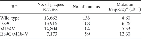
Overall mutation frequencies.The fidelity of DNA synthesis by wild-type, E89G, M184V, and E89G/M184V HIV-1 RTs was measured in an M13 phage gap-filling assay. Copying er-rors resulting in altered function of the phage lacZareporter gene were phenotypically scored. Gap-filling DNA synthesis reactions (two independent syntheses per enzyme) were per-formed with purified wild-type, E89G, M184V, and E89G/ M184V HIV-1 RTs, and completion of the reaction was con-firmed via agarose gel electrophoresis (data not shown). M13 DNA was electroporated into host E. coli, and plaques were generated by plating the transformants on a bacterial indicator lawn. Mutation frequencies for each enzyme were compiled from ratios of total mutant to total wild-type plaques obtained from one to three electroporations per gap-filling reaction per enzyme. The observed mutation frequencies for the wild-type, E89G, M184V, and E89G/M184V HIV-1 RTs were 8.60 3 1023, 6.2631023, 5.5331023, and 12.3031023, respectively
(Table 1). These represent background-corrected values, ad-justed by using the spontaneous mutation frequency of un-copied gapped DNA of 1.53 1023. These results indicated
that in terms of overall polymerase fidelity, the wild-type, E89G, M184V, and E89G/M184V HIV-1 RTs exhibit similar levels of fidelity (less than twofold differences). Of these, the E89G and M184V RTs were slightly less error prone than wild-type RT, while the E89G/M184V double mutant was slightly more error prone than wild-type RT.
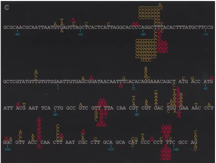
Analysis of mutation spectra of the variant RTs.DNA se-quence analysis of the phage mutants generated by the indi-vidual RTs revealed significant differences in the distribution and specificity of the mutations induced (Fig. 1). Mutations induced by wild-type, E89G, and M184V RTs were fairly well distributed throughout the target region, with some clustering at hotspots (sites with at least five mutations), while those produced by E89G/M184V RT were almost exclusively clus-tered at hotspots (see below). Close inspection of hotspot distribution shows that certain hotspots are shared by all RTs (Table 2). For example, the base substitution hotspot at posi-tion236 is well represented in the spectra of all of the RTs studied here, as is the frameshift hotspot at positions 137 to 139 (Fig. 1; Table 2). In contrast, the base substitution hotspot observed in the mutation spectrum of wild-type RT at position 112 and the deletion hotspot at positions 106 to 108 (Fig. 1) are conspicuously absent from the spectra of the mutant RTs. In fact, with the mutant RTs, these sites are nearly devoid of mutations (Fig. 1). Similarly, the frameshift hotspot found at positions234 to236 in the spectra of the E89G and E89G/ M184V RTs is completely absent in the spectra of wild-type and M184V RTs (P,0.0001 for both E89G [versus wild-type and M184V RTs] and E89G/M184V [versus wild-type and M184V RTs]) (Fig. 1). Furthermore, a greater percentage of total mutations induced by E89G/M184V (83.8%) are within hotspots compared to mutations induced by the other RTs (wild type, 53.6% [P , 0.0001]; E89G, 60.2% [P5 0.0002]; M184V, 60.6% [P5 0.0003]). For the E89G/M184V variant, four sites (positions236,234 to236, 70 to 73, and 137 to 139) account for almost 84% (83/99) of all mutations, with the234 to 236 positions alone accounting for 68% (67/99) of total mutations observed (Fig. 1c; Table 2).
Sequence analysis also revealed that wild-type and M184V RTs generated predominantly base substitution mutations, roughly 75% (103/138) and 77% (80/104), respectively, of all mutations (Table 3). On the other hand, E89G RT generated substitution and frameshift mutations (mainly single-base de-letions) at almost equal proportions (57:51) (Table 3). Inter-estingly, the E89G/M184V double mutant had a phenotype intermediate to that of single mutants with respect to base substitution/frameshift ratio, with approximately 67% (66/99) of RT-induced mutations being of the substitution class (Table 3).
[image:2.612.308.549.81.147.2]Most of the frameshift mutations detected involved the gain (11) or loss (21) of one base. Frameshift mutations involving the deletion of two bases were seen only with E89G (four occurrences out of 51 frameshift mutations; Fig. 1a) and E89G/
TABLE 1. Overall mutation frequencies
RT No. of plaquesscreened No. of mutants frequencyMutationa(1023)
Wild type 13,662 138 8.60
E89G 13,916 108 6.26
M184V 14,804 104 5.53
E89G/M184V 7,173 99 12.30
aCorrected for background by subtracting the spontaneous background
fre-quency of 1.5031023from the frequency observed for a given RT. Spontaneous
background was measured by using uncopied gapped M13mp2 DNA.
on November 9, 2019 by guest
http://jvi.asm.org/
on November 9, 2019 by guest
http://jvi.asm.org/
M184V RTs (one occurrence out of 33 frameshifts; Fig. 1c). Frameshift mutations generated by E89G also included one deletion involving three bases (Fig. 1a). Major deletions (.50 bp) were also induced; however, they were uncommon and seen only with wild-type (2 of 138 mutants scored) and E89G (3 of 108 mutants scored) RTs.
DISCUSSION
Viral mutation rates are the products of the combined ef-fects of overall polymerase (copying) error rates, replication rates, and biological selection (5). Presumably, increases or decreases in any of these factors could result in corresponding changes in mutation rate. This notion has previously not been tested for any variant of HIV-1 RT. The variants of HIV-1 RT that were recently reported to possess increased fidelities of dNTP insertion and primer extension in in vitro assays (6, 11, 20, 21, 24, 31) appeared suitable to examining the contribu-tions of polymerase fidelity to overall mutation rate in vivo. Thus, we have determined the overall copying fidelity of the
E89G, M184V, and E89G/M184V RT variants. We had earlier reported increases of three- to sixfold in average nucleotide insertion fidelity for the M184V and E89G RTs (6, 31). When changes in the fidelity of formation of specific mispairs are considered, the increases of 2- to 45-fold (6, 21, 24, 31) have been seen. With respect to terminal mismatch extension effi-ciency, increases in fidelity of primer extension, ranging from 3- to 66-fold greater (for extension from specific mispairs) than for wild-type RT, have been reported for E89G and M184V RTs (11, 20, 24). In contrast to these large increases in fidel-ities of dNTP insertion and primer extension previously ob-served for specific types of errors, the M184V and E89G RTs in the current study showed only slightly decreased overall polymerase error rates whereas the E89G/M184V variant rate was slightly increased (less than twofold differences when com-pared to the wild type) (Table 1). This discrepancy could be a result of the influence of certain features of the M13 forward mutation assay, such as host repair and the ability to detect slippage-mediated errors in addition to nucleotide misinser-tion and mispair extension. In fact, a substantial propormisinser-tion of
FIG. 1. (a) Spectrum of mutations induced by wild-type HIV-1 RT and its E89G variant. Comparison of mutations generated in the lacZatarget by wild-type HIV-1Hxb2and E89G HIV-1 RTs is shown. Mutations induced by wild-type HIV-1Hxb2RT are represented above the template sequence (white), and mutations induced
by E89G HIV-1 RT are shown below the template sequence. Substitutions are indicated by the letter corresponding to the new base (in yellow) above or below the base in the template sequence it is replacing. Nucleotide deletions are indicated by an upright (red) triangle over or under the corresponding run of template bases; nucleotide additions are indicated by an inverted (red) triangle over or under the corresponding run of template bases. Triangles with numerals (2 or 3) underneath them indicate deletions involving more than one base (the bases deleted or are thought to be involved in the deletion are underlined in red). A two-base deletion with the asterisk indicates where it was not possible to determine whether bases 140 and 141 or 141 and 142 were deleted. Nucleotide positions are indicated below the template sequence. (b) Spectrum of mutations induced by wild-type and the M184V mutant HIV-1 RTs. Comparison of mutations generated in the lacZatarget by wild-type HIV-1Hxb2and M184V HIV-1 RTs is shown. Mutations induced by wild-type HIV-1Hxb2RT are represented above the template sequence (white), and
mutations induced by M184V HIV-1 RT are shown below the template sequence. (c) Spectrum of mutations induced by wild-type and E89G/M184V mutant RTs. Comparison of mutations generated in the lacZatarget by wild-type HIV-1Hxb2and E89G/M184V HIV-1 RTs is shown. Mutations induced by wild-type HIV-1Hxb2
RT are represented above the template sequence (white), and mutations induced by E89G/M184V HIV-1 RT are shown below the template sequence.
on November 9, 2019 by guest
http://jvi.asm.org/
[image:4.612.79.519.68.401.2]the total mutations observed occurred within the homonucle-otide runs in the template sequence which are primary sites for slippage-mediated mutagenesis. Thus, in light of the modest differences observed in overall error rates, it appears that any increases in fidelity of these mutants that may be afforded by the increased dNTP insertion/primer extension fidelity are muted by the contributions of other mutagenic mechanisms.
The lack of increase in fidelity for M184V RT seen in this study would appear to be concordant with the biological data regarding viral evolution of M184V mutant viruses. Drug-re-sistant variants of such viruses were found to emerge in cell culture at rates equal to those of wild-type viruses (1, 13), suggesting that mutation rates were unaffected by the RT mu-tation. It is likely that compensatory factors similar to those operating in the M13 forward assay (as described above) were modulating the effect of the viral 184V substitution.
Although both E89G and M184V mutations increase nucle-otide insertion/primer extension fidelity, it seems likely that they exert their effects via distinct mechanisms. The residues affected by these mutations lie within the enzyme active site in or near the dNTP-binding pocket (12, 29). Structural data place these residues at opposing sides of the active site, with the E89 residue contacting the phosphate-deoxyribose back-bone of the template strand and the M184 residue interacting with the sugar moiety of the primer terminus (12, 29). Conse-quently, as mutations at these residues alter different surfaces of the dNTP-binding pocket, it might be anticipated that the
[image:5.612.48.548.80.262.2]effects of these modifications on the dNTP-binding pocket conformation would differ. Such differences are reflected in the divergence seen in the distribution and specificity of mutations induced by the variants. For instance, the E89G mutation had an enhancing effect on frameshift mutagenesis (both run and non-run associated). In contrast, the M184V mutation had a minimal effect on this class of errors (Table 3). It is generally thought that most frameshift mutations (insertions and dele-tions) involving few (one to three) nucleotides arise from tran-siently misaligned or slipped template-primer intermediates (28). The E89 residue is located within theb5a strand of the palm region of RT, where it is thought to help the polymerase grip the template (12). It is conceivable that the glycine sub-stitution at this residue, by virtue of glycine’s smaller side chain, results in a looser grip, which might enable the primed template to more easily form misaligned intermediates while still bound to the enzyme. Alternatively, a looser template grip stemming from the 89G mutation might allow the RT to more readily dissociate from the template, providing greater oppor-tunity for the formation of misaligned template-primers. A determination of dissociation rates using purified RTs and model template-primers may help differentiate between the two possibilities. The M184 residue participates in dNTP bind-ing (21, 32) rather than template-primer grip. Thus, it is pos-sible that substitutions at this position affect utilization of frameshift intermediates rather than the enzyme’s ability to promote their formation.
TABLE 2. Error rates at selected mutational hotspots
Site Wild type E89G M184V E89G/M184V
No. of errors Error rate No. of errors Error rate No. of errors Error rate No. of errors Error rate
Base substitutions
236 26 1/370 35 1/296 37 1/305 55 1/87.5
235 8 1/1,203 0 CDd 1 1/11,284 0 CD
84 5 1/1,926 0 CD 1 1/11,284 0 CD
89 5 1/1,926 4 1/2,588 5 1/2,257 1 1/4,829
112 5 1/1,926 0a CD 0a CD 0a CD
145 3 1/3,209 1 1/10,351 5 1/2,257 2 1/2,415
Frameshifts
234 to236 0b CD 23b 1/1,350 0b CD 12b 1/1,207
70 to 73 5 1/7,702 2 1/20,702 0 CD 6 1/3,220
88 to 90 5 1/5,777 1 1/31,054 0 CD 2 1/7,244
106 to 108 6 1/4,814 1c 1/31,054 1c 1/33,852 1c 1/14,488
137 to 139 9 1/3,209 7 1/4,436 16 1/2,116 10 1/1,449
Overall 138 1/17,651 108 1/24,249 104 1/27,450 99 1/12,341
aIn comparison of each mutant RT to wild type, P values ranged from 0.069 to 0.076.
bIn comparison of E89G to wild type or M184V as well as in comparison of E89G/M184V to wild type or M184V, P values were,0.0001 each. cIn comparison of each mutant RT to wild type, P values ranged from 0.139 to 0.244.
dCD, cannot determine.
TABLE 3. Summary of error rates for wild-type, E89G, M184V, and E89G/M184V RTs by class
Mutation type Wild type E89G M184V E89G/M184V
No. of errors Error rate No. of errors Error rate No. of errors Error rate No. of errors Error rate
All classes 138 1/17,651 108 1/24,249 104 1/27,450 99 1/12,341
Base substitutions 103 1/11,876 57 1/23,041 80 1/17,921 66 1/9,294
Frameshifts 35a 1/40,650 51 1/30,030 24a 1/69,444 33a 1/21,645
At runs 32 1/23,753 43 1/19,011 24 1/37,037 33 1/11,567
At non-runs 3 1/222,222 8 1/89,286 0 CDb 0 CD
aIn comparison of E89G to wild type, M184V, and E89G/M184V, P values were 0.0004, 0.0003, and 0.048, respectively. bCD, cannot determine.
on November 9, 2019 by guest
http://jvi.asm.org/
[image:5.612.51.549.628.708.2]To date, the E89G substitution has been found only in viral isolates bearing the M184V mutation as well. Therefore, the E89G/M184V double mutant was studied in order to under-stand the cumulative effect of these two mutations on error rate. Curiously, when present independently, the individual mutations increased polymerase fidelity slightly while a com-bination of the two led to a slight decrease in the overall polymerase fidelity (Table 1). More unexpected was the highly clustered distribution of the mutations induced by the E89G/ M184V RT, considering that both E89G and M184V RTs generated mutations that were fairly well distributed. Yet the influence of both RT mutations appears to be seen in the mutational specificity of the double mutant. Hotspots common to both single variants (positions 236 and 137 to 139) are reproduced by the E89G/M184V variant. The strong represen-tation of the deletion hotspot at the234 to236 site seems to suggest some dominance of the 89G mutation; however, the lack of non-run-associated frameshifts for the double mutant appears to indicate a significant contribution by the 184V res-idue to frameshift specificity. Thus, the error specificities of each single mutation are preserved to a degree in the double mutant while the effects of each individual substitution on polymerase fidelity are somewhat modulated by the other. Since these two residues occupy opposite sides of the active site, it may be possible that steric constraints imposed by these substitutions are additive, thus further restricting the range of stable premutational intermediates. If this is so, it may offer an explanation for the limited mutation distribution exhibited by E89G/M184V RT.
Since the influence of these mutations on overall RT fidelity appears minimal, could differences in error specificity have a significant biological effect on viruses harboring these muta-tions? It is difficult to predict any specific effects of 184V alteration since the change observed in overall fidelity was small. In the case of E89G variant viruses, perhaps the en-hancement in frameshift mutagenesis associated with the 89G substitution when expressed in vivo results in a higher propor-tion of virions with single-base inserpropor-tions and delepropor-tions which are far more likely to be lethal than base substitutions. This may partially account for the failure of this mutation to be seen in a viral isolate independent of the 184V substitution. The ability of the M184V mutation to reduce the frameshift genicity of E89G could permit a variant HIV with both muta-tions to replicate and to be recovered via in vitro selection protocols (7).
Finally, it is known that sequence context contributes sub-stantially to the mutagenic potential of a given nucleotide site (2, 18, 23), and this likely results in the site bias that produces hotspots. The studies reported here were conducted in the context of the lacZagene, which may not be representative of viral sequences. Therefore, these RT mutations could impart to replicating viral genomes overall fidelities higher or lower than those observed here. In this context, the double mutant E89G/M184V RT in particular needs to be tested for its effect on viral mutation rate since it appears to display few mutations outside the hotspots which are so characteristic of lacZagene. It should be possible through single-cycle infection studies with HIVs containing these mutations to establish the in vivo sig-nificance of our findings.
ACKNOWLEDGMENTS
We thank Clyde A. Hutchison III (University of North Carolina, Chapel Hill) for providing phasmids of the M184V mutant RT cDNA, B. D. Preston for providing the M13mp2 phage, T. A. Kunkel (Na-tional Institute for Environmental Health Sciences) for providing the bacterial strains for the M13 forward mutation assay, Kenneth Curr,
Clark Choi, and Jayanthi Manne for technical assistance, Ganjam V. Kalpana, William Franklin, and Lisa Rezende for critically reading the manuscript, and the Oligonucleotide Synthesis Facility of the Albert Einstein College of Medicine’s Cancer Center for DNA oligonucleo-tides.
This work was supported by Public Health Service grants AI-30861 and AI-40375 (to V.R.P.). W.C.D. acknowledges support from insti-tutional training grant T32-AI07501.
REFERENCES
1. Balzarini, J., H. Pelemans, A. Karlsson, E. De Clercq, and J. P. Kleim. 1996. Concomitant combination therapy for HIV infection preferable over sequen-tial therapy with 3TC and non-nucleoside reverse transcriptase inhibitors. Proc. Natl. Acad. Sci. USA 93:13152–13157.
2. Bebenek, K., J. Abbotts, J. D. Roberts, S. H. Wilson, and T. A. Kunkel. 1989. Specificity and mechanism of error-prone replication by human immunode-ficiency virus-1 reverse transcriptase. J. Biol. Chem. 264:16948–16956. 3. Bebenek, K., and T. A. Kunkel. 1995. Analyzing fidelity of DNA
poly-merases. Methods Enzymol. 262:217–232.
4. Boyer, P. L., C. Tantillo, A. Jacobo-Molina, R. G. Nanni, J. Ding, E. Arnold,
and S. H. Hughes.1994. Sensitivity of wild-type human immunodeficiency virus type 1 reverse transcriptase to dideoxynucleotides depends on template length; the sensitivity of drug-resistant mutants does not. Proc. Natl. Acad. Sci. USA 91:4882–4886.
5. Coffin, J. 1996. HIV viral dynamics. AIDS 10:S75–S84.
6. Drosopoulos, W. C., and V. R. Prasad. 1996. Increased polymerase fidelity of E89G, a nucleoside analog-resistant variant of human immunodeficiency virus type 1 reverse transcriptase. J. Virol. 70:4834–4838.
7. Gao, Q., Z. Gu, M. A. Parniak, J. Cameron, N. Cammack, C. Boucher, and
M. A. Wainberg.1993. The same mutation that encodes low-level human immunodeficiency virus type 1 resistance to 29,39-dideoxyinosine and 29,39 -dideoxycytidine confers high-level resistance to the (2) enantiomer of 29,39 -dideoxy-39-thiacytidine. Antimicrob. Agents Chemother. 37:1390–1392. 8. Gu, Z., Q. Gao, X. Li, M. A. Parniak, and M. A. Wainberg. 1992. Novel
mutation in the human immunodeficiency virus type 1 reverse transcriptase gene that encodes cross-resistance to 29,39-dideoxyinosine and 29,39 -dideoxy-cytidine. J. Virol. 66:7128–7135.
9. Hahn, B. H., G. M. Shaw, M. E. Taylor, R. R. Redfield, P. D. Markham, S. Z.
Salahuddin, F. Wong-Staal, R. C. Gallo, E. S. Parks, and W. P. Parks.1986. Genetic variation in HTLV III/LAV over time in patients with AIDS or at risk for AIDS. Science 232:1548–1553.
10. Hamburgh, M. E., W. C. Drosopoulos, and V. R. Prasad. The influence of 3TC-resistance mutations M184V and E89G in the human immunodefi-ciency virus reverse transcriptase on mispair extension effiimmunodefi-ciency. Submitted for publication.
11. Hsu, M., P. Inouye, L. Rezende, N. Richard, Z. Li, V. R. Prasad, and M. A.
Wainberg.1997. Higher fidelity of RNA-dependent DNA mispair extension by M184V drug-resistant than wild type reverse transcriptase of human immunodeficiency virus type 1. Nucleic Acids Res. 25:4532–4536. 12. Jacobo-Molina, A., J. Ding, R. G. Nanni, A. D. J. Clark, X. Lu, C. Tantillo,
R. L. Williams, G. Kamer, A. L. Ferris, P. Clark, A. Hizi, S. Hughes, and E. Arnold.1993. Crystal structure of human immunodeficiency virus reverse transcriptase complexed with double stranded DNA at 3.0 Å resolution shows bent DNA. Proc. Natl. Acad. Sci. USA 90:6320–6324.
13. Keulen, W., M. Nijhuis, R. Schuurman, B. Berkhout, and C. Boucher. 1997. Reverse transcriptase and HIV variation. Science 275:229. (Comment.) 14. Kew, Y., L. Olsen, A. Japour, and V. R. Prasad. Insertions into the b3-b4
hairpin loop of HIV-1 reverse transcriptase reveal a role for fingers subdo-main in processive polymerization. J. Biol. Chem., in press.
15. Kew, Y., Q. Song, and V. Prasad. 1994. Subunit selective mutagenesis of Glu89 residue in human immunodeficiency virus reverse transcriptase. J. Biol. Chem. 269:15331–15336.
16. Kunkel, T. A., and P. S. Alexander. 1986. The base substitution fidelity of eucaryotic DNA polymerases. Mispairing frequencies, site preferences, in-sertion preferences, and base substitution by dislocation. J. Biol. Chem.
261:160–166.
17. Kunkel, T. A., and A. Soni. 1988. Exonucleolytic proofreading enhances the fidelity of DNA synthesis by chick embryo DNA polymerase-gamma. J. Biol. Chem. 263:4450–4459.
18. Mendelman, L. V., M. S. Boosalis, J. Petruska, and M. F. Goodman. 1989. Nearest neighbor influences on DNA polymerase insertion fidelity. J. Biol. Chem. 264:14415–14423.
19. Meyerhans, A., R. Cheynier, J. Albert, M. Seth, S. Kwok, J. Sninsky, L.
Morfeldt-Manson, B. Asjo, and S. Wain-Hobson.1989. Temporal fluctua-tions in HIV quasispecies in vivo are not reflected by sequential HIV isola-tions. Cell 58:901–910.
20. Oude Essink, B. B., N. K. T. Back, and B. Berkhout. 1997. Increased poly-merase fidelity of the 3TC-resistant variants of HIV-1 reverse transcriptase. Nucleic Acids Res. 25:3212.
21. Pandey, V. N., N. Kaushik, N. Rege, S. G. Sarafianos, P. N. S. Yadav, and
M. J. Modak.1996. Role of methionine 184 in human immunodeficiency
on November 9, 2019 by guest
http://jvi.asm.org/
virus type 1 reverse transcriptase in the polymerase function and fidelity of DNA synthesis. Biochemistry 35:2168–2179.
22. Prasad, V. R., I. Lowy, T. De Los Santos, L. Chiang, and S. P. Goff. 1991. Isolation and characterization of a dideoxyguanosine triphosphate-resistant HIV-1 reverse transcriptase expressed in bacteria. Proc. Natl. Acad. Sci. USA 88:11363–11367.
23. Ricchetti, M., and H. Buc. 1990. Reverse transcriptases and genomic vari-ability: the accuracy of DNA replication is enzyme specific and sequence dependent. EMBO J. 9:1583–1593.
24. Rubinek, T., M. Bakhanashvili, and A. Hizi. 1997. The fidelity of 39 misin-sertion and mispair extension during DNA synthesis exhibited by two drug-resistant mutants of the reverse transcriptase of human immunodeficiency virus type 1 with Leu74Val and Glu89Gly. Eur. J. Biochem. 247:238. 25. Saag, M. S., B. H. Hahn, J. Gibbons, Y. Li, E. S. Parks, W. P. Parks, and
G. M. Shaw.1988. Extensive variation of human immunodeficiency virus type-1 in vivo. Nature 334:440–444.
26. Schinazi, R. F., R. M. Lloyd, Jr., M. Nguyen, D. L. Cannon, A. McMillan, N.
Ilksoy, C. K. Chu, D. C. Liotta, H. Z. Bazmi, and J. W. Mellors.1993. Characterization of human immunodeficiency viruses resistant to Oxathio-lane-cytosine nucleosides. Antimicrob. Agents Chemother. 37:875–881. 27. Schuurman, R., M. Nijhuis, R. van Leeuwen, P. Schipper, D. de Jong, P.
Collis, S. A. Danner, J. Mulder, C. Loveday, and C. Christopherson.1995. Rapid changes in human immunodeficiency virus type 1 RNA load and
appearance of drug-resistant virus populations in persons treated with lami-vudine (3TC). J. Infect. Dis. 171:1411–1419.
28. Streisinger, G., Y. Okada, J. Emrich, J. Newton, A. Tsugita, E. Terzaghi, and
M. Inouye.1966. Frameshift mutations and the genetic code. Cold Spring Harbor Symp. Quant. Biol. 31:77–84.
29. Tantillo, C., J. Ding, A. Jacobo-Molina, R. G. Nanni, P. L. Boyer, S. H.
Hughes, R. Pauwels, K. Andries, P. A. Janssen, and E. Arnold.1994. Loca-tions of anti-AIDS drug binding sites and resistance mutaLoca-tions in the three-dimensional structure of HIV-1 reverse transcriptase. Implications for mech-anisms of drug inhibition and resistance. J. Mol. Biol. 243:369–387. 30. Tisdale, M., S. D. Kemp, N. R. Parry, and B. A. Larder. 1993. Rapid in vitro
selection of human immunodeficiency virus type 1 resistant to 39-thiacytidine inhibitors due to a mutation in the YMDD region of reverse transcriptase. Proc. Natl. Acad. Sci. USA 90:5653–5656.
31. Wainberg, M. A., W. C. Drosopoulos, H. Salomon, M. Hsu, G. Borkow, M.
Parniak, Z. Gu, Q. Song, J. Manne, S. Islam, G. Castriota, and V. R. Prasad.
1996. Enhanced fidelity of 3TC-selected mutant HIV-1 reverse transcriptase. Science 271:1282–1285.
32. Wilson, J. E., A. Aulabaugh, B. Caligan, S. McPherson, J. K. Wakefield, S.
Jablonski, C. D. Morrow, J. E. Reardon, and P. A. Furman.1996. Human immunodeficiency virus type-1 reverse transcriptase. Contribution of Met-184 to binding of nucleoside 59-triphosphate. J. Biol. Chem. 271:13656– 13662.