A STUDY ON CLINICAL SPECTRUM OF CEREBRAL VENOUS
THROMBOSIS
Dissertation Submitted to
THE TAMILNADU DR. M.G.R. MEDICAL UNIVERSITY
Chennai.In partial fulfillment of the regulations For the award of the degree of
M.D. (GENERAL MEDICINE) BRANCH – I
GOVT. STANLEY MEDICAL COLLEGE & HOSPITAL
CHENNAI
CERTIFICATE
This is to certify that this dissertation entitled “A STUDY ON
CLINICAL
SPECTRUM OF
CEREBRAL VENOUS THROMBOSIS ” submitted by Dr.E.THIRULOGACHANDAR,to the Tamil Nadu Dr. M.G.R. Medical University Chennai is in partial fulfillment of the regulations for the award of M.D. DEGREE BRANCH –I (General Medicine) and is a bonafide research work carried out by him under direct supervision and guidance.
Prof. Dr. MAGESH KUMAR, M.D.,
Professor and Head, Dept. of Medicine, Govt. Stanley Medical College and Hospital
Chennai-600001.
Prof. Dr.K.H.NOORUL AMEEN,M.D.,
Chief UNIT VI & Guide, Dept of Medicine, Govt. Stanley Medical College and Hospital
Chennai-600001.
Prof. Dr. C.VAMSADHARA, M.D., Ph.D The Dean
DECLARATION
I Solemnly declare that the dissertation titled“A STUDY ON CLINICAL SPECTRUM OF CEREBRAL VENOUS THROMBOSIS ” was done by me at Stanley Medical College and Hospital during 2008-2010 under guidance and supervision of Prof.Dr.K.H.NOORUL AMEEN, M.D.,
The dissertation of submitted to the Tamil Nadu Dr. M.G.R. Medical University towards the partial fulfillment of requirement for the award of M.D. DEGREE (BRANCH-I) in General Medicine.
Place : Chennai
ACKNOWLEDGEMENT
I owe my thanks to Dr.C.VAMSADHARA M.D.,Phd., Dean, Stanley Medical College , for allowing me to avail the facilities needed for my dissertation work.
I also thankDr.A.Priya M.S., Medical Superintent ,Government Stanley Hospital for permitting me to use available resources from the hospital for my dissertation.
I am grateful toProf. Dr.S.MAGESHKUMAR , M.D., Professor and Head of the Department of Medicine, Stanley Medical College for permitting me to do the study and for his encouragement.
I have great pleasure in expressing my deep sense of gratitude and respect for Prof.Dr. K.H.NOORUL AMEEN, M.D., Professor and my Unit Chief, Department of Medicine for his valuable guidance and suggestions in preparing this dissertation.
I also thank my former Professor and Unit ChiefDr.S.Tito M.D., for his valuable contributions for this dissertation.
I am extremely thankful toDr. G.VASUMATHI, M.D., Registrar Department of Medicine my unit assistant professors and Dr. V.MOHANRAO, M.D., and Dr.S.RAMALINGAM, M.D., for their valuable guidance and constant encouragement.I am also grateful to my former Assistant Professors
Dr.T.Arun M.D., andDr.R.Arun M.D.,for their assistance in preparing this dissertation.
CONTENTS
S.NO TITLE P.NO
1. INTRODUCTION 1
2. AIM OF STUDY 2
3. REVIEW OF LITERATURE 4 4. MATERIALS AND METHODS 44
5. RESULTS 45
6. DISCUSSION 53 7. CONCLUSIONS 60
ANNEXURE
I BIBLIOGRAPHY
II PROFORMA
INTRODUCTION
Cerebral venous thrombosis (CVT)—ie, thrombosis of the intracranial veins and sinuses—is a rare type of cerebrovascular disease that affects about 5 people per million and accounts for 0·5 % of all stroke. CVT was rst recognised at the beginning of the 19th century1
and it was long thought to be an infective disorder that commonly a ected the superior sagittal
sinus and resulted in bilateral or alternating focal de cits, seizures, and coma, which usually led to death1. At that time, CVT was commonly diagnosed at autopsy and usually showed haemorrhagic lesions, which, by analogy with arterial stroke, was thought to contraindicate the use of heparin.
In the past 25 years, the widespread use of neuroimaging has aided early diagnosis of CVT and has thus completely modi ed the current information we have on this disorder. CVT is now typically recognised as a disorder with various clinical presentations and a usually favourable outcome, with mortality well below 10%. MRI and magnetic resonance angiography are the best diagnostic methods for diagnosis and heparin is the rst-line treatment. However, the diagnosis of CVT is still commonly overlooked or delayed because of the remarkable diversity of its clinical symptoms, mode of onset, and neuroimaging signs; furthermore, a cause cannot be found in about 15-30% of cases, the individual outcome may still be di cult to
AIM OF THE STUDY
REVIEW OF LITERATURE
Cerebral veins contain about 70% of the total cerebral blood volume, but cerebral venous sinus thrombosis (CVT) occurs less often than arterial stroke. Arterial and venous stroke cause different neurological deficits and occur in people of different ages. About half of the patients with an arterial stroke are older than 75 years, whereas CVT most often affects young adults and children. It is associated with a prothrombotic state due to (inherited) thrombophilia, other blood disorders, dehydration, infectious diseases, cancer, or more rare causes. In about a quarter of the patients with CVT the cause remains unknown.
The annual incidence is currently estimated to be 3–4 cases per million people. 3 out of 4 people with CVT are women. 1 out of 8 patients will die or remain handicapped as a result of CVT 2 . CVT constitutes about 10-15% of young strokes. During the past decade, modern neuroimaging techniques have improved the diagnostic process and together with the increased awareness this will probably result in increased recognition in the future. CVT has received far less attention in clinical research than arterial stroke. The small number of patients with CVT limits the performance of large epidemiological studies and clinical trials on a scale comparable to arterial stroke. .
ANATOMY OF CEREBRAL VENOUS SYSTEM
Introduction
The veins of the brain have no muscular tissue in their thin walls and possess no valves. They emerge from the brain and lie in the subarachnoid space. They pierce the arachnoid mater and the meningeal layer of the dura and drain into the cranial venous sinuses.
Cerebral venous system can be divided into a superficial and a deep system. The superficial system comprises of sagittal sinuses and cortical veins and these drain the superficial surfaces of both cerebral hemispheres. The deep system comprises of lateral sinus, straight sinus and sigmoid sinus along with draining deeper cortical veins. Both these systems mostly drain themselves into internal jugular veins. The veins draining the brain do not follow the same course as the arteries that supply it. Generally, venous blood drains to the nearest venous sinus, except in those draining from the deepest structures, which drain to deep veins. These drain, in turn, to the venous sinuses.
The superficial cerebral veins can be subdivided into three groups. These are interlinked with anastomotic veins of Trolard and Labbe. However, the superficial cerebral veins are very variable. They drain to the nearest dural sinus. Thus the superolateral surface of the hemisphere drains to the superior sagittal sinus while the posteroinferior aspect drains to the transverse sinus. The veins of the posterior fossa are variable in course and angiographic diagnosis of their occlusion is extremely difficult.
drains into the straight sinus. With the exception of wide variations of basal vein, the deep system is rather constant compared to the superficial venous system. Hence their thrombosis is easy to recognize. Cerebral venous system can be divided into two basic components.3,4
A) Superficial System:
The superficial system comprises of sagittal sinuses and cortical veins and these drain superficial surfaces of both cerebral hemispheres.
The superficial cerebral veins can be divided into three collecting systems5.First, a mediodorsal group draining into superior sagittal sinus (SSS) and the straight sinus (SS); Second, a lateroventral group draining into the lateral sinus; and Third, an anterior group draining into the cavernous sinus.
These veins are linked by the great anastomotic vein of Trolard, which connects the SSS to the middle cerebral veins. These are themselves connected to the lateral sinus (LS) by the vein of Labbe6.
The cavernous sinuses drain blood from the orbits, the inferior parts of the frontal and parietal lobe and from the superior and inferior petrosal sinuses. Blood from them flow into the internal jugular veins.
The straight sinus is formed by the union of inferior sagittal sinus and the great vein of Galen. The inferior sagittal sinus runs in the free edge of falx cerebri 8and unites with the vein of Galen to form the straight sinus. It runs backwards in the center of the tentorium cerebelli at the attachment of the falx cerebri, emptying into the torcula Herophili at the internal occipital protuberance.
Schematic presentation of cerebral venous system.1.Superior saggital sinus
2.Vein of Trolard 3.Inferior saggital sinus 4.superficial middle cerebral vein
5.Vein of Labbe 6.Straight sinus 7.Torcula herophili 8.Transverse sinus
The veins of the posterior fossa may again be divided into three groups:
1) Superior group draining into the Galenic system,
2) Anterior group draining into Petrosal sinus and
3) Posterior group draining into the torcular Herophili and neighbouring transverse sinuses.6
B) Deep System:
The deep system comprises of lateral sinus, straight sinus and sigmoid sinus along with draining deeper cortical veins. Both these systems mostly drain themselves into internal jugular veins.
The deep cerebral veins are more important than superficial veins from the angiographic point of view.10 Three veins unite just behind the interventricular foramen of Monro to form the internal cerebral vein . These include choroid vein, septal vein and thalamostriate vein. The Choroid vein runs from the choroid plexus of the lateral ventricle. The Septal vein runs from the region of the septum pellucidum in the anterior horn of the lateral ventricle and the thalamostriate vein runs anteriorly in the floor of the lateral ventricle in the thalamostriate
groove between the thalamus and lentiform nucleus. The point of union of these veins is called the venous angle.
The internal cerebral veins of each side run posteriorly in the roof of the third ventricle and unite beneath the splenium of the corpus callosum to form the great cerebral vein. The internal cerebral veins, which lie within 2 mm of the midline, are the most important deep veins since they can be used to diagnose midline shifts.11 The great cerebral vein of Galen is a short (1-2 cm long), thick vein that passes posterosuperiorly behind the splenium of corpus callosum in the quadrigeminal cistern. It receives the basal veins and the posterior fossa veins and drains to the anterior end of the straight sinus where this unites with the inferior sagittal sinus. The basal vein of Rosenthal begins at the anterior perforated substance by the union of anterior cerebral vein, middle cerebral vein and the striate vein.12 The basal vein on each side passes around the midbrain to join the great cerebral vein.
Functional Anatomic Features of Cerebral Venous System
1. The cerebral veins and sinuses neither have valve nor tunica muscularis. Because they lack valves, blood flow is possible in different directions.
2. The cortical veins are linked by numerous anastamoses, allowing the development of a collateral circulation and probably explaining the good prognosis of some cerebral venous thromboses.
3. Lack of tunica muscularis permits veins to remain dilated. This is important in understanding the huge capacity to compensate even an extended occlusion. Venous sinuses are located between two rigid layers of duramater15. This prevents their compression, when intracranial pressure rises.
4. Superficial cortical veins drain into SSS against the blood flow in the sinus, thus causing turbulence in the blood stream that is further aggravated by the presence of fibrous septa at the inferior angle of the sinus. This fact explains greater prevalence of SSS thrombosis.
6. The dural sinuses especially the SSS contain most of the arachnoid villi and granulations, in which absorption of CSF takes place. So dural sinus thrombosis blocks villi and leads to intracranial hypertension and papilloedema.
7. The deep cortical veins form venous circle around the mid brain and get engorged in superior sagittal sinus thrombosis.
INCIDENCE OF CEREBRAL VENOUS SINUS THROMBOSIS
Cerebral vein thrombosis can affect adults as well as children including neonates. The estimated annual incidence of cerebral venous thrombosis is 3–4 cases per million in adults and 7 cases per million inchildren or neonates 17,18. Among pediatric patients, neonates are the most commonly affected age group. Cerebral venous thrombosis is frequently reported in preschool children. Until the mid-60s, men and women were reported to be equally affected 19. More recently, cerebral vein thrombosis has been reported to be more common in women, particularly in the age group between 20 to 35 years. Most Indian studies have a large number of cases thereby suggesting that the incidence here is not as rare as previously thought of. It has been reported to cause 10-20% of young strokes in India31. Cerebral venous thrombosis occurring in puerperium is 10-12 times more frequent in India than Western countries.20
AETIOLOGY
Several inherited and acquired predisposing factors to cerebral venous thrombosis are recognized. However, the cause of cerebral venous thrombosis remains undefined in about one third of the cases. In a series of patients with objectively diagnosed cerebral venous thrombosis, no underlying cause could be identified in 20–35% of patients, even after extensive investigation 22-24. An initial distinction should be made between infective and noninfective causes.
Among the infective causes, infections of the orbit, mastoid, middle ear or face and meningitides are the conditions most commonly associated with cerebral venous thrombosis. Infections of the mastoid or face, in particular, are predisposing factors for a venous thrombosis in the lateral sinuses (transverse or sigmoid). Staphylococcus aureus, Gram-negative bacilli and fungi, such aspergillus, are the most commonly isolated microorganisms in patients with otitis and mastoiditis. Thrombosis of the cavernous sinus is nearly always caused by the infection of paranasal sinuses (ethmoid and sphenoid) or of the orbits. Infective causes probably occur less commonly with modern aggressive antibiotic treatment and nowadays account for no more than 10% of cases23.
CAUSES OF CEREBRAL VENOUS SINUS THROMBOSIS
1.LOCAL
a.Head injury b.Neurosurgery c.Meningitides
f.Space-occupying lesions g.Jugular catheterization
2.SYSTEMIC
a.Dehydration (diabetic ketoacidosis) b.Septicemia
c.Pregnancy and puerperium d.Inflammatory bowel disease e.Malignancy
f.Sarcoidosis
g.Collagen disease (Behcet’s syndrome, SLE, Sjögren’s syndrome) h.Hyperhomocysteinemia
i.Nephrotic syndrome j.Autoimmune thyroiditis
3.DRUGS
a.Oral contraceptives
b.Hormone replacement therapy c.Androgens d.L-asparaginase e.Ecstasy 4.BLOOD DYSCRASIAS a.Leukemia b.Myeloproliferative disorders c.Thrombocythemia d.Sickle-cell trait
5.COAGULOPATHIES
a.Protein S, protein C, antithrombin III deficiency b.Factor V Leiden mutation
c. Antiphospholipid antibodies.
Among the non-infective causes of cerebral venous thrombosis, the most common are cancer, myeloproliferative disorders, dehydration, oral contraceptives, disorders of blood coagulation, collagen diseases and pregnancy or puerperium.
The mechanical causes such as head trauma, neurosurgical procedures or jugular catheterization are considered to be conditions potentially predisposing to cerebral venous thrombosis. A lumbar puncture can also lead to thrombotic complication of the cerebral veins. A plausible explanation seems to be the reduction in the cerebrospinal fluid pressure due to the procedure where it can generate a dislocation of the brain with traction of its venous structures.
Cerebral venous thrombosis can occur in patients with both inherited and acquired thrombophilic states. Among the inherited conditions, factor V Leiden and prothrombin gene mutations, deficiencies of protein C, S and antithrombin are the most common. It is estimated that these deficiencies account for 10–15% of cases 8, 9. It has been reported that hyperhomocysteinemia is associated with a 4-fold increased risk of cerebral vein thrombosis27. Among the acquired conditions, the antiphospholipid antibody syndrome is associated with an increased risk of cerebral venous thrombosis25.
factor X, fibrinogen and pro-thrombin fragment 1 + 2 and decrease in the level of factor V during the use of oral contraceptives. These findings were more pronounced in women on third-generation oral contraceptives (containing desogestrel or gestodene).
The association of acquired predisposing conditions with genetic abnormalities increases the risk of developing cerebral venous thrombosis. The use of oral contraceptives in carriers of thrombophilic abnormalities appears to be associated with increased risk of cerebral venous thrombosis 27. De Bruijn et al. 27 reported a 30-fold increased risk for cerebral venous thrombosis in women with a combination of thrombophilic abnormalities and use of oral contraceptives, as compared to women without either risk factor27. A case report described cerebral venous thrombosis associated with the use of androgens in an otherwise healthy young man13
During pregnancy or puerperium, a significant increase in the risk of cerebral venous thrombosis was associated with caesarian delivery, increasing maternal age, presence of comorbidities including hyperemesis, intercurrent infections and maternal hypertension. A cumulative effect of resistance to activated protein C during pregnancy and decreased protein C levels following caesarian delivery has been recently postulated28,29
and puerperial CVT. These include dehydration, anemia, cesarean delivery, hypertension, infections, the mother’s age (more common in women aged 15–24 years than in those aged 25– 34 years), and thrombophilia . Some studies imply that anesthesia (dural puncture) performed for cesarean delivery may be responsible for some cases .
Physiological changes predisposing to CVST in pregnancy and puerperium
a.Increased Von-Willebrand factor
b.Increased factor I,VII,VIII,X
c.Increased fibrinogen
d.Protein C resistance & reduced protein S concentration
e.Incresed plasminogen activator inhibitors 1 and 2
f.Anaemia of pregnancy
g.Fluid restriction leading to dehydration and changes in blood flow
Cerebral vein thrombosis, although uncommonly, may be a manifestation of collagen disorders (such as systemic lupus erythematosus, Behcet’s disease, Sjögren’s syndrome) or inflammatory bowel disease.
nocturnal hemoglobinuria sickle cell disease , idiopathic hypereosinophilic syndrome, hemolytic anemia , and thrombocythemia .
In a case-control study30, severe anemia was significantly more frequent in CVT patients .Severe anemia was associated with thrombocytosis (81%) and was microcytic in (63%). There are three proposed hypotheses why iron deficiency anemia causes thrombosis/ischemia.
1. thrombocytosis occurs secondary to iron deficiency anemia and may be associated with a hypercoagulable state;
2. iron deficiency is thought to contribute to a hypercoagulable state by altering flow patterns in vessels because of reduced red cell deformity and increased viscosity;
3. hypoxia secondary to iron deficiency anemia can occur in situations of increased metabolic stress, such as dehydration and infection.
PATHOGENESIS
hypertension because of an impaired absorption of cerebrospinal fluid. The ventricles do not dilate and no hydrocephalus occurs because there is no gradient of pressure
CLINICAL PRESENTATION OF CEREBRAL VENOUS SINUS
THROMBOSIS
Cerebral venous pathology is significantly less common than arterial pathologies. Symptoms are often nonspecific and are related to the specifically involved venous structure, to the presence of valid collateral system and to intracranial hypertension. Unlike in arterial stroke, the symptom onset in cerebral venous thrombosis is usually subacute (2 days to 1 month; 50– 80%), in some cases it can be acute (2 days or less) and simulate arterial stroke (20–30%). In patients presenting with isolated intracranial hypertension, the onset of symptoms can be chronic (more than 2 months; 10–20%)32. Occasionally, symptoms can progress over more than 6 months33.
Signs and symptoms (%) in patients with cerebral venous thrombosis35
Headache 92%
Papilledema 45%
Motor deficit 42%
Seizures 37 %
Isolated intracranial hypertension 29%
Delirium 25%
Aphasia 18%
Stupor/Coma 13%
Sensorial deficit 11%
Oculomotor nerve palsy 10%
Visual deficit 9%
Meningeal signs 5%
Hemianopia 4%
Cerebellar signs 4%
Cerebral venous thrombosis has a more progressive development, strong tendency to fluctuate, association with other manifestations of increased intracranial hypertension and seizures. Other differences are: venous cerebral infarct usually does not fit into an arterial territory, more often it is bilateral and hemorrhagic transformation of the lesion is quite frequent.
syndromes34: isolated intracranial hypertension with headache and papilledema; headache plus focal neurological deficits and focal seizures; isolated cranial nerve lesions with headache; subacute unspecific encephalopathy; sinus cavernous syndrome with chemosis, protrusion of bulbi and painful ophthalmoplegia;.
1.HEADACHE
It is the most common symptom and it is present in up to 80 -90% of all patients. Headache can present as migraine in migraine patients or tension type headache, but it is usually progressive, continuous and refractory to medical treatment. The mechanism of the headache remains unknown in most cases. The two plausible hypotheses are stretching of nerve fibres in the walls of the occluded sinuses and local inflammation. Most patients who are admitted for other neurological symptoms often complain of headache at admission or report a history of headache of unusual type that started a few days or weeks earlier.
Headache attributed to CVT: Diagnostic criteria of International
classification of Headache Disorders.
23A.Any new headache, with or without neurological signs,fulfilling criteria C and D
B.Neuroimaging evidence of CVT
C.Headache (and neurological signs if present) in close temporal relation to CVT
D.Headache resolves within 1 month after appropriate treatment.
2.VISUAL DISTURBANCES
Patients affected by cerebral venous thrombosis can present with threatened vision, visual obscuration, visual loss and constriction of the visual field. 38. These symptoms can be related to either the presence of cerebral infarct (involving optic radiations or occipital cortex) or to papilledema, due to intracranial hypertension. Papilledema on fundoscopy can be initially evidenced as optic disc swelling, elevating and blurring. Over a few days, retinal exudates, splinter hemorrhages and infarcts can be observed. Papilledema frequency ranges from 45 to 86% in all cerebral venous thrombosis cases but this finding is less common in acute cases. Furthermore, papilledema is commonly associated with other signs of intracranial hypertension such as headache, vomiting and bradycardia. In the absence of treatment, papilledema is known to lead to optic atrophy39.
3. ISOLATED INTRACRANIAL HYPERTENSION
hypertension as the only sign of cerebral venous thrombosis. Cerebral venous thrombosis can be present with all the classical criteria for idiopathic intracranial hypertension, including normal brain computed tomography (CT) with normal cerebrospinal fluid (CSF) content. Then magnetic resonance imaging (MRI), with magnetic resonance venography should be performed, when necessary, also in patients with isolate intracranial hypertension.
4. FOCAL CEREBRAL SYMPTOMS
Seizure frequency has been reported to range from 10 to 60% in published series. Seizures occur more frequently in children (58%) and neonates (71%)40. Early symptomatic seizures were found in 44.3% of all patients while status epilepticus was found to occur in 12.8% of patients 41. Motor deficit, intracranial hemorrhage, and cortical vein thrombosis are independent predictors of early epileptic seizures. Seizures are about equally divided between focal and generalized types; the association of both types is very common. Seizures are usually generalized in patients with isolated intracranial hypertension; by contrast, they are common and often partial in patients who have focal deficit. The antiepileptic treatment should be started when the seizures are part of the clinical picture. Prolonged treatment with antiepileptic drugs for 1 year could be reasonable for patients with early seizures and hemorrhagic lesions on CT scan42.
are common and among their focal symptoms the most common is hemiparesis or hemiplegia present in 34–43% 32. Motor deficits are known to worsen over days and predominantly affect the legs. Patients with motor or sensory deficits, associated with parenchymal lesions accompanied by an involvement of sagittal sinus and cortical vein thrombosis tend to have more epileptic seizures 43. In rare cases, focal neurological deficits are transient and mimic transient ischemic attack44.
Disturbance of consciousness as the initial sign of cerebral venous thrombosis is rare, although it can be present when the thrombosis affects the deep venous system. Patients may be comatose when a large unilateral infarcts or hemorrhages compress the diencephalons and brainstem, when thrombosis involves the deep grey matter of the thalamus and corpus striatum, hypothalamus, ventral corpus callosum, medial occipital lobe and the upper part of the cerebellum.
Cranial nerve palsies are reported in 12% of all cases of cerebral venous thrombosis. The cranial nerves that have been described to be involved are III, IV, V, VI, VII, VIII, IX, X and XI, and the involvement can be multiple or single.
An isolated peripheral facial nerve palsy was described for segmental occlusion of the ipsilateral transverse sinus 45 . In the case of thrombosis of the petrosal sinuses, it is mainly characterized by a V nerve palsy for the superior sinus and a VI nerve palsy for the inferior one
46, 47
The involvement of the III, IV, V and VI cranial nerves can be due to the thrombosis of the anterior cavernous sinus. An involvement of the IX, X and XI cranial nerves is possible when there is thrombosis is the posterior cavernous sinus or the internal jugular vein, or the deep venous system or the cerebellar veins. In this latter case, other symptoms due to the involvement of the brainstem, such as limb or gait ataxia and impaired consciousness, may be present.
5.NEUROPSYCHOLOGICAL DEFICITS
Impaired anterograde memory, dementia, akinetic mutism, and abnormal movements such as athetoid movements and dystonia, can in rare cases be symptoms of deep cerebral venous thrombosis.
6.SUBACUTE DIFFUSE ENCEPHALOPATHY
A generalized encephalopatic illness without localizing signs or recognizable features of raised intracranial pressure is another pattern of presentation 48. A depressed level of consciousness is the most constant finding, varying from drowsiness to deep coma. This type of presentation is extremely misleading
7.UNUSUAL PRESENTATION OF CVT
b..Isolated psychiatric symptoms such as irritability, anxiety, depression, psychosis, delirium and amnesia are known to be the prevailing symptoms of cerebral venous thrombosis. They can be misleading in the postpartum period.
c..Reversible parkinsonism and MRI diffusion abnormalities50 have been described as a presenting symptom of cortical venous thrombosis.
d..Specific occasional manifestations such as trigeminal neuralgia have been reported in cerebral venous thrombosis. In this case report51 trigeminal-like neuralgia was associated with intracranial hypertension secondary to thrombosis of the deep cerebral venous system.
e..Acute visual loss , acute micrographia and hypophonia , migraine like phenomena, hearing loss , ocular flutter (intermittent bursts of conjugate horizontal saccades without intersaccadic interval) and dizziness when eating have been described as the only symptoms of cerebral venous thrombosis.52-57
TOPOGRAPHIC PATTERN
The location of the thrombosis can determine characteristic clinical patterns.
1.The superior sagittal sinus thrombosis is present in from 72 to 92% 22,23 of all of cerebral venous thrombosis cases. Patients can present with typical symptoms of isolated intracranial hypertension with headache, foggy vision, visual loss, nausea, vomiting and cranial nerve palsy Yet, more frequently, thrombosis of the superior sagittal sinus can lead to deficiency syndromes with cortical signs such as aphasia, hemianopia and neglect. At the same time, superior sagittal sinus thrombosis can produce cranial nerve palsy, hypoesthesia and hemiparesis that normally worsen over time.
2.Lateral sinus is involved in about 10% of the cases. When isolated, it is known to be asymptomatic and headache can be the exclusive symptom. When this type of thrombosis extends to the contiguum sinuses (superior sagittal sinus, deep venous system, superior and inferior sinuses),intracranial hypertension, depressed consciousness, focal cerebral signs and cranial nerve palsies (IX-X-XI)23 may be present.
3. When thrombosis extends to the cerebellar vein, patients with isolated headache develop vomiting and limb or gait ataxia. Involvement of cranial nerves (IX, X) can indicate the involvement of the internal jugular vein. Cortical vein thrombosis is rare (2–5%), usually asymptomatic.
5. Occlusion of the deep cerebral veins is the clinically most obscure of the venous syndromes. Thrombosis of the deep venous system should be strongly suspected in patients presenting with headache, nausea, vomiting, nystagmus, paresis, limb or gait ataxia and impaired consciousness. Isolated depression and disorientation due to bilateral thalamic lesions has been described 59. Thrombosis of the deep venous system has a poor outcome; neuropsychological deficits such as impaired anterograde memory, dementia, hemiparesis, akinetic mutism, and abnormal movements as athetoid movements and dystonia represent frequent sequelae60.
6.Cavernous sinus thrombosis is rare and represents about 0.5–2% of all cerebral venous thrombosis ; it can have infective etiology especially in younger patients Onset in the anterior cavernous sinus thrombosis is abrupt with headache, ocular pain, chemosis, proptosis, ocular nerve palsy (III, IV, VI and the ophthalmic division of V) and fever in the case of infective etiology.
CVT IN ELDERLY
In ISCVT, 8.2% of patients were aged 65 years or over. When cerebral venous thrombosis affects elderly patients 61 ,isolated intracranial hypertension syndrome and severe headache are less common, whereas depressed consciousness and mental status changes are more frequent. The prognosis for elderly patients is usually considerably worse. There were no differences between the two groups in the site and number of occluded sinuses and the presence and type of parenchymal lesions.
DIAGNOSIS OF CVT
COMPUTATED AXIAL TOMOGRAPHY
INDIRECT SIGNS
1. Erosion of middle ear structures and changes in the mastoid region .These changes are especially seen in patients with septic lateral sinus thrombosis.
2. Hydrocephalus and compression of the fourth ventricle. These changes may be seen in patients with cerebellar sinus thrombosis and venous infarcts.
3. Parenchymal changes include venous infarct localized or diffuse brain edema and sulcal effacement and enhancement of the falx and tentorium. Venous infarcts can be hemorrhagic or nonhemorrhagic. They are present in up to 40% of patients with CVST, the majority of which are hemorrhagic62,63
The following red flags should raise suspicion for venous infarcts: (1) multiplicity; (2) non-arterial territory; (3) subcortical localization; (4) ill-defined appearance, and (5) bilateral involvement of the thalami or basal ganglia 17, 62.. Hypodensity around hypertensive and non-CVT related hemorrhages due to oedema develops gradually during a 24- to 72-hour period and is usually symmetrical surrounding the hematoma.
4. The ventricles are sometimes small and slit-like as a result of increased intracranial pressure and associated edema. Occasionally, isolated gyral or linear enhancement is seen, and may be misinterpreted as ‘subarachnoid hemorrhages’.
DIRECT SIGNS
2.The dense delta (filled triangle) sign. This is seen on plain CT, as a dense triangle (from hyperdense thrombus) within the superior sagittal sinus. It is seen in up to 60% of patients. However, this is not specific62.
3.The empty delta (empty triangle) sign. This is seen on CT after contrast administration, as a bright triangle surrounding a central hypodense core. It represents contrast enhancement of the dilated collaterals surrounding the clot. It is seen in 25–52% of patients with sagittal, straight, and lateral sinus thrombosis64. This sign must be carefully interpreted, since false negatives and false positives are not uncommon. The presence of both the empty and dense delta signs increases the likelihood of the diagnosis of CVT.
MAGNETIC RESONANCE IMAGING
MRI has become the imaging modality of choice for the diagnosis of suspected CVT. Its multiplanar imaging capabilities and lack of bone artifacts make it more sensitive than CT to detect parenchymal abnormalities, petechial hemorrhages, thrombus formation, and blood flow. MRI, therefore, is superior to CT in providing definitive evidence for CVT. MRI findngs depend on the sequence used and stage (age) of the thrombosis.
STANDARD SPIN ECHO T1- AND T2-WEIGHTED MRI
At a very early acute stage (day 1–5), there is an absence of flow void and the thrombi appear isointense on T1W and hypointense on T2Wweighted images due to the presence of oxyhemoglobin in the intact red blood cells. A hypointense signal using T2W was detected in 90% of sites of venous thrombosis at the rst MRI investigation.The better sensitivity of T2W was clearly significant within the rst 3 days of symptom onset, with respective frequencies of hyposignal on T2W in over 90% and. T2W is particularly useful in isolated cortical venous thrombosis and during the very early days of acute CVT .
At the subacute stage (day 6–21), the thrombus becomes hyperintense, initially on T1-(day 6–9) then on T2-weighted images T1-(day 10–15), due to the conversion of oxyhemoglobin to methemoglobin. Absence of flow void persists. In large sinuses, hyperintensity of the thrombus proceeds from the periphery inwards. This can be seen as a hypointense thrombus surrounded by a circumferential hyperintense rim, ‘the target sign’. Increased signal intensity on both T1 and T2 images is the most frequent MRI finding in patients with CVT.
At the chronic stage (21–35 days), the MRI pattern is more variable. The thrombosed sinus can either remain totally or partially occluded or can recanalize. In most patients, the chronic thrombus appears heterogenous, becoming progressively isointense on T1 images and isointense to hyperintense on T2 images.
MAGNETIC RESONANCE VENOGRAPHY
stationary tissue. Absence of flow signal within a sinus and its non opacification suggest intraluminal thrombosis .The occluding thrombus often appears hyperintense.
CEREBRAL CT VENOGRAPHY
Recently, dynamic spiral CT techniques have been utilized to study the cerebral venous circulation. Spiral cerebral CT venography can be easily and rapidly performed in the acute setting, immediately following noncontrast CT.Filling defects within the affected sinuses, sinus wall enhancement and abnormal venous collaterals are the usual findings . This modality can be particularly useful in hospitals where timely access and availability of MRI or conventional angiography is limited. CTV images are not impaired by in-plane flow signal loss seen with MRV, and may be superior to MRV in visualizing sinuses or smaller veins with low flow67.
Conventional Angiography
Transcranial Doppler
Ultrasound techniques may have a role in diagnosing CVT68 Thrombosis of the superior sagittal sinus or deep basal veins of Galen, Labbé or Rosenthal can be associated with increased flow velocities in the deep venous system. It has been suggested that serial transcranial Doppler (TCD) evaluations may be useful in monitoring changes in venous flow and response to treatment. However, available data about the utility of TCD in CVT are limited, and the reliability of TCD findings needs confirmation.
D-Dimer
TREATMENT OF CEREBRAL VENOUS THROMBOSIS
Recent guidelines have been published for the treatment of CVT, which combines causal treatment to speci cally manage the various causes, antithrombotic treatment, and symptomatic treatment—ie, treatment of intracranial hypertension, seizures, headache, and visual failure. The aims of antithrombotic treatment in CVT are to recanalise the occluded sinus or vein, to prevent the propagation of the thrombus, and to treat the underlying prothrombotic state—in order to prevent venous thrombosis in other parts of the body, such as pulmonary embolism—and to prevent the recurrence of CVT.
HEPARIN THERAPY
Heparin as a treatment for CVT was advocated by Martin and Sheehan 60 years ago and is still considered to be the standard therapy. Antithrombotic agents may limit the spread of thrombus and promote its dissolution. Low molecular weight heparin or heparinoids when compared with unfractionated heparin have a longer half life, predictable response to xed dosing, and a lower incidence of thrombocytopenia and haemorrhagic complications.
Intravenous heparin should be the rst-line treatment for CVT, even in the presence of haemorrhagic infarction, provided there are no general contraindications to its use. If the patient deteriorates despite adequate heparinisation or presents moribund with coma, selective catheter-guided local thrombolysis may be an option, in spite of the increased haemorrhagic risk. This should be followed by 3–6 months of oral anticoagulation.
at least be doubled. Continuous treatment using an intravenous infusion system is started with 1,000–1,200 IU per hour, followed by an increase of 100–200 IU per hour every 6–8 h until aPTT is doubled. The required heparin dosage varies significantly among patients but administration of very high dosages (2,000–3,000 IU/h) may reflect antithrombin III deficiency or. Alternatively, LMWH (e.g. nadroparin in a dose of 180 antifactor Xa U/kg/24 h administered by two subcutaneous injections daily) can be given particularly in uncomplicated cases. A meta-analysis which compared the efficacy of fixed dose subcutaneous LMWH versus adjusted dose unfractionated heparin for extracerebral venous thromboembolism found a superiority for LMWH and significantly less major bleeding complications 71. .Further advantages of LMWH include the route of administration which increases the mobility of patients and the lack of laboratory monitoring and subsequent dose adjustments. Heparin therapy should be continued until remission of the acute stage of the disease (normalizing level of consciousness or remission of mental confusion, improvement of headache and focal neurological deficits).
ORAL ANTICOAGULANTS
to ineffective AC during coumarin or warfarin adjustment. However, oral AC should be stopped if clinical deterioration continues.
If CVT occurs during pregnancy, oral AC should be avoided due to its possible teratogenic effect and ability to pass the placenta. In these cases, AC should be continued with heparin. However, placenta hemorrhage with subsequent placenta insufficiency may also appear during heparin therapy. Recanalization occurs within the first 4 months after CVT irrespective of further AC.
Oral AC with a target INR of 2.0–3.0 may be given for 3 months if CVT was secondary to a transient (reversible) risk factor and for 6–12 months if it was idiopathic 72. However, the risk of recurrence of CVT may be lower than that of extracerebral venous thrombosis. In the ISCVT, 2.2% of all patients had a recurrent sinus thrombosis with a median follow-up of 16 months 72 and prolonged AC may expose some patients to an unnecessary bleeding risk, although there was also a risk of 4.3% for other thrombotic events during follow-up including 2.5% of pelvic or limb venous thrombosis and 0.5% of pulmonary embolism.
based on individual hereditary and precipitating factors as well as on the potential bleeding risks of long-term AC. Regular follow-up visits should be performed after termination of AC and patients should be informed about early signs (headache) indicating a possible relapse.
SYMPTOMATIC THERAPY
Symptomatic therapy includes the use of antiepileptic drugs (AED), management of increased intracranial pressure (ICP), the control of psychomotor agitation and psychotic features if present, analgesic treatment and the use of antibiotics in patients with septic CVT.
TREATMENT OF SEIZURES
Focal sensory and motor deficits, the presence of parenchymal lesions and intracranial hemorrhage on admission CT/MRI and cortical vein thrombosis are independent predictors of early symptomatic seizures 74. Although data are insufficient to give recommendations, these findings suggest that prophylactic treatment with AED may be a therapeutic option for those patients, whereas it is not warranted when there are no focal neurological deficits and no focal parenchymal lesions on brain scan (e.g. patients with isolated intracranial hypertension).
TREATMENT OF RAISED ICP
Antiedematous treatment is necessary in only 20% of patients and should be carried out according to general principles of therapy of raised ICP (head elevation at about 30, hyperventilation with a target PaCO2 30–35mm Hg, intravenous application of osmotic
diuretics).
In patients with isolated intracranial hypertension and threatened vision, a lumbar puncture with sufficient CSF removal to obtain a normal closing pressure should be performed before starting anticoagulants 24 h after the puncture. Acetazolamide may be considered in patients with persistent papilloedema. In few patients, vision continues to deteriorate despite repeated lumbar punctures and/or acetazolamide. In these cases, shunting procedures (lumboperitoneal, ventriculoperitoneal shunts or optic nerve fenestration) should be considered
73
. The use of tris-hydroxy-methyliminomethane which decreases ICP after intravenous administration via an alkalotic vasoconstriction may be a therapy option in ventilated patients.
Steroids are not generally recommended for treatment of elevated ICP since their efficacy is unproven and they may be harmful through their promotion of the thrombotic process. No benefit of steroids was found in a case-control study of the ISCVT72.
CVT-ENDOVASCULAR THROMBOLYTIC THERAPY
Endovascular thrombolytic therapy is reported as a safe and effective treatment for CVT in some uncontrolled case series, unless pretreatment cerebral haemorrhages are present. Because it is invasive and potentially dangerous it should be considered only in patients with a poor prognosis. Thrombolytic agents deployed were streptokinase,and recombinant tissue plasminogen activator (rtPA).The benefit to risk ratio of these treatments is still unknown.Local thrombolysis is believed to better restore venous blood flow than heparin alone.Hemorrhagic risk is higher in thrombolysis compared to heparin, especially when a pretreatment hemorrhage is already present.Thrombolysis is also used when the patient’s condition worsens despite heparin and symptomatic treatment. Clinical deterioration due to thrombosis progression in properly anticoagulated patients is rarely observed. When it is observed, a thrombolytic treatment should be considered as a valid option.
PROGNOSTIC FACTORS
Ferro and colleagues61 found that age of more than 37 years, male sex, coma, seizure, mental status disorder, deep CVT, right intracranial hemorrhage, posterior fossa lesion, worsening of previous focal or de novo focal de cits, central nervous system infection, and cancer were predictors of death or dependence.Cancer, coma, and intracranial hemorrhage were con rmed as predictors of poor outcome in other studies.
PROGNOSIS
Overall prognosis of CVT is fairly good, at least for independent survival of 83%. Between 57 and 86% of patients have complete functional recovery. Mortality ranks between 5.5% and 18% in recent series. Even though there appears to be no clear correlation between disease severity and outcome, several factors are associated with a poorer prognosis. These are, most importantly, infancy and advanced age, rapid onset with coma and focal de cits, and thrombosis affecting largely the deep venous system. The underlying condition, particularly sepsis, malignancy, and paroxysmal nocturnal haemoglobinuria adversely affect outcome.
FUTURE PREGNANCIES
MATERIALS AND METHODS
The study design was prospective observational study involving patients admitted in Medicine and Neuromedicine wards at Government Stanley Hospital, Chennai. Patients admitted and diagnosed to have cerebral venous sinus thrombosis based on CT/ MRI & MRV above the age of 12 years were included in study group.
METHODOLOGY
1.Various clinical features of the patients in a predesigned proforma one allotted for each patient.
2.Detailed history about risk factors for CVT was obtained from patients or their relatives.
3.Detailed general examination and neurological examination including, Fundus examination was also done and were recorded in the individual proforma.
4.CT / MRI & MRV findings were also recorded.
5.All the patients were done Fasting Serum Homocysteine and analysed.
RESULTS AND OBSERVATION
[image:50.595.60.527.259.330.2]A total of 50 patients of cerebral venous sinus thrombosis were admitted during our study period and analysed. Of these Males were 27(54%) and Females were 23(46%) ranging from 18 to 68 years.
Table 1. Total cases of CVT
Male
Female
Total
27(54%)
23(46%)
50
Majority of the cases affected were in the age range of 21-40 years,constituting a total of 44 of 50 cases( 88%), 2cases(4%) were aged <20 years,3 cases(6%) were between 41-60 years and 1 case(2%)was found aged >60 years. Median age of occurrence of cerebral venous sinus thrombosis was 28 years. Among the age group distribution in 21-40 years,22 cases(56%) were males and 22(44%) females.
Table 2. Age Group Distribution of CVT cases
Age group
Male
Female
Total
20 yrs
1
1
2(4%)
21-40 yrs
22
22
44(88%)
41-60 yrs
3
-
3(6%)
In our study,among the clinical features the most common symptom was headache. It was present in 42 of 50 patients (84%). The next common presentation was seizures in 26 cases (52%). Other symptoms noted were vomiting in 23 cases(46%), focal neurological deficit in 23 cases (46%), Right hemiparesis in 11 (22%) cases, Left hemiparesis occurred in 7 (14%),quaiparesis in 2 (4%) patients, altered sensorium in 16 cases (32%),visual disturbances in 14 cases(28%),neuropsychiatric disturbances in 3 case(6%) and cerebellar disturbances in 2 cases (4%). Right hemisensory loss was present in 3 (6%) cases.
[image:51.595.57.514.462.590.2]Among those who presented with seizures, focal seizures was observed in 7 patients(14%), generalized seizures in 15 cases(30%) and status epilepticus occurred in 4 case(8%). Among 50 patients mode of onset was acute within 2 days in about 13(26%) patients, subacute 3-28 days onset in 34(68%) patients and chronic >28 days in about 3(6%) patients.
Table 3. Mode of onset of CVT
Mode of onset
No. of patients
Acute <2 days
13(26%)
Subacute 3-28 days
34(68%)
Chronic >28 days
3(6%)
History of recent diarrhea was present in one patient about 2 days prior to onset of symptoms. History of smoking was present in 15 males (55%) and alcohol consumption was present in 13 patients (48%).Previous deep vein thrombosis occurred in a patient (2%).
On admission Glascow Coma Scale was assessed. GCS was 8/15 in 2 patients (5%), 9-13/15 in 20 patients (40%) and more than 9-13/15 in 28 patients (56%).
Table 4.GCS Distribution of cases
Glascow coma scale 8/15 9-13/15 >13/15
Table 5.SIGNS AND SYMPTOMS OF CVT
Symptoms/signs No of
patients
Percent age
Headache 42 84%
Vomiting 23 46%
Seizures Total 26 52%
Focal seizure 7 14%
Generalized seizure 15 30%
Status epilepticus 4 8%
Motor deficit Total 20 40%
Right hemiparesis 11 22%
Left hemiparesis 7 14%
Quadriparesis 2 4%
Cranial nerve palsy Total 19 38%
Papilloedema 29 58%
Altered sensorium 16 32%
Visual disturbance 14 28%
Neuropsychiatric disturbance 3 6%
Cerebellar disturbance 2 4%
Cranial nerve palsies occurred in 19(38%) patients. Of the cranial nerves, most commonly affected were CN VI and CN VII. VIth cranial nerve involvement was seen in 11 (22%) patients. VIIthcranial nerve was involved in 9 (18%) patients. IX and X cranial nerves were affected when Sigmoid sinus was affected. Papilloedema was found in 29(58%) patients. Motor deficits occurred in 20(40%) patients. Rt.hemiparesis was found in 11 patients (22%).Left hemiparesis was found in 7 (14%) patients. Quadriparesis was present in two cases (4%).Three patients had sensory loss(6%). Cerebellar signs were present in 2 (4%) patients. Isolated intracranial hypertension occurred in 13 (26%) patients.
Haemoglobin of lessthan 10 gm/dl was present in 14 (28%) of patients.
CT Brain performed in 43 patients suggested abnormalities in 34 (79%) patients but was normal in 9(21%) patients. Venous infarct occurred in 6(14%) patients. Infarct with associated hemorrhage was observed in 13(30%) patients. Direct signs of cerebral venous sinus thrombosis in CT Brain occurred in in 15 (38%) patients. Intracerebral haemorrghage was present in 2
patient (5%).
Table 6. CT Features of CVT( n=43)
CT findings
No. of cases
Normal
9(21%)
Venous infarct
6(14%)
Infarct with hemorrhage
13(30%)
Intracerebral hemorrhage
2(5%)
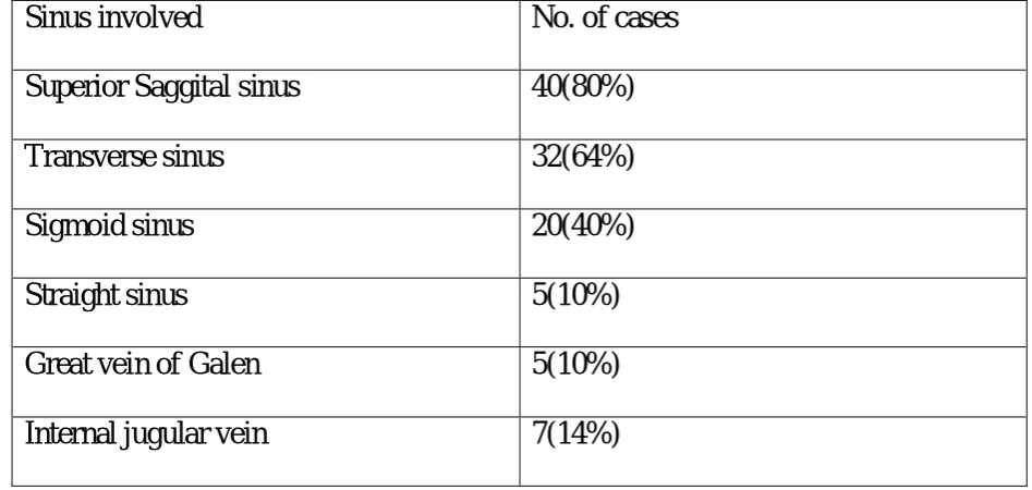
MRI Brain was done in all 50 cases. Among the sinuses involved Superior Saggital Sinus was involved in 40 (80%).Next common sinuses involved were Transverse sinus in 32(64%),Sigmoid Sinus in 20 (40%),Straight sinus in 5 (10%) Internal Jugular Vein in 7 (14%)and Great vein of Galen in 5 (10%).Both Superior Saggital and Transverse sinus were involved in 28 (56%) of cases.
Table 7. Sinuses involved in CVST (n=50)
Sinus involved
No. of cases
Superior Saggital sinus
40(80%)
Transverse sinus
32(64%)
Sigmoid sinus
20(40%)
Straight sinus
5(10%)
Great vein of Galen
5(10%)
Serum Homocysteine was done in 50 patients. It was elevated in 11 (22%) patients. Of these males were 10 and females constituted one patient.Stastical significance was found for homocysteine and CVT with a P value of less than o.o5
Table 8. Serum Homocysteine and CVT
TOTAL CASES
50
HYPERHOCYSTEINEMIA
MALE
27
10
FEMALE
23
1
P value <0.05 significant
Table 9.Risk Factors for CVT
Risk factors for CVT
Male
Female
Total
Postpartum state
-
14(55%)
14(28%)
H/o chicken pox
1(2%)
-
1(2%)
H/o recent diarrhoea
2(4%)
-
2(4%)
H/o previous DVT
-
1(2%)
1(2%)
CNS infection
1(2%)
-
1(2%)
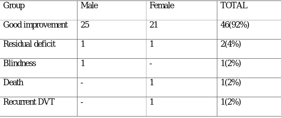
[image:56.595.61.510.458.713.2]Outcome at one month was assessed. Mortality was noted in 1 patient(2%),permanent blindness was noted in 1 patient(2%) and 2 patients(4%) developed permanent motor deficit. 46 patients(92%) showed significant improvement at the end of first month without any residual deficit.A female patient who had residual deficit developed recurrent deep vein thrombosis at third week of followup.
Table 10. Outcome of CVT at 1 month
Group
Male
Female
TOTAL
Good
improvement 25
21
46(92%)
Residual deficit
1
1
2(4%)
Blindness
1
-
1(2%)
Death
-
1
1(2%)
DISCUSSION
Cerebral venous sinus thrombosis is a challenging condition because of its variability of clinical symptoms and signs. A high index of clinical suspicion is needed to diagnose this uncommon condition so that appropriate treatment can be initiated.
DEMOGRAPHY(Table 1 &2)
A total of 50 patients of cerebral venous thrombosis admitted in our institution were taken into our study.Among them, 27 were males (54%) and 23 (46%) were females, with an age range of 18 years to 68 years. Cerebral venous thrombosis is usually a disease of female preponderance with a male to female ratio of 40:60. But our study had a slight male preponderance. It may be due to the fact that female patients delivered in our hospital might have got admitted with puerperial CVT in some other hospitals or due to the rising incidence of CVT among males because of high prevalence of both smoking and alcoholism. In our study smoking and alcohol consumption was seen in about 55% and 48% of males respectively. Whether this could have been a confounding factor has to confirmed by further large studies. Bhojo A. Khealani et al76in his study of 109 cases of CVT reported a female to male ratio of 57%:43%. Ameri and Bousser et al 23 had found a female to male ratio of 1.29:1 Female preponderance was mainly due to pregnancy and puerperium and oral contraceptives. In our study, none of the females gave history of oral contraceptives intake.
In our study,among the patients affected majority were in the age group of 21-40 years which constituted a total of 44 cases(88%), 2 cases(4%) were aged <20 years, 3 cases(6%) were between 41-60 years and 1 case(2%)was aged >60 years. Among the age group of 21-40 years,males were 22(44%) and females were 22(44%). It shows that CVT is the disease of young adults. Ameri and Bousser et al23 reported a uniform age distribution in men with cerebral venous thrombosis, while 61% of women with CVT were aged 20-35 years. This may be related to pregnancy or the use of oral contraceptives.
GLASCOW COMA SCALE(Table 4)
On admission Glascow Coma Scale (GCS) was assessed. It was 8/15 in 2 patients(4%) who presented to us after 10 days of onset of symptoms. Glascow Coma Scale of more than 9-13/15 was found in 20 patients (40%). Glascow Coma Scale more than 13 was found in 28 patients(56%). deBruijn et al 37 in his study of 59 cases has found Glascow Coma Scale 8 in 9(15%) patients on admission which was considered a poor prognostic indicator. In our study GCS 8 was present in 4% patients who had a poor outcome.
CLINICAL FEATURES(Table 5)
Ferro JM et al35 also has reported the presentation of CVST as variable and the incidence of headache was 92%,motor deficit 42%, seizures 37%, papillodema 45%, sensory deficit of 11%, delirium 25%, cerebellar signs 4% and stupor/coma in 13% of patients.
In our study among the patients with seizures, focal seizures occurred in 7 patients(14%),generalized seizures occurred in 15 patients (30%) and status epilepticus occurred in 4 patients (8%). Ferro JM et al 43 in a consecutive series of patients with cerebral venous thrombosis, observed that early symptomatic seizures were found in 44.3% of all patients while status epilepticus was found to occur in 12.8% of patients . Seizures are more typical in patients with parenchymal lesions, sagittal sinus and cortical vein thrombosis, and motor or sensory defects61. Motor deficit, intracranial hemorrhage, and cortical vein thrombosis are independent predictors of early epileptic seizures61.
RISK FACTORS (Table 9)
Out of the 23 female patients, 14(61%) had recent delivery . One patient had undergone caesarean section.. Among these cases 5 had pregnancy induced hypertension (36%) during their antenatal period..
34 years)77, and thrombophilia78. Pregnancy/puerperium-related CVT may more commonly have a sudden or acute onset and a progressive course that tends to become stable in a few days and a better prognosis than nonobstetric CVT22 .
Among the risk factors recent diarrhea was present in 2 patients (4%). Past history of deep vein thrombosis was present in one patient, one patient had recent chickenpox infection and one patient had CNS infection in the form of temporal lobe abscess.
FOCAL NEUROLOGICAL DEFICIT (Table 5)
In our study cranial nerve palsies occurred in 19 (38%) patients. Of the cranial nerves most commonly affected were CN VI and CN VII. VIth cranial nerve was the most common cranial nerve to be involved in 11(22%) patients. VIIth cranial nerve was involved in 9(18%) patients. Kuehnen J et al reports cranial nerve palsies in 12% of all cases of cerebral venous thrombosis. The cranial nerves that have been described to be involved are III, IV,V, VI, VII, VIII, IX, X and XI, and the involvement can be multiple or single. cases,
Papilloedema was found in 29 (58%) patients. Ferro JM et 35 al has also reported papillodema in 45% of patients with CVT. Bhojo A. Khealani et al 76 has found in a series of 59 patients, papillodema in a frequency of 35%.
Isolated intracranial hypertension
Isolated intracranial hypertension occurred in 13(26%) patients.Ferro JM et al35has found about isolated intracranial hypertension was the only presentation in about 29% of patients with CVT. deBrujin et al 37 also has reported about 20% of patients presenting with only isolated intracranial hypertension in the absence of any focal neurological deficit and normal CT Brain for whom the prognosis is generally good.
Haemoglobin of lessthan 10 gm/dl was present in 14 (28%) of patients. Anaemia was associated with increased risk of CVT as the study by Stolz et al30.
CT IMAGING(Table 7)
CT Brain done suggested abnormalities in 34 (80%) of patients. It was normal in 9(21%) patients. Provenzale JM et al65 has reported that CT Brain can be misinterpretated as normal in upto 40% of patients. Cantu et al22 has described normal CT Brain in about 9.47% of patients in his study. Contrast administration is almost always required to increase the reliability of CT in diagnosing CVT. In our study venous infarct occurred in 6(14%) patients, infarct with associated haemorrghe in 13(30%) patients. Direct signs of cerebral venous sinus thrombosis in CT Brain occurred in 15(38%) patients. Intracerebral haemorrhage was present in two patients (5%).Thalamic infarct occurred in 2 (5%) patients.
MRI IMAGING(Table 8)
sinus in 5(10%) cases, Internal Jugular Vein in 7(14%) cases and Great vein of Galen in 5(10%) cases. Both Superior Saggital and Transverse sinus were involved in 28(56%) of cases. Bhojo A. Khealani et al76 has reported Superior sagittal sinus involvement in 77(71%) cases, Transverse sinus involvement in 51(47%) cases, Sigmoid sinus involvement in 34(31%) cases, Straight sinus involvement in 11(10%) cases, Deep cerebral veins involvement in 8(3%) cases, Cortical veins in 7 (6%) cases and Internal jugular vein in 4(4%) cases in a series of 109 patients.
SERUM HOMOCYSTEINE(Table 9)
In our study fasting serum Homocysteine was done in all fifty patients. It was elevated in 11(22%) patients. Bhojo A. Khealani et al76 has reported hyperhomocysteinemia in about 29% of patients tested for serum homocysteine. Ida Martinelli et al 79 has reported a incidence of hyperhomocysteinemia in 27% of patients in a study of 121 patients. Hyperhomocysteinemia is associated with a 4-fold increased risk of cerebral vein thrombosis79.
OUTCOME AT 1 MONTH(Table 10)
In our study 2(5%) cases developed residual motor deficits. Biousse et al81 has reported in series of 160 patients about 5% had developed permanent motor deficit and became dependent. This correlates with our study.
In our study one patient developed permanent blindness(2%) due to optic atrophy. In the follow-up of ISCVT study 72, 4(1%) cases had severe visual loss. This complication was also rare in the study by Preter et al.82 where 2 patients (2.6%) were left with blindness due to optic atrophy. In the Lille study, 2(3.6%) patients had a decreased visual acuity due to optic nerve atrophy83. Biousse et al.81 systematically examined 59 out of 160 CVT consecutive patients with the isolated intracranial hypertension syndrome. 3(5%) cases developed optic atrophy with severe visual loss. In our study all the other patients (90%) had improved significantly well.
CONCLUSION
1. A total of 50 cases of CVT were studied.CVT is a disease of young adults.About 44 (88%) of patients were in the age group of 21-40 years.CVT has to be considered in differential diagnosis of young stroke as it affects often people in the age group of 21-40 years.56% of them were males and 44% were females.
2. Mean age of affected patients was 28 years.
3. CVT is more common in females in postpartum period when they have associated anaemia, and hypertension.
3. CVT has to be suspected in any patient with symptoms such as headache, seizures and focal neurological deficit as they are the most common clinical feature.
4. CVT can also present only with isolated intracranial hypertension
5. CT Brain is normal in upto 9(21%) of patients. Normal CT Brain doesnot ruleout CVT. So to confirm the diagnosis of CVT, MRI has to be done in all suspected cases.
6. Among the sinuses Superior Saggital Sinus is most commonly involved in 80% of patients followed by Transverse Sinus in 64% and Sigmoid sinus in 40% of cases.
7. Risk factors for CVT found are recent diarrhea in two(4%) patients, recent chicken pox in one (2%) patient, previous DVT one 2(%) patient, hyperhomocysteinemia in about 11(22%) of patients and CNS infection in one (2%) patient.
8. Initiation of heparin therapy followed by oral anticoagulants favours early recovery of neurological deficit and prevents further complications.
9. Followup at one month one patient has died with a mortality rate of 2%.One had permanent blindness and two patients had residual deficit.
Sex distribution of CVT cases
Male
Female
0 5 10 15 20 25 30 35 40 45
Age distribution of CVT cases
0 5 10 15 20 25 30 35 40 45
Symptoms & Signs of CVT
0 5 10 15 20 25 30 35
40
CT Picture of CVT Cases
0 10 20 30 40 Superior sagital sinus
Transverse sinus Sigmoind sinus Straight sinus Internal Juglar vein Great vein of galen
Sinus involvement in CVT
CT BRAIN CONTRAST-EMPTY DELTA SIGN
REFERENCES
1.Ribes F. Des rechereches faites Sur la phebite. Rev. Medi (Paris) 1825; 3:5- 41.
2. Ferro JM, Canhao P, Stam J, Bousser MG, Barinagarrementeria F: Prognosis of cerebral vein
and dural sinus thrombosis: results of the International Study on Cerebral Vein and Dural Sinus
Thrombosis (ISCVT). Stroke 2004;35:664–670.
3. Sutton D., Stevens J.: Vascular Imaging in Neuroradiology in Textbook of radiology and
Imaging, volume 2 by Churchill Livingstone New York 2003, pp1682-87.
4. Kido DK, Baker RA, Rumbaugh Calvin L. Normal Cerebral VascularAnatomy. In: Abrams
Angiography, Vascular and Interventional Radiology by Abrams HL, Third Edition. Little,
Brown and Company, Boston. USA. 1983 pp 257-68.
5. Meder JF, Chiras J. Roland J, Guinet P, Bracard S, Bargy F. Venous territories of the brain. J
Neuroradiol 1994; 21:118 - 33.
6. Einhaupl KM, Masuhr F.Cerebral Venous and Sinus thrombosis - an update Eur J Neurol
1994; 1: 109 - 26.
7. Huang, Y.P., and Wolf, B.S. Angiographic features of fourth ventricle tumors with special
reference to the posterior inferior cerebellar artery. Am J Radiol. 1969; 107:543.
8. Hacker H: Normal Supratentorial veins and dural sinuses. In: Newton TH, Potts DG, eds:
Radiology of Skull and Brain. Angiography. Book 3. St Louis:
10. Dora F and Zileli T: Common Variations of the lateral and occipital sinuses at the
confluence sinuum. Neuroradiology 1980; 20 : 23 - 7.
11. Taveras J.M..: Angiography in Neuroradiology Third Edition. Baltimore:
Williams & Wilkins. 1996 pp 998.
12. Wolf, B.S., Newman, C.M. and Schlesinger, B. The diagnostic value of the
deep cerebral veins in cerebral angiography. Am J Radiol. 1962;87:322.
13. Wolf B S, Huang Y P, Newman C.M. The superficial Sylvian venous drainage
system. Am J Radiol. 1963; 89:398.
14. Wolf B S, Huang YP. The subependymal veins of the lateral ventricles. Am J
Roentgenol Radium Ther Nucl Med. 1964; 91:406-26.
15. Parkash C, Bansal BC. Cerebral venous thrombosis. J Indian Acad Clin Med.
2000; 5: 55 - 61.
16. Kalbag RM, Woolf AL. Etiology of cerebral venous thrombosis in cerebral venous
thrombosis publ. Ed Kalbagh RM, Wolf AL. Volume1. Oxford
University Press London, 1967; pp 238.
17. Stam J: Thrombosis of the cerebral veins and sinuses. N Engl J Med 2005;352:1791–1798.
19.Krayenbuhl HA: Cerebral venous and sinus thrombosis. Clin Neurosurg 1966;14:1–24.
20.K.Srinivasan,Angiology,November vol.34no.11 731-746
21. Kellett MW, Martin PJ, Enevoldson TP, et al: Cerebral venous sinus thrombosis connected
with 20210, a mutation of the prothrombin gene. J Neurol Neurosurg Psychiatry 1998;65:611–
612.
22.Cantu C, Barinagarrementeria F: Cerebral venous thrombosis associated with pregnancy and
puerperium. A review of 67 cases. Stroke 1993;24:1880–1884.
23. Ameri A, Bousser MG: Cerebral venous sinus thrombosis. Neurol Clin 1992;10:87–111.
24 Daif A, Awada A, Al-Rajeh S, et al: Cerebral venous thrombosis in adult. A study of 40
cases from Saudi Arabia. Stroke 1995;26:1193.
25. Deschiens M, Canard J, Horellou M, et al: Coagulation studies, factor V Leiden and
anticardiolipin antibodies in 40 cases of cerebral venous sinus thrombosis. Stroke
1996;27:1724–1730.
26. Martinelli I, Battaglioli T, Pedotti P, et al: Hyperhomocysteinemia in cerebral vein
thrombosis.Blood 2003;102:1363–1366.
27. de Bruijn SF, Stam J, Koopman MM, et al: Case-control study of risk of cerebral sinus
thrombosis with third generation-oral contraceptives. Cerebral Venous Sinus Thrombosis Study
29. Cumming AM, tait RC, Fildes S, et al: Development of resistance to activated protein C
during pregnancy. Br J Hematol 1995;90:725–727.
30. Stolz E, Valdueza JM, Grebe M, Schlachetzki F, Schmitt E, Madlener K, Rahimi A,
Kempkes-Matthes B, Blaes F, Gerriets T, Kaps M: Anemia as a risk factor for cerebral venous
thrombosis?An old hypothesis revisited: results of a prospective study. J Neurol 2007;254:729–
734.
31. Pillai LV, Ambike DP, Nirhale S, Husainy SMK, Pataskar S, et al. Cerebral venous
thrombosis: An experience with anticoagulation with low molecular weight heparin. Indian J
Crit Care Med 2005; 9:14-18.
32. Bousser MG, Ross Russel RW: Cerebral Venous Thrombosis. London: W.B. Saunders,
1997.
33. Bousser MG, Chiras J, Bories J, et al: Cerebral venous thrombosis. A review of 38 cases.
Stroke 1985;16:199–213.
34. Kuehnen J, Schwartz A, Neff W, Hennerici M: Cranial nerve syndrome in thrombosis of the
transverse/sigmoid sinuses. Brain 1998;121:381–388.
35. Ferro JM: Cerebral venous thrombosis. J Neuroradiol 2002;29:231–239.;89:530.
36. Biousse V, Ameri A, Bousser MG: Isolated intracranial hypertension as the only sign of
cerebral venous thrombosis. Neurology 1999;53:1537.
37. De Bruijn SF, de Haan RJ, Stam J: Clinical features and prognostic factors of cerebral
38. Purvin VA, Trobe JD, Kosmorsky G: Neuro-ophthalmic features of cerebral venous
obstruction. Arch Neurol 1995;52:880–886.
39. Crassard I, Bousser MG: