Clinical
and
Imaging
Features
of
Transient
Ischaemic
Attack
and
the
Utility
of
ABCD
2
Score
Dissertation submitted to comply with the requirements of the degree:
D.M. NEUROLOGY (BRANCH – I)
ofTHE TAMILNADU Dr.M.G.R. MEDICAL UNIVERSITY CHENNAI
AUGUST 2010
CERTIFICATE
This is to certify that this dissertation entitled “Clinical and Imaging Features of Transient Ischaemic Attack and the Utility of ABCD2 Score” submitted by Dr
Karthik S N appearing for D.M., Degree examination in August 2010 is a bona fide record of work done by him under my direct guidance and supervision in partial fulfillment of regulations of the Tamil Nadu Dr. M.G.R. Medical University, Chennai. I forward this to the Tamil Nadu Dr.M.G.R. Medical University, Chennai, Tamil Nadu, India.
Prof. V SUNDAR MCh Prof. R M BOOPATHY MD., DM PROFESSOR & HEAD PROFESSOR OF NEUROLOGY INSTITUTE OF NEUROLOGY INSTITUTE OF NEUROLOGY Madras Medical College & GGH Madras Medical College & GGH Chennai – 600 003 Chennai – 600 003
Prof. J MOHANASUNDARAM MD, DNB, PhD DEAN
DECLARATION
I, Dr KARTHIK S N, do solemnly affirm that this dissertation titled
“Clinical and Imaging Features of Transient Ischaemic Attack and the Utility of ABCD2 Score” is a bona fide work done by me at Institute of Neurology, Madras Medical College & Govt. General Hospital,
Chennai, during 2007-2010 under the guidance and supervision of
Dr R M Boopathy M.D., D.M., Professor of Neurology, Institute of Neurology
The dissertation is submitted to The Tamilnadu Dr. M.G.R. Medical
University towards the partial fulfillment of requirements for the award
of D.M., degree in Neurology.
Place: Chennai
Date: 27/05/2010
KARTHIK S N
SPECIAL ACKNOWLEDGMENT
I gratefully acknowledge and sincerely thank
ACKNOWLEDGEMENT
My sincere thanks to Prof.V.Sundar, Prof and Head, Institute of Neurology for his immense kindness in allowing me to use the services of the department and to use and obtain all of the information needed for this study.
I thank Prof.R.M.Boopathy, Professor of Neurology, Institute of Neurology, with profound gratitude for his constant guidance, motivation, advice and valuable criticism, kindness and encouragement which enabled me to complete this work.
I thank Prof. C.Mutharasu, Prof. K.Bhanu, Prof. Gopinathan, Professors, Institute of Neurology for their constant guidance and encouragement.
I would also like to take this opportunity to thank my former Professor Dr V Natarjan. This work could not have been accomplished without his ideas and
encouragement.
I thank with gratitude, Dr.V.Kamaraj, Dr.S.Arunan, Dr.S.Jawahar and Dr.P.Muthukumar for their cooperation and guidance.
I thank my postgraduate friends for their constant support. I thank the Faculty and Staffs of Departments of Radiology , all the technical & non technical staffs of the Institute of Neurology, for their cooperation.
TABLE
OF
CONTENTS
PAGE
1. Introduction
1
2. Aims and Objectives
3
3. Literature Review
4
4. Materials and Methods
35
5. Results
40
6. Discussion
60
7. Conclusion
72
8. References
75
9. Master Chart
[image:6.612.112.504.201.572.2]
ABBREVIATIONS AND ACRONYMS
¾ ACA : Anterior Cerebral Artery.
¾ AICA : Anterior Inferior Cerebellar artery
¾ AF : Atrial Fibrillation.
¾ BA : Basilar Artery
¾ BP : Blood Pressure
¾ DM : Diabetes mellitus
¾ DWI : Diffusion Weighted imaging
¾ HDL : High Density Lipoproteins
¾ HT : Hypertension
¾ ICA : Internal Carotid Artery
¾ LDL : Low Density Lipoproteins
¾ MCA : Middle Cerebral Artery
¾ MRI : Magnetic Resonance Imaging
¾ mRS : Modified Ranikin Score
¾ NECT : Non Enhanced Computerized Tomogram
¾ NIHSS : National Institute of Health Stroke Scale
¾ PCA : Posterior Cerebral Artery
¾ PICA : Posterior Inferior Cerebellar artery
¾ SCA : Superior Cerebellar Artery
¾ TIA: : Transient Ischemic Attacks
KEY
TO
MASTER
CHART
A : AphasiaAF : Atrial Fibrillation
AD : Arterial Disease
Amx : Amaurosis Fugax
Atx : Ataxia
B : Both
CD : Cardiac Disease
D : Dysarthria
DM : Diabetes Mellitus
Dip : Diplopia
FB : Facio Brachial Monoparesis
F : Female
HA : Hemianopia
HT : Hypertension
L : Left
LOC : Loss of Consciousness
M : Male
N : No
N & V : Nausea and Vomiting
P : Posterior Circulation
R : Right
U : Undetermined
Vt : Vertigo
INTRODUCTION
Cerebrovascular disease is the third leading cause of death in developed
countries after heart disease and cancer; the overall prevalence is 794 per 100,000. It is
estimated that more than 700,000 patients have a stroke each year in the United States.
The loss of these patients from the work force and the extended hospitalization they
require during recovery make the economic impact of the disease one of the most
devastating in medicine.
About 15-20% of patients with stroke have a preceding TIA, making it one of the
common neurologic problems. Four to 20 percent of patients who have a TIA, experience
a stroke within the following 90 days; one half of those strokes occur within 48 hours.
Promptly recognizing patients who are at high risk of progressing to stroke provide us a
golden opportunity for stroke prevention. This depends upon accurately identifying the
cause of symptoms, and the nature, location, and severity of causative cardiac,
hematologic, and cerebrovascular abnormalities.
TIA being a clinical diagnosis, there is a significant variation among
physicians and neurologists in diagnosing this condition. In one series, majority (81%) of
the TIA referrals from general practitioners to neurology clinics were for nonvascular
events. Even the percent agreement among two neurologists for the diagnosis of TIA by
history varies from 42% to 86%. In such a situation development of a scoring system like
further assessing these patients. Although there are studies regarding stroke, currently
there are no big studies from India regarding the clinical and imaging features in patients
with TIA. Also ABCD2 score as a tool is not being routinely used even by the
Neurologist.
Considering the limited information available from India, this study was
undertaken to determine the utility of ABCD2 scoring system in Indian population and to
determine the differences in risk factors and clinical profile if any, when compared to
Aims
and
Objectives
1. To analyze the risk factors and clinical profile of transient ischemic attack.
2. To study the imaging feature in transient ischemic attack.
3. To evaluate the utility of ABCD2 score in predicting long-term survival and
stroke risk after TIA
4. To assess the treatment outcome of transient ischemic attack.
LITERATURE
REVIEW
INTRODUCTION — Transient ischemic attack (TIA) is a brief episode of neurologic
dysfunction resulting from focal temporary cerebral ischemia not associated with cerebral
infarction.TIA was originally defined clinically by the temporary nature (< 24 hours) of
the associated neurologic symptoms. However, the arbitrary nature of the 24-hour time
limit and lack of specific pathophysiologic meaning hampered the clinical and research
utility of the term "TIA." Recognition of these problems led to a change to a tissue-based
definition of TIA. The change was driven by advances in neuroimaging that enabled very
early identification of ischemic brain injury.
MODERN DEFINITION — As endorsed by 2009 guidelines from the American Heart
Association and American Stroke Association (AHA/ASA), transient ischemic attack
(TIA) is now defined as a transient episode of neurologic dysfunction caused by focal
brain, spinal cord, or retinal ischemia, without acute infarction [1]. In keeping with this
definition of TIA, ischemic stroke is now defined as an infarction of central nervous
system tissue.
TIA was originally defined as a sudden onset of a focal neurologic symptom and/or sign
lasting less than 24 hours, presumably brought on by a transient decrease in blood supply,
which rendered the brain ischemic in the area producing the symptom. However, this
classic definition of TIA was inadequate for several reasons. Most notably, there is risk of
last less than one hour. Thus, the benign connotation of "TIA" has been replaced by an
understanding that even relatively brief ischemia can cause permanent brain injury.
The advantages of modern tissue-based definitions of TIA and stroke include the
following [1,2]
• The defined end point is biologic (tissue injury, as confirmed or excluded by
neuroimaging) rather than arbitrary (24 hours).
• The definition encourages use of neurodiagnostic tests to identify brain injury and
its cause.
• The presence or absence of ischemic brain is more accurately reflected.
An earlier proposal for a tissue-based TIA definition noted that clinical symptoms of TIA
typically last less than one hour [2]. While this is true, the AHA/ASA did not incorporate
the phrase "typically less than one hour" in the new definition of TIA because there is no
time cutoff that reliably distinguishes whether a symptomatic ischemic event will result
in ischemic infarction [1].
Use of the new definitions in epidemiologic studies is likely to modestly alter the
incidence and prevalence rates of TIA and stroke, but these changes are encouraged by
the AHA/ASA because they should reflect more accurate diagnosis [1]. One study
estimated that switching from the classic to the tissue-based definition of TIA could
reduce the annual incidence of TIA in the United States by 33 percent (range 19 to 44
percent) and increase the annual incidence of stroke by 7 percent (range 4 to 10 percent)
OTHER TERMINOLOGY— The terms "acute neurovascular syndrome" and
"transient symptoms with infarction" (or "cerebral infarction with transient signs") have
been proposed to supplement TIA in the description of transient symptoms related to
ischemia.
Acute neurovascular syndrome — With the new tissue-based definitions of stroke and
TIA, there may be uncertainty regarding the diagnosis if immediate neuroimaging is not
available to detect infarction when transient symptoms of brain ischemia occur [1]. The
AHA/ASA has proposed (but not formally endorsed) consideration of a term such as
"acute neurovascular syndrome" that can be used in this setting if a diagnostic evaluation
is not performed or until the diagnostic evaluation is completed.
Transient symptoms with infarction — The awareness that a classically defined TIA
(<24 hours in duration) can be associated with irreversible ischemic brain injury led to a
proposal to label these events as "transient symptoms associated with infarction" (TSI) or
"cerebral infarction with transient signs" and to distinguish them from transient
symptoms without infarction [4].
While TSI in general has smaller infarct volumes than classically defined ischemic stroke
(where neurologic deficits persist for ≥ 24 hours), there is no unique size that
differentiates TSI from ischemic stroke [4].
Patients with TSI have a higher short-term risk of recurrent ischemic stroke than patients
who have transient symptoms without infarction. This conclusion is supported by a
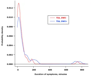
RELATIONSHIP OF SYMPTOM DURATION AND INFARCTION — A
classically defined TIA with symptoms lasting for as little as a few minutes can be
associated with infarction, whereas a spell lasting for many hours may rarely cause no
signal changes on diffusion-weighted MRI imaging (DWI).
Some reports suggest that increased duration of classically defined TIA (<24 hours in
duration) is associated with a higher probability of infarction on DWI, but the association
is not absolute [4,9-12]. A systematic analysis of patients with classically defined TIA
found that symptom duration was not a reliable predictor for the presence of infarction
(figure 1), even though the mean duration tended to be significantly longer in patients
with infarction than in those without infarction [4].
One potential caveat is that abnormalities on initial imaging such as diffusion-weighted
(DWI) MRI obtained during or soon after symptoms, may actually be reversible injuries.
However, most patients with TIA seek medical attention after their symptoms fully
resolve; a low proportion (≤ 7 percent) of patients with classically defined TIA are
admitted and scanned at the height of their symptoms [5,9-11]. Therefore, infarcts
observed in patients with classically defined TIA most likely represent permanent brain
injury, as the probability of DWI reversibility decreases as the time from symptom onset
Temporal behavior of symptoms in patients with transient ischemic attack (TIA)
Figure 1. The probability density function curve of symptom duration for transient symptoms associated with infarction (TSI) indicates the absence of continuity within the first 24 hours. The probability density function is the probability that the variable takes a value in a given interval and is equal to 1 over its entire range of values. The area under curve is almost equal to 1 at around 200 minutes. Also note that the curves for TIA with or without infarction overlap (p = 0.82). The distribution of duration of symptoms as seen here suggests that symptom duration is not a reliable feature to be used for predicting whether a transient neurological spell is associated with infarction.
DWI: diffusion-weighted magnetic resonance imaging.
DWI is advantageous for evaluating patients who have transient symptoms because it is
highly sensitive for detecting infarction, thereby confirming an ischemic cause. A
systematic review found that DWI detects corresponding appropriate ischemic lesions in
16 to 67 percent of patients with classically defined TIA [13]. Also, the combination of
DWI also has an advantage in differentiating acute infarction from chronic lesions. One
study estimated that the amount of error potentially imposed by the use of conventional
MRI in identifying the clinically responsible infarct in patients with classically defined
TIA could be as high as 50 percent when compared with DWI [5]. This is, in large part,
because infarctions associated with classically defined TIA are often very small. A
volumetric study of TIA-related infarcts showed that 96 percent of all infarcts were
smaller than 1 mL [4]. The smallest single lesion that was associated with a classically
defined TIA was 0.17 mL in volume. The mean infarct volume was 0.66 ± 1.20 mL.
Perfusion-weighted MR imaging (PWI), when used in combination with DWI, may
improve the accuracy of diagnosis in patients with transient ischemic symptoms [14,15].
This is illustrated by a prospective series of 43 patients with classically defined TIA who
had both studies performed within 48 hours of symptoms onset [15]. Lesions on DWI
only, PWI only, or both DWI and PWI were observed in 19, 16, and 16 percent of
patients, respectively. Thus, objective evidence of brain ischemic with either DWI or
PWI lesion was present in 51 percent.
INCIDENCE — Transient ischemic attack (TIA) is a common neurologic problem. In a
mainly white population from Rochester Minnesota, a population-based study reported
that the incidence rate of TIA was 68 per 100,000 [16]. In the Cincinnati and Northern
Kentucky region of the United States, where the ethnic and socioeconomic demographics
are similar to that of the United States as a whole, another population-based study found
men had significantly higher rates of TIA than whites and women [17]. From these data,
it was estimated that 240,000 TIAs occur each year in the United States.
The incidence of TIA in other populations has generally been lower than that reported in
the United States, ranging from about 10 to 56 per 100,000 in various studies worldwide
[18-21].
PATHOPHYSIOLOGIC MECHANISMS — A transient ischemic attack (TIA) should
be considered a syndrome. These syndromes are conveniently divided into three
pathophysiologic mechanisms: Large artery low-flow TIA (true TIA) Embolic TIA,
which may be artery-to-artery, or due to a cardioaortic or unknown source Lacunar or
small penetrating vessel TIA
Large artery low flow TIA — Large artery low-flow TIAs are brief (usually minutes to
a few hours), recurrent, and stereotyped. They are often associated with a tightly stenotic
atherosclerotic lesion at the internal carotid artery origin or in the intracranial portion of
the internal carotid artery (siphon) when collateral flow from the circle of Willis to the
ipsilateral middle or anterior cerebral artery is impaired (figure 2 and figure 3). Other
important causes include atherosclerotic stenotic lesions in the middle cerebral artery
stem (figure 4) or at the junction of the vertebral and basilar artery. Any obstructive
vascular process in the extracranial or intracranial arteries can cause a low-flow TIA
syndrome if collateral flow to the potentially ischemic brain also is impaired.
Anatomy of the cerebral arterial circulation
Embolic TIA — Embolic TIAs are characterized by discrete, usually single, more
that divided patients with TIAs into those with symptoms of short duration (less than 60
minutes) or long duration (60 minutes or greater), the latter group was much more likely
to have an embolic source (86 versus 46 percent) [22]. The embolus may arise from a
pathologic process in an artery, usually extracranial, or from the heart (eg, atrial
fibrillation or left ventricular thrombus). An ischemic stroke with infarction has occurred
if symptoms or signs persist beyond 24 hours. However, as previously mentioned,
symptoms that last less than 24 hours (often only as long as one hour) also may be
associated with some infarction. If the primary pathologic process is thought to be
embolic, a diligent search for its source is necessary before therapy to prevent future
stroke can be initiated.
Lacunar TIA — Lacunar or penetrating or small vessel TIAs are due to transient
cerebral ischemia induced by stenosis of one of the intracerebral penetrating vessels
arising from the middle cerebral artery stem, the basilar or vertebral artery (figure 5), or
the circle of Willis (figure 2 and figure 3). Occlusion of these small intracerebral
penetrating vessels usually is due to lipohyalinosis from hypertension, but also may arise
because of atheromatous disease at their origin. Occasionally, recurrent stereotyped TIAs
occur; in this setting, the term lacunar or small vessel TIAs seems appropriate.
CLINICAL MANIFESTATIONS — The symptoms of a transient ischemic attack
(TIA) depend upon the pathophysiologic subtype.
Low-flow TIA — Low-flow TIAs usually are short-lived (minutes) and often recurrent.
They may occur as little as several times per year but typically occur more often (once
Low-flow TIAs are generally stereotyped, especially when they are due to
hemodynamically significant stenotic lesions at the origin of the internal carotid artery, at
the siphon portion of the internal carotid artery where collateral flow to the circle of
Willis is inadequate, or in the middle cerebral artery stem. Symptoms due to ischemia
from these lesions often include hand, arm, leg, face, tongue, or cheek numbness or
weakness together, or a combination of one or more. Recurrent aphasic syndromes appear
when there is focal ischemia in the dominant hemisphere, and recurrent neglect occurs in
the presence of focal or nondominant hemisphere ischemia.
In contrast, recurrent symptoms are often not stereotyped when the stenotic lesion that
obstructs flow involves the vertebrobasilar junction or the basilar artery. The many
closely packed neuronal structures in the brainstem preclude consistent manifestations of
recurrent focal ischemia in this area.
Nevertheless, certain generalizations about recurrent low-flow TIA symptoms in the
posterior circulation can be made. Obstructive lesions in the distal vertebral artery or at
the vertebrobasilar junction usually cause disorganized dizziness that may or may not
include spinning or vertigo. The patient may complain that the room is tilting or that the
floor is coming up at them, rather than spinning dizziness. Patients most often use the
word dizziness to describe a myriad of symptoms, not necessarily spinning. Other
symptoms may include numbness of one side of the body or face, dysarthria, or diplopia.
Ischemia in the pons from stenotic lesions in the proximal to midbasilar artery can cause
bilateral leg and arm weakness or numbness and a feeling of heaviness in addition to
may speak of a feeling of impending doom. Ischemia in the territory of the top of the
basilar artery or proximal posterior cerebral artery may present with all of the above
recurrent symptoms as well as overwhelming drowsiness, vertical diplopia, eyelid
drooping, and an inability to look up. Transient ischemia at the top of the basilar artery is
usually due to embolism rather than low-flow TIA.
Embolic TIA — Embolic TIAs typically last hours rather than minutes as in low-flow
TIAs. They may be infrequent since they are the result of emboli from a specific source
(eg, a one, two, or three-time phenomenon). When the source of the embolus is in a
proximal vessel, recurrent emboli can lodge in different branches of the parent vessel
giving different symptoms.
Emboli are subject to natural thrombolysis and migration since they typically break off of
fresh thrombus. They may produce transient ischemia on many occasions, but an element
of silent infarction remains. Emboli may be better referred to as acceptable minor
embolism (ACME), a term coined by C Miller Fisher.
Embolic TIAs are best divided into those in the anterior cerebral circulation (carotid,
ACA, MCA territory) and those in the posterior cerebral circulation (vertebrobasilar,
posterior cerebral artery territory). Symptoms in both circulations depend upon the size of
the embolic fragment in relation to the size of the artery occluded.
Embolic TIAs in the anterior circulation may be large enough to occlude the middle
cerebral artery stem, producing a contralateral hemiplegia secondary to ischemia in the
deep white matter and basal ganglion/internal capsule lenticulostriate territory (figure 6).
inadequate. These include aphasic syndromes in the dominant hemisphere and
anosognosia or neglect in the nondominant hemisphere.
Smaller emboli that occlude branches of the middle cerebral artery stem result in more
focal symptoms, including hand alone or arm and hand numbness, weakness, and/or
heaviness induced by ischemia to the frontal area of the contralateral frontal lobe motor
system (figure 4). The symptoms also may be as specific as thumb or hand numbness or a
swollen feeling, suggesting focal ischemia in the hand area of the sensory strip or parietal
association cortex. Transient unilateral visual obstruction often signifies atherothrombotic
disease in the internal carotid artery proximal to the ophthalmic artery takeoff.
Atherothrombotic disease is most often responsible for these syndromes, although carotid
dissection and embolism from the aorta, heart, or an unknown source also should be
considered.
Posterior circulation territory embolic TIAs are generally produced by emboli arising
from atherothrombotic disease at the origin or distal segment of one of the vertebral
arteries or of the proximal basilar artery. Emboli arising from the aortic arch, the heart, an
unknown source, or from a dissecting lesion in the vertebral artery should also be
considered.
Symptoms vary according to the vertebral or basilar artery branch in which the emboli
lodges (figure 7). Emboli can produce transient ataxia, dizziness, diplopia, dysarthria,
quadrantanopsia, hemianopsia, numbness, crossed face and body numbness, and focal
hearing loss. When the top of the basilar artery is embolized, sudden, overwhelming
rostral midbrain reticular activating system ischemia. Emboli in the more distal branches
of the posterior cerebral artery may result in a homonymous field defect or in memory
loss (inferior medial temporal lobe ischemia).
Lacunar or small vessel TIA — Lacunar or small vessel TIAs are thought to be caused
by atherothrombotic obstructive lesions at the origin of the penetrating vessel or
lipohyalinosis distally. Embolism is rarely proposed as the mechanism. These small
vessel TIAs cause symptoms that are similar to the lacunar strokes that are likely to
follow. Thus, face, arm, and leg weakness or numbness due to ischemia in the internal
capsule, pons, or thalamus may occur, similar to the symptoms due to ischemia from
embolism or large vessel atherothrombotic disease or dissection. As a result, serious
disease in the parent vessel must be excluded before the diagnosis of lacunar or small
vessel TIA can be established with confidence.
Lacunar infarcts may be preceded by lacunar TIAs consisting of brief repetitive
stereotyped clinical symptoms and signs, and lacunar stroke onset may be stepwise and
progressive rather than abrupt [23-25]. Such a pattern of TIAs, or nonsudden onset in
association with a lacunar syndrome, is highly suggestive of small vessel lipohyalinotic
etiology [26].
IMPORTANT PATHOLOGIC PROCESSES — There are four pathologic processes
that give rise to low-flow "true" TIAs or embolic TIAs and that can produce sudden
devastating stroke if not recognized and treated. Atherothrombotic stenotic lesions at the
origin of the internal carotid artery that are narrowed more than 70 percent Intracranial
distal vertebral artery/vertebrobasilar junction/proximal basilar artery Emboli to the top
of the basilar artery or the middle cerebral artery stem that come from a source below,
either arterial, aortic, or cardiac Dissection lesions at the origin of the petrous portion of
the internal carotid artery or at the C1-2 level of the vertebral artery as it enters the
foramen transversarium
Internal carotid artery TIA — An atherothrombotic stenotic lesion at the origin of the
internal carotid artery that is narrowed to more than 70 percent of its normal lumen
diameter poses a threat of embolic or low-flow TIA or stroke [27-30]. Even a 50 percent
stenosis may be important when considering carotid endarterectomy for prevention of a
secondary stroke or of a primary stroke when a TIA has occurred.
Prospective natural history studies of asymptomatic atherothrombotic disease at the
origin of the internal carotid artery (mostly asymptomatic carotid artery bruits) suggest
that the rate of ipsilateral stroke increases dramatically when the residual lumen diameter
narrows to greater than 70 percent stenosis (figure 8 and figure 9) [31-33]. In one series
of 500 patients, for example, the incidence of stroke was 1.7 percent per year overall but
5.5 percent per year in those with more than a 75 percent carotid artery stenosis [32].
This degree of stenosis corresponds to a residual lumen diameter of 1.5 mm, the precise
point at which pressure drops across the stenotic lesion [34,35]. When the pressure drops,
flow to the ipsilateral middle cerebral artery stem is in part supplied by collateral
circulation from the circle of Willis and from the external carotid to ophthalmic to distal
internal carotid artery system (figure 2 and figure 3). Less flow is provided by the internal
thrombus formation at the site of the stenosis and subsequent embolism. When the circle
of Willis is compromised, low-flow "true" TIA ensues.
Intracranial atherothrombotic disease — Intracranial atherothrombotic disease that
produces low-flow or embolic TIA most commonly occurs at the distal vertebral
artery/vertebrobasilar junction/proximal basilar artery site. The potential of this lesion to
precipitate a disastrous stroke by thrombosis, thrombus propagation, and embolism is
extremely important. The other two most important, but less common, sites include the
siphon portion of the internal carotid artery and the middle cerebral artery stem. The
common carotid origin and the vertebral artery origin are much less problematic since
they only rarely give rise to artery-to-artery emboli.
The ability to noninvasively diagnose and follow these intracranial arterial lesions with
precision through MRI angiography, duplex Doppler, and transcranial Doppler flow
assessment allows for important preventive therapeutic considerations.
Arterial, aortic, or cardiac sources of emboli — Emboli at the top of the basilar artery or
in the middle cerebral artery stem that come from a source below — arterial, aortic, or
cardiac — are extremely important to recognize since they may produce fluctuating
symptoms or TIAs prior to a devastating stroke. Transient focal symptoms due to an
embolus at these sites occur because blood flow reestablishes itself around the embolus.
The symptoms may return in abundance and produce a stroke when the embolus itself
causes a thrombus that further occludes the artery. This can occur hours or even days
after the embolus has lodged at the site because it did not migrate or lyse. Acute
propagation and provide the time for spontaneous thrombolysis, although this has not
been proven definitively.
Dissection lesions — Dissection lesions at the origin of the petrous portion of the internal
carotid artery or at the C1-2 level of the vertebral artery as it enters the foramen
transversarium cause symptoms of cerebral ischemia due to low flow or embolism, which
occur within minutes, hours, or even days prior to a devastating stroke. Modern Doppler
and neurologic imaging technology can establish the diagnosis noninvasively with the
necessary precision to permit potentially stroke-saving therapeutic strategies (eg,
intravenous heparin).
Coronal section of cerebral hemisphere
[image:27.612.99.331.134.268.2][image:27.612.129.479.334.499.2]
Figure 3.Representation of the territories of the major cerebral vessels circulation
Severity of carotid stenosis predicts stroke risk
Figure 8. Relation between the degree of carotid artery stenosis and the annual risk of stroke. Data from Barnett, HJ, Eliasziw, M, Meldrum, HE, Taylor, DW, Neurology 1996; 46:603.
Figure 9. Incidence of ischemic events in 500 patients with asymptomatic carotid artery bruits according to the severity of carotid artery stenosis on initial Doppler ultrasonography. Patients with 75 percent stenosis were at significantly increased risk (P<0.0001). Data from Chambers, BR, Norris, JW, N Engl J Med 1986; 315:860.
Initial evaluation and management of transient ischemic attack and minor stroke INITIAL EVALUATION — Patients who have had a suspected TIA require urgent
evaluation due to the high stroke risk associated with TIA [1]. Furthermore, immediate
[image:31.612.78.376.330.486.2]The initial evaluation of suspected TIA and minor nondisabling ischemic stroke includes
basic laboratory studies that are suggested by the history and physical examination, an
electrocardiogram, brain imaging, and neurovascular imaging. Laboratory testing is
helpful in ruling out metabolic and hematologic causes of neurologic symptoms,
including hypoglycemia, hyponatremia, and thrombocytosis.
Several neurologic disorders give rise to transient focal neurologic symptoms, and these
should be considered before establishing a diagnosis of TIA. In addition to TIAs, the
most important and frequent causes of discrete self-limited attacks include: Seizures
Migraine auras Syncope
Less frequent causes include pressure- or position-related peripheral nerve or nerve root
compression that causes transient paresthesias and numbness; peripheral vestibulopathies
that cause transient episodic dizziness; and metabolic perturbations such as hypoglycemia
and hepatic, renal, and pulmonary encephalopathies that can produce temporary
aberrations in behavior and movement.
Hospitalization versus ambulatory evaluation — Whether hospitalization is required
for TIA evaluation is not clear, but urgent assessment and management is essential
regardless of inpatient or outpatient status [1, 35-39].
Possible advantages of hospitalization include facilitated early use of thrombolytic
therapy and other medical management if symptoms recur, expedited TIA evaluation, and
expedited institution of secondary prevention [39].
The 2009 American Heart Association and American Stroke Association (AHA/ASA)
patients with TIA who present within 72 hours of symptom onset and meet any of the
following criteria [1] :
• ABCD^2 score (show table 1) of ≥ 3
• ABCD^2 score of 0 to 2 and uncertainty that the diagnostic workup can be
completed within two days as an outpatient
• ABCD^2 score of 0 to 2 and other evidence that the event was caused by focal
ischemia
The ABCD^2 score (ie, ABCD squared, for Age, Blood pressure, Clinical features,
Duration of symptoms, and Diabetes) is a simple prognostic assessment tool with
moderate predictive accuracy that was designed to identify patients at high risk of
ischemic stroke in the first two days after TIA, as discussed later in detail (table 1).
The 2006 National Stroke Association (NSA) guidelines systematically reviewed,
critically evaluated, and updated prior published guidelines for the management of TIA
[39]. The following consensus recommendations regarding initial management were
proposed, based mainly on evidence from observational studies and clinical experience:
Hospitalization should be considered for patients with a first TIA within the past 24 to 48
hours, and is generally recommended for patients with the following conditions:
- Crescendo TIAs
- Duration of symptoms >1 hour
- Symptomatic internal carotid artery stenosis >50 percent
- Known cardiac source of embolus such as atrial fibrillation
- High risk of early stroke after TIA
Patients who need urgent evaluation and are not hospitalized should have rapid access to
the following studies:
- Brain imaging with head CT and/or MRI
- Neurovascular studies such as CT angiography (CTA), MR angiography (MRA),
and/or ultrasound
- Electrocardiogram (ECG)
All patients with a TIA within the past two weeks who are not hospitalized should
undergo investigations within 24 to 48 hours to determine the mechanism of ischemia
and subsequent preventive therapy Patients who are not admitted should be informed that
they need to return to the hospital immediately if symptoms recur
PROGNOSIS — TIA is a neurologic emergency because patients with TIA and minor
stroke are at increased risk of recurrent stroke [40-47]. This risk is illustrated by the
following studies:
• A meta-analysis of 11 observational studies published through December 2006
found that the risk of stroke at 2 days, 30 days, and 90 days after TIA was 3.5, 8.0,
and 9.2 percent, respectively [45]. In the three studies that used active
ascertainment of stroke outcome (ie, face-to-face evaluation by a practitioner at
three months rather than use of administrative data), the 2, 30, and 90 day risk of
stroke after TIA was even higher (9.9, 13.4, and 17.3 percent, respectively).
Similar findings were reported in a meta-analysis of 18 cohorts published through
• A prospective observational study of 1380 patients with TIA and 3855 patients
with ischemic stroke found that subsequent stroke incidence during the hospital
stay was 8 percent for patients with TIA and 7 percent for patients with ischemic
stroke [49]. During the first six months after the initial ischemic event, recurrent
stroke incidence was 13 percent for both groups. Two percent of patients with TIA
died during hospital stay, and 17 percent were dependent at follow-up.
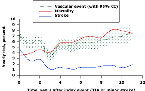
• A well-designed prospective cohort study of 2447 participants from the Dutch TIA
trial found that the risk for major vascular events and stroke was highest shortly
after TIA or minor stroke, declined to its lowest point at about three years, and
then progressively increased over the remainder of the 10-year follow-up (figure
10) [50]. In contrast, the risk for mortality gradually rose throughout the study. By
10 years, 60 percent had died and 54 percent had experienced new vascular events
(stroke and myocardial infarction). Event-free survival was 48 percent. Predictive
factors for risk of vascular events and death included age over 65 years, diabetes,
claudication, previous vascular surgery, and pathologic Q waves on baseline
electrocardiogram.
The urgency associated with TIA derives also from the observation that TIAs are most
likely to occur in the hours and days immediately preceding ischemic stroke. As an
example, a study that analyzed four cohorts of patients who had recent ischemic stroke
found that TIAs occurred most often in the 48 hours prior to the stroke [51]. Another
study found that the risk of ischemic stroke occurring within 24 hours of a probable or
days after a first TIA, 42 percent occurred within the first 24 hours. This may be an
overestimate related to the difficulty distinguishing a single ischemic event (stroke) with
fluctuating symptoms from separate events (TIA followed by stroke) within a short
period of time. Nevertheless, these observations underscore the high early risk of
developing a permanent deficit after transient ischemic symptoms and the importance of
urgent assessment, risk stratification, and treatment.
Given this short time window and high risk of stroke — 4 to 10 percent in the first 48
hours after TIA [45] — neurologic evaluation of and intervention for TIA should occur
urgently. Furthermore, clinical TIAs associated with evidence of infarction by
neuroimaging may be a marker of particularly high risk for ischemic stroke.
Recognition and urgent evaluation of TIAs can identify patients who may benefit from
preventive therapy or from revascularization of large vessels such as the carotid artery.
As examples, premonitory carotid territory TIAs occur in approximately 50 to 75 percent
of patients with ischemic stroke from extracranial carotid disease [53-55],and
vertebrobasilar TIAs are associated with a risk of subsequent stroke or death that is
similar to or possibly higher than that seen with carotid TIAs [56]. In addition, the large
artery atherosclerosis subtype of TIA appears to be associated with a higher risk of stroke
recurrence at 7 and 90 days after TIA than other subtypes (cardioembolism, small vessel
disease, undetermined, or other determined cause) [57].
Predicting stroke risk after TIA — Methods that can reliably assess the risk of stroke
Preliminary evidence suggests that a simple assessment called the ABCD2 score (ie,
ABCD squared, for Age, Blood pressure, Clinical features, Duration of symptoms, and
Diabetes) can be used to identify patients at high risk of ischemic stroke in the first two
days after TIA [58]. The ABCD2 score is tallied as follows:
• Age (≥ 60 years = 1 point)
• Blood pressure elevation when first assessed after TIA (systolic ≥ 140 mmHg or
diastolic ≥ 90 mmHg = 1 point)
• Clinical features (unilateral weakness = 2 points; isolated speech disturbance = 1
point; other = 0 points)
• Duration of TIA symptoms (≥ 60 minutes = 2 points; 10 to 59 minutes = 1 point;
<10 minutes = 0 points)
• Diabetes (present = 1 point)
The ABCD2 score was based upon two earlier prognostic scores for TIA — the California
score [43] and the ABCD score [59] — and was derived and validated using independent
study populations (two derivation and four validation cohorts) from the US (California)
and the UK (Oxford) that included 4809 patients with TIA [58]. The new unified
ABCD^2 score was a slightly more accurate predictor of stroke risk than either of its
predecessors in these populations.
Estimated two-day stroke risks determined by the ABCD2 score in the combined
derivation and validation cohorts were as follows [58] : Score 6 to 7: High two-day stroke
risk (8.1 percent) Score 4 to 5: Moderate two-day stroke risk (4.1 percent) Score 0 to 3:
No patient in any of the cohorts with an ABCD2 score of ≤ 1 had a stroke within two days
[58]
This study supports the idea that TIA is a high-risk condition and that, in the right clinical
context, prognostic scores may identify individuals who are most likely to have an
imminent stroke [58].
While the ABCD2 score may be a useful clinical tool, its predictive performance was
generally lower in hospital settings (where clinical decisions are resource intensive or
involve risk) compared with population based settings, potentially limiting its utility [58].
In addition, there was considerable variation in the stroke risk associated with higher
ABCD2 scores among the six population cohorts in which the score was tested. Of
concern, a retrospective population-based study found that patients classified as low risk
by an ABCD2 score ≤ 4 had a higher rate of stroke (6 percent) within seven days of TIA
than low risk patients in the initial report (1 percent) [60]. Twenty-five percent of
subsequent stroke events occurred in patients with scores ≤ 4.
Risk models that combine information from acute diffusion-weighted MRI and presumed
TIA etiology in addition to the clinical ABCD2 score may improve the accuracy of stroke
risk prediction after TIA [61,62]. As an example, the CIP model incorporated
diffusion-weighted MRI findings with a dichotomized ABCD2 score [61]. The result was improved
accuracy, compared with the ABCD2 score alone, for stroke risk predictions at both two
days and seven days after TIA. However, the absence of external validation and the
In conclusion, further refinement and validation of stroke risk scores in diverse settings is
needed before making clear-cut recommendations based on these scores. Until then, rapid
etiologic evaluation and institution of secondary preventive measures are essential for all
patients with TIA.
URGENT TREATMENT — The preferred approach to treatment of TIA and ischemic
stroke is to determine the pathophysiology of the event so that specific stroke preventive
therapy can be prescribed. An overview of the treatment of specific causes of TIA and
ischemic stroke is discussed elsewhere. In addition to specific treatment, accumulating
evidence suggests that immediate intervention after a TIA or minor ischemic stroke can
reduce the risk of recurrent stroke compared with delayed intervention. This point is
illustrated by the following reports:
• The prospective EXPRESS study evaluated the impact of expediting outpatient
treatment for TIA or minor ischemic stroke [63]. In order to compare traditional
with expedited treatment, the study was conducted in two phases. In phase one,
323 patients were seen in a traditional clinic setting where evaluation required a
scheduled appointment and treatment recommendations were made to referring
physicians. In phase two, 297 patients were seen in an urgent walk-in stroke clinic
without having to arrange an appointment, and treatment was implemented
immediately by clinic practitioners. In both phases, treatment of confirmed TIA or
stroke was individualized according to patient characteristics, but generally
included antiplatelet or anticoagulant therapy, statin therapy, antihypertensive
The following observations were reported [63] :
- The median delay to assessment in the outpatient clinic was significantly reduced
from phase one to phase two (3 days versus <1 day), as was the median delay to first
prescription of treatment (20 days versus 1 day)
- The risk of recurrent stroke at 90 days was significantly lower for patients seen in
phase two than for those seen in phase one (2.1 versus 10.3 percent; adjusted hazard ratio
0.20, 95% CI 0.08-0.49)
Although EXPRESS was not a randomized trial, the study was nested in an ongoing
population-based study of stroke and TIA, thus minimizing the potential problems of
incomplete ascertainment and selection bias that complicate observational studies.
• The observational SOS-TIA study analyzed the rapid assessment of 1085 patients
with suspected TIA in a hospital-based clinic with 24 hour access [64]. Patients
were evaluated within four hours of admission, and those with a final diagnosis of
confirmed or possible TIA (n = 845) received immediate treatment with a stroke
prevention program that included antiplatelet or anticoagulant treatment and/or
carotid revascularization as appropriate. At 90 days, the observed stroke rate was
much lower than an expected stroke rate predicted by the ABCD^2 scores (1.24
versus 5.96 percent).
The results of this study should be interpreted with caution because of methodologic
limitations, including the use of ABCD^2 scores to predict stroke risk, rather than
Early evaluation and intervention for symptomatic carotid artery disease may be an
important aspect of stroke prevention. Supporting evidence comes from a pooled analysis
of the NASCET and ECST trials, which found that early carotid endarterectomy (within
two weeks of a nondisabling stroke or TIA) significantly improved outcome compared
with later surgery [66]. Thus, early identification of symptomatic carotid disease is
critical.
Given these data, it is recommend that appropriate diagnostic evaluation and stroke
prevention treatment be implemented without delay, preferably within one day of the
[image:41.612.77.368.351.534.2]ischemic event, for patients who present with TIA or minor ischemic stroke.
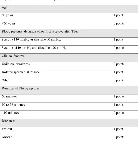
Table 1. ABCD2 score.
The ABCD 2 score can be used to identify patients at high risk of ischemic stroke in the first two
days after TIA. The score is tallied as follows:
Age:
60 years 1 point
<60 years 0 points
Blood pressure elevation when first assessed after TIA:
Systolic 140 mmHg or diastolic 90 mmHg 1 point
Systolic <140 mmHg and diastolic <90 mmHg 0 points
Clinical features:
Unilateral weakness 2 points
Isolated speech disturbance 1 point
Other 0 points
Duration of TIA symptoms:
60 minutes 2 points
10 to 59 minutes 1 point
<10 minutes 0 points
Diabetes:
Present 1 point
Absent 0 points
Materials
and
Methods
Methodology
This study was a longitudinal, prospective, observational study.
Sample Size
We prospectively analyzed 249 consecutive patients with TIAs who were referred
to our out-patient department within 72 hrs of TIA onset between Oct 2007 and March
2010. Of these, 36 patients were excluded as they did not meet inclusion criteria while 7
patients were excluded because of improper documentation. In effect we could analyze
206 patients. Of these 116 were men and 90 women with a mean age of 54.4 ± 10.1
years. A diagnosis of TIA was made in cases of clinical deficits lasting less than 24 hours
regardless of an infarction seen on cerebral imaging scans. ABCD2 scoring of all patients
was recorded at the time of their first visit. Computed tomography (CT) was performed
on all patients within two weeks of onset of the TIA in order to exclude nonischemic
brain lesions such as brain hemorrhage, chronic subdural hematoma, and brain tumors.
Methods
Inclusion Criteria
Patients referred to Neurology OPD with reversible episodes of neurologic deficits of
vascular origin that resolve completely within 24 hours.
Exclusion Criteria
1. Seizures at onset
2. Severe Cognitive impairment
3. Signs and symptoms not consistent with neuroanatomical and vascular distribution
Hospitalization: According to 2006 National Stroke Association (NSA) guidelines,
patients with crescendo TIAs, duration of symptoms >1 hour, symptomatic internal
carotid artery stenosis >50 percent, known cardiac source of embolus such as atrial
fibrillation, known hypercoagulable state, high risk of early stroke after TIA were
admitted and these patients received a five day course of subcutaneous heparin in
addition to aspirin and statin.
Information on symptoms during the TIAs was obtained from the patients or their
families. The presence or absence of the following symptoms was assessed: disturbance
of consciousness, speech disturbance, nausea/vomiting, vertigo/dizziness, visual
speech disturbance, a careful history was taken so as to distinguish aphasia from
dysarthria.
The following baseline and clinical characteristics were evaluated: 1) age and
gender: 2) duration of TIA (<1 hour, and 1-24 hours): 3) number of TIAs: 4) use of
antiplatelet agents or anticoagulants: 5) past history of brain infarction, TIA, myocardial
infarction, or definite angina pectoris: 6) risk factors for stroke, including hypertension,
diabetes mellitus, hyperlipidemia, and current smoking: 7) significant arterial pathologies
in the carotid system: and 8) potential cardiac source of emboli.
The criteria for stroke risk factors were as follows: 1) use of antihypertensive agents,
systolic blood pressure (SBP) >160 mm Hg or diastolic blood pressure (DBP) >90
mmHg: 2) use of oral hypoglycemic agents, insulin, or glycosylated hemoglobin
(HbA1C) >6.4%: 3) use of antihyperlipidemic agents, or serum cholesterol level >200
mg/dl and serum triglyceride level 150 mg/dL: and 4) current smoking defined as a
history of smoking in the preceding three months. Information on risk factors and other
comorbidity was obtained primarily from the patient; additional information was
collected from relatives. All patients gave in-formed consent and follow-up was by either
direct interview or through mobile phone. Patients were directly followed up on 2nd, 7th,
30th, and 90th day.
ABCD2 score (ie, ABCD squared, for Age, Blood pressure, Clinical features, Duration
of symptoms, and Diabetes) was used to identify patients at high risk of ischemic
• Age (≥ 60 years = 1 point)
• Blood pressure elevation when first assessed after TIA (systolic ≥ 140 mmHg or
diastolic ≥ 90 mmHg = 1 point)
• Clinical features (unilateral weakness = 2 points; isolated speech disturbance = 1
point; other = 0 points)
• Duration of TIA symptoms (≥ 60 minutes = 2 points; 10 to 59 minutes = 1 point;
<10 minutes = 0 points)
• Diabetes (present = 1 point)
For study purpose the patients were grouped according to:
1) duration of TIAs into intervals of less than 10 minutes, 10 minutes to less than 2
hours, and 2 hours to less than 24 hours
2) the etiological classification, they were subsequently grouped as Atrial Fibrillation and
Non-Atrial Fibrillation groups.
3) the presence or absence of CT findings in patients with no prior history of stroke.
4) treatment, as heparin and non heparin groups
To detect potential cardiac sources of emboli (emboligenic cardiac diseases), all
patients underwent 12-lead electrocardiography (ECG) and transthoracic
echocardiography. AF included both paroxysmal and persistent AF and was identified
during hospitalization. Emboligenic cardiac diseases included non-valvular AF: acute
valve disease: prosthetic cardiac valve: implantation of a pacemaker: and dilated
cardiomyopathy.
We performed color-flow duplex ultrasonography in all patients in order to evaluate
significant arterial pathologies in the carotid and vertebral system. The grade of stenosis
of the internal carotid artery (ICA) was determined by the method used in the North
American Symptomatic Carotid Endarterectomy Trial. The lesions were considered
significant if the ICA showed >70% stenosis or if an ulceration was evident in the carotid
bifurcation.
Ethics
Data for the study was collected following the local ethical guidelines. The identity of the
individual patients was completely anonymous. All patients signed an informed consent.
There was no delay in any of the therapeutic interventions in order to carry out the
present study.
Statistical Analysis
Statistical analysis was performed using PASW version 18.0 statistical software (SPSS
Results
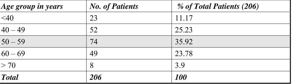
AGE DISTRIBUTION:
The maximum number of patients were in the age group between 50 and 59 years
followed by the age group between 40 and 49, 60 and 69 years. Table 1 shows the age
[image:48.612.70.548.290.428.2]distribution in this study.
TABLE - 1: AGE DISTRIBUTION
Age group in years No. of Patients % of Total Patients (206)
<40 23 11.17
40 – 49 52 25.23
50 – 59 74 35.92
60 – 69 49 23.78
> 70 8 3.9
Total 206 100
SEX DISTRIBUTION:
There were 116 males (56.3%) and 90 females (43.7%) among the 206 patients in this
TABLE 2: SEX DISTRIBUTION IN THIS STUDY
Sex No. of Patients % of Total Patients (206)
Males 116 56.3
Females 90 43.7
Total 206 100
AGE AND SEX DISTRIBUTION:
Males predominated in the age group between 50 and 59 years followed by 40 to 49
years. Females predominated in the age group between 50 and 59 years followed by 60 to
69 years.
Around two thirds of males (70.7%) were in the age group between 40 and 59 years and
two-third of females (61.1%) were in the age group between 50 and 69 years. The Table
[image:49.612.69.548.526.708.2]3 shows age distribution based on sex.
TABLE 3: AGE DISTRIBUTION BASED ON SEX
Age Group in Years Males (116) Females (90)
<40 13 (11.2%) 10 (11.1%)
40 – 49 31 (26.7%) 21 (23.4%)
50 – 59 41 (35.4%) 33 (36.7%)
60 – 69 27 (23.2%) 22 (24.4%)
> 70 4 (3.5%) 4 (4.4%)
RISK FACTORS
HYPERTENSION:
Among 206 patients, 129 patients had hypertension (62.6%)
TABLE -4: PREVALENCE OF HYPERTENSION IN THIS STUDY
Duration in years No. of patients % of total Patients (206)
Detected now 21 10.2
0 - 1 year 22 10.8
1 - 5 years 43 20.8
> 5 years 43 20.8
Total 129 62.6
DIABETES:
Out of 206 patients, 92 patients were diabetic (44.6%)
TABLE -5: PREVALENCE OF DIABETES MELLITUS IN THIS STUDY
Duration No. of patients % of Total Patients (206)
Detected now 9 4.36
0 - 1 years 14 6.77
1 - 5 years 29 14.07
> 5 years 40 19.4
Total 92 44.6
SMOKING:
93 patients were smokers (45.1%), (91 males and 2 females).
TABLE -6: PROPORTION OF SMOKING IN THIS STUDY
Duration No. of patients % of Total Patients (206)
0 - 5 years 7 3.4
5 - 10 years 16 7.8
> 10 years 70 33.9
Total 93 45.1
ALCOHOLISM:
73 patients were alcoholic (35.4%) (67 were males, 6 were females).
TABLE -7: PREVALENCE OF ALCOHOL INTAKE IN THIS STUDY
Duration No. of patients % of Total Patients (206)
0 - 5 years 5 2.4
5 - 10 years 17 8.25
> 10 years 51 24.75
SERUM LIPID PROFILE: It was done in all patients.
TOTAL CHOLESTEROL (TC):
TABLE -8: RANGE OF TOTAL CHOLESTEROL VALUES IN THIS STUDY
TC range (mg/dl) No. of Patients % of total Patients (206)
< 150 33 16
150 – 199 89 43.2
200 – 249 44 21.4
> 250 40 19.4
Total 206 100
Above cut off in 40%
TRIGLYCERIDES:
TABLE - 9: RANGE OF TRIGLYCERIDES VALUES IN THIS STUDY
TGL range No. of Patients % of total Patients (206)
< 150 29 14.02
150 – 199 75 36.5
200 – 399 101 49
> 400 1 0.48
HDL CHOLESTEROL:
TABLE -10: RANGE OF HDL CHOLESTEROL VALUES IN THIS STUDY
HDL range (mg/dl) No. of Patients % of total Patients (206)
> 40 84 40.77
< 40 122 59.23
Total 206 100
ECHO CARDIOGRAPHY:
It was done in all patients (206) (Table: 11)
Echo No % of total Patients (206)
LVH with Normal EF 120 58.4
Normal study 28 13.6
LV systolic dysfunction 31 15
LV Global dysfunction 2 0.96
Rheumatic Heart disease 9 4.3
MVPS with MR 1 0.48
MVPS without MR 9 4.3
LA clot 4 1.9
Aortic valve sclerosis 1 0.48
Prosthetic valve 1 0.48
CLINICAL FEATURES:
The clinical features of all the patients were studied in detail. The commonest
presentation was the weakness of the extremities with or without speech, language,
sensory and gait disturbances.
MOTOR WEAKNESS:
It was the commonest clinical presentation and seen in 135 patients among the 206
patients (65.53%) in this study. Among the 135 patients, Right side weakness was found
TABLE - 12: DISTRIBUTION OF WEAKNESS
Side of weakness No. of Patients % of total Patients (206)
Right 53 25.72
Left 82 39.81
Total 135 65.53
In addition, facio brachial monoparesis was seen in 15 patients, of which 13 were right
sided and 2 were left sided.
APHASIA:
Aphasia was seen in 33 patients among the 206 patients (16.2%). Of these 33 patients, 21
patients (66.6%) had right side weakness and 3 patients (9.1%) had left side weakness.
Aphasia as the only clinical manifestation was seen in 4 patients while 5 patients had
other symptoms in addition to aphasia
DYSARTHRIA:
Among the 206 patients in this study, 51 patients had dysarthria.
TABLE -14: DISTRIBUTION OF DYSARTHRIA
No. of Patients % of total Patients (206)
Anterior Circulation 26 12.6
Post Circulation 25 12.1
[image:55.612.68.558.107.213.2]SENSORY DISTURBANCE:
It was seen in 30 patients out of the 206 patients (14.56%). Table 14 shows the
distribution of sensory disturbance in this study. Sensory disturbance as the only
[image:56.612.67.547.432.689.2]symptom occurred in 2 patients.
TABLE -15: DISTRIBUTION OF SENSORY DISTURBANCE
Side of sensory disturbance No. of Patients % of total Patients (206)
Right side 14 6.8
Left side 16 7.76
Total 30 14.56
OTHER CLINICAL FEATURES:
TABLE -15: DISTRIBUTION OF OTHER CLINICAL FEATURES
S.No Clinical features No. of Patients % of total Patients
(206)
1. Visual Disturbance 17 8.25
2. Nausea & Vomiting 18 8.7
3. Dizziness/Vertigo 25 12.1
4. Gait Disturbance 48 23.3
5. LOC 38 18.4
6. Headache 6 2.9
7. TIA in the past 67 28
TERRITORIAL DISTRIBUTION: TABLE – 16
135 out of 206 (55.82%) patient had symptoms related to carotid territory while 63
(30.58%) had symptoms of vertebrobasilar territory. 8 patients (3.38%) had mixed
symptoms. While territory was uncertain in 20 (9.72%)
Territory No. of patients % of total Patients (206)
Carotid 115 55.82
Vertebrobasilar 63 30.58
Mixed 8 3.88
Uncertain 20 9.72
Total 206 100
DURATION OF SYMPTOM:
The symptom duration was reported as follows: <10 minutes in 64 patients, ≥ 10 and <30
minutes in 21, ≥ 30 and <60 minutes in 17, ≥ 1 and < 2 hours in 31, ≥ 2 and < 12 h 30 in
30, and ≥ 12 hours in the remaining 43 patients. In effect 64 patients had symptoms
lasting < 10 min, 38 had symptoms between 10 and <60 minutes while 104 patients had
symptoms lasting > 60 minutes. There was no significant difference between short
duration (< 60 min, 102 patients) and long duration (> 60 min, 104 patients) TIAs in
terms of the proportion. For this study we classified the patients into three groups for
further analysis: 64 patients with TIAs <10 minutes (Group 1); 69 patients with TIAs ≥10
DEMOGRAPHIC CHARACTERISTICS, COMORBIDITY, AND RISK FACTORS WITH RESPECT TO DURATION OF SYMPTOM: TABLE - 17
Group 1 <10 min (n=64)
Group 2 >10 min to <2 h (n=69)
Group 3 2 h to <24 h (n=73) Total (n=206) P value
Age, Years 57.2 58.1 58.7 58 0.521
Sex, M % 56.25 (36) 53.6 (39) 56.1 (41) 55.1 (116) 0.993
Prior TIA 36 (23) 21.8 (15) 26 (19) 28 (67) 0.002
Prior stroke 9.3 (6) 20.2 (14) 31.5 (23) 20.8 (43) 0.0153
Atrial fibrillation 4.6 (3) 8.7 (6) 20.5(15) 11.7 (24) 0.039
Arterial hypertension 64 (41) 60.8 (42) 63 (46) 62.6 (129) 0.86
Hypercholesterolemia 43.6 (28) 37.2 (26) 39.5 (30) 40.7 (84) 0.653
Diabetes mellitus 43.7 (28) 47.8 (33) 42.4 (31) 44 (92) 0.895
Smoking 43.7 (28) 46.3 (32) 46.5 (34) 45.1 (93) 0.788
Cardiac Disease 20.3 (13) 24.6 (17) 21.9 (16) 22.3 (46) 0.68
Arterial disease 6.2 (4) 5.7 (4) 6.8 (5) 6.3 (13) 0.986
All values in percentage. Values in bracket are absolute numbers.
A history of cerebral infarction, atrial fibrillation was found more frequently in Groups 2
and 3 than it was in Group 1. While history of prior TIA was more frequent in Group 1.
No other differences in history or risk factors were observed among the three groups
DEMOGRAPHIC CHARACTERISTICS, COMORBIDITY, AND RISK FACTORS WITH RESPECT TO ETIOLOGY: TABLE - 18
AF Group (n=24) Non-AF Group (n=182) P Value
Age, years 59.7 56.3 0.015
Sex, M (%) 56.2 54.4 0.834
TIA Duration <1 hours 1 to 24 hours
12 90 0.971
12 92 Number of TIAs
1 2 to 3 4 to 9
20 116 0.267
4 40
0 26 Medication at TIA,
Antiplatelet Anticoagulation
4 44 0.538
4 4 Past History TIA Brain Infarction Angina Myocardial Infarction
8 59 0.419
8 69 0 13 0 9 Risk Factor Arterial hypertension Hypercholesterolemia Diabetes mellitus Smoking
12 117 0.32
6 78 4 88
12 79
Arterial Disease 4 86 0.032
Embologenic Cardiac Diseases other than AF
RHD, MVP others