White Rose Research Online URL for this paper:
http://eprints.whiterose.ac.uk/141443/
Version: Published Version
Article:
Males, Alexandra and Davies, Gideon J. orcid.org/0000-0002-7343-776X (2019) Structural
studies of a surface-entropy reduction mutant of O-GlcNAcase. Acta crystallographica.
Section D, Structural biology. pp. 70-78. ISSN 2059-7983
https://doi.org/10.1107/s2059798318016595
[email protected] https://eprints.whiterose.ac.uk/ Reuse
Items deposited in White Rose Research Online are protected by copyright, with all rights reserved unless indicated otherwise. They may be downloaded and/or printed for private study, or other acts as permitted by national copyright laws. The publisher or other rights holders may allow further reproduction and re-use of the full text version. This is indicated by the licence information on the White Rose Research Online record for the item.
Takedown
If you consider content in White Rose Research Online to be in breach of UK law, please notify us by
Received 24 September 2018 Accepted 21 November 2018
Edited by M. Czjzek, Station Biologique de Roscoff, France
Keywords:O-GlcNAc; O-GlcNAcase; neurodegeneration; surface-entropy reduction; crystallization.
PDB reference: surface-entropy reduction mutant of O-GlcNAcase, 6hki
Supporting information:this article has supporting information at journals.iucr.org/d
Structural studies of a surface-entropy reduction
mutant of O-GlcNAcase
Alexandra Males and Gideon J. Davies*
Department of Chemistry, University of York, York YO10 5DD, England. *Correspondence e-mail: [email protected]
The enzyme O-GlcNAcase catalyses the removal of the O-GlcNAc co/post-translational modification in multicellular eukaryotes. The enzyme has become of acute interest given the intimate role of O-GlcNAcylation in tau modification and stability; small-molecular inhibitors of human O-GlcNAcase are under clinical assessment for the treatment of tauopathies. Given the importance of structure-based and mechanism-based inhibitor design for O-GlcNAcase, it was sought to test whether different crystal forms of the human enzyme could be achieved by surface mutagenesis. Guided by surface-entropy reduction, a Glu602Ala/Glu605Ala variant [on the Gly11–Gln396/Lys535–Tyr715 construct; Rothet al.(2017),Nature Chem. Biol.13, 610–612] was obtained which led to a new crystal form of the human enzyme. An increase in crystal contacts stabilized disordered regions of the protein, enabling 88% of the structure to be modelled; only 83% was possible for the wild-type construct. Although the binding of the C-terminus was consistent with the wild type, Lys713 in monomerAwas bound in the 1 subsite of the symmetry-related monomerAand the active sites of the B monomers were vacant. The new crystal form presents an opportunity for enhanced soaking experiments that are essential to understanding the binding mechanism and substrate specificity of O-GlcNAcase.
1. Introduction
The regulation of O-GlcNAc cycling on thousands of nuclear and cytoplasmic proteins is coordinated by two enzymes. O-GlcNAc transferase (OGT) catalyses the addition of GlcNAc, derived from UDP-GlcNAc, to serine and threonine residues, and O-GlcNAcase (OGA; CAZY database family GH84) cleaves O-GlcNAc (Torres & Hart, 1984; Holt & Hart, 1986; Kreppel et al., 1997; Dong & Hart, 1994; Lubas et al., 1997; Hartet al., 2007). Two isoforms, OGA-L and OGA-S, are localized to the nucleus/cytoplasm (Comtesseet al., 2001) and to the surface of lipid droplets, respectively. The reciprocal relationship between O-phosphorylation and O-glycosylation on the particular protein tau has been keenly studied in the context of neurodegeneration (Arnoldet al., 1996; Yuzwa et al., 2008, 2014; Shenet al., 2012; Griffith & Schmitz, 1995; Gao et al., 2001; Liu et al., 2004). In patients with Alzheimer’s disease, tau undergoes hyperphosphorylation, causing it to dissociate from microtubules and aggregate into paired helical filaments (PHF) and neurofibrillary tangles (NFTs) (Grundke-Iqbalet al., 1986; Marottaet al., 2015). O-GlcNAc cycling has also been implicated in tumorigenesis owing to its significant role in orchestrating a vast number of cellular processes, for example transcriptional and cytoskeletal regulation, cell signalling and division, and metabolism (Slawson & Hart, 2011).
Structural characterization of the human O-GlcNAcase orthologue (HsOGA-L/HsOGA) showed dimer formation
both in solution and in the crystal where it has been shown to be promoted by helix-exchange (Rothet al., 2017) in contrast to the non-helix-exchanged bacterial dimers (Dennis et al., 2006; Schimplet al., 2010; Raoet al., 2006). Composed of two ordered domains, the N-terminal glycoside hydrolase domain forms a (/)8 barrel (Li et al., 2012), while the C-terminal
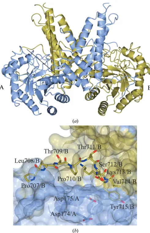
stalk domain (Tolemanet al., 2004; Heet al., 2014) is composed of six-helices.17, consisting of Glu676–Pro694, undergoes a dimer ‘swap’ (Rothet al., 2017), playing a structural role that contributes to dimerization (Li, Li, Luet al., 2017; Elsenet al., 2017; Rothet al., 2017). Located in a V-shaped cleft formed between the N-terminal domain of monomer A and the C-terminal domain of monomerB(Fig. 1a) are the catalytic residues, Asp174 and Asp175, that initially act as a general acid and a general base through a two-step retaining mechanism (Macauley et al., 2005). The terminal domain, which is not present in OGA-S, has a ‘pseudo’-histone acetyltransferase activity but was not included in the crystal-lized construct in the threeHsOGA structures as it has a high degree of disorder.
The recognition mode for glycopeptide substrates has been explored by obtaining structures of the catalytically inactive Asp175Asn mutant of HsOGA in complex with a variety of glycosylated peptides (Li, Li, Luet al., 2017; Li, Li, Huet al., 2017). The peptides bound in each structure can be segregated into two binding modes with forward or reverse orientations of the peptide (amino to carboxyl or carboxyl to amino, respectively) within the binding site. Initially, crystallographic peptide studies were conducted using an orthologue from the bacterium Clostridium perfringens (Schimpl et al., 2012; Mariappa et al., 2015) in complex with TAB1, lamin B1 and p53 glycosylated peptides. These peptides bound in the forward direction. Later, Li, Li, Huet al.(2017) compared the same glycosylated peptides withHsOGA and they were found to act in the same way. However, -crystallin B and ELK1 bound in the reverse direction, strengthening the interest in determining how OGA selects its target (Li, Li, Hu et al., 2017).
In the ‘apo’ structure, HsOGA11–396+535–715 (thus named
owing to the co-expression of two plasmids containing resi-dues 11–396 and 535–715), which was published by Rothet al. (2017) and deposited as PDB entry 5m7r, the C-terminus of monomerAfrom residues Pro707 to Tyr715 can be traced in the reverse direction into the active-site groove of the symmetry-related molecule of monomer B (Fig. 1b). When overlaid with theHsOGA–p53 complex (PDB entry 5un8; Li, Li, Luet al., 2017), the position of the Tyr715 hydroxyl group lies directly on top of the O6 of O-GlcNAc.
To conduct biochemical/biophysical studies and rational drug design, complete and coherent structures are required. However, the published structures ofHsOGA have stretches of residues that are incomplete (Elsenet al., 2017; Li, Li, Luet al., 2017; Li, Li, Huet al., 2017). For example, the structure described by Roth et al. (2017) has regions within the N-terminus (Glu11–Arg58 and Lys341–Thr370) and the C-terminus (Asp696–Pro706) that are not modelled in the structure. Expulsion of the C-terminus ofHsOGA11–396+535–715
from the active site is required before a competing compound can bind, therefore making it challenging to conduct crystal-soaking experiments; this is a problem for weak-binding compounds/inhibitors.
The work in this paper utilized surface-entropy reduction (SER) to enhance the structural characterization ofHsOGA and to contribute towards the hypothesis for the substrate-recognition mode of OGA, in which either the O-GlcNAc moiety or the peptide sequence is important for recognition. Using the SERp online server (Goldschmidt et al., 2007), potential pairs of mutations were identified forHsOGA. The rational design of mutating clusters of residues on a protein is a favourable strategy, with the aim of making the protein more susceptible to crystallization (Derewenda, 2004a,b; Dere-wenda & Vekilov, 2006; Cooperet al., 2007). Surface-entropy reduction (SER) is a concept in which flexible, solvent-exposed residues, primarily lysine and glutamate, are mutated to alanine to reduce the entropic loss during the packing of the
[image:3.610.316.561.309.688.2]Acta Cryst.(2019). D75, 70–78 Males & Davies Surface-entropy reduction mutant of O-GlcNAcase
71
Figure 1
protein into a crystalline lattice (Longenecker et al., 2001; Matejaet al., 2002; Vekilov, 2003; Vekilovet al., 2002).
Following the co-expression strategy of Rothet al. (2017) using pACYC-Duet-1 Gly11–Gln396 and pET-YSBLIC3C Lys535–Tyr715 plasmids, the successful pair of mutations was Glu602Ala/Glu605Ala (HsOGAE602AE605A). Unlike
HsOGA11–396+535–715, the C-terminus of monomer A can be
traced into the active site of the symmetry-related monomer A, with Lys713 binding in the position of O-GlcNAc. Furthermore, previously disordered loops had become ordered and could be built into the final model. Additionally, the activities and secondary-structure profiles of full-length HsOGA (HsOGAFL),HsOGA11–396+535–715andHsOGAE602AE605A
were examined to verify the loss of the ‘pseudo-HAT’ domain and that the SER mutation did not alter the activity. The results showed that the SER mutant exhibited similar Km
values to the full-length enzyme, since the mutation is distant from the active site, highlighting the potential of SER variants for studying the structural and ligand-binding characteristics ofHsOGA.
2. Materials and methods
2.1. Macromolecular production
The cloning of the constructs has been described previously (Rothet al., 2017). Homologous DNA for the Lys535–Tyr715 construct, in the vector pET-YSBL-LIC-3C, was mutated using a Q5 Site-Directed Mutagenesis Kit with Q5 Hot Start High-Fidelity DNA Polymerase. The forward primer, A GAT AGC gct AAA ATC gct GAA TGG, was designed to mutate the primary sequence A GAT AGC GAA AAA ATC GAA GAA; the reverse primer was TTA CCC TTG CAG TTA ACC GAA. NEB 5-alpha competent Escherichia coli cells were transformed with the Lys535–Tyr715 E602AE605A construct. The DNA was extracted and sequenced to verify the mutation.
The Gly11–Gln396 and Lys535–Tyr715 E602AE605A constructs in the vectors pACYCDUET-1 and pET-YSBL-LIC-3C, respectively, were co-transformed into E. coliBL21 (DE3) cells. Luria–Bertani broth was inoculated with a cell suspension and was incubated at 37C until the OD
600reached
1.0. The cells were induced with 1 mMIPTG and incubated at 16
C for 20 h.
The purification of HsOGAE602AE605A followed the same
purification protocol as that ofHsOGA11–396+535–715described
previously (Roth et al., 2017; Supplementary Figs. S1a and S1b).
2.2. Crystallization
HsOGAE602AE605Awas initially crystallized by sitting-drop
vapour diffusion at 15 mg ml 1 under condition E11 of the PACT premierscreen from Molecular Dimensions (Newman et al., 2005): 0.2M sodium citrate tribasic, 20% PEG 3350. Further optimization of the conditions to 0.2Msodium citrate tribasic pH 7.5, 17% polyethylene glycol 3350 in a 48-well MRC MAXI optimization plate improved the crystal shape.
The total volume of the drop was 1ml and the protein: reservoir solution ratio was 1:1; the total volume in the reservoir was 100ml.
2.3. Data collection and processing
Diffraction images were collected on beamline I04-1 at Diamond Light Source (DLS). After data collection, the diffraction images were integrated using the-3diioption in
xia2 (Winter, 2010) and reintegrated usingAIMLESS(Evans, 2006; Evans & Murshudov, 2013) from the CCP4 software suite (Winn et al., 2011). Data-collection and processing statistics are given in Table 1.
2.4. Structure solution and refinement
Molecular replacement against the coordinates of PDB entry 5m7r was conducted using MOLREP (Vagin & Teplyakov, 2010). Refinement of the model was conducted using multiple rounds ofREFMAC (Murshudov et al., 1997, 2011; Pannuet al., 1998; Winnet al., 2003; Vaginet al., 2004; Nicholls et al., 2012) and manual model building in Coot (Emsleyet al., 2010). Waters were added using Find Waters in Cootand validated. The data were processed to a resolution of 3.3 A˚ (Table 2).
2.5. Michaelis–Menten kinetics
Michaelis–Menten kinetics were assayed using HsOGAFL
and HsOGA11–396+535–715 as positive controls against the
mutantHsOGAE602AE605A. In a 200ml reaction volume, 50 nM
protein solution and a serial dilution of the ligand 4-nitro-phenylN-acetyl--d-glucosaminide (pNP-GlcNAc) from 1500 to 11.7mM [dissolved in 2.5% DMSO (final concentration)] was added to PBS buffer at pH 7.4.
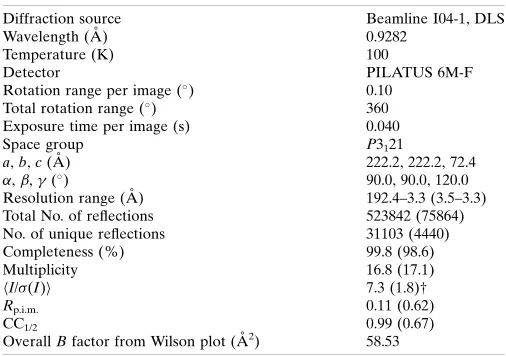
[image:4.610.313.566.106.284.2]The reaction was monitored at 405 nMcontinuously using a Molecular Devices SpectraMax M5 spectrophotometer. The Table 1
Data collection and processing.
Values in parentheses are for the outer shell.
Diffraction source Beamline I04-1, DLS
Wavelength (A˚ ) 0.9282
Temperature (K) 100
Detector PILATUS 6M-F
Rotation range per image () 0.10
Total rotation range () 360
Exposure time per image (s) 0.040
Space group P3121
a,b,c(A˚ ) 222.2, 222.2, 72.4
,,() 90.0, 90.0, 120.0
Resolution range (A˚ ) 192.4–3.3 (3.5–3.3) Total No. of reflections 523842 (75864) No. of unique reflections 31103 (4440) Completeness (%) 99.8 (98.6)
Multiplicity 16.8 (17.1)
hI/(I)i 7.3 (1.8)†
Rp.i.m. 0.11 (0.62)
CC1/2 0.99 (0.67)
OverallBfactor from Wilson plot (A˚2) 58.53
experiment, which was conducted at 25C, was duplicated and
each ligand concentration was repeated in triplicate.
GraphPad Prism v.5 was used to process the data, with nonlinear regression of Michaelian saturation curves. The initial velocities were calculated from the linear range of the reaction-progress curve. A standard curve of 4-nitrophenol was used to extract a molar extinction coefficient.
2.6. Circular-dichroism spectroscopy
The protein samples were dialysed overnight into 25 mM sodium phosphate pH 8.0 and diluted to 0.1 mg ml 1. The spectra were recorded at 21
C in a QS 248 0.2 mm cuvette with 0.5 s per point and 78 s per read. The wavelength ranged from 195 to 320 nm. The background for each protein was measured immediately before the experiment in the same cuvette and values were taken as averages from triplicate reads.
3. Results and discussion
3.1. Comparison of the mutant crystal structure with that of the wild type
To incorporate protein molecules into a crystal, a thermo-dynamic cost is endured to bury hydrophobic residues into a constrained conformation and from the immobilization of the prevalent flexible hydrophilic side chains on the surface (Avbelj & Fele, 1998). Reducing the entropic shield can lead to an increase in the variety of conditions, morphologies and crystallographic space groups (Parthasarathyet al., 2008; Kim et al., 2005). Therefore, crystallization conditions were re-screened using the HsOGAE602AE605A variant; crystals were
obtained in 17% polyethylene glycol 3350, 0.2M sodium citrate pH 8.0. This is comparable to the wild-type crystal-lization condition of 0.1–0.2Mtriammonium citrate pH 6.5– 7.5, 16–24% PEG 3350.
In a different crystal form, flexible loops can become ordered by making backbone crystal contacts or adopting preferential conformations (Derewenda, 2004a). The space group wasP3121, which is a lower symmetry group compared
with P43212 for the HsOGA11–396+535–715structure. The data
statistics are presented in Tables 1 and 2.
Theoretically, SER should lead to an improvement in the resolution and hence the overall quality of the structure (Parthasarathy et al., 2008). However, the structure was determined to a resolution of 3.3 A˚ , which was a lower reso-lution compared with the HsOGA11–396+535–715 and
catalyti-cally inactive mutant structures, possibly owing to the thin-rod crystal morphology compared with the large trigonal bi-pyramid wild-type crystals.
A solvent channel can be observed through the crystal structure (Fig. 2a). The dimers form a trigonal spring such that the C-terminus of monomer A binds into the active-site monomer A of a symmetry-related dimer, etc.(Figs. 2a and 2b). Owing to the different crystal contacts made on the surface of the protein, regions of high disorder could be built into the structure. 88% of the structure was complete, in comparison with 83% of the HsOGA11–396+535–715 structure.
The regions of highest disorder in both monomers were between Lys341 and Thr370, in addition to loops on the protein surface. After multiple rounds of refinement, the confidence for the inclusion of residues Lys341–Asp347 increased, enabling further visualization of the disordered region (Figs. 2cand 2d). The residues that were still disordered in monomers A and B were Gly11–Gly56, Ser348–Glu369, Lys535 and Pro707–Tyr715 for monomerBonly. In the protein structures described by Elsenet al.(2017) and Li, Li, Luet al. (2017) the residues Lys341–Asp371 in monomerA(PDB entry 5uhk) and Asn335–Val372 in monomerA(PDB entry 5tke), respectively, were also not observed.
When the N-terminal domains of monomer A from HsOGAE602AE605AandHsOGA11–396+535–715were overlapped,
the r.m.s.d. of the N-terminal domains was 1.5 A˚ and the r.m.s.d. for the C-terminal domains was 7.1 A˚ , indicating a high degree of flexibility in the latter domain (Fig. 2e).
As mentioned, the C-terminus of monomerAwas found to bind into the active site of a symmetry-related monomerA, aiding the formation of the new crystal packing (Figs. 2band 3a). Initially, the residues of17 interact with monomerBin a domain swap; the residues from Pro694 to Phe703 then bend back towards the residues of the respective monomer, with Gln704–Tyr715 binding into the active site (Figs. 3aand 3b). Electrostatic interactions between the C-terminus of monomer A and other residues of monomer A, B and a symmetry-related moleculeBstabilize this formation. Pro707 ofHsOGAE602AE605Ahas drastically moved position and faces
in the opposite direction. There are three consecutive prolines that facilitate the change in direction. Hence, the C-terminus binds to the active site of the symmetry-related monomerA rather than monomerB(as in the wild type). The C-terminus of monomerBcould not be built in from Pro707, indicating that it does not bind into an active site because of the crystal packing. InHsOGA11–396+535–715, residues Asp696–Pro707 in
monomer A are missing; therefore, the direction of the peptide is ambiguous. In the natural human sequence, the pseudo-histone acetyltransferase domain is connected to the
[image:5.610.314.566.107.275.2]Acta Cryst.(2019). D75, 70–78 Males & Davies Surface-entropy reduction mutant of O-GlcNAcase
73
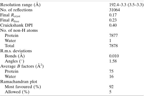
Table 2
Structure solution and refinement.
Values in parentheses are for the outer shell.
Resolution range (A˚ ) 192.4–3.3 (3.5–3.3)
No. of reflections 31064
FinalRcryst 0.17
FinalRfree 0.23
Cruickshank DPI 0.40
No. of non-H atoms
Protein 7877
Water 1
Total 7878
R.m.s. deviations
Bonds (A˚ ) 0.010
Angles () 1.58
AverageBfactors (A˚2)
Protein 75
Water 16
Ramachandran plot
Most favoured (%) 92
Figure 2
(a,b) Crystal symmetry ofHsOGAE602AE605A. (a) The connections made between the dimers show the trigonal solvent channel, with 50% solvent as
calculated from the Matthews coefficient of 2.51 A˚3Da 1(Kantardjieff & Rupp, 2003; Matthews, 1968). (b) Side view of the repeating trigonal dimers showing the linking C-terminus of monomerAbinding into monomerAof the next dimer complex. The monomers are labelledAandB, with asterisks indicating the C-termini. (c,d) Disordered loop regions were stabilized in the new crystal form. MonomerAis shown in sea green and monomerBin brown. (c) The regions in purple were built into theHsOGAE602AE605Astructure using PDB entry 5m7r as the template model and were missing from the
wild-type structure. (d) Residues Lys341–Gly347 and the maximum-likelihood/A-weighted 2Fobs Fcalcmap shown in red contoured at 0.12 e A˚ 3
. (e) Overlap of the N-terminal monomersAfrom chainAfor bothHsOGA11–396+535–715(monomerA, blue; monomerB, gold) andHsOGAE602AE605A
(monomerA, sea green; monomerB, brown) as calculated byCCP4mg Superposemodels. The residue range selected for superposition was Arg59/A– His395/A.
stalk domain; therefore, binding of the C-terminus in the active site or alternatively C-termini disorder are possible artefacts of the removal of the HAT domain and of crystal packing.
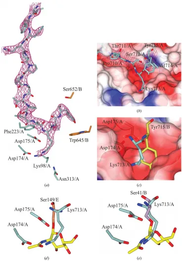
The C-terminal residues Lys713/Aand Tyr715/A hydrogen-bond to and make electrostatic interactions with residues of the active site (Fig. 3b). Interestingly, the NZ atom of Lys713 is in the same position as the N2 atom of O-GlcNAc and is 2.97 A˚ away from the Oatom of Asp175/Aon the symmetry-related monomer (Fig. 3d). This pushes Tyr715 into the +2 subsite, where it interacts with the O
symmetry-related monomer (Fig. 3a). In comparison, the Tyr715/Ahydroxyl group of the wild type lies above O6 of O-GlcNAc and hydrogen-bonds to the O1
atom of Asp285/B (Fig. 3c).
In comparison to the crystal structure of HsOGA in complex with-crystallin B and ELK1 (PDB entries 5vvv and 5vvt, respectively; Li, Li, Lu et al., 2017), the C-terminal residues are in the same reverse direction as the glycosylated
[image:7.610.122.490.143.669.2]Acta Cryst.(2019). D75, 70–78 Males & Davies Surface-entropy reduction mutant of O-GlcNAcase
75
Figure 3
The C-terminus of monomerAbinds into the active site of the symmetry-related monomerA. (a) Neighbouring residues in the active site of symmetry-related monomerAofHsOGAE602AE605A, with the C-terminal residues bound and the maximum-likelihood/A-weighted 2Fobs Fcalcmap shown in red
contoured at 0.17 e A˚ 3
. (b) Surface representation of the active site with the C-terminus bound in a negatively charged pocket. (c) Overlay of Lys713/A from HsOGAE602AE605A and Tyr715/B from HsOGA11–396+535–715 (in gold) in the binding pocket. (d) Overlay of the C-terminus of chain A of
HsOGAE602AE605Aand that ofHsOGA in complex with glycosylated p53 peptide (PDB entry 5un8), showing Ser149/Ein coral. O-GlcNAc is shown in
yellow. (e) Overlay of Lys713/AfromHsOGAE602AE605AwithHsOGA in complex with glycosylated-crystallin B (PDB entry 5vvv), showing Ser41/Bin
peptides (Fig. 3e). This is in contrast to the structure containing a glycosylated p53 peptide shown in Fig. 3(d).
The density for all available HsOGA peptide-complex structures supports the notion that OGA can bind peptides in both the forward and reverse directions. Comparison between the different peptide structures shows the versatility of the active site for different peptides.
3.2. Comparison of the constructs and mutants
To ensure that the structural stabilization had not occurred owing to a change in the secondary structure of HsOGAE602AE605Aand that the mutation had not affected the
activity of the enzyme, the full-length protein (HsOGAFL),
HsOGA11–396+535–715 and HsOGAE602AE605A underwent
kinetic and secondary-structure analyses.
Comparison of the results for HsOGAFL and
HsOGA11–396+535–715shows that the split construct has similar
activity to the full-length variant, suggesting that co-expres-sion of the two domains does not affect the activity (Fig. 4). Therefore, the ligand-binding data should be an accurate representation of binding to the full-length protein. When comparing HsOGA11–396+535–715 and HsOGAE602AE605A, the
values are very similar. The Vmax is higher, indicating an
increase in the reaction rate (Table 3). The increase inVmax
may be owing to a discrepancy in the enzyme concentration ([E]), asV0is directly proportional to [E]. The similarity of
VmaxandKmfor the mutant and the wild type suggest that the
mutation did not alter the substrate-binding affinity or the enzyme activity of the protein for its substrate.
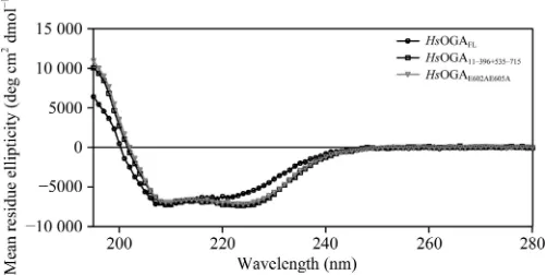
The CD spectra show that all of the constructs are fully folded and the spectra for HsOGA11–396+535–715 and
HsOGAE602AE605Aare very similar, suggesting that there is no
change in the composition of the mutant (Fig. 5). The spectral profile ofHsOGAFLdiffers from those of the split constructs
in that it has a less pronounced minimum in the 222 nm region and an overall blue-shifted spectrum in the <210 nm region. This suggests that there could be a lower proportion of -helical structure and/or higher structural disorder. This links to the inclusion of the pseudo-HAT domain that is connected to the C-terminal stalk domain. Since the structure of the human homologue of this domain is unknown owing to the inability ofHsOGAFLto crystallize, the structure can only be
inferred from the structural homologues from Oceanicola granulosusandStreptomyces sviceus(Heet al., 2014; Raoet al., 2013). An estimate of the secondary-structure content of the proteins suggests a decrease of 6.4% in-helical components and an increase of 3.7% in-sheet components inHsOGAFL
compared with HsOGA11–396+535–715 (Supplementary Table
S1). Homology modelling using crystal structures of the HAT domain from the bacterial homologueO. granulosussuggests a similar overall structure minus the acetyltransferase activity (Raoet al., 2013). However, the data could be skewed by the inclusion of the linker region to this domain and the potential difference in the homology model structure. The secondary-structure contents ofHsOGA11–396+535–715andHsOGAE602AE605A
are consistent, further confirming that the mutation did not affect the overall structure.
4. Conclusions
[image:8.610.45.296.92.151.2]In this study, surface-entropy reduction has been utilized to produce further structural information on O-GlcNAcase by the incorporation of residues Ala57–Arg58, Lys341–Asp347, Thr370, Glu536, Cys596–Gly598, Gly674–Asp675 and Asp696–Pro707, an increase in the number of observed resi-dues of 5%. Although the binding of the C-terminus to the active site may be an artefact of crystallization, it reveals further details regarding the substrate specificity of OGA, as peptides have been shown to bind in a bidirectional yet conserved conformation. The results described in this study Figure 5
Circular-dichroism spectra for the different constructs. The experimental data in millidegree units were converted to mean residue ellipticity values with units of deg cm2
dmol 1
[image:8.610.45.296.338.517.2]using the equation given in Ishtikharet al. (2014).
Figure 4
[image:8.610.45.296.572.698.2]Michaelis–Menten curves for the kinetic assay of theHsOGA mutant. pNP-GlcNAc was used as the substrate at concentrations up to seven times higher than theKm.
Table 3
Kinetics analysis comparing differentHsOGA constructs.
Construct HsOGAFL HsOGA11–396+575–715HsOGAE602AE605A
Vmax(mMmin 1) 2.960.07 2.500.06 4.620.15
Km(mM) 29815 22713 23020
kcat(min 1) 59.11.4 49.91.2 92.53.1
present an opportunity for further investigation of the binding orientation of peptides within an SER-modified OGA enzyme. Given the progression of hOGA inhibitors into clinical trials, different surface mutants of the enzyme may afford new routes to drug development.
Acknowledgements
CD data collection was conducted by Dustin King and assis-tance in conducting the kinetics experiments and analysing the data was provided by Matthew Alteen, both of whom are employed by Professor David Vocadlo at Simon Fraser University. We thank Diamond Light Source for access to beamline I04-1 (proposal No. mx-13587) that contributed to the results presented here, and Johan Turkenburg and Sam Hart for coordinating data collection.
Funding information
This research was funded by the Biotechnology and Biological Sciences Research Council (grant BB/M011151/1) for the support of AM. GJD is the Royal Society Ken Murray Research Professor.
References
Arnold, C. S., Johnson, G. V. W., Cole, R. N., Dong, D. L.-Y., Lee, M. & Hart, G. W. (1996).J. Biol. Chem.271, 28741–28744.
Avbelj, F. & Fele, L. (1998).J. Mol. Biol.279, 665–684.
Comtesse, N., Maldener, E. & Meese, E. (2001).Biochem. Biophys. Res. Commun.283, 634–640.
Cooper, D. R., Boczek, T., Grelewska, K., Pinkowska, M., Sikorska, M., Zawadzki, M. & Derewenda, Z. (2007).Acta Cryst.D63, 636– 645.
Dennis, R. J., Taylor, E. J., Macauley, M. S., Stubbs, K. A., Turkenburg, J. P., Hart, S. J., Black, G. N., Vocadlo, D. J. & Davies, G. J. (2006).Nature Struct. Mol. Biol.13, 365–371.
Derewenda, Z. S. (2004a).Structure,12, 529–535.
Derewenda, Z. S. (2004b).Methods,34, 354–363.
Derewenda, Z. S. & Vekilov, P. G. (2006).Acta Cryst.D62, 116–124.
Dong, D. L.-Y. & Hart, G. W. (1994).J. Biol. Chem.269, 19321–19330.
Elsen, N. L., Patel, S. B., Ford, R. E., Hall, D. L., Hess, F., Kandula, H., Kornienko, M., Reid, J., Selnick, H., Shipman, J. M., Sharma, S., Lumb, K. J., Soisson, S. M. & Klein, D. J. (2017).Nature Chem. Biol.
13, 613–615.
Emsley, P., Lohkamp, B., Scott, W. G. & Cowtan, K. (2010). Acta Cryst.D66, 486–501.
Evans, P. (2006).Acta Cryst.D62, 72–82.
Evans, P. R. & Murshudov, G. N. (2013).Acta Cryst.D69, 1204–1214. Gao, Y., Wells, L., Comer, F. I., Parker, G. J. & Hart, G. W. (2001).J.
Biol. Chem.276, 9838–9845.
Goldschmidt, L., Cooper, D. R., Derewenda, Z. S. & Eisenberg, D. (2007).Protein Sci.16, 1569–1576.
Griffith, L. S. & Schmitz, B. (1995).Biochem. Biophys. Res. Commun.
213, 424–431.
Grundke-Iqbal, I., Iqbal, K., Tung, Y.-C., Quinlan, M., Wisniewski, H. M. & Binder, L. I. (1986).Proc. Natl Acad. Sci. USA,83, 4913–
4917.
Hart, G. W., Housley, M. P. & Slawson, C. (2007).Nature (London),
446, 1017–1022.
He, Y., Roth, C., Turkenburg, J. P. & Davies, G. J. (2014).Acta Cryst. D70, 186–195.
Holt, G. D. & Hart, G. W. (1986).J. Biol. Chem.261, 8049–8057.
Ishtikhar, M., Khan, S., Badr, G., Osama Mohamed, A. & Hasan Khan, R. (2014).Mol. Biosyst.10, 2954–2964.
Kantardjieff, K. A. & Rupp, B. (2003).Protein Sci.12, 1865–1871. Kim, A.-R., Dobransky, T., Rylett, R. J. & Shilton, B. H. (2005).Acta
Cryst.D61, 1306–1310.
Kreppel, L. K., Blomberg, M. A. & Hart, G. W. (1997).J. Biol. Chem.
272, 9308–9315.
Li, B., Li, H., Hu, C.-W. & Jiang, J. (2017).Nature Commun.8, 666.
Li, B., Li, H., Lu, L. & Jiang, J. (2017).Nature Struct. Mol. Biol.24,
362–369.
Li, J., Li, Z., Li, T., Lin, L., Zhang, Y., Guo, L., Xu, Y., Zhao, W. & Wang, P. (2012).Biochemistry (Mosc.),77, 194–200.
Liu, F., Iqbal, K., Grundke-Iqbal, I., Hart, G. W. & Gong, C.-X. (2004).Proc. Natl Acad. Sci. USA,101, 10804–10809.
Longenecker, K. L., Garrard, S. M., Sheffield, P. J. & Derewenda, Z. S. (2001).Acta Cryst.D57, 679–688.
Lubas, W. A., Frank, D. W., Krause, M. & Hanover, J. A. (1997).J. Biol. Chem.272, 9316–9324.
Macauley, M. S., Whitworth, G. E., Debowski, A. W., Chin, D. & Vocadlo, D. J. (2005).J. Biol. Chem.280, 25313–25322.
Mariappa, D., Selvan, N., Borodkin, V. S., Alonso, J., Ferenbach, A. T., Shepherd, C., Navratilova, I. H. & van Aalten, D. M. F. (2015). Biochem. J.470, 255–262.
Marotta, N. P., Lin, Y. H., Lewis, Y. E., Ambroso, M. R., Zaro, B. W., Roth, M. T., Arnold, D. B., Langen, R. & Pratt, M. R. (2015). Nature Chem.7, 913–920.
Mateja, A., Devedjiev, Y., Krowarsch, D., Longenecker, K., Dauter, Z., Otlewski, J. & Derewenda, Z. S. (2002).Acta Cryst.D58, 1983–
1991.
Matthews, B. W. (1968).J. Mol. Biol.33, 491–497.
Murshudov, G. N., Skuba´k, P., Lebedev, A. A., Pannu, N. S., Steiner, R. A., Nicholls, R. A., Winn, M. D., Long, F. & Vagin, A. A. (2011). Acta Cryst.D67, 355–367.
Murshudov, G. N., Vagin, A. A. & Dodson, E. J. (1997).Acta Cryst. D53, 240–255.
Newman, J., Egan, D., Walter, T. S., Meged, R., Berry, I., Ben Jelloul, M., Sussman, J. L., Stuart, D. I. & Perrakis, A. (2005).Acta Cryst. D61, 1426–1431.
Nicholls, R. A., Long, F. & Murshudov, G. N. (2012).Acta Cryst.D68, 404–417.
Pannu, N. S., Murshudov, G. N., Dodson, E. J. & Read, R. J. (1998). Acta Cryst.D54, 1285–1294.
Parthasarathy, G., Cummings, R., Becker, J. W. & Soisson, S. M. (2008).Acta Cryst.D64, 141–148.
Rao, F. V., Dorfmueller, H. C., Villa, F., Allwood, M., Eggleston, I. M. & van Aalten, D. M. F. (2006).EMBO J.25, 1569–1578.
Rao, F. V., Schuttelkopf, A. W., Dorfmueller, H. C., Ferenbach, A. T., Navratilova, I. & van Aalten, D. M. F. (2013).Open Biol.3, 130021.
Roth, C., Chan, S., Offen, W. A., Hemsworth, G. R., Willems, L. I., King, D. T., Varghese, V., Britton, R., Vocadlo, D. J. & Davies, G. J. (2017).Nature Chem. Biol.13, 610–612.
Schimpl, M., Borodkin, V. S., Gray, L. J. & van Aalten, D. M. F. (2012). Chem. Biol.19, 173–178.
Schimpl, M., Schu¨ttelkopf, A. W., Borodkin, V. S. & van Aalten, D. M. F. (2010).Biochem. J.432, 1–7.
Shen, D. L., Gloster, T. M., Yuzwa, S. A. & Vocadlo, D. J. (2012).J. Biol. Chem.287, 15395–15408.
Slawson, C. & Hart, G. W. (2011).Nature Rev. Cancer,11, 678–684. Toleman, C., Paterson, A. J., Whisenhunt, T. R. & Kudlow, J. E.
(2004).J. Biol. Chem.279, 53665–53673.
Torres, C. R. & Hart, G. W. (1984).J. Biol. Chem.259, 3308–3317.
Vagin, A. A., Steiner, R. A., Lebedev, A. A., Potterton, L., McNicholas, S., Long, F. & Murshudov, G. N. (2004).Acta Cryst. D60, 2184–2195.
Vagin, A. & Teplyakov, A. (2010).Acta Cryst.D66, 22–25.
Vekilov, P. G. (2003).Methods Enzymol.368, 84–105.
Vekilov, P. G., Feeling-Taylor, A., Yau, S.-T. & Petsev, D. (2002).Acta Cryst.D58, 1611–1616.
Winn, M. D., Ballard, C. C., Cowtan, K. D., Dodson, E. J., Emsley, P., Evans, P. R., Keegan, R. M., Krissinel, E. B., Leslie, A. G. W.,
McCoy, A., McNicholas, S. J., Murshudov, G. N., Pannu, N. S., Potterton, E. A., Powell, H. R., Read, R. J., Vagin, A. & Wilson, K. S. (2011).Acta Cryst.D67, 235–242.
Winn, M. D., Murshudov, G. N. & Papiz, M. Z. (2003). Methods Enzymol.374, 300–321.
Winter, G. (2010).J. Appl. Cryst.43, 186–190.