Pharmacological Properties and Physiological Function
of a P2X-Like Current in Single Proximal Tubule Cells
Isolated from Frog Kidney
John P. Davies• Louise Robson
Received: 22 July 2010 / Accepted: 22 September 2010 / Published online: 23 October 2010 ÓThe Author(s) 2010. This article is published with open access at Springerlink.com
Abstract Although previous studies have provided evi-dence for the expression of P2X receptors in renal proximal tubule, only one cell line study has provided functional evidence. The current study investigated the pharmaco-logical properties and physiopharmaco-logical role of native P2X-like currents in single frog proximal tubule cells using the whole-cell patch-clamp technique. Extracellular ATP activated a cation conductance (P2Xf) that was also Ca2?
-permeable. The agonist sequence for activation was ATP=ab-MeATP[BzATP=2-MeSATP, and P2Xf
was inhibited by suramin, PPADS and TNP-ATP. Activa-tion of P2Xfattenuated the rundown of a
quinidine-sensi-tive K?conductance, suggesting that P2Xfplays a role in
K?channel regulation. In addition, ATP/ADP apyrase and inhibitors of P2Xf inhibited regulatory volume decrease
(RVD). These data are consistent with the presence of a P2X receptor that plays a role in the regulation of cell volume and K? channels in frog renal proximal tubule cells.
Keywords Purinergic receptor physiology in epithelia
Renal physiologyIon channelPotassium ion channel
Volume regulation in epithelial cells
Introduction
P2 purinoceptors are a class of ATP-activated receptors that play a critical role in a variety of cellular processes in both electrically excitable and nonexcitable cells. They are subdivided into two distinct classes, P2X and P2Y recep-tors (Burnstock and Kennedy 1985; North and Barnard
1997). P2X receptors are ATP-gated, nonselective, Ca2? -permeable cation channels that, on activation, allow extracellular Ca2? to enter the cell, leading to a rise in intracellular Ca2?. The P2Y receptors are G protein-cou-pled. On activation, some P2Y receptors cause a rise in intracellular Ca2?, via the release of Ca2? from intracel-lular stores.
At the molecular level a number of mammalian P2X and P2Y receptors have been identified, P2X1–7 and
P2Y1,2,4,6,11–14, with only two amphibian P2X receptors
identified, P2X4 (Juranka et al. 2001) and a P2X5-like
current (Jensik et al.2001). These receptors can act as ho-momeric channels and form heteromeric channels. To date, in heterologous systems, combinations of a number of P2X receptors have been observed, including P2X2/3, P2X1/5and
P2X4/6(Le et al. 1998,1999; Radford et al. 1997; Torres
et al. 1999). There is also evidence for the presence of P2X2/3 heteromers in rat nodose neurons (Lewis et al. 1995). A more recent study has provided evidence for P2X4/7receptors (Guo et al.2007). The properties of these
heteromers are determined by their receptor composition, with properties taken from both receptor types. This means that heteromeric channels demonstrate very different properties from the single cloned receptors.
Both P2X and P2Y family members are found in the kidney, with the majority of work on renal P2 receptors to date focusing on their role in the distal tubule and col-lecting duct. Previous work has demonstrated that both
Electronic supplementary material The online version of this article (doi:10.1007/s00232-010-9308-8) contains supplementary material, which is available to authorized users.
J. P. DaviesL. Robson (&)
Department of Biomedical Science, University of Sheffield, Sheffield S10 2TN, UK
P2X and P2Y receptors regulate the activity of the epi-thelial Na?channel (ENaC). P2Y2receptors inhibit ENaC
function (Pochynyuk et al.2008), while basolateral P2X4
and heteromeric P2X4/6have been shown to activate ENaC
(Wildman et al. 2008; Zhang et al. 2007). In inner med-ullary collecting duct cells there is evidence for a role for P2X1, P2X3, P2X4, P2Y1and P2Y2 (McCoy et al. 1999;
Xia et al.2004) in regulating Na? and Cl- transport. In addition, P2 receptors regulate aquaporin-2-mediated water reabsorption and K?channel activity in the collecting duct (Lu et al. 2000; Wildman et al. 2009). In the thick ascending limb P2 receptors also play a regulatory role, with P2Y2and an as yet unidentified P2X receptor involved
(Jensen et al.2007; Silva and Garvin2009). P2Y2receptors
are also thought to be important in macula densa cell sig-naling (Liu et al.2002) and in the modulation of apoptosis of human mesangial cells (Solini et al. 2007). In the proximal tubule a significant body of work has concen-trated on P2Y receptors, with molecular and functional approaches indicating the presence of P2Y1, P2Y2, P2Y4
and P2Y6(Bailey2004; Bailey et al.2001; Cha et al.1998;
Chan et al.1998; Lee et al.2005). These P2Y receptors are thought to play an important role in regulating Na? -glu-cose transport activity, HCO3-reabsorption and the
regu-lation of basolateral Cl- channels (Bailey et al. 2001; Bouyer et al.1998; Lee et al.2005). In contrast, very few studies have identified P2X receptors at either the molec-ular or the functional level in proximal tubule. The few studies completed have used cell lines and primary cultures and provide evidence for the expression of P2X1, P2X4,
P2X5and P2X6(Filipovic et al.1998; Leipziger and Unwin 2003; Takeda et al. 1998). At a functional level, only studies in LLC-PK1cells have shown ATP-activated,
P2X-mediated currents (Filipovic et al.1998). In these cells, the pharmacological properties of the current were consistent with P2X2, although RT-PCR identified a fragment that
closely resembled rat P2X1. To date, no study has shown
evidence for the existence of functional P2X receptors in native proximal tubule cells. Therefore, the aim of the current study was to examine the physiological function and pharmacological properties of an ATP-activated cur-rent in freshly isolated frog single proximal tubule cells.
Methods
Cell Isolation
Single proximal tubule cells were isolated from Rana temporaria using an enzyme digestion technique (Hunter
1989). Frogs were killed by stunning, and the brain and spinal cord were destroyed prior to removal of the kidneys, in accordance with U.K. legislation. Proximal tubule cells
were identified by their ‘‘snowman’’ appearance (Robson and Hunter 1994c).
Cell Length Experiments
Cell length was measured using two different techniques that utilize changes in light intensity at the cell membrane/ bath interface. The first technique used a photodiode array-based system as described previously (Mounfield and Robson 1998), while the second technique used a digital camera-based system (Soft Cell; Cairn Research, Kent, UK). Cells were initially superfused with frog Ringer that contained (in mM) 50 NaCl, 3 KCl, 2 CaCl2, 1 MgCl2, 10
HEPES (titrated to pH 7.4 using NaOH) and 89 mannitol. Hypotonic shock was then induced by the removal of 40 mM mannitol. This was repeated in unpaired cells in the presence of varying P2X receptor antagonists (camera-based system) or in the presence of ATP/ADP apyrase (array-based system), which breaks down ATP. Thus, if ATP release is important in volume regulation, then ATP/ ADP apyrase should inhibit the regulatory response. Antagonist or apyrase was present in both control and hypotonic solutions. All test solutions were compared to day-matched controls.
Patch Experiments
Approximately 20ll of the cell suspension was placed in a Perspex bath on the stage of an inverted microscope (IX70; Olympus, Tokyo, Japan). Standard patch-clamp techniques were employed to investigate whole-cell currents (Hamill et al.1981), with voltage protocols driven from a computer equipped with a Digidata interface (Axon Instruments, Foster City, CA). Data were obtained and analyzed using pClamp (Axon Instruments). Recordings were made using a List EPC-7 amplifier (HEKA, Lambrecht, Germany). On achieving the whole-cell configuration via the basolateral aspect of the cell, currents were saved directly onto the hard disk of the computer following low-pass filtering at 5 kHz. Microsoft (Redmond, WA) Excel 2000 was used to determine average steady-state currents at each potential. Cell area was calculated from the capacity transients seen in response to a 20-mV potential step, with membrane capacitance assumed to be 1lF/cm2. Except where stated, the pipette contained a high-Na? solution (in mM) 100 NaCl, 2 MgCl2, 0.5 EGTA and 10 HEPES (titrated to pH
7.4 with NaOH) and the bath contained (in mM) 100 NaCl, 0.5 CaCl2, 0.5 MgCl2and 10 HEPES (titrated to pH 7.4
concentrations of agonists, osmolality was maintained by substitution of mannitol.
Two different voltage-clamp approaches were used. In one set of experiments clamp potential was held constant at
-100 mV and changes in current at this potential were recorded over time. At various time points potential was ramped to between-100 and?20 mV. In the second set of experiments whole-cell potential was clamped at-40 mV and then stepped to between ?20 and -100 mV in
-20 mV steps. The reversal potential (Vrev) of currents
obtained using this voltage protocol was determined using polynomial regression analysis. The ATP-activated con-ductance (GATP) was taken over the potential range -100
to -20 mV. For K? current studies, outward (Gout) and
inward (Gin) chord conductances were calculated from
outward and inwardly directed currents, respectively.
Effect of ATP and BzATP
Patches were exposed to 100lM, 200lM, 500lM and 2 mM 30-O-(4-benzoyl)benzoyl ATP (BzATP) and ATP. The order of exposure to the two agonists was varied to ensure that there was no effect of desensitization. To examine possible desensitization, whole-cell patches were exposed to an agonist (either 2 mM BzATP or 500lM ATP) three times. The dose response to these agonists was determined using the following bath solution (in mM): 85 NaCl, 0.5 CaCl2, 0.1 MgCl2, 25 mannitol and 10 HEPES
(titrated to pH 7.4 with NaOH). Cells were exposed to either ATP or BzATP (10, 8, 4, 2, 1 and 0.5 mM).
Agonist Potency and Effect of Antagonists
Patches were exposed to 500lM ATP, followed by 500lM of a second agonist, either BzATP, 2-methylthio ATP (2-MeSATP) ora,bmethylene ATP (ab-MeATP). To examine the effect of antagonists, patches were exposed to 500lM ATP alone, then ATP in the presence of one of the following P2X receptor antagonists: suramin, 20,30
-O-(2,4,6-trinitrophenyl)-ATP (TNP-ATP) or pyridoxal-5-phosphate-6-azophenyl-20,40-disulfonic acid (PPADS). The effect of the P2X7antagonists brilliant blue G (BBG)
(Jiang et al. 2000) and KN-62 (Humphreys et al. 1998) were examined by exposing patches to 2 mM BzATP and then BzATP in the presence of the antagonist.
Current Properties and Relative Permeability of ATP-Activated Current
Whole-cell patches were obtained with the standard high-Na?pipette solution. The bath contained (in mM) 100 NaCl, 0.5 CaCl2, 0.1 MgCl2and 10 HEPES (titrated to pH 7.4 with
NaOH). Current activation was achieved using 2 mM ATP.
To examine the effect of ivermectin, an antiparasitic agent known to enhance activation of P2X4 (Priel and
Silberberg2004), whole-cell patches were obtained with a bath solution containing (in mM) 20 NaCl, 0.5 CaCl2, 0.1
MgCl2, 160 mannitol and 10 HEPES (titrated to pH 7.4
with NaOH). The effect of 10lM ivermectin was deter-mined in paired cells, with patches exposed to 2 mM ATP initially and then after incubation with ivermectin for 1 min.
To determine the cation to anion permeability ratio, a dilution protocol was used. ATP-activated currents were recorded in paired cells in both 100 and 20 mM NaCl. Osmolality was maintained by the addition of 160 mM mannitol to the 20 mM solution. The junction potential shift induced by reducing bath NaCl fivefold was measured using a flowing 3 M KCl reference electrode, and all 20 mM NaClVrevvalues were corrected for this value. The
monovalent cation permeability ratio was determined in paired cells by substituting 100 mM NaCl with either RbCl, CsCl or LiCl. To determine the relative permeability for Na? to NMDG?, whole-cell patches were obtained with 100 mM NaCl, 0.5 mM EGTA and 10 mM HEPES (titrated to pH 7.4 using NaOH) in the pipette and 100 mM NMDG-Cl and 10 mM HEPES (titrated to pH 7.4 using NMGD?) in the bath. The ATP-activated current was determined (addition of 4 mM ATP to the bath). To determine the Ca2? to Na? permeability ratio, the extra-cellular solution was then exchanged for one that contained 5 mM CaCl2, in addition to NMDG-Cl and HEPES. For all
permeability ratio calculations the Goldman-Hodgkin-Katz voltage equation was used, with a modified form utilized in the Ca2?to Na?calculation (Laycock et al.2009).
Effect of Extracellular Cations
The bath contained (in mM) 100 NaCl and 10 HEPES (titrated to pH 7.4 with NaOH). Patches were exposed to 2 mM ATP under the control circumstance and then in the presence of either 100lM Zn2?or 50 lM Cu2?. In Ca2? experiments the effect of ATP was examined with either 0.5 (control) or 2 (test) mM Ca2?in the bath. The effect of extracellular pH was examined using the standard bath solution, with patches exposed to 2 mM ATP at pH 7.4 and either pH 6.3 (addition of HCl) or pH 8.3 (addition of NaOH).
Effect of P2X Activation on Whole-Cell K?Conductance
To determine the effect of activation of the frog P2X receptor on K1 channels, whole-cell K? currents were measured in the absence and presence of ATP. The pipette contained (in mM) 100 KCl, 2 MgCl2, 10 HEPES (titrated
alkaline phosphatase, which was included to reduce K? channel rundown. The bath contained (in mM) 92 NaCl, 3 KCl, 1 CaCl2, 0.5 MgCl2, 10 HEPES (titrated to pH 7.4 with
NaOH) and 20 mannitol. On achieving the whole-cell configuration, 1 mM quinidine was added to the bath and the current sensitive to quinidine was taken as the magni-tude of the K? currents. Quinidine has previously been shown to inhibit two K?-selective currents in frog renal proximal tubule cells (Robson and Hunter 1997). The quinidine-sensitive current was then determined again after 5 min. To examine the effect of P2X receptor activation, whole-cell patches were obtained as described previously. However, after the first exposure to quinidine and sub-sequent wash, cells were exposed to 4 mM ATP. ATP was left in the bath for 5 min, and the quinidine-sensitive current was determined after this time.
Chemicals and Solutions
The osmolality of all solutions was measured (Roebling osmometer) and adjusted to within 1 mosmol/kg water of 215 mosmol/kg water with water or mannitol as appro-priate. Chemicals were obtained from Sigma (St. Louis, MO) and were of analytical grade.
Statistics
Results are given as means±1 SEM, with the number of experiments in parentheses. Except where stated in the text, significance was tested using paired Student’s t-test. Additional tests included unpaired Student’s t-test, ANOVA, Fisher’s exact probability test and correlation coefficient as appropriate. For all, significance was assumed at the 5% level.
Results
Effect of ATP and BzATP
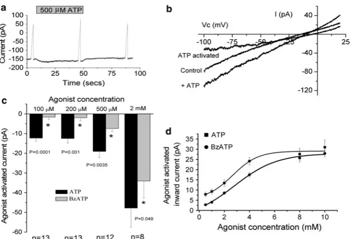
Both ATP and BzATP increased whole-cell currents at
-100 mV, although ATP gave a greater increase compared to BzATP. ATP increased whole-cell currents at all con-centrations tested (PB 0.001 for all). BzATP increased whole-cell currents at 2 mM (P=0.004) and 500lM (P=0.0004) but was without effect at 200lM (P=0.267) and 100lM (P=0.192). Figure1a, b shows typical traces from cells exposed to 500lM ATP. Mean agonist-activated currents are shown in Fig.1c. At all concentrations the response to ATP was greater than the response to BzATP (unpaired Student’st-test). The BzATP and ATP activated current did not demonstrate desen-sitization. The 2 mM BzATP-activated currents were
-12.5±1.67, -16.0±4.52 and -11.51±1.92 pA (n =13) for first, second and third exposures, respectively. The mean 500lM ATP activated currents were-10.6±
2.96, -8.77±1.78 and -9.08±1.23 pA (n=13) for first, second and third exposures, respectively. There was no significant difference between these: F2,36=0.69 and
F2,36=0.23, for BzATP and ATP, respectively (ANOVAs).
There was also no correlation between current activation and exposure number (r2=0.04 andr2=0.08).
ATP activated the whole-cell current in a dose-depen-dent manner (Fig. 1d). Half-maximal activation was observed with 2.77±0.24 mM ATP (n =9), with a Hill coefficient of 3.89±0.57 and maximal current of
230.1±3.68 pA (r2=0.992±0.002). BzATP also activated the currents in a dose-dependent manner. Half-maximal activation was observed with 4.00 ±0.38 mM BzATP (n =7), with a Hill coefficient of 2.00 ±0.07 and maximal current of 232.1±2.31 pA (r2=0.997±
0.001). The ATP concentration required for half-maximal activation was significantly smaller than the half-maximal concentration of BzATP (P=0.01, unpaired Student’s
t-test). There was no significant difference between the maximal current activated by either agonist (P=0.62, unpaired Student’st-test).
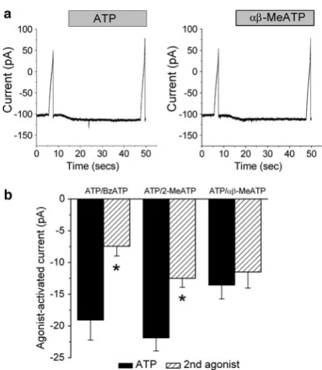
Agonist Potency and Effect of Antagonists
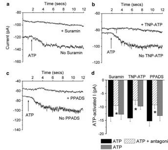
At 500lM, ATP, BzATP, 2-MeSATP andab-MeATP all increased whole-cell currents (PB0.0001). In paired cells the effect of ATP on whole-cell current was greater than the effect of either BzATP (n=12,P=0.003) or 2-MeSATP (n =24, P\0.0005) (Fig.2). In contrast, ATP and ab-MeATP increased currents by comparable amounts (n =13,P=0.30) (Fig.2). The agonist potency sequence was ATP=ab-MeATP[BzATP=2-MeSATP. In the presence of 100lM suramin (n=16), 30 nM TNP-ATP (n =16) or 10 lM PPDAS (n=21), the ATP-activated current was inhibited by 30%, 44% and 34%, respectively (P\0.008 for all) (Fig.3). The inhibition by suramin and PPADS was reversible, but that by TNP-ATP was not. It has previously been demonstrated that inhibition by PPADS can be increased after incubation for 10 min. However, in paired cells the ATP-activated current in the presence of PPADS at 1 min, -9.00±1.82 pA (n=9), was not sig-nificantly different from that at 10 min, -10.2±4.12 pA (n =9, P =0.65). PPADS (100lM) inhibited the ATP-activated current by 65%, -11.2±1.91 pA (n=9) vs.
-3.94±1.12 pA, in the absence and presence of PPADS, respectively (P=0.01). On wash the response to ATP recovered, -11.1±1.64 pA. P2X7 antagonists were
respectively (P=0.91). In the second set of experiments, BzATP increased whole-cell current by-32.0±7.29 pA (n=6). In the presence of KN-62 this was unchanged,
-27.8±7.24 pA (P=0.27).
Current Properties and Relative Permeability of ATP-Activated Current
Figure4a shows typical traces obtained in the absence and presence of 2 mM ATP. Addition of ATP to the bath increased whole-cell currents (Fig.4) (P\0.001). The inward chord conductance under the control circumstance was 30.2±2.66lS/cm2 (n =14). On addition of ATP this increased to 39.7±3.18lS/cm2, a mean increase of 9.53±0.90lS/cm2. The ATP-activated point conduc-tance from these 14 cells is shown in Fig.4c. Conductance and potential demonstrated a significant negative correla-tion (r2= -0.86, P\0.05), indicating that the ATP-activated current demonstrated inward rectification. The ATP-activated current was potentiated by 1-min incubation with 10lM ivermectin. Initially, ATP increased the cur-rent at -100 mV from -710±216 to -809±232 pA (n=7). However, after incubation of the same patches in ivermectin, the current increased from-657±220 to
-785 ±239 pA (n=7). The mean ATP-activated current
in the presence of ivermectin, which enhances the activa-tion of P2X4, was significantly greater,-98.1±23.2 vs.
-127±23.1 pA (n=7, P=0.025), in the absence and presence of ivermectin, respectively.
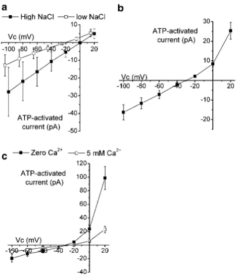
With 100 mM NaCl in the bath, the mean Vrev of the
ATP-activated current was ?1.29±2.01 mV (n=10). On reducing bath NaCl to 20 mM NaCl, theVrevshifted to 224.4±3.26 mV (P\0.001), a mean shift of225.6±
2.27 mV. This corresponded to a cation to anion perme-ability ratio of 15.5±6.75 (Fig.5a). Substitution of NaCl for RbCl or CsCl shifted the Vrev by ?5.65±1.35 mV
(n =9, P=0.002) and ?4.22±1.04 mV (n=8, P=
0.003) with RbCl and CsCl, respectively. LiCl was without effect onVrev,?1.37±2.14 mV (n=8,P=0.52). These
shifts corresponded to cation to Na? selectivity ratios of 1.28±0.07, 1.22±0.04 and 1.08±0.10, for Rb?, Cs? and Li?, respectively. TheVrevof the ATP-activated current with Na? in the pipette and NMDG? in the bath was
229.4±1.62 mV (n=7) (Fig.5b). This corresponded to an NMDG?:Na?permeability ratio of 0.32±0.02. Addition of 5 mM Ca2?to the extracellular solution shifted theVrevby
?5.72±1.90 mV (n=7, P=0.017) (Fig.5c). This corresponded to a Ca2?:Na? permeability ratio of 6.56±2.98. These data correspond to a selectivity sequence of Ca2?[Rb?=Cs?[Na?=Li?[NMDG?.
Fig. 1 Effect of different concentrations of ATP and BzATP. Clamp potential was held at-100 mV. At three time points (control, plus ATP and wash) a ramp protocol was run between-100 and?20 mV. aA typical trace (current in picoamperes against time in seconds), showing the effect of 500lM ATP.Solid barindicates when ATP was present in the bath.b Typical I–V traces showing the current recorded under the control circumstance, in the presence of 500lM ATP and the ATP-activated current. Vc is the command voltage.
[image:5.595.118.479.54.299.2]Effect of Extracellular Cations
Zn2? increased the ATP-activated conductance (GATP)
(n =10,P=0.004) (Fig.6). The Zn2?-mediated enhance-ment ofGATP was reversible, withGATPreturning to
pre-Zn2?levels on washout. In the Cu2?experiments the effect of ATP was attenuated (n =7, P=0.003) (Fig.6). The Cu2?-mediated inhibition of GATP was reversible, with GATP retuning to pre-Cu2? levels on washout. With
0.5 mM Ca2? in the bath GATP was 6.24±0.74lS/cm2
(n=16). This was significantly reduced when bath Ca2? was increased to 2 mM (P=0.03) (Fig.6). However, this reduction inGATP was not reversible. Acidification
inhib-ited, while alkalinization activated, GATP (mean data
shown in Table1).
Effect of P2X Activation on Whole-Cell K? Conductance
Whole-cell potential was clamped at 240 mV and then stepped to between?20 and2100 mV in220-mV steps. In control cells at time zero, quinidine inhibited a whole-cell current,IQuin(Fig.7a) (P\0.001). The quinidine-sensitive
outward and inward conductances (GQout andGQin) were
7.65±1.09 and 12.2±1.56lS/cm2, respectively (n =15). TheVrevofIQuinwas241.4±3.84 mV (Fig.7e), consistent with K?-selective currents. However, IQuin
decreased after 5 min (Fig. 7b).GQoutandGQinwere
sig-nificantly reduced at 5.26±0.60 and 7.38±0.99lS/cm2, respectively (n =15,P\0.001). TheVrevwas unchanged, 239.16±4.98 mV (P=0.46) (Fig.7e). In a separate population of test cells the responses to quinidine before and after the addition of ATP were determined. The initialGQout
and GQin in the absence of ATP were 13.2±2.18 and
23.6±3.73lS/cm2, respectively (n =17). After 5-min exposure to ATP, these were unchanged,GQout9.5±1.47
and GQin 22.3 ±3.50lS/cm2 (P[0.12) (Fig.7f). The Vrev of IQuin was 242.3±4.07 and 241.8±6.49 mV,
initially and after 5-min exposure to ATP, respectively (P=0.93) (Fig.7f). Addition of ATP to the extracellular solution increased the outward and inward conductances by 8.26±1.67 and 15.7±2.20lS/cm2, respectively, after 5 min (n =17,P\0.0001) (Fig.7c). TheVrevof the
ATP-activated current was 225.7 ±4.11 mV (Fig.7d). This was significantly depolarized in comparison to theVrev of IQuin(P=0.01).
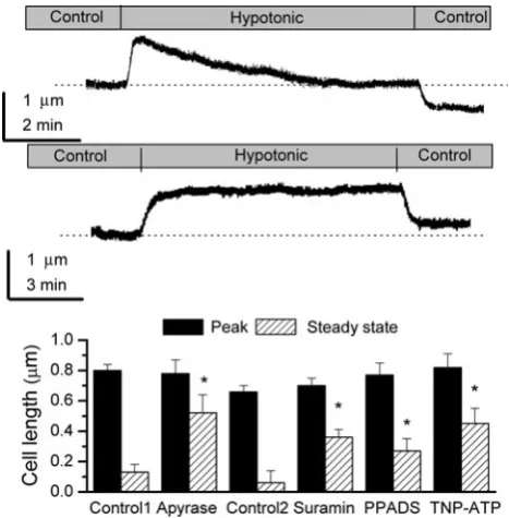
Role of P2X Receptors in Volume Regulation
Hypotonic shock elicited two responses, as described previ-ously (Robson and Hunter 1994c). In 29 cells (42%), cell length increased to a peak, with recovery observed when cells were placed back in control Ringer. These were designated ‘‘non-RVD’’ cells (Fig.8a, lower trace) and represent cells that require HCO3-for regulatory volume decrease (RVD)
(Robson and Hunter1994c). These cells were not considered further in the context of this study. In the remaining 40 cells (58%) cell length increased to a peak, followed by recovery toward the preshock level. These were designated ‘‘RVD’’ cells (Fig.8a, upper trace). The initial length of the RVD cells was 21.2±0.34lm (n =40). Hypotonic shock increased this by 0.80±0.04lm (P\0.0001), followed by recovery. Steady-state length was 0.13±0.05lm above the preshock level at steady state after volume regulation.
A possible role for extracellular ATP in RVD was examined by exposing cells to ATP/ADP apyrase. Neither the proportion of cells undergoing RVD (64%, n=7, Fisher’s exact probability test) nor the initial length of the cells (22.4±0.76lm, n =7, unpaired Student’s t-test) was different from control RVD cells. However, ATP/ADP apyrase inhibited RVD in comparison to control cells (unpaired Student’st-test). Figure8b shows the increase to peak and steady-state length after RVD relative to initial length for apyrase and day-matched controls (control 1).
In a second series of experiments, two types of response were observed in control cells, as described previously,
[image:6.595.52.286.52.321.2]RVD (47%) and non-RVD cells (53%). In RVD cells initial length was 22.2±0.46lm (n =22). On exposure to a hypotonic shock, length increased by 0.66±0.04 lm, to a
peak of 22.9 ±0.48lm (P\0.001), followed by RVD. Length was 0.06 ±0.08lm above the preshock level at steady state after volume regulation. Under the experi-mental conditions the proportions of cells undergoing RVD were 75% (21) for 100lM suramin, 63% (12) for 10lM PPADS and 61% (14) for 30 nM TNP-ATP. The number of RVD cells was significantly increased for suramin but not PPADS or TNP-ATP (Fisher’s exact probability test). The initial lengths of RVD cells were 22.9±0.47 (n=21), 21.7±1.06 and 22.2±0.54 (n=14)lm in the presence of suramin, PPADS and TNP-ATP, respectively. Figure8b shows the increase to peak and steady-state length after RVD relative to initial length for all experimental condi-tions. Suramin, TNP-ATP and PPADS all inhibited RVD in comparison to the control circumstance, F3,65=5.57 (all
tested using ANOVA).
Discussion
These data provide evidence for the functional expression of a P2X-like current in frog renal proximal tubule cells, P2Xf. P2Xfwas cation-selective, did not discriminate well
between monovalent cations, had a poor permeability to NMDG and was around six times more permeable for Ca2? over Na?. It was activated by a variety of P2X agonists,
Fig. 3 Effect of antagonists on the 500lM ATP-activated current. Cells were held at -100 mV and then exposed to 500lM ATP in the absence and then the presence of the antagonist. This was followed by a second control exposure to ATP after washout of the antagonist.a–cTypical whole-cell traces at-100 mV from three different cells in the absence and presence of 100lM suramin, 30 nM TNP-ATP or 10lM PPADS, respectively.Arrowsindicate when 500lM ATP was added to the bath.dMean ATP-activated currents. * Significant reduction compared to the first response to ATP.Black bars indicate ATP,hatched bars indicate ATP plus antagonist andgray barsindicate ATP
Fig. 4 Effect of ATP on whole-cell currents. Cells were clamped at
-40 mV and then potential stepped to between?20 and-100 mV in
-20-mV steps. aTypical traces obtained from the same cell in the
[image:7.595.192.537.55.363.2] [image:7.595.51.289.389.592.2]including ATP, BzATP, 2MeSATP and ab-MeATP, and was sensitive to some P2X antagonists. P2Xfalso
demon-strated inward rectification, a property shared by some P2X receptors (Evans et al. 1996). These properties are con-sistent with P2Xf being attributable to a P2X receptor.
However, one difference from P2X receptors was that it was activated only by high concentrations of extracellular ATP, 100lM to 10 mM. In contrast, most of the cloned P2X receptors require only micromolar levels of ATP (Torres et al. 1998; Valera et al. 1994; Virginio et al.
1998b), although P2X7is activated by high levels of ATP
(Rassendren et al. 1997). Consistent with LLC-PK1 cells
(Filipovic et al. 1998), activation of P2Xf was observed
with 100 lM ATP, although maximal activation required millimolar levels. The reason for this difference is not clear, but it may reflect the fact that the current study involves an amphibian P2X receptor. Certainly, a study on cloned Xenopus P2X4 also used high concentrations of
ATP (Juranka et al.2001). In addition, frog P2X receptors in aorta require similarly high concentrations of agonists for activation (Knight and Burnstock1996), with maximal activation not observed with 3 mM ATP. Alternatively, it is known that the proximal tubule membrane contains ecto-ATPases that break down ATP (Huang et al. 2006). Therefore, another explanation could be that the ATP in the extracellular solution next to the membrane was at a lower concentration than the bulk solution. The Hill coefficients for ATP and BzATP were similar to other studies (Jiang et al.2003), with the larger coefficient for ATP suggesting greater cooperativity of binding compared to BzATP. In terms of the in vivo luminal ATP concentration in the proximal tubule, there is a general lack of information, with one study suggesting a maximal concentration of around 275 nmol/l in rats (Vekaria et al.2006). This concentration would not be sufficient to activate P2Xfand, indeed, is also
on the low side for activation of mammalian P2X recep-tors. However, this concentration reflects the mean in the tubular fluid, and one suggestion is that ATP concentra-tions closer to the membrane could be much higher. If this is the case, then it is possible that sufficiently high con-centrations are reached for activation of both mammalian and amphibian P2X receptors. In addition, as discussed later, it is clear that activation of P2Xfcan impact on the
physiological function of the frog renal proximal tubule. What are the properties of P2Xf, and how do these
compare to the different cloned P2X receptors? Two amphibian cloned receptors have been identified and show 67% homology with rat P2X4and P2X5(Jensik et al.2001;
Juranka et al.2001). P2Xfshows some similarities to these
cloned receptors and to some heteromeric mammalian receptors (Table2). The two amphibian P2X receptors can be activated by levels of ATP, below 100lM. This is
Fig. 5 Selectivity of ATP-activated current. Cells were clamped at
-40 mV and then potential stepped to between?20 and-100 mV in
-20-mV steps.Vcis the command voltage.aMean currents recorded
in high (filled square) or low (open square) NaCl in paired cells (n=10). Note the shift along thexaxis of the low-NaCl data due to junction potential correction.bMean ATP-activated current recorded with NaCl in the pipette and NMDG-Cl in the bath (n=7).cMean ATP-activated currents recorded with bath NMDG-Cl in the absence of Ca2? (filled square) or in the presence of 5 mM Ca2? (open square) in paired cells (n=7)
Fig. 6 Effect of divalent cations on GATP. Cells were clamped at
-40 mV and then potential stepped to between?20 and-100 mV in
-20-mV steps. The graph shows the effect of Zn2?, Cu2?and Ca2?.
[image:8.595.53.288.55.329.2] [image:8.595.68.269.478.636.2]different from P2Xf, where 100lM ATP was needed for
activation. However, as neither study performed a dose response to ATP, a definitive comparison cannot be made.
In divalent free conditions neither cloned P2X receptor demonstrated rapid desensitization on exposure to ATP, similar to P2Xf. However, although desensitization was not
observed with P2Xfwhen bath Ca2?was low, an apparent
desensitization was observed when the cells were exposed to 2 mM Ca2?. Under this circumstance poor recovery of the response to ATP was observed on washout of Ca2? from the bath. This decrease in the response to ATP during several exposures was not observed with low Ca2?and is consistent with the Ca2?-dependent desensitization observed in amphibian P2X receptors. The agonist potency sequence of P2Xf, ATP =abmeATP[BzATP=
2Me-SATP, is similar to the frog aorta P2X receptor (Knight and Burnstock1996). This suggests that P2Xfis not attributable
to P2X7(Rassendren et al.1997), although variations in the
response to BzATP are seen at P2X7receptors from
[image:9.595.53.289.129.508.2]dif-ferent species (Fonfria et al. 2008). It is therefore not
Table 1 Effect of extracellular pH on the ATP-activated current
* Significant difference from pH 7.4 (P\0.029)
IATP-100 mV (pA) (n=9) IATP-100 mV (pA) (n=9)
pH 7.4 -21.4±2.94 pH 7.4 -19.0±4.03
pH 6.3 -12.2±2.73* pH 8.3 -30.2±6.45*
pH 7.4 -20.1±4.23 pH 7.4 -24.8±5.01
Fig. 7 Effect of quinidine and ATP on whole-cell currents. Cells were clamped at-40 mV and then potential stepped to between?20
and-100 mV in -20-mV steps.Vc is the command voltage.a, b
Mean whole-cell currents recorded in the absence of ATP initially on achieving the whole-cell configuration (a) and after 5 min (b). In both figuresfilled square indicates currents recorded in the absence of quinidine, while filled circle indicates currents recorded in the presence of quinidine (n=15 for all). c Effect of addition of extracellular ATP on mean whole-cell current.Filled square, currents recorded in the absence of ATP;open circle, currents recorded after 5-min exposure to ATP (n=17).dMean ATP-activated current.e, f Quinidine-sensitive currents in the absence of ATP (e) or presence of ATP (f). In both graphs filled square indicates currents recorded initially after achieving the whole-cell configuration, while filled circleindicates currents recorded 5 min later
[image:9.595.308.542.156.394.2]possible to absolutely rule out P2Xfbeing attributable to a
P2X7-like receptor, although the P2X7antagonists KN-62
and BBG were without effect on P2Xf(Humphreys et al. 1998; Jiang et al. 2000). P2Xf was sensitive to 100lM
suramin, 10lM PPADS and 30 nM TNP-ATP (30%, 44% and 34% inhibition, respectively). This sensitivity to PPADS is similar to amphibian P2X4 (50% inhibition)
(Juranka et al.2001) and P2X4/6(40% inhibition) (Le et al. 1998), although it is different from the bullfrog P2X receptor, which is completely blocked by both 100lM suramin and PPADS (Jensik et al.2001). The sensitivity to PPADS was similar to amphibian P2X4and P2X4/6.
TNP-ATP is a potent inhibitor of P2X1/5, P2X1/4and P2X2/3(Le
et al.1999; Nicke et al.2005; Virginio et al.1998b), with *80% inhibition observed with 30 nM. This is higher than the sensitivity of P2Xf. Like P2Xf, the bullfrog cloned
receptor also shows inhibition by Ca2?(Jensik et al.2001). P2Xf was potentiated by ivermectin, which is known to
enhance activation of P2X4(Priel and Silberberg2004) and
was also potentiated by extracellular Zn2?and inhibited by extracellular Cu2?and H?.
Overall, P2Xfwould appear to share greatest similarity
with P2X2/3 or P2X4/6, although its properties are not
entirely consistent with these heteromeric P2X receptors. Given the fact that previous expression studies have shown the presence of P2X1, P2X4, P2X5and P2X6in the renal
proximal tubule, it is likely that P2Xfrepresents a P2X4/6
-like P2X receptor.
P2Xf is clearly a native proximal tubule P2X-like
receptor, and on activation it would be expected to lead to an influx of Ca2?into the cell. However, the magnitude of the P2Xf-mediated currents was small. In comparison to
P2X receptor expression studies this is to be expected as in those studies the magnitude of the P2X currents reflects the fact that the channels have been overexpressed. Looking at studies of native P2X receptor currents provides a variable
picture in terms of current magnitude. In LLC-PK1cells,
currents were on the order of a few hundred picoamperes (Filipovic et al. 1998). In contrast, studies in neurons, airway ciliated cells and Leydig cells show small P2X-mediated currents, similar to P2Xf(Chaves et al.2006; Ma
et al. 2006; Mori et al. 2001). It is clear, therefore, that physiologically relevant P2X native currents can be small. What is the physiological role of P2Xfin the renal
proxi-mal tubule? The data presented in the current study suggest that P2Xfplays an important role in volume regulation and
K?channel activation.
Frog proximal tubule cells have the ability to regulate their volume in response to cell swelling, RVD (Robson and Hunter 1994c). Such volume regulation plays an important role in diverse cellular process such as cell growth and proliferation, osmoregulation and cellular metabolism. There is a clear role for P2 activation in reg-ulating proximal tubule cell proliferation (Lee and Han
[image:10.595.53.545.72.210.2]2006), while a role for P2 receptor activation in RVD in hepatocytes has been proposed (Wang et al. 1996). This suggests that P2 receptors and volume regulation may be important for normal cell function. Previous work in frog proximal cells has demonstrated a role for K? and Cl -channels in RVD (Robson and Hunter 1994c, 2005). Whole-cell patch-clamp experiments have identified bar-ium-sensitive K?and DIDS-sensitive Cl-currents that are volume sensitive (Robson and Hunter 1994a, 2005). Unpublished studies demonstrate that quinidine-sensitive K? currents are also volume-sensitive, with quinidine-sensitive conductance reduced in the presence of a hyper-tonic bath solution (19.2±4.38 vs. 7.31 ±1.46lS/cm2in the presence of control and hypertonic solutions, respec-tively, n=12 for each group). For the Cl-channels vol-ume activation is mediated by protein kinase C (PKC) (Robson and Hunter 1994a). The mechanism underlying volume regulation of the K? currents has not been
Table 2 Comparison of the properties of P2Xfwith cloned amphibian and mammalian heteromeric receptors
Amphibian P2X4 Amphibian P2X5 P2X2/3 P2X1/4 P2X1/5 P2X4/6 References
Agonist potency X r r r X Jensik et al. (2001)
Juranka et al. (2001) Le et al. (1998) Le et al. (1999) Liu et al. (2001) Nicke et al. (2005) Stoop et al. (1997) Virginio et al. (1998a) Wildman et al. (1998)
Suramin [ [ [ [ r
40%
PPADS r [ [ [ [ r
TNP-ATP [ [ [
Zn2? r r
Ca2? r r X
pH X r r
Percentages indicate percentage inhibition values close to those observed for P2Xf
elucidated but does not appear to involve PKC. In addition, RVD is inhibited in the absence of extracellular Ca2?, consistent with a Ca2? influx pathway playing a critical role (Robson and Hunter1994c). The specific mechanism by which Ca2? enters the renal proximal is unknown; however, a number of candidates have been proposed. One of these is stretch-activated, Ca2?-permeable cation chan-nels (SACs), as the SAC inhibitor gadolinium (Gd3?) blocks RVD (Robson and Hunter1994c) and two Gd3?and volume-sensitive cation conductances have been identified in these cells (Robson and Hunter1994b). However, sub-sequent work has indicated that the volume-sensitive Cl -channels are also inhibited by Gd3? (Robson and Hunter
1994a). Therefore, the effect of Gd3? on RVD could simply reflect inhibition of Cl- efflux rather than Ca2? influx. A second possibility is that, on cell swelling, there is release of ATP from the cells, with ATP subsequently leading to Ca2?influx via the activation of P2X receptors. This is supported by the current study, which demonstrated that in the presence of apyrase RVD was inhibited, sug-gesting that the release and presence of ATP are important in initiating volume regulation. RVD was also inhibited by the P2X antagonists suramin, TNP-ATP and PPADS. This supports a role for P2X receptor activation in RVD. The degree of inhibition of RVD was similar to that observed with P2Xf.
These data support a role for P2Xf activation in RVD
and suggest that it may provide the Ca2?influx pathway. This influx of Ca2?would be expected to activate down-stream efflux pathways, such as the K?and Cl-channels described earlier. A clear link exists to activation of the Cl-channels as this is PKC-mediated. For the activation of K? channels a rise in intracellular Ca2? could directly activate channels or work indirectly via Ca2?-dependent signalling systems. The whole-cell K? current data described here suggest that P2Xfplays an important role in
the activation of quinidine-sensitive K? channels previ-ously observed in the cells. In the absence of extracellular ATP, whole-cell quinidine-sensitive K?currents decreased over 5 min. However, in the presence of extracellular ATP this rundown was absent, indicating that extracellular ATP was able to inhibit the rundown process. TheVrev of the
quinidine-sensitive currents, around 240 mV, suggests that the K?channel regulated by extracellular ATP was a previously identified K?conductance (Robson and Hunter
1997). The total ATP activated current demonstrated inward rectification, similar to P2Xf. Interestingly, theVrev
of the total ATP-activated current was more positive than the K?currents but more negative than theVrevfor P2Xf.
This suggests that at least part of the ATP-activated current may reflect activation of K?-selective channels. Activation of K? channels via P2X receptor activation has been observed in rat osteoclasts and toad gastric smooth muscle
cells (Weidema et al. 1997; Zou et al. 2001), with P2Y2
-mediated inhibition of K?channels in the mouse cortical collecting duct (Lu et al. 2000).
In conclusion, the current study provides the first report of a native P2X receptor in renal proximal tubule cells. The receptor, P2Xf, was cation-selective, did not discriminate
between cations and was Ca2?-permeable. P2Xfwas
acti-vated by the purines ATP=abmeATP[BzATP=
2MeSATP, did not demonstrate fast desensitization and was inhibited by suramin, PPADS and TNP-ATP. P2Xf
-mediated currents were enhanced in the presence of Zn2? or ivermectin and inhibited in the presence of Cu2?or on acidification. These properties are consistent with P2Xf
being attributable to a P2X receptor and suggest that P2Xf
may be attributable to a heteromeric receptor, with P2X4/6
a possible candidate. The evidence presented suggests that activation of P2Xf plays a role in the regulation of cell
volume and K? channels in frog renal proximal tubule cells.
Acknowledgement This work was funded by the Wellcome Trust. J. P. D. was supported by a University of Sheffield studentship.
Open Access This article is distributed under the terms of the Creative Commons Attribution Noncommercial License which per-mits any noncommercial use, distribution, and reproduction in any medium, provided the original author(s) and source are credited.
References
Bailey MA (2004) Inhibition of bicarbonate reabsorption in the rat proximal tubule by activation of luminal P2Y1 receptors. Am J Physiol Renal Physiol 287:F789–F796
Bailey MA, Imbert-Teboul M, Turner C, Srai SK, Burnstock G, Unwin RJ (2001) Evidence for basolateral P2Y6 receptors along the rat proximal tubule: functional and molecular characteriza-tion. J Am Soc Nephrol 12:1640–1647
Bouyer P, Paulais M, Cougnon M, Hulin P, Anagnostopoulos T, Planelles G (1998) Extracellular ATP raises cytosolic calcium and activates basolateral chloride conductance in Necturus proximal tubule. J Physiol 510:535–548
Burnstock G, Kennedy C (1985) Is there a basis for distinguishing two types of P2-purinoceptor? Gen Pharmacol 16:433–440 Cha SH, Sekine T, Endou H (1998) P2 purinoceptor localization
along rat nephron and evidence suggesting existence of subtypes P2Y1 and P2Y2. Am J Physiol Renal Physiol 274:F1006–F1014 Chan CM, Unwin RJ, Bardini M, Oglesby IB, Ford APDW, Townsend-Nicholson A, Burnstock G (1998) Localization of P2X1 purinoceptors by autoradiography and immunohistochem-istry in rat kidneys. Am J Physiol Renal Physiol 274:F799–F804 Chaves LAP, Pontelli EP, Varanda WA (2006) P2X receptors in mouse Leydig cells. Am J Physiol Cell Physiol 290:C1009– C1017
Filipovic DM, Adebanjo OA, Zaidi M, Reeves WB (1998) Functional and molecular evidence for P2X receptors in LLC-PK1 cells. Am J Physiol Renal Physiol 274:F1070–F1077
Fonfria E, Levy DS, Goodwin JA, Roman S, Smith GD, Condreay JP, Michel AD (2008) Cloning and pharmacological characteriza-tion of the guinea pig P2X7 receptor orthologue. Br J Pharmacol 153:544–556
Guo C, Masin M, Qureshi OS, Murrell-Lagnado RD (2007) Evidence for functional P2X4/P2X7 heteromeric receptors. Mol Pharma-col 72:1447–1456
Hamill OP, Marty A, Neher E, Sakmann B, Sigworth FJ (1981) Improved patch-clamp techniques for high-resolution current recording from cells and cell-free membrane patches. Pflugers Arch 391:85–100
Huang DY, Vallon V, Zimmermann H, Koszalka P, Schrader J, Osswald H (2006) Ecto-50-nucleotidase (cd73)-dependent and -independent generation of adenosine participates in the medi-ation of tubuloglomerular feedback in vivo. Am J Physiol Renal Physiol 291:F282–F288
Humphreys BD, Virginio C, Surprenant A, Rice J, Dubyak GR (1998) Isoquinolines as antagonists of the P2X7 nucleotide receptor: high selectivity for the human versus rat receptor homologues. Mol Pharmacol 54:22–32
Hunter M (1989) Isolation of single proximal cells from frog kidneys. J Physiol 416:13P
Jensen MEJ, Odgaard E, Christensen MH, Praetorius HA, Leipziger J (2007) Flow-induced [Ca2?]
i increase depends on nucleotide
release and subsequent purinergic signaling in the intact nephron. J Am Soc Nephrol 18:2062–2070
Jensik PJ, Holbird D, Collard MW, Cox TC (2001) Cloning and characterization of a functional P2X receptor from larval bullfrog skin. Am J Physiol Cell Physiol 281:C954–C962 Jiang L-H, Mackenzie AB, North RA, Surprenant A (2000) Brilliant
blue G selectively blocks ATP-gated rat P2X7 receptors. Mol Pharmacol 58:82–88
Jiang L-H, Kim M, Spelta V, Bo X, Surprenant A, North RA (2003) Subunit arrangement in P2X receptors. J Neurosci 23:8903–8910 Juranka PF, Haghighi AP, Gaertner T, Cooper E, Morris CE (2001) Molecular cloning and functional expression ofXenopus laevis oocyte ATP-activated P2X4 channels. Biochim Biophys Acta 1512:111–124
Knight GE, Burnstock G (1996) The effects of purine compounds on the isolated aorta of the frogRana temporaria. Br J Pharmacol 117:873–878
Laycock S, Taylor HC, Haigh C, Lee AT, Cooper GJ, Ong ACM, Robson L (2009) A novel dephosphorylation-activated conduc-tance in a mouse renal collecting duct cell line. Exp Physiol 94:914–927
Le K-T, Babinski K, Seguela P (1998) Central P2X4 and P2X6 channel subunits coassemble into a novel heteromeric ATP receptor. J Neurosci 18:7152–7159
Le K-T, Boue-Grabot E, Archambault V, Seguela P (1999) Functional and biochemical evidence for heteromeric ATP-gated channels composed of P2X1 and P2X5 subunits. J Biol Chem 274:15415–15419
Lee YJ, Han HJ (2006) Role of ATP in DNA synthesis of renal proximal tubule cells: involvement of calcium, MAPKs, and CDKs. Am J Physiol Renal Physiol 291:F98–F106
Lee YJ, Park SH, Han HJ (2005) ATP stimulates Na?-glucose cotransporter activity via cAMP and p38 MAPK in renal proximal tubule cells. Am J Physiol Cell Physiol 289:C1268–C1276 Leipziger J, Unwin RJ (2003) Purinergic receptors in the kidney. In:
Schwiebert EM (ed) Purinergic receptors and signalling. Aca-demic Press, San Diego, pp 369–394
Lewis C, Neidhart S, Holy C, North RA, Buell G, Surprenant A (1995) Coexpression of P2X2 and P2X3 receptor subunits can
account for ATP-gated currents in sensory neurons. Nature 377:432–435
Liu M, King BF, Dunn PM, Rong W, Townsend-Nicholson A, Burnstock G (2001) Coexpression of P2X3 and P2X2 receptor subunits in varying amounts generates heterogeneous popula-tions of P2X receptors that evoke a spectrum of agonist responses comparable to that seen in sensory neurons. J Pharma-col Exp Ther 296:1043–1050
Liu R, Bell PD, Peti-Peterdi J, Kovacs G, Johansson A, Persson AEG (2002) Purinergic receptor signaling at the basolateral membrane of macula densa cells. J Am Soc Nephrol 13:1145–1151 Lu M, MacGregor GG, Wang W, Giebisch G (2000) Extracellular
ATP inhibits the small-conductance K channel on the apical membrane of the cortical collecting duct from mouse kidney. J Gen Physiol 116:299–310
Ma W, Korngreen A, Weil S, Cohen EB-T, Priel A, Kuzin L, Silberberg SD (2006) Pore properties and pharmacological features of the P2X receptor channel in airway ciliated cells. J Physiol 571:503–517
McCoy DE, Taylor AL, Kudlow BA, Karlson K, Slattery MJ, Schwiebert LM, Schwiebert EM, Stanton BA (1999) Nucleotides regulate NaCl transport in mIMCD-K2 cells via P2X and P2Y purinergic receptors. Am J Physiol Renal Physiol 277:F552– F559
Mori M, Heuss C, Gahwiler BH, Gerber U (2001) Fast synaptic transmission mediated by P2X receptors in CA3 pyramidal cells of rat hippocampal slice cultures. J Physiol 535:115–123 Mounfield PR, Robson L (1998) The role of Ca2? in volume
regulation induced by Na?-coupled alanine uptake in single proximal tubule cells isolated from frog kidney. J Physiol 510:145–153
Nicke A, Kerschensteiner D, Soto F (2005) Biochemical and functional evidence for heteromeric assembly of P2X1 and P2X4 subunits. J Neurochem 92:925–933
North RA, Barnard EA (1997) Nucleotide receptors. Curr Opin Neurobiol 7:346–357
Pochynyuk O, Bugaj V, Rieg T, Insel PA, Mironova E, Vallon V, Stockand JD (2008) Paracrine regulation of the epithelial Na? channel in the mammalian collecting duct by purinergic P2Y2 receptor tone. J Biol Chem 283:36599–36607
Priel A, Silberberg SD (2004) Mechanism of ivermectin facilitation of human P2X4 receptor channels. J Gen Physiol 123:281–293 Radford KM, Virginio C, Surprenant A, North RA, Kawashima E
(1997) Baculovirus expression provides direct evidence for heteromeric assembly of P2X2 and P2X3 receptors. J Neurosci 17:6529–6533
Rassendren F, Buell GN, Virginio C, Collo G, North RA, Surprenant A (1997) The permeabilizing ATP receptor, P2X7. Cloning and expression of a human cDNA. J Biol Chem 272:5482–5486 Robson L, Hunter M (1994a) Role of cell volume and protein kinase
C in regulation of a Cl-conductance in single proximal tubule cells ofRana temporaria. J Physiol 480:1–7
Robson L, Hunter M (1994b) Volume-activated, gadolinium-sensitive whole-cell currents in single proximal cells of frog kidney. Pflugers Arch 429:98–106
Robson L, Hunter M (1994c) Volume regulatory responses in frog isolated proximal cells. Pflugers Arch 428:60–68
Robson L, Hunter M (1997) Two K?-selective conductances in single proximal tubule cells isolated from frog kidney are regulated by ATP. J Physiol 500:605–616
Solini A, Santini E, Chimenti D, Chiozzi P, Pratesi F, Cuccato S, Falzoni S, Lupi R, Ferrannini E, Pugliese G, Virgilio FD (2007) Multiple P2X receptors are involved in the modulation of apoptosis in human mesangial cells: evidence for a role of P2X4. Am J Physiol Renal Physiol 292:F1537–F1547
Stoop R, Surprenant A, North RA (1997) Different sensitivities to pH of ATP-induced currents at four cloned P2X receptors. J Neuro-physiol 78:1837–1840
Takeda M, Kobayashi M, Endou H (1998) Establishment of a mouse clonal early proximal tubule cell line and outer medullary collecting duct cells expressing P2 purinoceptors. Biochem Mol Biol Int 44:657–664
Torres GE, Haines WR, Egan TM, Voigt MM (1998) Co-expression of P2X1 and P2X5 receptor subunits reveals a novel ATP-gated ion channel. Mol Pharmacol 54:989–993
Torres GE, Egan TM, Voigt MM (1999) Hetero-oligomeric assembly of P2X receptor subunits. Specificities exist with regard to possible partners. J Biol Chem 274:6653–6659
Valera S, Hussy N, Evans RJ, Adami N, North RA, Surprenant A, Buell G (1994) A new class of ligand-gated ion channel defined by P2x receptor for extracellular ATP. Nature 371:516–519 Vekaria RM, Unwin RJ, Shirley DG (2006) Intraluminal ATP
concentrations in rat renal tubules. J Am Soc Nephrol 17:1841–1847
Virginio C, North RA, Surprenant A (1998a) Calcium permeability and block at homomeric and heteromeric P2X2 and P2X3 receptors, and P2X receptors in rat nodose neurons. J Physiol 510:27–35
Virginio C, Robertson G, Surprenant A, North RA (1998b) Trinitrophenyl-substituted nucleotides are potent antagonists selective for P2X1, P2X3, and heteromeric P2X2/3 receptors. Mol Pharmacol 53:969–973
Wang CZ, Namba N, Gonoi T, Inagaki N, Seino S (1996) Cloning and pharmacological characterization of a fourth P2X receptor subtype widely expressed in brain and peripheral tissues including various endocrine tissues. Biochem Biophys Res Commun 220:196–202
Weidema AF, Barbera J, Dixon SJ, Sims SM (1997) Extracellular nucleotides activate non-selective cation and Ca2?-dependent K?channels in rat osteoclasts. J Physiol 503:303–315 Wildman SS, King BF, Burnstock G (1998) Zn2?modulation of
ATP-responses at recombinant P2X2 receptors and its dependence on extracellular pH. Br J Pharmacol 123:1214–1220
Wildman SSP, Marks J, Turner CM, Yew-Booth L, Peppiatt-Wildman CM, King BF, Shirley DG, Wang W, Unwin RJ (2008) Sodium-dependent regulation of renal amiloride-sensitive currents by apical P2 receptors. J Am Soc Nephrol 19:731–742 Wildman SSP, Boone M, Peppiatt-Wildman CM, Contreras-Sanz A, King BF, Shirley DG, Deen PMT, Unwin RJ (2009) Nucleotides downregulate aquaporin 2 via activation of apical P2 receptors. J Am Soc Nephrol 20:1480–1490
Xia S-L, Wang L, Cash MN, Teng X, Schwalbe RA, Wingo CS (2004) Extracellular ATP-induced calcium signaling in mIMCD-3 cells requires both P2X and P2Y purinoceptors. Am J Physiol Renal Physiol 287:F204–F214
Zhang Y, Sanchez D, Gorelik J, Klenerman D, Lab M, Edwards C, Korchev Y (2007) Basolateral P2X4-like receptors regulate the extracellular ATP-stimulated epithelial Na?channel activity in renal epithelia. Am J Physiol Renal Physiol 292:F1734–F1740 Zou H, Ugur M, Drummond RM, Singer JJ (2001) Coupling of a