Bmp and Nodal Independently Regulate
lefty1
Expression to Maintain Unilateral Nodal Activity during
Left-Right Axis Specification in Zebrafish
Kelly A. Smith1.¤
, Emily Noe¨l1.
, Ingrid Thurlings1, Holger Rehmann2, Sonja Chocron1, Jeroen Bakkers1,3*
1Hubrecht Institute, Royal Netherlands Academy of Arts and Sciences (KNAW) and University Medical Center Utrecht, Utrecht, The Netherlands,2University Medical Center Utrecht, Utrecht, The Netherlands,3Interuniversity Cardiology Institute of the Netherlands, Utrecht, The Netherlands
Abstract
In vertebrates, left-right (LR) axis specification is determined by a ciliated structure in the posterior region of the embryo. Fluid flow in this ciliated structure is responsible for the induction of unilateral left-sided Nodal activity in the lateral plate mesoderm, which in turn regulates organ laterality. Bmp signalling activity has been implied in repressing Nodal expression on the right side, however its mechanism of action has been controversial. In a forward genetic screen for mutations that affect LR patterning, we identified the zebrafish linkspoot (lin) mutant, characterized by cardiac laterality and mild dorsoventral patterning defects. Mapping of the lin mutation revealed an inactivating missense mutation in the Bmp receptor 1aa(bmpr1aa) gene. Embryos with a mutation inlin/bmpr1aaand a novel mutation in its paralogue, bmpr1ab,
displayed a variety of dorsoventral and LR patterning defects with increasing severity corresponding with a decrease in
bmpr1adosage. In Bmpr1a-deficient embryos we observed bilateral expression of the Nodal-related gene,spaw, coupled with reduced expression of the Nodal-antagonistlefty1in the midline. Using genetic models to induce or repress Bmp activity in combination with Nodal inhibition or activation, we found that Bmp and Nodal regulatelefty1expression in the midline independently of each other. Furthermore, we observed that the regulation oflefty1by Bmp signalling is required for its observed downregulation of Nodal activity in the LPM providing a novel explanation for this phenomenon. From these results we propose a two-step model in which Bmp regulates LR patterning. Prior to the onset of nodal flow and Nodal activation, Bmp is required to inducelefty1expression in the midline. When nodal flow has been established and Nodal activity is apparent, both Nodal and Bmp independently are required forlefty1expression to assure unilateral Nodal activation and correct LR patterning.
Citation:Smith KA, Noe¨l E, Thurlings I, Rehmann H, Chocron S, et al. (2011) Bmp and Nodal Independently Regulatelefty1Expression to Maintain Unilateral Nodal Activity during Left-Right Axis Specification in Zebrafish. PLoS Genet 7(9): e1002289. doi:10.1371/journal.pgen.1002289
Editor:Mary C. Mullins, University of Pennsylvania School of Medicine, United States of America
ReceivedApril 7, 2011;AcceptedJuly 30, 2011;PublishedSeptember 29, 2011
Copyright:ß2011 Smith et al. This is an open-access article distributed under the terms of the Creative Commons Attribution License, which permits unrestricted use, distribution, and reproduction in any medium, provided the original author and source are credited.
Funding:KAS was supported by a Concordia fellowship from Stichting Vrienden van het Hubrecht (http://www.hubrecht.eu/information/foundation.html). Research in JB’s laboratory was supported by the Netherlands Organisation for Scientific Research (NWO/ALW) grant 864.08.009 (http://www.nwo.nl/nwohome. nsf/pages/SPPD_7AK3L2_Eng). The bmpr1absa0028 zebrafish mutant was generated as part of the ZF-MODELS Integrated Project in the 6th Framework Programme (Contract No. LSHG-CT-2003-503496) funded by the European Commission (http://cordis.europa.eu/fp6/dc/index.cfm?fuseaction = UserSite. FP6HomePage). The funders had no role in study design, data collection and analysis, decision to publish, or preparation of the manuscript.
Competing Interests:The authors have declared that no competing interests exist.
* E-mail: [email protected]
.These authors contributed equally to this work.
¤ Current address: Institute for Molecular Biology, Brisbane, Australia
Introduction
In vertebrates the internal organs are positioned asymmetrically along the left-right (LR) axis. For example, in humans, the heart is positioned on the left side, as is the stomach whilst the liver is positioned on the right side. Within organs LR asymmetry also exists. For example, the two lungs appear identical however they are divided into lobes with 3 on the right lung and 2 on the left. Animals with situs inversus totalis (a LR reversal of all organs) have no pathological features [1] however severe medical problems occur in infants with a partial reversal in a subset of organs (situs ambigious or heterotaxia). These heterotaxic phenotypes occur during early embryonic development and can have both genetic as well as environmental causes [2,3].
A ciliated organ at the posterior end of the embryo is required for LR-axis specification in the embryo. In this LR organ, the node
in mouse or Kupffer’s vesicle (KV) in zebrafish, cilia rotate and create a directional fluid flow from the right to left side of the embryo. This directional nodal flow induces a unilateral and asymmetric expression ofNodalin the left lateral plate mesoderm (LPM) directing organ laterality. Unilateral expression ofNodalis essential for correct LR-axis specification, a function that has been highly conserved from human to snails [2,4,5]. Although unilateral expression ofNodalis highly conserved and essential for LR–axis specification, there is still very little understanding of how this unilateral Nodal expression is initiated by nodal flow and maintained in the LPM.
which after associating with Smad4 protein is translocated to the nucleus to activate transcription of downstream target genes. Extracellular antagonists such as Lefty and Cerberus can inhibit Nodal signalling either by direct interaction with Nodal or by competing with Nodal for binding to the receptor. The activity of Lefty proteins, Lefty1 and Lefty2, is controlled at the level of transcription. In most tissues Lefty expression is dependent on Nodal signalling [6]. During LR-axis formation in mouse embryos Lefty1andLefty2have reciprocal expression patterns. WhileLefty1 is expressed strongly in the presumptive floor plate and only weakly in the left LPM,Lefty2is expressed strongly in the left LPM and only weakly in the presumptive floorplate [7]. During LR axis formation in zebrafish embryos lefty1 is expressed in the notochord. Only after LR patterning has been established are lefty1andlefty2expressed in the left cardiac field [8]. Nodal likely activates its own expression via a positive feedback loop while it also activates expression of its own antagonistsLefty1and Lefty2. Genetic experiments in mouse demonstrated that Lefty1 is the more important antagonist and is essential for LR-axis formation [9]. It is believed thatLefty1 expression in the midline prevents Nodal from crossing the midline, blocking activation of Nodal signalling in the right LPM. Indeed loss ofLefty1expression caused the ectopic expression of Nodaland other left-sided genes in the right LPM and resulted in various laterality defects. It has been suggested that Nodal and Lefty maintain the L/R asymmetry by a self-enhancement and lateral-inhibition (SELI) mechanism [10]. With the SELI model it is possible to explain how a small difference between two separated regions is converted into a robust difference through local activation and long-range inhibition [11].
Bmps have been implicated in LR patterning but data on their precise role has been contradictory [12–22]. This is partly due to Bmp ligands acting in opposite fashions, depending on the time and place of action during LR-axis specification [13,16]. Bmp proteins are members of the Tgf-ß superfamily of growth factors. Extracellular antagonists of Bmp signalling are Noggin, Chordin and Follistatin. Upon interaction with their serine/threonine
kinase type I and II Bmp receptors, Bmp ligands induce intracellular phosphorylation of Smad1, 5 or 8 proteins [23]. Mouse embryos deficient for the type I Bmp receptor Bmpr1a/ Alk3 or Acvr1/Alk2 fail to form mesoderm, which has hampered the study of their role during LR-axis specification [24–26].
In the current work we describe the identification of thelinkspoot (lin)mutant from a forward genetic screen for laterality mutants. A missense mutation in thebmpr1aagene is responsible for the LR defect oflinmutant embryos. Due to a genome duplication event, there is a second gene encoding a Bmpr1a (bmpr1ab) in the zebrafish genome. By screening an ENU-mutagenized zebrafish library we identified a nonsense allele in thebmpr1abgene. Genetic analysis reveals that a reduction in Bmpr1a activity results in left isomerism of the viscera, demonstrating an essential and early role in LR-axis specification. Previous genetic data has provided evidence that Bmp signalling is required to repress Nodal activation in the right LPM but various direct and indirect models have been proposed to explain this activity [12–22,27]. Here we provide evidence that Bmp signalling via Bmpr1a inhibits Nodal activation in the right LPM indirectly by inducinglefty1expression in the midline, offering a new model of the interactions between Nodal, Bmp and Lefty in induction and maintenance of LR asymmetry.
Results
Identification of the laterality mutant,linkspoot, in a forward genetic screen
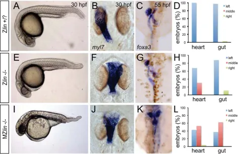
From an ENU-mutagenesis screen, we identified a unique mutant,linkspoot(linhu4087), that displayed a reduced ventral tail fin in combination with a heart-specific laterality defect (Figure 1A, 1B, 1E and 1F). At 30 hours post fertilization (hpf), 24.6% (n = 464) of the embryos derived from an incross of two lin heterozygous carriers displayed the small but noticeable reduc-tion of the ventral tail fin (Figure 1A and 1E). Whilst the majority oflinmutant embryos with the ventral tail fin reduction had no other obvious morphological defects and survived to adulthood, 29% (33 out of 114linmutant embryos) showed cardiac defects resulting in cardiac failure and death at around 5 days post fertilization (dpf) (Figure 1F, 1G). Examination of the cardiac defect inlinmutant embryos revealed a midline positioning of the heart in contrast to a leftward positioning in wild-type siblings at 28 hpf. Furthermore at 48 hpf, when the heart in wild-type sibling embryos has completed looping toward the right, heart looping in these lin mutant embryos was incomplete (n = 6/8) (data not shown). Despite the aberrant heart looping in almost 30% of thelin mutant embryos, patterning of the myocardium and endocardium was grossly normal. Expression of tbx2b and has2in the atrioventricular canal myocardium and endocardium, respectively, was comparable between lin mutant and sibling embryos (Figure S1). In addition, bmp4 expression was still restricted (although slightly expanded) to the venous pole, atrioventricular canal and arterial pole (Figure S1).
We observed that the laterality of the other visceral organs (direction of gut looping, positioning of the liver and pancreas) was unaffected inlinmutant embryos (30 out of 34) (Figure 1C, 1D, 1G and 1H). Sincelinmutant embryos that did not display the cardiac defects described above survived up to adulthood we crossed homozygous lin mutant females with heterozygous lin carrier males. The resulting maternal and zygotic (MZ) lin mutant embryos displayed a reduction of ventral structures such as the tail fin and blood islands (Figure 1I). Such phenotypes have been associated with aberrant dorsoventral patterning of the embryo [28]. In addition, we observed in MZlin mutants, uncoordinated Author Summary
Although vertebrates are bilaterally symmetric when observed from the outside, inside the body cavity the organs are positioned asymmetrically with respect to the left and right sides. Cases where all the organs are mirror imaged, known as situs inversus, are not associated with any medical defects. Severe medical problems occur however in infants with a partial organ reversal (situs ambigious or heterotaxia), which arises during embryonic development. Left-right asymmetry in the embryo is established by unilateral expression of Nodal, a member of the Tgf-ß superfamily of secreted growth factors, a role that has been conserved from human to snails. By performing a genetic screen in zebrafish for laterality mutants, we have identified the linkspootmutant, which displayed partial defects in asymmetric left-right position-ing of the internal organs. The gene disrupted in the
laterality defects in the viscera (Figure 1J–1L). Aberrant position-ing of the heart and other viscera can be caused by defects in formation or function of the Kupffer’s vesicle, resulting in disrupted LR patterning. We therefore examined cilia rotation in the KV, and found that both in Zlinand MZlinmutant embryos with a midline positioning of the heart, cilia rotation in the Kupffer’s vesicle was unaffected (Figure S2 and Videos S1, S2, S3), suggesting that the laterality defect was not due to a disruption of Kupffer’s vesicle function. Together, these results suggest that the affected gene product inlinmutants is required for dorsal-ventral and left-right axis specification.
linkspoot encodes Bmpr1aa
To better understand the molecular nature of the lin mutant phenotype, we positionally cloned the gene that is disrupted in the linmutant. Using bulk segregant analysis with SSLP markers we placed thelinmutation onto chromosome 13. Mapping of thelin locus using 570 mutant embryos resulted in the identification of a chromosomal region containing a zebrafish orthologue of the mammalianBmpr1a/Alk3gene, encoding a Bmp Type I receptor (Figure 2A). Since Bmp signalling is instructive for cardiac laterality as well as ventral tail fin formation [13,29,30], we sequenced the coding region of thebmpr1aagene for mutations. We identified a base pair substitution (T .G) at position 1538 resulting in a leucine to arginine substitution at position 337
(L337R) in the kinase domain of the Bmpr1aa protein (Figure 2B). The T1538G polymorphism was invariably linked with the mutant phenotype (n = 570). No other non-synonymous substitutions were identified in the coding region ofbmpr1aathat were linked with the mutant phenotype. Modelling of the corresponding region of human BMPR1A suggested that the L312R (corresponding to zebrafish L337R) substitution is incompatible with proper folding of this region and thereby likely destabilizes the entire kinase domain (Figure 2C, 2D).
[image:3.612.60.533.62.368.2]To address the functional consequence of the L337R substitu-tion, we introduced the lin mutation in the bmpr1aa gene and generated synthetic mRNA for injection into embryos. Surpris-ingly, injection of wild-typebmpr1aamRNA into wild-type 1-cell stage embryos resulted in a loss of the ventral tail fin (Table 1). Injection ofbmpr1aa L337RmRNA had a stronger inhibition of Bmp signalling since more of the injected embryos displayed a dorsalised phenotype, which was also stronger in its effect (Table 1). These results suggest that increasing wild-type Bmpr1a beyond physiological levels has a negative effect on Bmp signalling, possibly by titrating out other components of the signalling pathway. The dominant-negative effect is stronger for the Bmpr1aa L337R most likely because Bmpr1aa L337R is still able to form a receptor complex and interact with Bmp but it can no longer phosphorylate the receptor Smad protein due to its mutation in the kinase domain. To test this hypothesis we injected Figure 1. Dorsoventral and laterality defects in zygotic and maternal zygoticlinmutant embryos.(A–D) Wild-type zygoticlinsiblings with normal ventral tail fin (A), left-positioned heart tube (B) and normal organ situs with liver on the left, pancreas on the right and left looped gut tube (C). (D) Quantification of heart position (n = 14) and direction of gut looping (n = 40). (E–H) Zygoticlin(Zlin) mutant embryos displayed a mild reduction of the ventral tail fin (n = 100/108) (E). In addition, in almost 30% of Zlinmutant embryos, the heart tube was positioned at the midline (F). Gut laterality was unaffected in Zlin mutant embryos (G). Quantification of heart position (n = 108) and direction of gut looping (n = 100). (I–L) Maternal zygoticlin(MZlin)mutant embryos derived from a cross of a homozygouslinmutant female and male showing the more severe posterior truncation (I) compared to a Zlinmutant embryo (E). In addition, most MZlinmutant embryos displayed a laterality defect in the heart (J), liver (bilateral, K) and in looping of the gut (K). (L) Quantification of heart positioning (n = 151) and direction of gut looping (n = 16).
doi:10.1371/journal.pgen.1002289.g001
a lower dose of the wild-typebmpr1aamRNA into embryos derived from an incross of two lin heterozygous carriers to determine whether we could rescue the tail fin defects oflinmutant embryos. Indeed we observed that injection of low levels of wild-type bmpr1aawas able to rescue the ventral tail fin defects in almost 50% oflinmutant embryos (Table 1). Consistent with our model that Bmpr1aa L337R has reduced signalling activity, we never observed a rescue of the tail fin defects oflinmutant embryos when we injected the bmpr1aa L337R mRNA. From these results we conclude that the gene that is disrupted inlinmutants encodes the Bmp receptor, Bmpr1aa, and that the lin mutation inactivates Bmpr1aa activity.
Bmpr1aa and Bmpr1ab are partially redundant during dorsal-ventral and left-right axis formation
[image:4.612.63.548.60.353.2]To further characterise the requirement for bmpr1a during zebrafish development, we analysed its expression. Interestingly, database searches revealed that due to a genome duplication event, a paralogue ofbmpr1aaexisted in the form ofbmpr1ab/alk3b (exhibiting 80% identity at the protein level). We, therefore, simultaneously analysed the expression pattern of these two closely related genes. ISH analysis revealed that bothbmpr1aparalogues
Figure 2. Genetic variations found in zebrafishbmpr1aaandbmpr1abgenes.(A) Thelinmutation was mapped to a region on chromosome 13 that includes thebmpr1aagene. (B) T.A basepair change that was found in alllinmutant embryos results in a Leu to Arg change at position 337 (L337R). (C) Crystal structure of human BMPR1B. The kinase domain from the human BMPR1B with the kinase inhibitor LDN-193189 (ball-and-stick representation) bound to the ATP binding site (pdb entry 3MDY). Leu 312 (corresponding to Leu 337 in fish) is shown in red. Structural elements providing residues to the hydrophobic core surrounding Leu 312 are highlighted in dark blue. (D) Detailed view of the hydrophobic core surrounding Leu 312 (in red). Black labels refer to the structure of human BMPR1A, the corresponding residues in fish are indicated by grey italic labels. Consequences of the L312R mutation are analyzed by replacing the leucine side chain in the structure model with arginine, of which five typical rotamers are shown (yellow to green). All rotamers cause serious clashes with surrounding residue, which are highly conserved in fish. (E) C.A basepair change in thebmpr1abgene that results in a premature stop codon at position 84 in the extracellular domain of the receptor. (F) A MZbmpr1abmutant embryo at 2 dpf with no obvious phenotypes in the heart or tail region (magnified). (G) Bmpr1a dose-dependent effect on dorsoventral and left-right patterning. Embryos derived from an incross ofbmpr1aa+/2;bmpr1ab+/2double carrier fish was analyzed and quantified for the dorsoventral phenotypes (classified as C1 (mild) to C4 (strong)) and position of the heart (left or midline) if present. n/a, not applicable since no heart tissue was present.
doi:10.1371/journal.pgen.1002289.g002
Table 1.Injection studies.
phenotype#
RNA injection
conc.
ng/ml genotype n wt (%)
C1 (%)
C2 (%)
C3 (%)
C4 (%)
{ (%)
bmpr1a 10 +/+ 64 83 3 8 2 0 4
bmpr1a 20 +/+ 54 72 9 13 0 0 6
bmpr1a L337R 10 +/+ 70 46 4 14 7 10 19
bmpr1a L337R 20 +/+ 62 47 10 3 1 23 16
— lin+/+,+/2 84 100 0 0 0 0 0
lin2/2 27 11 89 0 0 0 0 bmpr1a a 2 lin+/+,+/2 81 96 4 0 0 0 0 lin2/2 32 47 53 0 0 0 0 bmpr1a L337R 2 lin+/+,+/2 78 90 10 0 0 0 0
lin2/2 21 0 100 0 0 0 0
are expressed from the 2-cell stage, indicating maternal deposition of the transcripts (Figure S3). Each paralogue was expressed in a ubiquitous fashion up until the 10-somite stage, however the signal forbmpr1aawas more intense compared to the signal ofbmpr1ab suggesting different levels of expression. From 20-somites onwards, the expression of both paralogues became progressively restricted to anterior regions.
The similar expression patterns observed for bmpr1aa and bmpr1ab suggest comparable functions for the paralogues. To analyse this possibility further we screened a mutagenesis library for abmpr1abmutant. We identified a mutant harbouring a stop codon (TAC.TAA) in the second exon of the gene, truncating the protein 84 amino acids into the ligand-binding domain (Y84X) (Figure 2E). Although a DV patterning defect was reported upon morpholino knockdown of bmpr1ab [31], we observed no morphological phenotype in the majority of bmpr1ab zygotic mutants. Furthermore, maternal zygoticbmpr1ab mutants exhib-ited no observable phenotype (Figure 2F).
We next tested for possible redundancy between the two paralogues. By incrossing double heterozygous carriers for the two mutations (bmpr1aa+/2;bmpr1ab+/2), we observed a spectrum of dorsalised embryonic phenotypes, ranging from wild-type pheno-types to C4 dorsalisation in the most severe instances (categorisa-tion according to Mullins et al., [28]) (Figure 2G). Genotyping revealed that the severity of the dorsalisation phenotype correlates with decreasing gene dosage ofbmpr1aaandbmpr1ab, with double mutant embryos always exhibiting a C4 dorsalised phenotype. Importantly, this gene dosage effect was also observed on LR patterning, with 80% of embryos of genotype bmpr1aa2/2; bmpr1ab+/2 presenting with a cardiac laterality defect (Figure 2H). Interestingly, loss of the bmpr1aaparalogue affected phenotypic severity more robustly than loss of the bmpr1ab. Unfortunately, we were unable to score the cardiac laterality phenotype of double mutant embryos as no cardiac field was detected in these embryos (Figure S4), consistent with previous observations that Bmp signalling is required for cardiac specifica-tion [32,33]). These results demonstrate that thebmpr1aparalogues play partially redundant roles in both dorsoventral and LR patterning.
Bmp acts upstream of Nodal signalling during left-right patterning
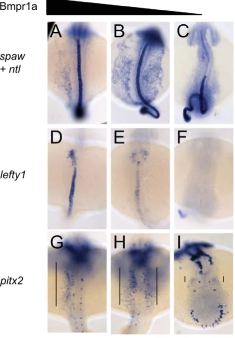
[image:5.612.317.554.65.406.2]Since no role for Bmp1a in LR axis formation has been reported thus far, we further investigated how Bmp signalling via Bmpr1a regulates LR patterning. We analysed the expression pattern of marker genes whose expression is controlled by LR patterning in embryos derived from an incross of bmpr1aa+/2; bmpr1ab+/2parental fish. Expression analysis of the Nodal-related genespawrevealed that embryos that retained at least one wild-type copy of bmpr1aa, displayed normal spaw expression (Figure 3A). However, embryos that had lost both wild-type copy of bmpr1aa and retained at least one wild-type copy of bmpr1ab displayed strong and bilateral expression ofspawin the entire LPM (Figure 3B). On the contrary, in embryos that had lost all wild-type copies of bmpr1aaand bmpr1abwe observed a reduction of spaw expression in the LPM by in situ hybridization (Figure 3C) and quantitative RT-PCR (Figure S5). The bmpr1aa/bmpr1ab double mutant embryos displayed a strong (C4) dorsalised phenotype resulting in a curling of the tail region. Although a Kupffer’s vesicle was present in these embryos (data not shown), the structure of the tail is suspected to have physically intervened with the potential of the Kupffer’s vesicle to activate and/or propagate spawexpression in the posterior LPM.
Figure 3. Dose-dependent effect of Bmpr1a on the expression of laterality genes.In situ hybridisation at 18-somites forspaw(in LPM) andno tail(ntl) (in midline) (A–C),lefty1at 23-somites (heart field and midline) (D–F) and pitx2 at 23-somites (in LPM) (G–I). (A,D,G) Embryos selected for normal ventral tail fin or C1 dorsalization (genotypes: bmpr1aa +/+ or +/2; bmpr1ab +/+ or +/2 or 2/2). (B,E,H) Embryos selected for C3 dorsalization (genotype bmpr1aa2/ 2;bmp1ab+/2). (C,F,I) Embryos selected for C4 dorsalization (genotype
bmpr1aa2/2;bmpr1ab2/2). All embryos are shown as dorsal views with anterior to the top and left to the left. Number of embryos examined is presented in Table 2.
doi:10.1371/journal.pgen.1002289.g003
Table 2.Expression pattern ofspaw, lefty1, andpitx2in
bmpr1aa/abgenotypes.
probe Bmpr1aa Bmpr1ab
+/+ +/+
+/+ +/2
+/+
2/2
+/2
+/+ +/2
+/2
+/2 2/2
2/2
+/+
2/2
+/2 2/2 2/2 n
spaw left 1 6 1 3 6 6 3 0 0 26
bilateral 1 0 0 1 1 0 1 11 0 15
absent 0 0 0 0 0 0 0 3 15 18
lefty1 Left 1 7 3 6 12 2 3 0 0 34
(heart) bilateral 0 0 0 0 1 0 0 5 0 6
absent 0 0 0 0 2 1 0 0 4 7
pitx2 left 3 7 3 6 7 7 1 0 0 34
(LPM) bilateral 1 0 0 0 1 2 1 14 0 19
absent 0 0 0 0 0 0 0 0 3 3
doi:10.1371/journal.pgen.1002289.t002
[image:5.612.318.556.568.721.2]Similar disruptions to asymmetric gene expression were observed upon analysis of lefty1 expression in the cardiac field and pitx2 expression in the gut region. Expression of lefty1 was restricted to the left cardiac field in embryos that retained at least one wild-type copy ofbmpr1aa(Figure 3D). Embryos that had lost both wild-type copy ofbmpr1aaand retained at least one wild-type copy ofbmpr1ab, however, displayed a clear bilateral expression of lefty1in the cardiac field (Figure 3E). Since embryos without any wild-type bmpr1a gene lack the entire cardiac field, no lefty1 expression was observed in the cardiac region of these embryos (Figure 3F). Furthermore,pitx2is expressed in the posterior LPM and its expression is regulated by Nodal activity; this expression was unaltered in embryos that still possessed at least one wild-type copy ofbmpr1aa(Figure 3G). Consistent with the observed spaw andlefty1expression,pitx2expression was also bilateral in the LPM of embryos that had lost both wild-type copy of bmpr1aa and retained at least one wild-type copy of bmpr1ab and was compromised in embryos that had lost all 4 copies of the wild-typebmpr1agene (Figure 3H, 3I).
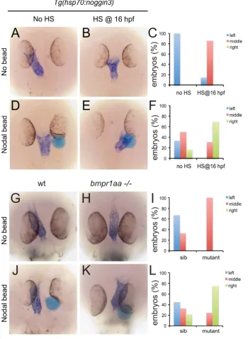
These results suggest that during LR patterning Bmp signalling via Bmpr1a regulates Nodal activity. To address the interrelation between Bmp and Nodal signalling we tested the possibility that Nodal acts downstream of Bmp signalling during cardiac laterality. Therefore we attempted to rescue the Bmp-dependent cardiac laterality defect by implanting Nodal-soaked beads in the anterior LPM (ALPM), in order to induce ectopic Nodal signalling. To block Bmp signalling,Tg(hsp70l:nog3)embryos were heat-shocked at 16 hpf which resulted in a cardiac laterality defect in almost all embryos (6 out of 7; Figure 4A–4C). Interestingly, when a Nodal bead was placed in the right ALPM of non-heat-shocked embryos the heart tube was displaced from the left side towards the midline in approximately 50% of the embryos (Figure 4C, 4F). This effect of the Nodal bead was even stronger when the bead was placed in heat-shockedTg(hsp70l:nog3) embryos. The cardiac tube in such embryos with reduced Bmp signalling was directed towards the right-sided bead in nearly 70% of cases (Figure 4D–4G). In a similar experiment using MZbmpr1aa mutant embryos we again observed that the cardiac tube was directed towards the Nodal bead in 75% of embryos examined (Figure 4H–4J). Together these results suggest that during generation of cardiac laterality Bmp and Bmpr1a act upstream of, or in parallel with, Nodal.
Expression oflefty1in the midline is regulated by Bmp
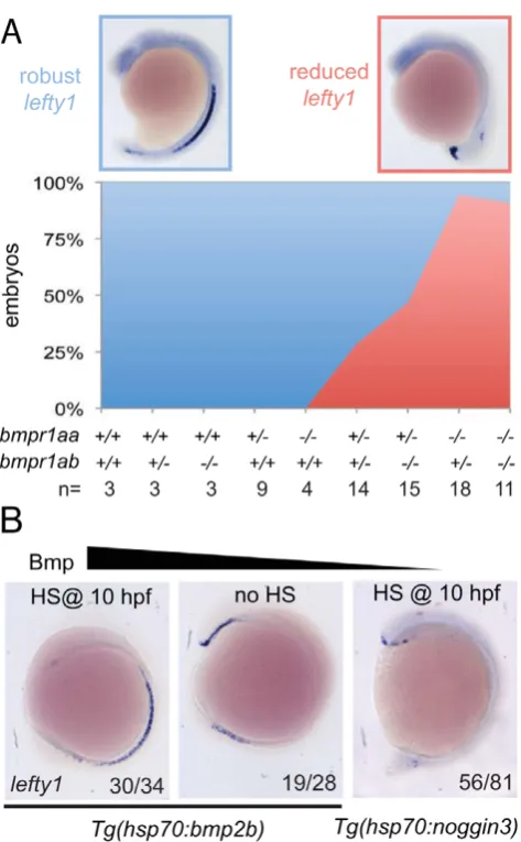
Our observation thatspawis ectopically expressed in the right LPM mesoderm in embryos that had lost 2–3 copies of their wild-typebmpr1aindicated that Bmpr1a is normally required to repress spawexpression in the right LPM. For this to be a direct effect of Bmp signalling it is expected that Bmp signalling is elevated in the right LPM, as recently reported studying mouse embryos [17]. Although we previously reported on elevated Bmp activity in the left anterior LPM before the cardiac tube is formed (22-somite stage)[34], we never observed enhanced Bmp activity in the right posterior LPM using an anti phospho-Smad1,5,8 antibody (data not shown). An alternative to the model in which Bmp activity directly regulatesspawexpression in the right LPM is a model in which Bmp activity regulates spaw expression in an indirect manner. It is well established that Lefty1 in the midline is required to prevent Nodal protein produced in the left LPM from crossing the midline and inducingNodalexpression ectopically in the right LPM [9]. We, therefore, systematically analysedlefty1expression in the midline of embryos with a gradual loss of Bmpr1a signalling. Doing so, we observed that embryos with 4 or 3 copies of the wild-typebmpr1agene displayed normal and robustlefty1expression in the embryonic midline (Figure 5A). Analysis oflefty1expression in
embryos that had lost 2 or 3 copies of the wild-type receptor gene, we observed an increase in the number of embryos with reduced lefty1expression levels in the midline. Embryos that had lost all 4 copies of the wild-typebmpr1agene consistently showed a near loss of alllefty1expression (Figure 5A).
To address whether a disruption of fluid flow in Kupffer’s vesicle might explain the reducedlefty1expression we analyzed the lrrc50hu255h mutant, a loss-of-function allele of a conserved cilia protein that is required for cilia motility [35]. We observed that in the majority of lrrc50hu255h mutant embryos lefty1 was robustly expressed in the midline (Figure S6), suggesting that the observed reduction oflefty1expression inbmpr1amutant embryos was not a consequence of a disruption in Kupffer’s vesicle function.
To test whether Bmp activity can regulate lefty1expression in the midline we analysed lefty1 expression in embryos with increased or reduced Bmp activity. We manipulated levels of Bmp signalling by performing heat-shock experiments on embryos carrying the Tg(hsp70l:bmp2b) or Tg(hsp70l:nog3) transgenes, allowing temporally controlled upregulation or downregulation of Bmp signalling, respectively. The embryos were heat-shocked after gastrulation to prevent strong effects on dorsal-ventral patterning due to altered Bmp signalling levels and lefty1 expression was analysed at somitogenesis stages. Consistent with the data from the bmpr1a mutant analysis, we observed an upregulation of lefty1 expression in embryos with ectopic Bmp activity, intermediate levels of lefty1 in wild-type embryos and reduced lefty1 expression in embryos with reduced Bmp activity (Figure 5B). These results demonstrate that Bmp signalling is both required and sufficient forlefty1expression in the midline.
Bmp and Nodal regulatelefty1expression independently
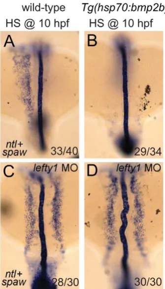
results demonstrate that when Bmp signalling is ectopically activated, lefty1 expression is induced independently of Nodal. When comparing the level of lefty1 induction in heat-shocked Tg(hsp70l:bmp2b) embryos with or without the SB treatment we observed less ectopic lefty1 expression in the presence of the SB inhibitor (comparing Figure 6B and 6D). This result suggests a synergistic effect of Nodal and Bmp onlefty1expression.
Thus far our results suggest that Nodal and Bmp regulatelefty1 expression in the midline independent from each other. To confirm such an independent regulation we tested whether Nodal
[image:7.612.58.412.64.543.2]can regulate lefty1 expression independent from Bmp signalling. To block Bmp activityTg(hsp70l:nog3)embryos were heat-shocked at tail-bud stage (10 hpf), which resulted in reduced expression of lefty1in the anterior midline at 18 hpf (Figure 6E, 6F). To induce Nodal in Bmp-depleted embryos, a Nodal bead was placed in the ALPM. As a consequence of Nodal bead implantation we observed restoration of the anteriorlefty1 expression even in the absence of Bmp signalling (Figure 6G, 6H). These results demonstrate that Nodal can activatelefty1expression independent from Bmp and confirm that Bmp and Nodal regulate lefty1 Figure 4. Rescue of Bmp-related cardiac laterality defects by Nodal beads.In situ hybridisation formyl7to highlight the position of the linear heart tube at 30 hpf.Tg(hsp70l:nog3)embryos with no heat-shock (A,D) or heat-shocked at 16 hpf (B,E). Beads (blue) preincubated with recombinant Nodal protein placed in the right ALPM of non-heat-shocked (D) or heat-shocked (E)Tg(hsp70l:nog3)embryos at 17–18 hpf. Control siblings (G,J) or MZbmpr1aamutant embryos (H,K). Beads (blue) preincubated with Nodal protein placed in the right ALPM of siblings (J) or
MZbmpr1aamutant embryos (K). Position of the inflow pole of the linear heart tube was determined for embryos without a Nodal bead (C) and for embryos in which a Nodal bead was placed on the right side (F). Embryos are shown as dorsal views with anterior to the top and left to the left. doi:10.1371/journal.pgen.1002289.g004
expression independent from each other. Together these results indicate thatlefty1expression is regulated by at least two parallel pathways involving Nodal and Bmp.
Lefty1 is required for the suppression ofspawexpression by Bmp
Finally, we wanted to address whether the observed effect of Bmp on spawexpression in the LPM is direct or indirect via its proposed role in regulating lefty1 expression. InTg(hsp70l:bmp2b) embryos that were heat-shocked at the tail bud stage, we observed a strong down-regulation of spaw expression in the LPM (Figure 7A, 7B), which was coupled with ectopiclefty1expression (Figure 5B). To test whether the upregulation of lefty1 in the
midline was responsible for the downregulation ofspawexpression in the LPM, we performedlefty1knock-down by injecting embryos with a previously published morpholino that effectively targets lefty1[39]. Interestingly, injection of thelefty1MO in heat-shock inducedTg(hsp70l:bmp2b)embryos resulted in restoration ofspaw expression in the left LPM, with ectopic expression also observed in the right LPM (Figure 7D), similar to non-heat-shocked embryos (Figure 7C). These results demonstrate that lefty1 expression in the midline is required for Bmp to repress spaw expression in the LPM and acts as an intermediary between Bmp signalling andspawexpression.
Discussion
[image:8.612.60.297.56.439.2]We describe here the identification of two novel zebrafish bmpr1amutants; abmpr1aamutant allele from a forward genetic screen for laterality mutants and a bmpr1ab mutant allele by screening a mutagenized library. By generating and analyzing compound heterozygous and double mutant embryos forbmpr1aa and bmpr1ab, we observed a strong correlation between the number of wild-typebmpr1agene copies being lost and the severity of the LR patterning defects observed. Most strikingly we observed a shift from the normal unilateral expression of the Nodal-related spawgene in the left LPM to a bilateralspawexpression in both the left and the right LPM. This shift was accompanied by a reduction in the expression oflefty1 at the midline. This demonstrates that Bmp signalling regulates normal unilateral Nodal activation in the LPM, an observation supported by Nodal bead implantation in the LPM that restored cardiac laterality in Bmp-deficient embryos. Mechanistically our data suggests that there are two parallel pathways, a Bmp and a Nodal dependent pathway, to promote lefty1 expression in the midline and regulate LR patterning (see Figure 8 for proposed model). This model also explains the observation made in several animal models that ectopic Bmp signalling downregulates Nodal activation, suggesting that Bmp signalling is required on the right side to repress Nodal activation [13,20,22,38]. Our data now demonstrates that, at least in zebrafish, this regulation of Nodal activity by Bmp is indirect and depends on the activation of lefty1 expression, as was demonstrated by knock-down oflefty1 in embryos with elevated Bmp signalling (Figure 7). Expanding the previous reaction-diffusion model of an agonist (Nodal) and antagonist (Lefty1), we can now include an additional level of regulation, in which Bmp induces Lefty1, which is required to establish unilateral Nodal activity in the LPM.
Lefty1 is essential for formation of the LR axis [9]. Loss ofLefty1 in mouse embryos results in a left-isomerism, whereby left-sided genes become expressed bilaterally. These described effects are very similar to those observed upon reducing Bmpr1a levels or Bmp signalling in the zebrafish embryo shown here. Others have reported that expression ofLefty1in the midline is dependent on Nodal activity from the LPM in both zebrafish and mouse embryos [5,36,38]. Detailed analysis of theLefty1promoter region by Saijoh and colleagues identified a 1.2 kb upstream region of the Lefty1gene that was sufficient to drive its midline expression [40]. In addition, it was reported that although Foxh1 binding sites are present in this upstream promoter region, these were not required to driveLefty1expression in the midline [36]. This suggests that, besides Nodal, additional factors are required for inducing midline Lefty1 expression. Indeed our data demonstrate that during zebrafish LR axis formation, Bmp signalling is required and sufficient to drivelefty1expression in the midline. Firstly, we found that in mutants with reduced copies of the wild-typebmpr1agene, lefty1 expression is gradually lost from the midline. Secondly, in Figure 5. Bmp via Bmpr1a regulateslefty1expression in the
midline.(A) In situ hybridisation forlefty1at 15-somites on embryos from an incross of bmpr1aa+/2;bmpr1ab+/2 double carrier fish. Embryos were analysed forlefty1 expression and classified as robust (blue boxed panel) or reduced (red boxed panel) expression after which the embryos were genotyped. Quantification of the results is shown in the stacked area graph (blue, robustlefty1; red reducedlefty1). (B) In situ hybridisation for lefty1 at 10-somite stage. Embryos shown are
Tg(hsp70l:bmp2b) embryos either heat-shocked at 10 hpf to induce
bmp2bexpression (left panel) or without heat-shock (middle panel) and
Tg(hsp70l:nog3) embryos heat-shocked at 10 hpf to inhibit Bmp signalling (right panel). Lateral view of 10-somite stage embryos with dorsal to the right and anterior up.
transgenic embryos that ectopically expressnoggin3, a potent Bmp antagonist,lefty1expression is diminished from the midline while Nodal signalling is still active (indicated by bilateral spaw expression). Thirdly, ectopic activation of the Bmp signalling pathway using a Tg(hsp70l:bmp2b) transgenic results in elevated and ectopic expression oflefty1in the midline.
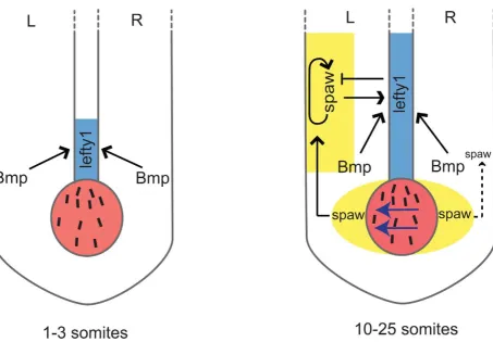
Our experiment using ectopic Bmp signalling in the absence of Nodal activity demonstrated that under conditions where Bmp signalling is sufficiently high, Nodal is not required to inducelefty1 in the midline. This might be important during early stages of LR axis formation. Based on the following observations, we hypoth-esize that at the initiation of LR axis formation,lefty1expression in the midline is initiated by Bmp signalling independently of Spaw activity. Firstly, in zebrafish embryos lefty1 expression in the midline was observed at the 1–3 somite stage whilespawexpression is initiated only at the 5-somite stage in the perinode region and at the 10-somite stage in the LPM ([5] and unpublished observations M. Verhoeven, E. Noe¨l and J. Bakkers). Secondly, this initiallefty1 expression was unaffected by the injection of MOs that efficiently targetedspaw[5]. Thirdly, at these early somite stages expression of Bmp ligands is very strong in the tail bud region [41]. When blocking all Bmp signalling at this early stage in heat-shocked Tg(hsp70l:nog3)embryos,lefty1expression was indeed not initiated in the midline. Together these results suggest that at the initiation of LR axis formation,lefty1expression in the midline is initiated by
Bmp while the maintenance of lefty1 expression in the midline requires both Nodal and Bmp (see model in Figure 8).
Non-redundant roles for Bmp1a and Acvr1l during LR patterning
[image:9.612.51.553.62.372.2]Although the zebrafish has been used extensively to identify new regulators by conducting forward genetic screens, there has been very limited success identifying novel mutants displaying LR patterning defects [42,43]. This might be due to the variability and mixture of the phenotypes that can be observed (situs inversus, situs ambigious or situs solitus) as well as the natural occurrence of these phenotypes in the commonly used wild-type strains. Alternatively, an earlier and essential function of the gene product in embryo development masking any LR defects would hamper the identification of such LR genes. In addition, redundancy with paralogous genes often present in the zebrafish genome can mask the full loss-of-function phenotype. In lin mutant embryos, two copies of the wild-typebmp1aagene are lost while the two wild-type bmpr1abcopies are still present. Thelin/bmpr1aamutant embryos displayed heart-specific laterality defects (although not fully penetrant) without displaying any gut laterality defects. Previously, we showed temporally distinct requirements for Bmp signalling functions during both LR axis formation and heart morphogenesis [13,34]. The heart-specific laterality defect oflin/bmpr1aamutant embryos (eg. loss of leftward cardiac jogging and rightward cardiac Figure 6. Bmp and Nodal inducelefty1independently.(A–D)Tg(hsp70l:bmp2b)embryos were left at 28uC (A,C) or heat-shocked at 10 hpf to inducebmp2bexpression (B,D). A subset of embryos were incubated in the presence of the Nodal inhibitor SB431542 directly after the heat-shock. Embryos were analysed by in situ hybridisation forlefty1expression at 15-somites. (E–H)Tg(hsp70l:nog3)embryos were left at 28uC (E,G) or heat-shocked at 10 hpf to inducenoggin3expression (F,H). In a subset of embryos a bead preincubated with recombinant Nodal was placed in the ALPM. Embryos were analysed by in situ hybridisation forlefty1expression at 18-somites. All embryos are shown as lateral views with dorsal to the right and anterior to the top. Arrows point to most anteriorlefty1expression domain. Numbers in lower right represents the number of embryos that displayed the phenotype represented in the panels.
doi:10.1371/journal.pgen.1002289.g006
looping) is very similar to the cardiac laterality defect previously observed in the lost-a-fin/alk8 mutant. This suggests that during these processes Bmpr1a/Alk3 and Acvrl1/Alk8 play non-redun-dant functions similar to those described for these receptors during dorsoventral patterning [31]. These results also imply that either the regulation of heart laterality is more sensitive to reducing Bmp signalling activity than the digestive system or that this process is less compensated by wild-type maternalbmpr1aaRNA present in the oocyte. In agreement with the latter suggestion are the observations thatbmpr1aais maternally provided in the oocyte and that maternal zygotic (MZ)lin/bmpr1aa mutant embryos (from survivinglin/bmpr1aahomozygous females) displayed an increase in the strength of the LR patterning defects, including gut laterality defects.
Conservation of the Nodal-Bmp-Lefty1 pathway
To our knowledge, this is the first report describing the requirement for Bmpr1a in regulating LR axis formation. Mouse Bmpr1amutant embryos do not form mesoderm at embryonic day 7.5 and subsequently die before embryonic day 9.5, preventing the study of LR axis formation in these mutants [26]. Interestingly, the closely related mouseAcvr1/Alk2gene has been implicated in LR patterning [16]. Since theAcvr1mutant mouse embryos also die early due to severe gastrulation defects, chimeric embryos were
produced and analysed for LR patterning. Depending on the relative contribution of mutant cells to the chimeric embryos, a variety of laterality defects were described. In chimeric embryos with a relative high contribution of Acvr1 mutant cells, bilateral expression of Nodal and Pitx2 in the LPM was observed in combination with reduced expression ofLefty1in the midline. The phenotypes described for the chimeric embryos withAcvr1mutant cells corroborate our observations in the Bmpr1a compound heterozygous/mutant embryos, suggesting a conserved role for Bmp type I receptors during LR axis formation.
The Bmp signal that regulatesLefty1expression in the midline does so independent of Smad1, one of the three Bmp-specific Smad proteins. Although Smad1 inactivation in mouse embryos resulted in the activation of Nodal expression in the right LPM, Lefty expression in the midline was unaffected in such embryos [15]. Alternatively, Smad5 could be responsible for transducing the Bmp signal. Embryos lacking Smad5 no longer expressLefty1 in the midline, which is accompanied by bilateralNodalandPitx2 expression in the LPM [12]. Several observations in mouse suggest that during LR axis specification, Bmp signalling can also repress Nodal activation in the right LPM more directly and indepen-dently from its regulation ofLefty1in the midline. As mentioned above, Smad1-deficient embryos showed bilateral Nodal expres-sion while Lefty expression in the midline was reported to be unaffected [15]. In a study by Mine and co-workers, elevated phospho-Smad1,5,8 levels in the right LPM compared with the left LPM of mouse embryos was reported [17]. In addition, an increase on the left side of phospho-Smad1,5,8 levels was observed inChordinand Noggin double mutant embryos, combined with a loss of Nodal and Lefty1,2 expression. However in Chordin;Noggin double mutant embryos, perinodal Nodal was also reduced and defects in the morphology of the node and the density of cilia were described, suggesting an additional defect in the transduction of a signal from the node to the LPM in such embryos. This defect in communication between the node and the LPM most likely also explains why we observed a complete lack ofspawexpression in the LPM of bmpr1aa;bmp1ab double mutant embryos. In zebrafish embryos, we did not observe a stronger phospho-Smad1,5,8 level in the right LPM compared to the left side during LR specification. However, at later stages we did observe the opposite in the anterior LPM where phospho-Smad1,5,8 levels were increased on the left side [34]. In addition, our observation that ectopic Bmp signalling in theTg(hsp70l:bmp2b) embryos can no longer repressspawactivation in the LPM when Lefty1 is absent makes it very unlikely that such a direct repression of Bmp signalling on spaw expression exists in the zebrafish embryo. Together this indicates that the regulation oflefty1by Nodal and Bmp during LR axis specification is conserved amongst various vertebrate species. However there are species-specific differences as to what other activities Bmp signalling has during this process. Possibly, differences in geometry or scale of the embryos and speed of their development might require additional regulatory mech-anisms to maintain the crucial but very unstable unilateral Nodal activation during LR axis specification.
Materials and Methods
Zebrafish strains and screen
[image:10.612.61.229.60.354.2]All zebrafish strains were maintained in the Hubrecht Institute using standard husbandry conditions. Animal experiments were approved by the Animal Experimentation Committee (DEC) of the Royal Netherlands Academy of Arts and Sciences. Thebmpr1ahu4087 mutant was identified during a forward genetic screen performed at the Hubrecht institute. ENU mutagenesis was performed as Figure 7. Lefty1 is required for Bmp induced repression of
spaw.In situ hybridisation ofspaw(in LPM) andntl(in midline) at 18-somites. Wild-type (A,C) orTg(hsp70l:bmp2b)(B,D) embryos were heat-shocked at 10 hpf to inducebmp2bexpression (B,D). Ectopic expression of bmp2bresulted in the loss ofspawexpression in the LPM (B). A subset of embryos were injected with alefty1MO (C,D), which resulted in bilateralspawexpression even in the presence of ectopicbmp2b(D). Embryos are shown as dorsal views with anterior to the top and left to the left.
previously described for the creation of the Hubrecht Institute target selected mutagenesis library [44]. F1 progeny of mutagenised males were outcrossed to create approximately 300 F2 families, which were then incrossed. F3 progeny were screened for cardiac laterality defects at 28–34 hpf. Thebmpr1ahu4087mutant can be identified using nested PCR with the following primers:
PCR1
Forward primer: AGCTCATCCGGAGAAGTATG Reverse primer: TCCACTTCATTTGTGTCACTG PCR2
Forward primer: TGTAAAACGACGGCCAGT ATATG-TACCCAGCCCTGATG
Reverse primer: AGGAAACAGCTATGACCAT AGCTTCA-GATTCAGATCAACAC
Thebmpr1absa0028mutant was identified from the mutagenesis
library at the Sanger institute by screening finclip DNA using nested PCR with the following primers:
PCR1:
Forward primer: CCAGACTACATGCTTCATG Reverse Primer: ATTGTGACAGGCCTACAATG PCR2:
Forward primer: TGTAAAACGACGGCCAGT CAGAA-GATGCCACAAACAAC
Reverse primer:
AGGAAACAGCTATGACCATGGTCA-CACCGAGTAATTTCC
Products were then sequenced with M13F or M13R primers. Published transgenic lines used were Tg(hsp70ll:nog3)fr14 and Tg(hsp70ll:bmp2b)fr13[13].
Genetic mapping and genotyping
Meiotic mapping of thelinkspootmutation was performed using standard simple sequence length polymorphisms. The primers used for SSLP can be found on www.ensembl.org.
Morpholino oligo and RNA synthesis
[image:11.612.70.524.56.371.2]The lefty1 morpholino was described previously [39]. The coding region of thebmpr1aagene was cloned into pCS2+ by PCR amplification. The lin mutation was introduced in the pCS2+bmpr1aaconstruct using the QuickChange kit (Stratagene). In vitrotranscription was performed from Acc65I digested template using the SP6 mMessage mMachine kit for all injected mRNA (Ambion).
Figure 8. Schematic representation oflefty1regulation during LR axis specification.Two phases oflefty1regulation can be distinguished. i) At the 1–3 somite stage the KV (shown in red) is formed but the nodal flow (indicated by blue arrows) has not yet been initiated. While at this stage
lefty1is already expressed in midline (shown in blue)spawexpression is still absent from the embryo. Thus, this earlylefty1expression is induced independent of Nodal but does depend on Bmp activity. Most likely, robustlefty1expression is required prior to the initiation of LR axis specification to prevent ectopic activation ofspawin the right LPM later on. ii) At the 5-somite stagespawexpression becomes apparent in the perinode region (yellow area flanking the KV) and from the 10-somite stage onward (up to the 25-somite stage)spawis expressed unilateral in the left LPM (yellow-boxed area). Our results demonstrate that at this second phase both Spaw/Nodal and Bmp activity are required independently to maintainlefty1
expression in the midline. Lefty1 in the midline antagonises Spaw and prevents it from crossing the midline where it would induce its own expression in the right LPM.
doi:10.1371/journal.pgen.1002289.g008
SB431542 treatment
SB431542 (Sigma) was resuspended in DMSO to a concentra-tion of 10 mM, and subsequently diluted to a working concen-tration of 150mM in embryo medium. Control embryos were treated with an equal volume of DMSO. 30 embryos were treated per 5 ml of SB/DMSO solution.
In situ hybridization
In situ hybridization was carried out as previously described [45]. Embryos were cleared in MetOH and mounted in benzylbenzoate/benzylalcohol (2:1) before pictures were taken. Riboprobes were generated by transcription from a linearized template in the presence of 11-UTP.
Bead implants
Agarose beads (Affigel blue, BioRad) were rinsed twice in PBS and incubated for 1 hr at 37uC with 50mg/ml recombinant mouse Nodal protein (R&D systems). Implants were performed as previously described [46].
Supporting Information
Figure S1 Formation of the cardiac atrioventricular canal is unaffected inbmpr1aamutant embryos. (A,B) In situ hybridization forbmp4in the heart of wild-type andbmpr1aamutant embryos at 48 hpf. Bmp4 is expressed in the inflow region, atrioventricular (AV) canal (arrow) and outflow region of the heart. Although cardiac looping was affected inbmpr1aamutant embryos, expression ofbmp4 was unaffected. (C,D) In situ hybridization for tbx2b, which was expressed in the AV canal in wild-type siblings (C) and bmpr1aa mutant embryos (D). (E,F) In situ hybridization forhas2, which was expressed in the endocardial cushion cells that will form the AV valves.Has2expression was unaffected inbmpr1aamutant embryos (F) compared to its wild-type siblings (E).
(PDF)
Figure S2 Cilia rotation in Kupffer’s vesicle of Zlin mutant is unaffected. Brightfield images of the heart of wt and zygotic lin mutants after imaging cilia in the KV. Zygoticlinmutants display defects in positioning of the heart, however cilia motility in the KV is unaffected (Videos S1 and S2), demonstrating cilia-independent heart defects.
(PDF)
Figure S3 Expression of bmpr1aa and bmpr1ab. In situ hybrid-ization for bmpr1aa (upper row) and bmpr1ab (lower row) at the indicated stages from 2-cells up to 24 hpf. Both maternalbmpr1aa mRNA and bmpr1ab mRNA was detected at the 2-cell stage. mRNA for both Bmp receptors was detected at the various developmental stages up to 24 hpf. Whilst expression of both Bmp receptors was distributed ubiquitously up to the 10-somite stage, it became progressively more intense in anterior structures at the 20-somite stages and later.
(PDF)
Figure S4 bmpr1aa/bmpr1abdouble mutant embryos lack myocar-dial tissue. In situ hybridization for myl7 (cmlc2) expressed in the
myocardium of wild-type, bmpr1aa mutant or bmpr1ab mutant embryos. Myl7 expression was not detected in bmpr1aa/bmpr1ab double mutant embryos. All embryos shown as dorsal views at 30 hpf. (PDF)
Figure S5 spawexpression is affected inbmpr1aa;bmpr1abembryos. RT-PCR analysis ofspawexpression in wild type – C2, C3 and C4 dorsalised embryos derived from an incross of bmpr1aa+/- and bmpr1ab+/-heterozygous fish (see Text S1 for detailed protocol). C3 dorsalised embryos (bmpr1aa-/-;bmpr1ab+/-) exhibit a 1.6-fold increase in spaw expression, while C4 dorsalised embryos (bmpr1aa-/-;bmpr1ab-/-) have a 6.9-fold decrease inspawexpression, consistent with in situ analysis ofspawexpression.MyoDexpression is gradually reduced in C3 and C4 dorsalised embryos when compared to controls, consistent with a reduction in tail structures. (PDF)
Figure S6 lefty1 expression in lrcc50 mutant embryos. In situ hybridization analysis oflefty1expression inlrrc50mutant embryos at 16 somites. The majority of wild type embryos express lefty1 from the posterior tip of the notochord anteriorly to around the middle of the trunk (A). The majority of lrrc50mutants express lefty1 in a similar domain to wild type embryos (B). A subset of lrrc50mutants either expresslefty1 in a domain restricted to the posterior tip of the notochord (C), or do not expressionlefty1(D). Lateral views, dorsal to the right.
(PDF)
Text S1 Supplemental methods. (DOC)
Video S1 High speed image of cilia rotation in the KV of a wild type embryo at 8 somites. See Text S1 for technical details related to the videos.
(AVI)
Video S2 High speed image of cilia rotation in the KV of a zygoticlin-/-embryo at 8 somites.
(AVI)
Video S3 High speed image of cilia rotation in the KV of a maternal-zygoticlin-/-embryo at 8 somites.
(AVI)
Acknowledgments
We thank Dr. Cuppen (Hubrecht Laboratory) and Dr. Stemple (Welcome Trust Sanger Institute) for providing thebmpr1absa0028 zebrafish mutant, which was generated as part of the ZF-MODELS Integrated Project in the 6th Framework Programme. We thank the Hubrecht screen team, M. Verhoeven, and M. Witte for technical assistance and M. Rebagliati for suggesting the Nodal-inhibitor experiment.
Author Contributions
Conceived and designed the experiments: KAS EN JB. Performed the experiments: KAS EN SC IT. Analyzed the data: KAS EN JB HR. Wrote the paper: KAS EN JB.
References
1. Ramsdell AF (2005) Left-right asymmetry and congenital cardiac defects: getting to the heart of the matter in vertebrate left-right axis determination. Dev Biol 288: 1–20. 2. Bamford RN, Roessler E, Burdine RD, Saplakog˘lu U, dela Cruz J, et al. (2000) Loss-of-function mutations in the EGF-CFC gene CFC1 are associated with human left-right laterality defects. Nat Genet 26: 365–369.
3. Kuehl KS, Loffredo C (2002) Risk factors for heart disease associated with abnormal sidedness. Teratology 66: 242–248.
4. Grande C, Patel NH (2009) Nodal signalling is involved in left-right asymmetry in snails. Nature 457: 1007–1011.
5. Long S, Ahmad N, Rebagliati M (2003) The zebrafish nodal-related gene southpaw is required for visceral and diencephalic left-right asymmetry. Development 130: 2303–2316.
7. Meno C, Ito Y, Saijoh Y, Matsuda Y, Tashiro K, et al. (1997) Two closely-related left-right asymmetrically expressed genes, lefty-1 and lefty-2: their distinct expression domains, chromosomal linkage and direct neuralizing activity in Xenopus embryos. Genes Cells 2: 513–524.
8. Thisse B, Pflumio S, Furthauer M, Loppin B, Heyer V, et al. (2001) Expression of the zebrafish genome during embryogenesis. ZFIN Direct Data Submission. 9. Meno C, Shimono A, Saijoh Y, Yashiro K, Mochida K, et al. (1998) lefty-1 is required for left-right determination as a regulator of lefty-2 and nodal. Cell 94: 287–297.
10. Nakamura T, Mine N, Nakaguchi E, Mochizuki A, Yamamoto M, et al. (2006) Generation of robust left-right asymmetry in the mouse embryo requires a self-enhancement and lateral-inhibition system. Dev Cell 11: 495–504.
11. Meinhardt H, Gierer A (2000) Pattern formation by local self-activation and lateral inhibition. Bioessays 22: 753–760.
12. Chang H, Zwijsen A, Vogel H, Huylebroeck D, Matzuk MM (2000) Smad5 is essential for left-right asymmetry in mice. Dev Biol 219: 71–78.
13. Chocron S, Verhoeven MC, Rentzsch F, Hammerschmidt M, Bakkers J (2007) Zebrafish Bmp4 regulates left-right asymmetry at two distinct developmental time points. Dev Biol 305: 577–588.
14. Fujiwara T, Dehart DB, Sulik KK, Hogan BLM (2002) Distinct requirements for extra-embryonic and embryonic bone morphogenetic protein 4 in the formation of the node and primitive streak and coordination of left-right asymmetry in the mouse. Development 129: 4685–4696.
15. Furtado MB, Solloway MJ, Jones VJ, Costa MW, Biben C, et al. (2008) BMP/ SMAD1 signaling sets a threshold for the left/right pathway in lateral plate mesoderm and limits availability of SMAD4. Genes & Development 22: 3037–3049.
16. Kishigami S, Yoshikawa S-I, Castranio T, Okazaki K, Furuta Y, et al. (2004) BMP signaling through ACVRI is required for left-right patterning in the early mouse embryo. Dev Biol 276: 185–193.
17. Mine N, Anderson RM, Klingensmith J (2008) BMP antagonism is required in both the node and lateral plate mesoderm for mammalian left-right axis establishment. Development 135: 2425–2434.
18. Monsoro-Burq A, Le Douarin NM (2001) BMP4 plays a key role in left-right patterning in chick embryos by maintaining Sonic Hedgehog asymmetry. Mol Cell 7: 789–799.
19. Piedra ME, Ros MA (2002) BMP signaling positively regulates Nodal expression during left right specification in the chick embryo. Development 129: 3431–3440.
20. Rodrı´guez Esteban C, Capdevila J, Economides AN, Pascual J, Ortiz A, et al. (1999) The novel Cer-like protein Caronte mediates the establishment of embryonic left-right asymmetry. Nature 401: 243–251.
21. Schlange T, Arnold H-H, Brand T (2002) BMP2 is a positive regulator of Nodal signaling during left-right axis formation in the chicken embryo. Development 129: 3421–3429.
22. Yokouchi Y, Vogan KJ, Pearse RV, Tabin CJ (1999) Antagonistic signaling by Caronte, a novel Cerberus-related gene, establishes left-right asymmetric gene expression. Cell 98: 573–583.
23. Shi Y, Massague´ J (2003) Mechanisms of TGF-ß signaling from cell membrane to the nucleus. Cell 113: 685–700.
24. Gu Z, Reynolds EM, Song J, Lei H, Feijen A, et al. (1999) The type I serine/ threonine kinase receptor ActRIA (ALK2) is required for gastrulation of the mouse embryo. Development 126: 2551–2561.
25. Mishina Y, Crombie R, Bradley A, Behringer RR (1999) Multiple roles for activin-like kinase-2 signaling during mouse embryogenesis. Dev Biol 213: 314–326.
26. Mishina Y, Suzuki A, Ueno N, Behringer RR (1995) Bmpr encodes a type I bone morphogenetic protein receptor that is essential for gastrulation during mouse embryogenesis. Genes & Development 9: 3027–3037.
27. Zhu L, Marvin MJ, Gardiner A, Lassar AB, Mercola M, et al. (1999) Cerberus regulates left-right asymmetry of the embryonic head and heart. Curr Biol 9: 931–938.
28. Mullins MC, Hammerschmidt M, Kane DA, Odenthal J, Brand M, et al. (1996) Genes establishing dorsoventral pattern formation in the zebrafish embryo: the ventral specifying genes. Development 123: 81–93.
29. Bauer H, Lele Z, Rauch GJ, Geisler R, Hammerschmidt M (2001) The type I serine/threonine kinase receptor Alk8/Lost-a-fin is required for Bmp2b/7 signal transduction during dorsoventral patterning of the zebrafish embryo. Develop-ment 128: 849–858.
30. Schilling TF, Concordet JP, Ingham PW (1999) Regulation of left-right asymmetries in the zebrafish by Shh and BMP4. Dev Biol 210: 277–287. 31. Little SC, Mullins MC (2009) Bone morphogenetic protein heterodimers
assemble heteromeric type I receptor complexes to pattern the dorsoventral axis. Nat Cell Biol 11: 637–643.
32. Kishimoto Y, Lee KH, Zon L, Hammerschmidt M, Schulte-Merker S (1997) The molecular nature of zebrafish swirl: BMP2 function is essential during early dorsoventral patterning. Development 124: 4457–4466.
33. Schultheiss TM, Burch JB, Lassar AB (1997) A role for bone morphogenetic proteins in the induction of cardiac myogenesis. Genes & Development 11: 451–462.
34. Smith KA, Chocron S, von der Hardt S, de Pater E, Soufan A, et al. (2008) Rotation and asymmetric development of the zebrafish heart requires directed migration of cardiac progenitor cells. Dev Cell 14: 287–297.
35. van Rooijen E, Giles RH, Voest EE, van Rooijen C, Schulte-Merker S, et al. (2008) LRRC50, a conserved ciliary protein implicated in polycystic kidney disease. J Am Soc Nephrol 19: 1128–1138.
36. Yamamoto M, Mine N, Mochida K, Sakai Y, Saijoh Y, et al. (2003) Nodal signaling induces the midline barrier by activating Nodal expression in the lateral plate. Development 130: 1795–1804.
37. Inman GJ, Nicola´s FJ, Callahan JF, Harling JD, Gaster LM, et al. (2002) SB-431542 is a potent and specific inhibitor of transforming growth factor-beta superfamily type I activin receptor-like kinase (ALK) receptors ALK4, ALK5, and ALK7. Mol Pharmacol 62: 65–74.
38. Wang X, Yost HJ (2008) Initiation and propagation of posterior to anterior (PA) waves in zebrafish left-right development. Dev Dyn 237: 3640–3647. 39. Feldman B, Concha ML, Sau´de L, Parsons MJ, Adams RJ, et al. (2002) Lefty
antagonism of Squint is essential for normal gastrulation. Curr Biol 12: 2129–2135.
40. Saijoh Y, Oki S, Ohishi S, Hamada H (2003) Left-right patterning of the mouse lateral plate requires nodal produced in the node. Dev Biol 256: 161–173. 41. Martı´nez-Barbera´ JP, Toresson H, Da Rocha S, Krauss S (1997) Cloning and
expression of three members of the zebrafish Bmp family: Bmp2a, Bmp2b and Bmp4. Gene 198: 53–59.
42. Chen JN, van Bebber F, Goldstein AM, Serluca FC, Jackson D, et al. (2001) Genetic steps to organ laterality in zebrafish. Comp Funct Genomics 2: 60–68. 43. Chen JN, van Eeden FJ, Warren KS, Chin A, Nu¨sslein-Volhard C, et al. (1997) Left-right pattern of cardiac BMP4 may drive asymmetry of the heart in zebrafish. Development 124: 4373–4382.
44. Wienholds E (2003) Efficient Target-Selected Mutagenesis in Zebrafish. Genome Research 13: 2700–2707.
45. Thisse C, Thisse B (2008) High-resolution in situ hybridization to whole-mount zebrafish embryos. Nat Protoc 3: 59–69.
46. von der Hardt S, Bakkers J, Inbal A, Carvalho L, Solnica-Krezel L, et al. (2007) The Bmp gradient of the zebrafish gastrula guides migrating lateral cells by regulating cell-cell adhesion. Curr Biol 17: 475–487.