0022-538X/05/$08.00⫹0 doi:10.1128/JVI.79.17.11434–11442.2005
Copyright © 2005, American Society for Microbiology. All Rights Reserved.
Large-Scale Analysis of Adeno-Associated Virus Vector Integration
Sites in Normal Human Cells†
Daniel G. Miller,
1Grant D. Trobridge,
2‡ Lisa M. Petek,
2Michael A. Jacobs,
3Rajinder Kaul,
3and David W. Russell
2,4*
Department of Pediatrics, Division of Genetics and Developmental Medicine,1Department of Medicine, Divisions of
Hematology2and Medical Genetics,3and Department of Biochemistry,4University
of Washington, Seattle, Washington
Received 17 February 2005/Accepted 26 May 2005
The integration sites of viral vectors used in human gene therapy can have important consequences for safety and efficacy. However, an extensive evaluation of adeno-associated virus (AAV) vector integration sites has not been completed, despite the ongoing use of AAV vectors in clinical trials. Here we have used a shuttle vector system to isolate and analyze 977 unique AAV vector-chromosome integration junctions from normal human fibroblasts and describe their genomic distribution. We found a significant preference for integrating within CpG islands and the first 1 kb of genes, but only a slight overall preference for transcribed sequences. Integration sites were clustered throughout the genome, including a major preference for integration in ribosomal DNA repeats, and 13 other hotspots that contained three or more proviruses within a 500-kb window. Both junctions were localized from 323 proviruses, allowing us to characterize the chromosomal deletions, insertions, and translocations associated with vector integration. These studies establish a profile of insertional mutagenesis for AAV vectors and provide unique insight into the chromosomal distribution of DNA strand breaks that may facilitate integration.
Successful gene therapy often requires the long-term trans-gene expression provided by integrating viral vectors. How-ever, integration can cause insertional mutagenesis and onco-gene activation, as demonstrated by two X-linked severe combined immune deficiency patients who developed leuke-mia after treatment with a retroviral vector that integrated near theLMO2proto-oncogene (14). Large-scale analyses of integration sites can shed light on the process of insertional mutagenesis, and studies of murine leukemia virus, human immunodeficiency virus, avian retroviruses, and vectors based on them have demonstrated distinct integration patterns (16, 27, 33, 40, 46). Of particular importance is the relationship of integration sites to genes, as this may determine the likelihood of oncogene activation. In this regard, human immunodefi-ciency virus vectors have been shown to integrate preferentially throughout transcription units, while murine leukemia virus vectors integrate preferentially near transcription start sites (40, 46).
Vectors based on adeno-associated virus (AAV) have a lin-ear, single-stranded DNA genome containing a transgene cas-sette flanked by viral inverted terminal repeats (ITRs). Trans-duction occurs by multiple pathways, including integration into the host genome (24), expression from linear and circular episomal forms (6, 30), and homologous recombination with chromosomal sequences (37). Definitive evidence for
integra-tion has come from sequencing vector-chromosome juncintegra-tions recovered from human cells (25, 26, 38, 47) and mouse tissues (28, 29), which demonstrated that AAV vectors integrate at nonhomologous chromosomal locations.
Chromosomal sequences surrounding vector proviruses can be deleted or rearranged (25, 26, 29), although it is not known if the AAV vector causes these changes. While integration may occur in only a subset of transduced cells, the large vector doses infused during in vivo gene delivery can lead to substan-tial numbers of integration events. In mouse models of liver transduction, integrated AAV vector genomes were present at roughly 0.05 copies/cell (32), a value that would correspond to 7.5⫻ 109 integration events in humans undergoing liver–di-rected gene therapy with AAV vectors (assuming 1.5⫻ 1011 cells/liver) (44). The potential consequences of billions of in-tegration events are largely unknown.
Integration requires that the linear AAV vector genome ligate to two chromosomal ends. Unlike retroviral vectors, AAV vectors do not contain an endonuclease to generate chromosomal ends, so they must rely on existing double-strand breaks or nicks. The deletions, insertions, and microhomolo-gies found at AAV vector-chromosome junctions suggest that integration occurs by the nonhomologous end-joining pathway of double-strand break repair (26), and AAV vectors will in-tegrate at a specific double-strand break when it is created in human cells (25). This dependency on host cell factors and chromosomal features allows us to interpret AAV vector inte-gration sites as chromosomal repair events tagged by a provi-rus.
Here we have performed a large-scale analysis of AAV vec-tor integration sites in normal human cells in the absence of selective pressure, including their relationship to genes, repet-itive DNAs, and other chromosomal features. We have
char-* Corresponding author. Mailing address: Dept. of Medicine, HSB K236A, University of Washington, 1705 NE Pacific St., Seattle, WA 98195-7720. Phone: (206) 616-4562. Fax: (206) 616-8298. E-mail: [email protected].
† Supplemental material for this article may be found at http://jvi .asm.org.
‡ Present address: Clinical Research Division, Fred Hutchinson Cancer Research Center, Seattle, Wash.
11434
on November 8, 2019 by guest
http://jvi.asm.org/
proviruses and identified several integration hotspots. Our re-sults establish the profile of insertional mutagenesis associated with AAV vectors, and they suggest that similar integration studies may be a valuable tool for understanding chromosome biology.
MATERIALS AND METHODS
Nucleic acid manipulations.Plasmids pDG (13) and pA2-TOA (25) have been described. Genomic DNAs were isolated by standard techniques using the Pure-gene kit (Gentra Systems, Minneapolis, MN). RNA was prepared for microarray studies by using the RNeasy and QIAshredder kits from QIAGEN (Valencia, CA). Plasmid DNAs used for vector production were purified from bacterial pellets by using QIAGEN plasmid maxi kits. Plasmid DNAs containing rescued proviruses were purified and sequenced according to the standard DNA se-quencing protocols used in our Genome Center (1, 22). Briefly, the plasmid DNAs were prepared on a QIAGEN Biorobot 3000 utilizing QIAprep 96 Turbo plasmid DNA preparation kit according to the protocols suggested by the man-ufacturer. The sequencing reactions were carried out with the Big Dye termina-tor chemistry V3.1 kit (Applied Biosystems, Foster City, CA) using left and right
sequencing primers 5⬘-GATAAG CTG TCA AAC ATG AGA ATT C and
5⬘-ATCACG AGG CCC TTT CGT CTT CAA G, respectively. Electrophoresis
of sequencing reactions was performed on ABI Prism 3700 capillary sequencers and the raw trace data were analyzed and viewed using PHRED/PHRAP/ CONSED software tools (8, 9, 12).
Cell culture.Cells were grown at 37°C in 5% CO2in Dulbecco’s modified
Eagle’s medium containing 4 g of glucose/liter (Gibco/Invitrogen, Carlsbad, CA), 10% heat-inactivated fetal bovine serum, penicillin, and streptomycin. Primary, normal male human fibroblasts (MHF2) were obtained from the Coriell Institute for Medical Research (Camden, NJ; catalog no. GM05387). 293T cells have been described (7). MHF2 cells were transduced with the AAV2-TOA vector by
seeding 6-cm tissue culture dishes with 5⫻105cells on day 0, replacing the
medium and infecting with 2.5⫻1010
genome-containing particles of
AAV2-TOA on day 1 (5⫻104genome-containing particles per cell), and replacing the
medium again on day 3. On day 6 cells were detached with trypsin and seeded to one 15-cm dish. On day 10 the cells in a single 15-cm dish were split to three 15-cm dishes. On day 14 genomic DNA was prepared from the 15-cm dishes,
except for 3⫻106cells that were used to seed another set of three 15-cm dishes.
This process was repeated every 6 days, and the majority of proviruses were isolated from DNA prepared on the fourth round of this procedure.
Vector preparation.The serotype 2 AAV vector AAV2-TOA was made by cotransfection of 293T cells with helper plasmid pDG and vector plasmid pA2TOA and purified by benzonase treatment of cell lysates, iodixanol step gradient, heparin affinity column chromatography (HiTrap, Amersham Bio-sciences, Uppsala Sweden), and HiTrap desalting column as described (48). AAV vector quantification was based on the amount of full-length single-stranded vector genomes detected by alkaline Southern blot analysis (18).
Shuttle vector rescue in bacteria.Rescue of AAV2-TOA proviruses was done
as described (25) with the following modifications: 20g of genomic DNA
containing integrated proviruses was digested with 80 units of MfeI, AvrII, or NcoI, extracted with phenol and chloroform, and precipitated with ethanol.
DNA fragments were resuspended in 355l of H2O and brought to 400l with
40l of 10x ligation buffer and 5l of T4 DNA ligase (400 U/l, New England
Biolabs, Beverly, MA). Ligations were incubated at 15°C overnight to circularize fragments, heat inactivated by incubation at 65°C for 20 min, brought to 50 mM NaCl, digested further with 80 units of DpnI for an additional 2 h to remove bacterial DNA, extracted with phenol and chloroform, and precipitated with ethanol.
The DNA pellets were resuspended in 5l of H2O, andEscherichia colistrain
DH10B (15) was transformed by electroporation with⬃4g (1l) of DNA at
a time. Transformed bacteria were grown on agar containing 50g/ml ampicillin
and colonies were replated to agar containing 12.5g/ml tetracycline. Bacteria
resistant to both ampicillin and tetracycline were grown in 96-well culture dishes
in freezing medium and then frozen at⫺80°C for future sequencing. Freezing
medium contains 10 g tryptone, 5 g yeast extract, 10 g NaCl, 6.3 g K2HPO4, 1.8 g
KH2PO4. 0.5 g sodium citrate, 0.9 g (NH4)2SO4, and 44 ml glycerol per liter of
H2O, brought to 10M MgSO4and supplemented with ampicillin after
auto-claving.
Microarray analysis of gene expression levels.We seeded 5⫻105
MHF2 cells in six 6-cm dishes on day 0. On day 1 fresh medium was added to the dishes, and
three dishes received 2.5⫻1010
genome-containing particles of AAV2-TOA. On day 3 RNA was harvested from confluent 6-cm dishes and all six samples were
processed independently. Labeling of 5g of total RNA was performed as
described by Affymetrix (Santa Clara, CA); 15g of cRNA was used per
Af-fymetrix HG-U133 Plus 2.0 array, which analyzes 47,400 transcripts and variants. Only the subset of probes that identify specific RefSeq gene transcripts (13,069) were used in our analysis. Probe sets that hybridized with more than one gene were excluded. Gene expression levels from all three uninfected cell samples were averaged and compared to those from all three infected cell samples. Where multiple probe sets reflect the transcription level of a single RefSeq gene, the average transcription level was used in rankings.
Database searches and comparisons with genomic features.DNA sequences were processed with computer programs interpreted by the PERL programming language. Sequences were truncated at bp 500, and expected vector-derived sequences were trimmed. The resulting junction sequences were aligned to build 35 of the human genome and three additional files containing AAV2-TOA vector sequence, nonvector sequence from plasmid pA2TOA, and the 43-kb human ribosomal DNA (rDNA) repeat (GenBank accession no. U13369) (10) using a stand-alone version of BLAT (21) that generates a BLAST alignment score.
The input script was as follows: blatchromosome_file query_file⫺out⫽
blast8-ooc⫽11.oocoutput_file. An additional 95% homology requirement and BLAST
score of⬎100 were used to establish genomic positions. Alignments were sorted
by BLAST score, and those with the five highest scores were saved for further processing. The average match length for all sequences was 383 bp. Nucleotide insertions were defined as sequence preceding the alignment with the highest BLAST score when the alignment did not start at position number 1 of the sequence query. Additional PERL programs were used to remove duplicate junction sequences, compare localized integration sites to various chromosomal features using tables available from the University of California–San Francisco database (20), and determine the positions of restriction enzyme sites in the human genome.
We produced a randomly localized set of genomic positions by generating random numbers between 1 and 5,941,037,819 (the size of the build 35 diploid male genome with chromosomes laid end to end) with the PERL “rand” func-tion. The buffer size had to be increased from 15 to 31 bits to avoid generating duplicate numbers. These random numbers were converted to chromosomal positions by splitting the numeric range of the diploid genome into separate chromosomes with each starting at base pair 1 of the p arm and extending the entire length of the chromosome. These chromosomal positions were used to extract 383 bp of sequence from build 35 of the human genome at each randomly determined position, and the resulting files were aligned with the genome using
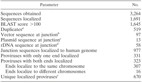
Sequences obtained 3,264
Sequences localized 1,691
BLAST score⬎100 1,645
Duplicatesa 519
Vector sequence at junctionb 97
Plasmid sequence at junctionc 13
rDNA sequence at junctiond 58
Junction sequences localized to human genome 977 Proviruses with only one end localized 331 Proviruses with both ends localized 323 Ends localize to the same chromosome 307 Ends localize to different chromosomes 16
Unique localized provirusese 670
a
Proviruses with junctions occurring at the exact same base as another se-quenced provirus.
b
Provirus sequence was joined to other sequences derived from the AAV vector genome.
c
Provirus sequence was joined to nonvector sequences derived from the AAV vector plasmid.
d
DNA sequences are not localized to specific chromosomal sites.
e
Counts proviruses with both ends sequenced once if localized to the same chromosome and twice if localized to different chromosomes, and assumes all proviruses with one end localized were distinct.
on November 8, 2019 by guest
http://jvi.asm.org/
[image:2.585.300.543.80.222.2]BLAT as described above. About 7% of these extracted sequences corresponded to gapped or repetitive sequence in the human genome, could not be reliably localized, and were discarded. A set of 10,000 localized positions was used as a control data set (calculated random integration events) for comparison with
AAV vector integration site positions. To analyze clustering and hotspots, we used similar sets of 499 and 670 random genomic positions as size-matched controls
To identify oncogenes, we searched several databases, including Entrez Gene
(http://www.ncbi.nlm.nih.gov/entrez/query.fcgi?db⫽gene), OMIM (http://www
.ncbi.nlm.nih.gov/entrez/query.fcgi?db⫽OMIM), the Tumor Gene Database
(http://www.tumor-gene.org/TGDB/tgdb.html), and the Retrovirus Tagged Can-cer Gene Database (http://rtcgd.ncifcrf.gov/).
Statistical analysis.In all cases statistical significance was determined using
the2
test to compare AAV vector integration site frequencies with those of
randomly generated genomic positions.Pvalues were determined using tables,
and those less than 0.01 were considered significant.
RESULTS
We infected normal human fibroblasts with the AAV shuttle vector AAV2-TOA containing a bacterial replication origin and antibiotic resistance genes, then rescued integrated
provi-FIG. 1. Location of junction sites in the AAV vector proviruses. In the top panel, the nucleotide sequence of an AAV vector ITR in the flop orientation is shown numbered from 1 to 145 beginning at the 3⬘ end. The locations of the most common vector-chromosome junctions are indicated by underlined, bold type. In the bottom panel, the per-cent of vector-chromosome junctions found at specific base pairs in the AAV ITR and adjoining vector sequence is shown. The percent found in three observed peaks is also indicated.
0 2 4 6 8 10
[image:3.585.46.283.66.312.2]FIG. 2. Chromosomal distribution of integration sites. (A) Local-ized AAV vector integration sites (n⫽670) and a calculated set of random sites (n⫽10,000) are graphed as a percentage of all integrants in each chromosome. Only one integration junction was included for AAV vector proviruses where both ends were localized to the same chromosome. Asterisks mark comparisons with P values of ⬍0.01. (B) A human chromosome ideogram is shown with AAV vector inte-gration sites (dots to the left of each chromosome) and hotspots where at least three integrants were found within 500 kb (boxed dots; see Table 3). Each dot represents a unique AAV vector integrant (n⫽ 670) and is 33% opaque to display multiple overlapping integrants. Ribosomal DNA repeats present on the p arm of chromosomes 13, 14, 15, 21, and 22 contained a significant number of AAV vector integrants that are described separately in Fig. 3.
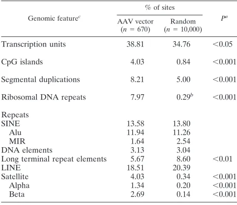
TABLE 2. Genomic features of integration sites
Genomic featurec
% of sites
Pa
AAV vector
(n⫽670)
Random
(n⫽10,000)
Transcription units 38.81 34.76 ⬍0.05
CpG islands 4.03 0.84 ⬍0.001
Segmental duplications 8.21 5.00 ⬍0.001 Ribosomal DNA repeats 7.97 0.29b ⬍0.001
Repeats
SINE 13.58 13.80
Alu 11.94 11.26
MIR 1.64 2.54
DNA elements 3.13 3.04
Long terminal repeat elements 5.67 8.60 ⬍0.01
LINE 18.51 20.39
Satellite 4.03 0.34 ⬍0.001
Alpha 1.34 0.20 ⬍0.001
Beta 2.69 0.14 ⬍0.001
aP values of⬍0.05 are not shown and were not considered statistically
signif-icant.
bPercent of rDNA in the diploid human genome, assuming 400 rDNA repeats
of 43 kb.
cSINE, short interspersed nucleotide element; MIR, mammalian interspersed
repetitive element; LINE, long interspersed nucleotide element.
on November 8, 2019 by guest
http://jvi.asm.org/
[image:3.585.305.534.71.400.2] [image:3.585.42.283.469.678.2]ruses and flanking chromosomal DNA inE. coli. as bacterial plasmids. Of the 3,264 sequences obtained, 1,691 had flanking junction DNA that could be localized to build 35 of the human genome, rDNA, vector, or nonvector plasmid sequences, with an average alignment of 383 bp per query. After discarding presumed duplicates (junctions at the exact same nucleotide position), and eliminating sequence reads with BLAST scores
⬍100, a total of 977 unique integration junctions were local-ized to the human genome (Table 1). Both left and right junctions were obtained from 323 proviruses. Of the flanking sequences, 9.3% were from the vector genome or plasmid backbone, which may represent vector-vector recombination events, foldback priming of DNA synthesis at ITRs, or pack-aging of plasmid sequences into virions, as observed previously (26, 28, 38). None of the vector proviruses contained intact ITRs, and distinct preferences were noted for junction bases within the ITR secondary structure (Fig. 1).
The chromosomal features of vector integration sites are shown in Table 2. As a control, we generated a set of sequences localized to random positions in the human genome that were processed the same way as our provirus sequence reads (see Materials and Methods). Compared to this control set, AAV vectors preferentially integrated in CpG islands, segmental duplications, ribosomal repeats, and satellite DNA, and there were fewer integrants than expected in long terminal repeat elements. The preference for satellite DNA may be an artifact, as most satellite sequences are not localized and would there-fore not be included in our random set. There was a modest preference for integrations in transcription units that was not as statistically significant (P⬍0.05).
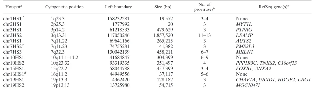
Vector proviruses were found in all human chromosomes (Fig. 2A). Relative to the set of calculated random integration events, some chromosomes had statistically significant differ-ences in vector integration frequencies. Chromosome 5 lacked integration events compared to controls (P ⬍0.01). Integra-tion hotspots, defined asⱖ3 independent proviruses within 500 kb (Fig. 2B), may explain why some chromosomes had statis-tically significant increases in integration frequencies. Chromo-somes 7 and 19 had more vector integrations than expected, with three and two hotspots, respectively. A list of all the
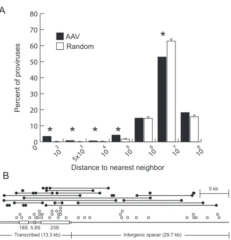
hotspots meeting these criteria is shown in Table 3, and in many cases, the hotspot size was significantly less than 500 kb. This tendency to integrate in clusters was further quantified by measuring the distances between neighboring integration sites (using data from the left sequencing primer only, to avoid counting sites identified by sequencing opposite ends of the same provirus). Of 499 unique sites, 3.41% or 9.02% of neigh-boring vector integration sites were within 1 kb or 100 kb, respectively, compared to 0.2% or 1.87%, respectively, of neighboring calculated random integrants (Fig. 3A). A major hotspot with almost 8% of all vector integrations occurred in rDNA repeats (Fig. 3B), which are not localized to the human genome sequence (Table 2). This 43-kb repeat is present at an estimated 400 copies in a diploid human genome (4) and con-stitutes approximately 0.29% of human DNA.
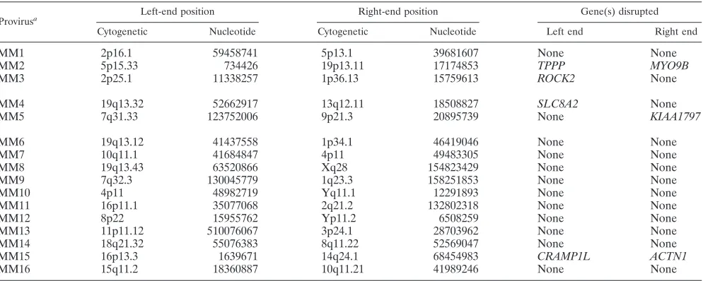
Of the 307 proviruses with both ends localized to the same chromosome, 70% had deletions of genomic DNA ranging in size up to⬃106bp (Fig. 4A) and 35% of junctions had inser-tions of DNA that also varied in size (Fig. 4B). Sixteen provi-ruses had left and right junctions where the best alignment scores were on different chromosomes (Table 4). As these represent possible chromosomal translocations, we used addi-tional criteria to establish their validity. In 11 of 16 proviruses with mismatched ends (MM6 to MM16), the second-best BLAST score was⬎90% of the best score for at least one end, raising the possibility that there may have been a localization error due to sequence repeats. Many of these junctions mapped to pericentromeric chromosomal regions rich in alpha satellite DNA. The remaining five proviruses had BLAST scores for both ends that were significantly above those of other possible alignments, and three of these had scores over 500 for both ends (MM1 to MM3). Even if one conservatively assumes that only the three translocations meeting the most rigorous criteria are real, this represents nearly 1% of all vector integrations (3 of 323).
Additional analyses were performed to assess the relation-ship of integration sites to transcription. CpG islands are fre-quently found near promoter regions and may regulate gene transcription (3, 23). AAV vectors had a 4.8-fold preference for integration in CpG islands, which did not extend into
sur-Hotspot Cytogenetic position Left boundary Size (bp)
provirusesb RefSeq gene(s)
chr1HS1d 1q23.3 158232281 19,572 3–4 None
chr2HS1 2p25.3 1777992 20 3 MYT1L
chr3HS1 3p14.2 61218533 479,629 3 PTPRG
chr3HS2 3q13.31 117050246 1,857,520 11–13 LSAMP
chr7HS1 7q11.22 69641166 265,215 3 AUTS2
chr7HS2d 7q11.23 74755281 41,382 3 PMS2L3
chr7HS3 7q32.3 130042139 458,211 6–7 MKLN1
chr10HS1 10q11.1–11.2 41684847 304,399 6–9 None
chr10HS2 10q23.32 93319335 351,497 4 PPP1R3C,TNKS2,C10orf13
chr15HS1 15q22.2 58044788 457,399 3–4 FOXB1,ANXA2
chr16HS1d 16q11.2 44949556 37,117 5–6 None
chr19HS1 19p13.3 4362420 128,182 3 CHAF1A,UBXD1,HDGF2,LRG1
chr19HS2 19p13.13 13725980 54,715 3 MGC10471
a
At least three independent proviruses within 500 kb.
b
Lower numbers assume that rescued proviruses with only one end localized are duplicates of rescued proviruses where only the other end was localized.
c
Protein function is described in Table S1 in the supplemental material.
d
Did not meet hotspot criteria when second-best BLAST score required to be⬍90% of best score.
on November 8, 2019 by guest
http://jvi.asm.org/
[image:4.585.38.542.81.227.2]rounding regions (Fig. 5A). We also analyzed the distribution of integration sites within transcription units and found a 3.6-fold preference for integrating within 1 kb downstream of the start site, but nowhere else within 100 kb (Fig. 5B). Interest-ingly, the percentage of calculated random integration events increased slightly near transcription start sites, perhaps reflect-ing the density of genes in the human genome. Another pos-sible bias comes from the distribution of restriction enzyme sites in the human genome. We analyzed the recognition site positions for each enzyme used in this study relative to CpG islands and transcription starts (Fig. S1 and S2 in the supple-mental material). While there were variations in enzyme site frequencies around these genomic elements, they did not cor-relate with the biases seen for AAV vector integration sites.
Microarray analysis was performed to determine if integra-tions occurred preferentially in expressed genes. We ranked 13,069 RefSeq genes by expression level and plotted the ranks from uninfected and infected fibroblasts, highlighting those where vectors integrated (Fig. 6). There was little overall
im-pact of AAV vector infection on gene expression, consistent with an earlier study (41). Two hundred sixty proviruses inte-grated within the transcribed region of a gene, and the average expression rank of those found on the chip (n ⫽ 195) was 1.09-fold above that of genes with calculated random inte-grants in both infected and uninfected cells. The average ex-pression rank of the genes where integrations occurred within 1 kb of transcription starts (14 found on the chip) was 1.14- to 1.16-fold above that of genes with calculated random inte-grants. These results suggest that gene expression did not have a major impact on integration. A more significant effect of transcription was observed for the rDNA genes transcribed by RNA polymerase I. Of the 58 proviruses found in rDNA re-peats, the integration frequency was 3.7-fold higher in tran-scribed versus nontrantran-scribed regions (2.71 and 0.74 provi-ruses/kb, respectively) (Fig. 3B).
DISCUSSION
In this report we have used an AAV shuttle vector system to rescue integrated proviruses as bacterial plasmids. This
[image:5.585.46.283.63.309.2]al-0 10 20 30 40 50 60 70 80
FIG. 3. Clustering of AAV vector integration sites and localization within the human ribosomal DNA. (A) The distance between each unique AAV vector integration site identified with one sequencing primer (n⫽499) and its nearest neighbor was determined and binned by size, and the percentage of proviruses within each bin was plotted. We used only left junctions in this analysis, to ensure that two different ends of the same provirus were not scored as neighbors. As a control, we performed a similar analysis on three size-matched sets of ran-domly distributed sites (n⫽499), and plotted means with standard deviations. Significant differences (P⬍0.01) are marked with asterisks. Each bar represents the number of clones with a nearest neighbor within the distance bounded by the values on thexaxis. (B) Each AAV vector integration junction localized to the 43-kb ribosomal DNA repeat unit is shown as a circle above a scale diagram of the repeat. Solid circles connected by lines represent the junctions of proviruses where both ends were localized (n ⫽ 21). Open circles represent proviruses where only one end was localized (n⫽37). The 13.3-kb transcribed region is drawn with open boxes, where the thick regions represent the 18S, 5.8S, and 23S rRNAs. The 29.7-kb intergenic spacer is depicted as a single bold line.
0 10 20 30 40 50
[image:5.585.315.528.71.385.2]0 5 10 15 20 25 30 35 40
FIG. 4. Deletions and insertions found at integration sites. (A) The sizes of chromosomal deletions associated with AAV vector proviruses (n⫽212) were sorted into bins and the percentage of proviruses in each deletion size range bounded by values shown on thexaxis was plotted. (B) The sizes of insertions found at AAV vector junctions (n⫽416) were sorted into bins and the percentage of proviruses with insertions in each size range bounded by values shown on thexaxis was plotted. Junctions linked to vector or plasmid sequence were not in-cluded.
on November 8, 2019 by guest
http://jvi.asm.org/
lowed us to recover hundreds of integration events from nor-mal human cells without selection in a scalable process, with automated plasmid purification and junction sequencing. Our approach has several advantages compared to more commonly used PCR-based methods. First, the size of junction sequences is not limited by the efficiency of PCR amplification, and the quality of sequence obtained from plasmids is high, so we were able to generate long sequence reads (average match length of 383 bp). This allows more accurate localization of junctions containing repetitive sequences, and if necessary additional portions of the plasmid can be sequenced. In contrast, the PCR methods used to sequence murine leukemia virus and human immunodeficiency virus integration junctions (40, 46) gener-ated sequences with average match lengths of 98 and 191 bp, respectively, when aligned by our criteria. Second, the shuttle vector allows one to recover both junctions from a provirus, which in the case of AAV is crucial for determining the chro-mosomal structure at the integration site. Third, while both approaches may be biased by the genomic distribution of re-striction sites used, PCR is also biased by parameters affecting amplification. The major drawback to the shuttle vector ap-proach is that the vector must include a selectable bacterial marker and replication origin.
An important issue regarding insertional mutagenesis is its relationship to chromosomal gene transcription. We found that AAV vectors had only a modest preference for integration within transcription units (38.81% versus 34.76% for calcu-lated random integrants), which was less than that reported for AAV vector integration in mouse livers, where 72% of inte-grants were in genes (29). This could reflect differences in the etiology of chromosomal breaks used for integration, where relatively low hepatocyte proliferation rates may decrease the impact of DNA replication on integration sites and increase the impact of transcription. AAV vectors had a more dramatic preference for integration at transcription start sites, with a
⬎3-fold enrichment within the first kb transcribed, but these accounted for just 2.1% of all integrants due to the small
window size. We found only a modest effect of transcription, with the average expression rank of genes containing provi-ruses at any transcribed position or within 1 kb of the start site only 1.09 to 1.16-fold greater than that of genes with calculated random integration events.
There was also a significant preference for integration in CpG islands (4.03% versus 0.84% for calculated random inte-grants), which typically act as transcriptional control elements for nearby genes (3, 23). Surprisingly, some of our results were similar to those of murine leukemia virus vectors, where inte-grations also occurred preferentially near transcription start sites and CpG islands, with only a slight overall preference for genes (46). Thus, despite the distinct life cycles of these viruses, they may share aspects of integration site selection.
AAV vectors do not contain an endonuclease, so integration must occur at chromosomal sites where free DNA ends form. This can take the form of a double-strand break (25) or per-haps a nick that is converted to a double-strand break during DNA replication. The preference for transcriptional start sites, CpG islands, and segmental duplications suggests that these regions may be prone to DNA damage that leads to breaks. In the case of transcription start sites, this damage could be due to local unwinding that initiates transcription and exposes bases. CpG islands can have altered chromatin structure and hypersensitivity to nucleases (42, 45), may act as replication origins (5), and, when methylated, can be mutagenic due to deamination of 5-methylcytosine (34), all of which could in-crease the likelihood of strand breakage. Segmental duplica-tions are recombinogenic areas of the human genome (39) that may also recombine with AAV vector DNA by similar mech-anisms. By the same reasoning, long terminal repeats may be relatively protected from DNA damage, as they were under-represented sites of AAV vector integration.
The integration hotspots we observed may also be damage-prone areas of the genome. The major hotspot in rDNA could reflect unique aspects of these repeats, which are frequently involved in recombination events with distinct mechanisms in
Provirusa
Cytogenetic Nucleotide Cytogenetic Nucleotide Left end Right end
MM1 2p16.1 59458741 5p13.1 39681607 None None
MM2 5p15.33 734426 19p13.11 17174853 TPPP MYO9B
MM3 2p25.1 11338257 1p36.13 15759613 ROCK2 None
MM4 19q13.32 52662917 13q12.11 18508827 SLC8A2 None
MM5 7q31.33 123752006 9p21.3 20895739 None KIAA1797
MM6 19q13.12 41437558 1p34.1 46419046 None None
MM7 10q11.1 41684847 4p11 49483305 None None
MM8 19q13.43 63520866 Xq28 154823429 None None
MM9 7q32.3 130045779 1q23.3 158251853 None None
MM10 4p11 48982719 Yq11.1 12291893 None None
MM11 16p11.1 35077068 2q21.2 132802318 None None
MM12 8p22 15955762 Yp11.2 6508259 None None
MM13 11p11.12 510076067 3p24.1 28703962 None None
MM14 18q21.32 55076383 8q11.22 52569047 None None
MM15 16p13.3 1639671 14q24.1 68454983 CRAMP1L ACTN1
MM16 15q11.2 18360887 10q11.21 41989246 None None
a
MM1 to MM3: BLAST score⬎500, second hit⬍90% of first hit. MM1 to MM5: BLAST score⬎100, second hit⬍90% of first hit.
on November 8, 2019 by guest
http://jvi.asm.org/
[image:6.585.45.542.80.279.2]transcribed and nontranscribed regions (11) that may account for the distribution of vector integrations we observed. In crus-taceans and insects, rDNA can be a preferred site of transpo-son insertions (19, 35). Other hotspots correlated with known areas of genomic instability. Hotspots chr7HS1 and chr7HS2 both flank the region where deletions occur in Williams-Beuren syndrome (2), and hotspot chr3HS1 lies near the com-mon fragile site FRA3B (17), an area where DNA gaps and breaks may form and a known integration site for plasmids and papillomavirus (36, 43). The study of AAV vector integration hotspots may lead to new insights into chromosome biology, as the integrated proviruses serve as tags for chromosomal dam-age at the nucleotide level. Large-scale integration surveys done in specific cell types or under different conditions may
help us understand how chromosome structures change during differentiation or genotoxic stress.
Our findings help to define the spectrum of insertional mu-tagenesis associated with AAV vectors, with implications for their use in gene therapy. Overall, we observed a broad distri-bution of integrations throughout the human genome, with significant clustering in several hotspots. The effects of inte-grating in the rDNA repeat hotspot are not known, but given that these genes are already highly expressed and present in multiple copies, there should be minimal phenotypic effects. A major concern at other hotspots is the potential for activating oncogenes by introducing promoters and/or enhancers, and although wild-type AAV has never been shown to cause can-cer, the transcriptional control elements are different in vec-tors. The hotspots we identified did not include known onco-genes, but given the broad distribution of integration sites, one must assume that a provirus could integrate near any gene.
The chromosomal changes associated with integration can be significant, as large deletions and even translocations were observed. Since DNA damage present at these sites presum-ably exposed free DNA ends prior to integration, a key re-maining question is whether the same chromosomal effects would have occurred in the absence of AAV. For example, would repair of damaged chromosomes produce the same de-letion sizes or translocations without vector integration? Our study also underscores the variability that occurs in proviral structures. All proviruses were deleted to some degree at their terminal repeats, and many sequence reads identified vector genomes joined to other vector or plasmid sequences that could affect transgene expression. Our results complement a recent report of AAV vector integration in murine hepatocytes with similar findings, albeit with a greater preference for tran-scribed genes (31). Further research will be required to deter-0
1 2 3 4 5
0.0 0.5 1.0 1.5 2.0 2.5
FIG. 5. Integration in CpG islands and at transcription start sites. (A) Localized AAV vector integration sites (n⫽670) and a calculated set of random sites (n⫽10,000) were mapped relative to those of CpG islands (identified as GC contentⱖ50%, length⬎200 bp, and ratio of observed to expected number of CpG dinucleotides⬎0.6). Integration sites within CpG islands or in twelve 0.75-kb windows flanking each island (the average size of a CpG island is 764 bp) were binned and plotted as the percentage of all sites. Significant differences (P⬍0.01) are marked with an asterisk. (B) AAV vector integration sites (n⫽ 670) and a calculated set of random sites (n⫽10,000) were mapped relative to the transcription start sites of RefSeq genes, binned into windows of increasing sequence size, and plotted as a percentage of all integrants per kb (to account for the different window sizes). Signifi-cant differences (P⬍0.01) are marked with an asterisk.
FIG. 6. RefSeq gene expression levels and AAV vector integration. RNA samples from uninfected normal human fibroblasts (yaxis) and those infected with AAV2-TOA (xaxis) were hybridized to the human U133A Plus 2.0 gene chip array (Affymetrix) and the expression levels of all RefSeq genes assayed in the array were ranked and plotted (light gray dots). RefSeq genes containing AAV vector integrants are circled in red. The average expression rank of RefSeq genes containing a calculated random integrant (n⫽2,930; yellow circle), those contain-ing AAV vector integrants (n⫽195; purple circle), and those contain-ing AAV vector integrants within 1 kb of transcription start sites (n⫽ 14; green square) are shown.
on November 8, 2019 by guest
http://jvi.asm.org/
preclinical animal models.
ACKNOWLEDGMENTS
We thank Maynard Olson and the University of Washington Ge-nome Center for helpful advice and sequencing and the Center for Expression Arrays for microarray analysis.
This work was supported by grants from the U.S. National Institutes of Health and the Child Health Research Center at Children’s Hos-pital, Seattle, Wash.
REFERENCES
1.Anonymous.2004. Finishing the euchromatic sequence of the human
ge-nome. Nature431:931–945.
2.Bayes, M., L. F. Magano, N. Rivera, R. Flores, and L. A. Perez Jurado.2003. Mutational mechanisms of Williams-Beuren syndrome deletions. Am. J.
Hum Genet.73:131–151.
3.Bird, A. P.1986. CpG-rich islands and the function of DNA methylation.
Nature321:209–213.
4.Bross, K., and W. Krone.1972. On the number of ribosomal RNA genes in
man. Humangenetik14:137–141.
5.Delgado, S., M. Gomez, A. Bird, and F. Antequera.1998. Initiation of DNA
replication at CpG islands in mammalian chromosomes. EMBO J.17:2426–
2435.
6.Duan, D., P. Sharma, J. Yang, Y. Yue, L. Dudus, Y. Zhang, K. J. Fisher, and J. F. Engelhardt.1998. Circular intermediates of recombinant adeno-asso-ciated virus have defined structural characteristics responsible for long-term
episomal persistence in muscle tissue. J. Virol.72:8568–8577.
7.DuBridge, R. B., P. Tang, H. C. Hsia, P. M. Leong, J. H. Miller, and M. P. Calos.1987. Analysis of mutation in human cells by using an Epstein-Barr
virus shuttle system. Mol. Cell. Biol.7:379–387.
8.Ewing, B., and P. Green.1998. Base-calling of automated sequencer traces
using phred. II. Error probabilities. Genome Res.8:186–194.
9.Ewing, B., L. Hillier, M. C. Wendl, and P. Green.1998. Base-calling of automated sequencer traces using phred. I. Accuracy assessment. Genome
Res.8:175–185.
10.Gonzalez, I. L., and J. E. Sylvester.1995. Complete sequence of the 43-kb human ribosomal DNA repeat: analysis of the intergenic spacer. Genomics
27:320–328.
11.Gonzalez, I. L., and J. E. Sylvester.2001. Human rDNA: evolutionary pat-terns within the genes and tandem arrays derived from multiple
chromo-somes. Genomics73:255–263.
12.Gordon, D., C. Abajian, and P. Green.1998. Consed: a graphical tool for
sequence finishing. Genome Res.8:195–202.
13.Grimm, D., A. Kern, K. Rittner, and J. A. Kleinschmidt.1998. Novel tools for production and purification of recombinant adenoassociated virus
vec-tors. Hum. Gene Ther.9:2745–2760.
14.Hacein-Bey-Abina, S., C. Von Kalle, M. Schmidt, M. P. McCormack, N. Wulffraat, P. Leboulch, A. Lim, C. S. Osborne, R. Pawliuk, E. Morillon, R. Sorensen, A. Forster, P. Fraser, J. I. Cohen, G. de Saint Basile, I. Alexander, U. Wintergerst, T. Frebourg, A. Aurias, D. Stoppa-Lyonnet, S. Romana, I. Radford-Weiss, F. Gross, F. Valensi, E. Delabesse, E. Macintyre, F. Sigaux, J. Soulier, L. E. Leiva, M. Wissler, C. Prinz, T. H. Rabbitts, F. Le Deist, A. Fischer, and M. Cavazzana-Calvo. 2003. LMO2-associated clonal T cell
proliferation in two patients after gene therapy for SCID-X1. Science302:
415–419.
15.Hanahan, D., J. Jessee, and F. R. Bloom.1991. Plasmid transformation of
Escherichia coli and other bacteria. Methods Enzymol.204:63–113.
16.Hematti, P., B. K. Hong, C. Ferguson, R. Adler, H. Hanawa, S. Sellers, I. E. Holt, C. E. Eckfeldt, Y. Sharma, M. Schmidt, C. von Kalle, D. A. Persons, E. M. Billings, C. M. Verfaillie, A. W. Nienhuis, T. G. Wolfsberg, C. E. Dunbar, and B. Calmels.2004. Distinct genomic integration of MLV and SIV vectors in primate hematopoietic stem and progenitor cells. PLoS Biol.
2:e423.
17.Huebner, K., and C. M. Croce.2001. FRA3B and other common fragile sites:
the weakest links. Nat Rev. Cancer1:214–221.
18.Inoue, N., and D. W. Russell.1998. Packaging cells based on inducible gene amplification for the production of adeno-associated virus vectors. J. Virol.
72:7024–7031.
19.Jakubczak, J. L., W. D. Burke, and T. H. Eickbush.1991. Retrotransposable elements R1 and R2 interrupt the rRNA genes of most insects. Proc. Natl.
Acad. Sci. USA88:3295–3299.
20.Karolchik, D., R. Baertsch, M. Diekhans, T. S. Furey, A. Hinrichs, Y. T. Lu, K. M. Roskin, M. Schwartz, C. W. Sugnet, D. J. Thomas, R. J. Weber, D. Haussler, and W. J. Kent.2003. The University of California–San Francisco
Genome Browser Database. Nucleic Acids Res.31:51–54.
21.Kent, W. J.2002. BLAT–the BLAST-like alignment tool. Genome Res.
12:656–664.
McKernan, J. Meldrim, J. P. Mesirov, C. Miranda, W. Morris, J. Naylor, C. Raymond, M. Rosetti, R. Santos, A. Sheridan, C. Sougnez, N. Stange-Tho-mann, N. Stojanovic, A. Subramanian, D. Wyman, J. Rogers, J. Sulston, R. Ainscough, S. Beck, D. Bentley, J. Burton, C. Clee, N. Carter, A. Coulson, R. Deadman, P. Deloukas, A. Dunham, I. Dunham, R. Durbin, L. French, D. Grafham, S. Gregory, T. Hubbard, S. Humphray, A. Hunt, M. Jones, C. Lloyd, A. McMurray, L. Matthews, S. Mercer, S. Milne, J. C. Mullikin, A. Mungall, R. Plumb, M. Ross, R. Shownkeen, S. Sims, R. H. Waterston, R. K. Wilson, L. W. Hillier, J. D. McPherson, M. A. Marra, E. R. Mardis, L. A. Fulton, A. T. Chinwalla, K. H. Pepin, W. R. Gish, S. L. Chissoe, M. C. Wendl, K. D. Delehaunty, T. L. Miner, A. Delehaunty, J. B. Kramer, L. L. Cook, R. S. Fulton, D. L. Johnson, P. J. Minx, S. W. Clifton, T. Hawkins, E. Branscomb, P. Predki, P. Richardson, S. Wenning, T. Slezak, N. Doggett, J. F. Cheng, A. Olsen, S. Lucas, C. Elkin, E. Uberbacher, M. Frazier, et al.
2001. Initial sequencing and analysis of the human genome. Nature409:860–
921.
23.Larsen, F., G. Gundersen, R. Lopez, and H. Prydz.1992. CpG islands as gene
markers in the human genome. Genomics13:1095–1107.
24.McLaughlin, S. K., P. Collis, P. L. Hermonat, and N. Muzyczka.1988. Adeno-associated virus general transduction vectors: analysis of proviral
structures. J. Virol.62:1963–1973.
25.Miller, D. G., L. M. Petek, and D. W. Russell.2004. Adeno-associated virus
vectors integrate at chromosome breakage sites. Nat. Genet.36:767–773.
26.Miller, D. G., E. A. Rutledge, and D. W. Russell.2002. Chromosomal effects
of adeno-associated virus vector integration. Nat. Genet.30:147–148.
27.Mitchell, R. S., B. F. Beitzel, A. R. Schroder, P. Shinn, H. Chen, C. C. Berry, J. R. Ecker, and F. D. Bushman.2004. Retroviral DNA integration: ASLV,
HIV, and MLV show distinct target site preferences. PLoS Biol.2:E234.
28.Nakai, H., Y. Iwaki, M. A. Kay, and L. B. Couto.1999. Isolation of recom-binant adeno-associated virus vector-cellular DNA junctions from mouse
liver. J. Virol.73:5438–5447.
29.Nakai, H., E. Montini, S. Fuess, T. A. Storm, M. Grompe, and M. A. Kay.
2003. AAV serotype 2 vectors preferentially integrate into active genes in
mice. Nat. Genet.34:297–302.
30.Nakai, H., T. A. Storm, and M. A. Kay.2000. Recruitment of single-stranded recombinant adeno-associated virus vector genomes and intermolecular re-combination are responsible for stable transduction of liver in vivo. J. Virol.
74:9451–9463.
31.Nakai, H., X. Wu, S. Fuess, T. A. Storm, D. Munroe, E. Montini, S. Burgess, M. Grompe, and M. A. Kay.2005. Large-scale molecular characterization of
adeno-associated virus vector integration in mouse liver. J. Virol.79:3606–
3614.
32.Nakai, H., S. R. Yant, T. A. Storm, S. Fuess, L. Meuse, and M. A. Kay.2001. Extrachromosomal recombinant adeno-associated virus vector genomes are
primarily responsible for stable liver transduction in vivo. J. Virol.75:6969–
6976.
33.Narezkina, A., K. D. Taganov, S. Litwin, R. Stoyanova, J. Hayashi, C. Seeger, A. M. Skalka, and R. A. Katz.2004. Genome-wide analyses of avian sarcoma
virus integration sites. J. Virol.78:11656–11663.
34.O’Neill, J. P., and B. A. Finette.1998. Transition mutations at CpG dinucle-otides are the most frequent in vivo spontaneous single-based substitution
mutation in the human HPRT gene. Environ. Mol. Mutagen.32:188–191.
35.Penton, E. H., B. W. Sullender, and T. J. Crease.2002. Pokey, a new DNA
transposon in Daphnia (Cladocera: Crustacea). J. Mol. Evol.55:664–673.
36.Rassool, F. V., T. W. McKeithan, M. E. Neilly, E. van Melle, R. Espinosa, 3rd, and M. M. Le Beau.1991. Preferential integration of marker DNA into the chromosomal fragile site at 3p14: an approach to cloning fragile sites.
Proc. Natl. Acad. Sci. USA88:6657–6661.
37.Russell, D. W., and R. K. Hirata.1998. Human gene targeting by viral
vectors. Nat. Genet.18:325–330.
38.Rutledge, E. A., and D. W. Russell.1997. Adeno-associated virus vector
integration junctions. J. Virol.71:8429–8436.
39.Samonte, R. V., and E. E. Eichler.2002. Segmental duplications and the
evolution of the primate genome. Nat. Rev. Genet.3:65–72.
40.Schroder, A. R., P. Shinn, H. Chen, C. Berry, J. R. Ecker, and F. Bushman.
2002. HIV-1 integration in the human genome favors active genes and local
hotspots. Cell110:521–529.
41.Stilwell, J. L., and R. J. Samulski.2004. Role of viral vectors and virion shells
in cellular gene expression. Mol. Ther.9:337–346.
42.Tazi, J., and A. Bird.1990. Alternative chromatin structure at CpG islands.
Cell60:909–920.
43.Wilke, C. M., B. K. Hall, A. Hoge, W. Paradee, D. I. Smith, and T. W. Glover.
1996. FRA3B extends over a broad region and contains a spontaneous HPV16 integration site: direct evidence for the coincidence of viral
integra-tion sites and fragile sites. Hum. Mol. Genet.5:187–195.
44.Wilson, Z. E., A. Rostami-Hodjegan, J. L. Burn, A. Tooley, J. Boyle, S. W. Ellis, and G. T. Tucker.2003. Inter-individual variability in levels of human microsomal protein and hepatocellularity per gram of liver. Br. J. Clin.
Pharmacol.56:433–440.
on November 8, 2019 by guest
http://jvi.asm.org/
45.Wolf, S. F., and B. R. Migeon.1985. Clusters of CpG dinucleotides impli-cated by nuclease hypersensitivity as control elements of housekeeping
genes. Nature314:467–469.
46.Wu, X., Y. Li, B. Crise, and S. M. Burgess.2003. Transcription start regions in the human genome are favored targets for MLV integration. Science
300:1749–1751.
47.Yang, C. C., X. Xiao, X. Zhu, D. C. Ansardi, N. D. Epstein, M. R. Frey, A. G.
Matera, and R. J. Samulski.1997. Cellular recombination pathways and viral terminal repeat hairpin structures are sufficient for adeno-associated virus
integration in vivo and in vitro. J. Virol.71:9231–9247.
48.Zolotukhin, S., B. J. Byrne, E. Mason, I. Zolotukhin, M. Potter, K. Chesnut, C. Summerford, R. J. Samulski, and N. Muzyczka. 1999. Recombinant adeno-associated virus purification using novel methods improves infectious
titer and yield. Gene Ther.6:973–985.