The lytic origin of DNA replication of Epstein-Barr virus,oriLyt, is a complex eukaryotic origin which is
activated during the lytic phase of the viral life cycle. It consists of at least two independent cis-acting
components, one of which plays a dual role in transcription and DNA replication. The binding of the viral factor BZLF1, a member of the AP1 family of transcription factors, to this upstream component is crucial for
oriLytfunction (A. Schepers, D. Pich, and W. Hammerschmidt, EMBO J. 12:3921–3929, 1993). The second
cis-acting element, the downstream component of oriLyt, is equally indispensable; however, its function is
unknown. In this study, the downstream component was found to be the binding target of several cellular proteins. One could be identified as Sp1 or as a related protein which binds twice to the downstream component oforiLyt. Mutational analysis indicated that Sp1 alone is not directly involved in mediating DNA replication; however, other factors which share the same binding sequence or bind closely to one of the Sp1 binding sites
are likely candidates to contribute to a replication protein complex at the downstream component oforiLyt. The
sequence requirements for the downstream component are remarkably stringent, indicating that at least one
of the putative factors is a sequence-specific DNA-binding protein which is required for the activation oforiLyt.
Epstein-Barr virus (EBV), a human herpesvirus, is able to infect human B lymphocytes in vitro and in vivo. These target cells for EBV become latently infected with the virus, and its genetic information is maintained in the dividing cell popula-tion as extrachromosomal multiple copies (for a review, see reference 18). The cis-acting element, which mediates DNA replication during the latent phase of the viral life cycle, has been identified as the plasmid origin of DNA replication, oriP, together with its viral transactivator (20, 25). In such latently infected cells, only 11 genes of more than 80 viral genes are found expressed and no virus is produced. In contrast, during the lytic phase of the viral life cycle, the viral DNA is amplified several hundred-fold via a different origin of DNA replication,
oriLyt (15), and most or all of the viral genes are expressed.
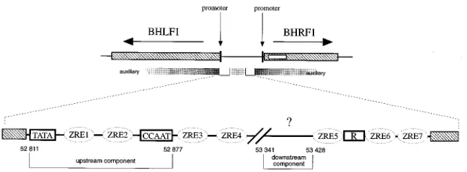
The structure of oriLyt is complex (Fig. 1) and is associated with two divergent promoters for the BHLF1 and BHRF1 genes (1). oriLyt consists of several regions which are required for its full activity. Auxiliary elements which are only poorly defined are nonessential but influence the efficiency with which
oriLyt-containing plasmids replicate in transient replication
ex-periments (15). Two essential or core elements have been identified which constitute the minimal origin of DNA repli-cation (15) and which have been dissected further into up-stream and downup-stream components (23). The upup-stream com-ponent (Fig. 1) colocalizes with the basic promoter region of the BHLF1 transcriptional unit which is directly transactivated by the viral gene product BZLF1. BZLF1 belongs to the AP1 family of transcription factors (8) and is a sequence-specific DNA-binding protein (3) which acts as a homodimer. It binds to seven sites, ZRE1 to ZRE7 (Fig. 1), within oriLyt; ZRE1 and ZRE2 are part of the oriLyt upstream component, ZRE3
and ZRE4 are located adjacent to this component, and ZRE5 to ZRE7 are distributed further downstream.
The BZLF1 protein is the molecular switch that induces the lytic phase of the viral life cycle in latently EBV-infected cells (4, 24). Moreover, this trans-acting element is directly involved in DNA replication of oriLyt through its ZRE sites (22). An additional viral protein, the R enhancer factor, which scores as a transcriptional activator of several viral promoters including the BHLF1 gene (13), binds to oriLyt (Fig. 1) but is probably not directly involved in oriLyt DNA replication (23).
It became obvious from our studies that none of these fac-tors is known to interact with the second essential element of
oriLyt, the downstream component (Fig. 1). This region, which
was mapped to be only 40 bp long (23), is located approxi-mately 530 bp downstream from the upstream component. In contrast to it, the downstream component is solely dedicated to the activation of oriLyt-mediated DNA replication and does not seem to be involved in the transcriptional regulation of either the BHLF1 or the BHRF1 gene. The fine mapping of the downstream component with a series of deletion and sub-stitution mutants of oriLyt plasmids identified a DNA sequence that is sensitive to any introduced mutation (see Fig. 7A) (23). We analyzed the downstream component in order to eluci-date its function. We hypothesized that its extreme sensitivity to sequence alteration underlies a role as a protein-binding motif with a narrow sequence specificity. We sought and were able to find several DNA-protein complexes that form with this region which are likely to be of cellular origin and include the transcription factor Sp1. We do not know yet whether these DNA-protein complexes act as repressors or as activators of
oriLyt-mediated DNA replication, but it is tempting to
specu-late that this origin of DNA replication is reguspecu-lated by both the virus and its host.
MATERIALS AND METHODS
Cell lines.D98HR1 cells were derived from a somatic cell hybrid between the EBV genome-positive Burkitt’s lymphoma cell line P3HR1 and the human epithelial cell line D98 (11). This cell line was maintained in Dulbecco’s modified
* Corresponding author. Mailing address: Institut fu¨r Klinische Molekularbiologie und Tumorgenetik, GSF-Forschungszentrum fu¨r Umwelt und Gesundheit GmbH, Marchioninistr. 25, D-81377 Munich, Germany. Phone: 49/89/7099-506. Fax: 49/89/7099-500. Electronic mail address: [email protected].
1878
on November 9, 2019 by guest
http://jvi.asm.org/
Eagle’s medium containing 5% fetal calf serum and 5% newborn calf serum. Cells from HH514, a het-free cell clone of the P3HR1 Burkitt’s lymphoma cell line (16), and DG75, an EBV-negative B-cell lymphoma cell line (12), were grown in RPMI 1640 medium supplemented with 10% fetal calf serum.
Transient replication assays.The plasmid p968.22, which carries a BamHI-SalI fragment with the complete oriLyt element (nucleotide coordinates 48848 to 56084 [1]) from the EBV strain B95-8, has been described in detail elsewhere (23). Oligonucleotide-directed mutations were introduced into this plasmid as described elsewhere (23) and were confirmed by DNA sequencing. The muta-tions relevant to this study are graphically shown in the figures which include the sequence information on specific mutations. Transient replication assays were performed in D98HR1 cells by cotransfections of wild-type (p968.22) or mutant oriLyt plasmids together with an internal standard and the plasmid pCMV-BZLF1, which efficiently induces the lytic cycle of EBV (15). The replication efficiencies of the different oriLyt mutant plasmids were measured with the aid of a PhosphorImager (Fuji) and were normalized against the replication efficiency of p968.22, which was set to 100%.
Proteins.Nuclear extracts were prepared from uninduced or tetradecanoyl phorbol acetate (TPA) and butyrate-induced EBV-positive HH514 cells, a clone of the Burkitt’s lymphoma cell line P3HR1 (16), and from DG75 cells, an EBV-negative cell line (12), according to the protocol described by Dignam et al. (7). Nuclear extracts from Sf9 cells either untransfected or transfected with an Sp1 expression vector (pPacSp1 [5]) were a gift of Christian Geltinger. The purified Sp1 protein was purchased from Promega.
DNA fragments and oligonucleotides.The 179-bp PvuII-PvuII fragment from nucleotide coordinates 53308 to 53486 of the EBV strain B95-8 (1) of plasmid p1181, which contains the oriLyt downstream element, was subcloned into the SmaI site in pBluescript II SK(2) and then excised as a 242-bp XhoI-XbaI fragment, which was used for the DNase I footprinting experiments. The syn-thetic oligonucleotides covering the oriLyt downstream element (Fig. 2) were similarly cloned in pBluescript II SK(2). A BamHI-EcoRI fragment from these different clones was used to prepare the probe for the electrophoretic mobility shift assays. The DNA fragments were singly 5932P end labeled with T4 polynu-cleotide kinase. The labeled fragments were purified on an 8% polyacrylamide gel and electroeluted.
Electrophoretic mobility shift assays.A 2.5-mg amount of protein was incu-bated with 23104
cpm of the different32
P-labeled fragments. The incubations were carried out in a buffer containing 1mM MgCl2, 10 mM N-2-hydroxyeth-ylpiperazine-N9-2-ethanesulfonic acid (HEPES)-KOH (pH 7.9), 0.5 mM dithio-threitol, 0.5 mM phenylmethylsulfonyl fluoride, 150 mM KCl, 10% glycerol, and 2 mg of poly(dIdC) at 208C for 30 min. The protein-DNA complexes were separated from the free probe by electrophoresis through a 4% polyacrylamide gel (acrylamide-bisacrylamide [29:1]) in 0.23Tris-borate-EDTA. Competitions were carried out by adding a defined molar excess of unlabeled double-stranded oligonucleotides immediately before the addition of the labeled probe. The Sp1 binding site oligonucleotide used in the competition assays contained the con-sensus sequence 59-ATTCGATCGGGGCGGGGCGAG-39. The effects of anti-bodies directed against Sp1 (Sp1 PEP2 from Santa Cruz Biotechnology, Inc.) were analyzed by preincubating the nuclear extract with 1ml of serum for 10 min before addition of the probe.
Chemical modification of DNA.Labeled DNA fragments were modified by the following procedures. For the guanine methylation procedure, 53105cpm of the probe was methylated using 1ml of dimethyl sulfate (DMS) (Merck) for 3 min at 188C in 200ml of 50 mM sodium cacodylate (pH 8.0)–1 mM EDTA. The reaction was stopped by the addition of 50ml of stop buffer (5 M sodium acetate [pH 7.0], 1 Mb-mercaptoethanol). The DNA was then precipitated with ethanol, resuspended in 50ml of water, and dialyzed for 2 h against water. For the
phosphate ethylation procedure, 53105
cpm of the probe in 100ml of 50 mM sodium cacodylate (pH 8.0) was mixed with an equal volume of a saturated solution of ethylnitrosourea (Sigma) in ethanol. After incubation for 30 min at 508C, 12ml of 5 M ammonium acetate and 200ml of ethanol were added to precipitate the DNA. After resuspension in water, the DNA was reprecipitated with ethanol. The electrophoretic mobility shift assays were carried out as de-scribed above, but with 23105
cpm of modified32
P-labeled probe. After autoradiography, the DNA was electroeluted from the bands corresponding to the different complexes (R) and the free, unbound probe (F). After precipitation with ethanol, the amounts of radioactivity of the bound and free probe were adjusted, and the DNA was cleaved at the chemically modified sites. The DNA fragments methylated at guanine positions were resuspended in 100ml of 1 M piperidine and were then incubated for 30 min at 908C. The DNA fragments ethylated at phosphate positions were resuspended in 45ml of 10 mM sodium phosphate (pH 7.0) and were cleaved by the addition of 7.5ml of 1 M NaOH for 30 min at 908C. The cleaved products from each reaction were separated by electrophoresis through a denaturing 8% polyacrylamide gel and were autora-diographed.
RESULTS
Cellular proteins bind to theoriLytdownstream component.
In order to know whether proteins specifically interact with the downstream component of oriLyt, we employed electrophore-tic mobility shift assays with nuclear extracts from either EBV-negative DG75 cells or EBV-positive HH514 or D98HR1 cells. In the case of EBV-positive cells, we prepared nuclear extracts from latently infected cells or from cells in which the lytic phase of EBV’s life cycle had been induced with a combination of TPA and butyrate. The TD element, which is part of the
oriLyt downstream sequence as shown in Fig. 2A, was
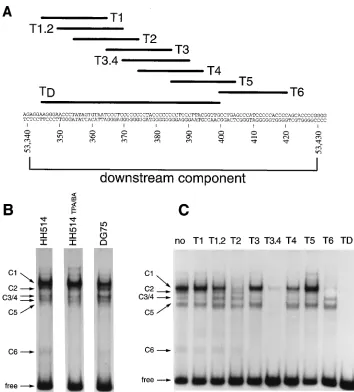
radio-actively labeled and incubated with nuclear extracts derived from the HH514 cell line. Several protein-DNA complexes with different mobilities, complexes C1 to C6 (Fig. 2B), were reproducibly detected. In some experiments, the complex C3 comigrated with the complex C4, as is the case in Fig. 2C; however, these two complexes differed in terms of competition and protein DNA contact points (see below). Two other com-plexes with higher electrophoretic mobilities were also occa-sionally detected. The same complexes with indistinguishable electrophoretic mobilities were seen with protein extracts from induced HH514 cells or from DG75 cells (Fig. 2B). These results demonstrated that cellular proteins interact with the
oriLyt downstream element. Since we were unable to detect
[image:2.612.139.478.70.195.2]any change in the DNA-protein complexes in this assay by comparing uninduced and induced cell extracts, we think that no EBV protein or EBV-induced factor interacts directly with this element in vitro and that its activation is mediated by proteins already bound to it during the latent phase of EBV’s
FIG. 1. Structure of oriLyt of EBV. The lytic origin is flanked by two divergent genes of BHLF1 and BHRF1, which are shown together with their promoter localizations and mRNA structures. The localizations of the two essential elements of oriLyt, the upstream and downstream components are shown in the upper part of the figure, together with those of the auxiliary regions which increase the efficiency with which oriLyt replicates. The fine structure of the central region of oriLyt encompassing the upstream and downstream components, the seven ZRE binding sites for the viral transactivator BZLF1, the binding sites for the R enhancer factor, and the consensus sequences for the TATA and CCAAT box-binding proteins are shown. The coordinates for the boundaries of the two essential components, which are approximately 540 bp apart, are shown.
VOL. 69, 1995 CELLULAR PROTEINS AT oriLyt 1879
on November 9, 2019 by guest
http://jvi.asm.org/
life cycle. It is also possible that the detected complexes are prevalent in our extracts and mask the detection of other still unidentified complexes.
To assess the specificity of the detected protein-DNA inter-actions in the electrophoretic mobility shift assays, we intro-duced specific DNA competitors which constitute small parts of the downstream component of oriLyt. Formation of the C1 complex could be inhibited with high efficiency by adding a 200-fold excess of oligonucleotide T2 and T6 (Fig. 2A and C). A 100-fold excess of the oligonucleotide T3.4 reduced the formation of the C2 and C4 complexes (data not shown); a 200-fold excess of T3.4 competed with the formation of all complexes (Fig. 2C). Less clearly visible in Fig. 2C, the T2 and T6 oligonucleotides could also inhibit the formation of com-plex C3 but not comcom-plex C4, which could be reproduced in a number of electrophoretic mobility shift assays. The C6 com-plex, which has the fastest electrophoretic mobility, can be easily inhibited by any GC-rich oligonucleotides such as T3,
[image:3.612.126.480.67.459.2]T3.4, T4, T5, T6, and TD. The T6 probe, which lies down-stream from the TD fragment (Fig. 2A), gave rise to at least two specific complexes (data not shown) which could be inhib-ited to various extents by a 100-fold molar excess of the oligo-nucleotides T2, T3.4, and TD. The observations that the T6 oligonucleotide, which is located outside the TD probe, could inhibit the formation of most of the complexes observed with the TD probe and, reciprocally, that complexes obtained with the T6 probe could be inhibited by oligonucleotide sequences present in TD (data not shown) indicate that identical or similar proteins bind to several sites within the oriLyt down-stream element. This interpretation was also indicated by ex-periments in which the complete oriLyt downstream element was used as a probe in the gel shift assays (data not shown). Depending on the molar excess of the competitors, T2 and T3.4 could compete for the formation of identical complexes, which suggested that the same protein(s) can interact with different affinities with different parts of the oriLyt downstream
FIG. 2. Cellular protein-DNA complexes formed on the oriLyt downstream element. (A) Nucleotide sequence of the oriLyt downstream component from nucleotide coordinates 53340 to 53430 (1). The positions of the probe (TD) used for the gel shift assays and of the different oligonucleotides used in the competition experiments are indicated at the top. (B) Electrophoretic mobility shift assay. The positions of the different complexes formed with the TD probe are indicated by arrows, with complexes C1 through C6 being indicated. The nuclear extracts were derived from an EBV-positive cell line, HH514, uninduced or induced with a combination of TPA and Na-butyrate (TPA/BA) to undergo the lytic cycle, and from an EBV-negative cell line, DG75. (C) Competition analysis. HH514 nuclear extract was incubated with the TD probe in the presence of a 200-fold molar excess of unlabeled double-stranded competitor oligonucleotides (labeled T1 to TD in panel A). The lane marked no did not contain any specific competitor oligonucleotide.
on November 9, 2019 by guest
http://jvi.asm.org/
element. Taken together, these results show that different cel-lular proteins interact specifically with the oriLyt downstream element in vitro and that three subsequences of this element (oligonucleotides T2, T3.4, and T6) are important for these interactions.
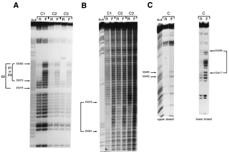
Characterization of the binding sites of different complexes. The contact points of these different complexes with the TD oligonucleotide were analyzed by chemical modifications of the labeled probe with dimethyl sulfate for the lower strand (methylation interference analysis) or, since there are no gua-nine residues on the upper strand, with ethylnitrosourea (ethy-lation interference analysis). The results of these experiments are shown in Fig. 3 (and are summarized in Fig. 9). The C1 and C3 complexes, which were inhibited by the T2 oligonucleotide,
showed identical contact points on the DNA sequence 59
-GGGGGAGGG-39from nucleotide positions 53365 to 53373.
The presence of complex C3, which varied between experi-ments, is possibly due to a degradation product of the protein present in complex C1, or, alternatively, the C1 complex is a multiprotein complex between the protein present in complex C3 and other cellular proteins. The contact points for complex C2 were less clear. Presumably, they colocalized to the same region, but three adjacent guanine residues seemed to partic-ipate in the interaction (Fig. 3A). The interference on these three additional residues extended to nucleotide position 53376 and was much weaker than those of the other guanines
in the 59 part of this site. Contact points for the C4 complex
could not be well established to draw a conclusion about the binding sites involved. The result of the ethylation interference on the upper strand (Fig. 3B) confirmed that complexes C1 and C3 had similar contact points and that the protein present in complex C2 had a binding site overlapping with that present in complex C1. For the C4 complex, no clear interference could be detected.
DMS interference assays with several complexes obtained with the T6 probe in electrophoretic mobility shift assays (data not shown) were indistinguishable (Fig. 3C shows an example). The region between nucleotide positions 53406 and 53417 on the lower strand and the position of guanine residues at posi-tion 53405 and, more weakly, at posiposi-tion 53403 on the upper strand indicated an additional protein complex at this position of the downstream component of oriLyt.
Sp1 or an Sp1-like protein binds to theoriLytdownstream
component.Since the Sp1 protein has been shown to interact
with DNA sequences similar to the sequence present on the
oriLyt downstream element, we wanted to examine the
[image:4.612.83.532.69.366.2]possi-bility that Sp1 or a related protein is present in one of the complexes. Different approaches were used to address this possibility. First, a consensus Sp1-binding site oligonucleotide could efficiently compete for the formation of the C1 complex in the gel shift assay, while an unrelated oligonucleotide with a similar but different GC-rich binding site for the transcription factor AP2 was ineffective (Fig. 4A). Second, preincubation of the nuclear extract with a rabbit serum directed against Sp1
FIG. 3. Contact point analysis of proteins that bind to the oriLyt downstream component. (A) Guanine residues required for protein-DNA interaction on the lower strand of the TD probe identified by DMS interference as described in Materials and Methods. (B) Phosphate residues required for protein-DNA interaction on the upper strand of the TD probe identified by ethylation interference as described in Materials and Methods. C1 to C3 refer to the different complexes described in the legend to Fig. 1. R, retarded complex; F, free probe. G1A, Maxam and Gilbert sequence reaction of the probe. The numbers on the left of the figure indicate the nucleotide coordinates of the contact point regions on the oriLyt sequence (1). (C) DMS interference assay on the T6 region of the oriLyt downstream component on the lower and upper DNA strand. A number of differently migrating DNA-protein complexes can be detected with the T6 oligonucleotide in gel shift assays. They all give identical DMS interference patterns (data not shown), and, therefore, only one of them is shown in this part of the figure. Guanine residues which are contacted by proteins, together with their nucleotide coordinates, are indicated.
VOL. 69, 1995 CELLULAR PROTEINS AT oriLyt 1881
on November 9, 2019 by guest
http://jvi.asm.org/
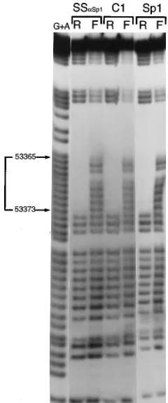
prior to the addition of the TD probe diminished the specific shift that was observed for the C1 complex but not for the others, and a new, supershifted complex (denoted SS) ap-peared (Fig. 4B). Preincubation of the nuclear extract with anti-Ets1 antiserum had no effect. As a control, we used a nuclear extract derived from Sf9 insect cells. It is known that Sp1 is not present in this insect cell line (5), and, indeed, no supershift was observed when the nuclear extract was preincu-bated with anti-Sp1 antiserum prior to the addition of the TD probe (Fig. 4B). However, the specific supershift with anti-Sp1 serum could easily be detected in nuclear extract from SF9 cells which had been transfected with the Sp1-expressing plas-mid pPacSp1 (Fig. 4B). Third, contact point analysis by meth-ylation or ethmeth-ylation interference revealed that the nuclear protein present in the C1 complex, purified Sp1 protein, and the protein present in the supershift complex with anti-Sp1 antiserum contacted identical guanine residues and were vir-tually indistinguishable (Fig. 5). Taken together, these obser-vations strongly support the interpretation that Sp1 or a closely related protein interacts with the oriLyt downstream element to form the C1 complex observed in vitro with different nuclear extracts.
There is more than one Sp1-binding site on theoriLyt
down-stream element.Competition assays carried out with the TD
[image:5.612.135.474.72.285.2]probe (see above and Fig. 2) revealed that the T6 oligonucle-otide competed efficiently for the formation of complex C1, in which Sp1 is most likely involved. This finding indicated that Sp1 can also interact with the T6 element. To test this idea, we used a DNase I footprint assay with the complete oriLyt down-stream sequence preincubated with the purified Sp1 protein (Fig. 6). As expected, a clear footprint is observed on the T2 subsequence from nucleotide positions 53358 to 53375. This finding is in complete agreement with the results described above. In addition, a clear footprint is also detected in the sequence overlapping the T6 oligonucleotide, which is very rich in guanine residues (nucleotide coordinates 53407 to
FIG. 4. Sp1 or an Sp1-related protein binds to the oriLyt downstream element. (A) Competition analysis. HH514 nuclear extract was incubated with the TD probe in the presence of a 25- or 50-fold molar excess of unlabeled competitor oligonucleotides corresponding to a canonical Sp1-binding site or an AP2-binding site. TD, competition with the unlabeled TD oligonucleotides; no, contains no specific competitor oligonucleotide; arrows, the positions of the complexes. (B) Analysis with an antiserum directed against Sp1. HH514 nuclear extract or SF9 nuclear extract, containing or lacking the Sp1 protein, was preincubated with an antiserum (a) directed against Sp1 or against the Ets protein prior to the addition of the TD probe. no Ab, preincubation reaction mixture without antiserum; SS, position of the specific super-shifted complex.
FIG. 5. Comparison of the contact points of proteins that bind the oriLyt downstream component. Guanine residues required for protein-DNA interac-tion on the lower strand of the TD probe are identified by DMS interference as described in Materials and Methods. C1 and SSaSp1, the HH514 complexes described in the legend to Fig. 4B; Sp1, the complex formed with a biochemically purified Sp1 protein; R, retarded complex; F, free probe; G1A, the Maxam and Gilbert sequence reaction of the probe. The numbers indicate the positions of the nucleotide residues on the oriLyt sequence which are necessary for the DNA-protein interaction.
on November 9, 2019 by guest
http://jvi.asm.org/
[image:5.612.374.494.362.649.2]53418). The binding of Sp1 to this sequence was confirmed by two different experiments. First, the canonical Sp1 binding site could compete for the fixation of a cellular protein on the T6 oligonucleotide used as a probe in gel shift assays, and, second, a clear supershift is detectable with the T6 probe and an anti-Sp1 serum (data not shown). Together, these observations confirmed that Sp1 or a very similar protein binds twice to two adjacent sites in the oriLyt downstream element.
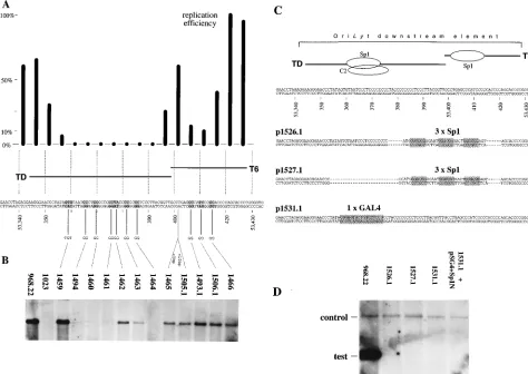
oriLytmutants and replication assays.Mutation analyses of the downstream component of oriLyt have revealed a distinct region which is sensitive to introduced mutations (23) (Fig. 7A for a compilation of the data). The TD probe which binds several proteins including Sp1 spans this very sensitive region, and the T6 probe covers the flanking sequence which lies downstream of TD (Fig. 7A). We tested whether any of the protein-DNA interactions found within the downstream com-ponent of oriLyt was crucial for DNA replication. We created 2-base exchange mutants within this region (Fig. 7A) which were embedded into the BamHI-SalI fragment of p968.22 (23). These oriLyt mutants were analyzed for their replication effi-ciencies in the transient replication assay. As Fig. 7B summa-rizes, four mutants (p1494, p1460, p1461, and p1464) were completely inactive in the replication assay; a fifth was much reduced (p1463). All of the other mutants were able to repli-cate, although most of them only at a level of 20 to 30% compared with the wild-type origin in p968.22. One mutant (p1459), which lies within the T1 region (Fig. 2A) in which no protein complex could be detected, was unchanged in its
ca-pacity to replicate. These data demonstrate that the formation of protein-DNA complexes and the location of important cis-acting sequences perfectly coincide in the TD region. Less dramatically, artificial spacing (p1465 and p1505.1) or alter-ations of the DNA sequence around the contact points of the putative Sp1 site within the T6 region led to a reduction in DNA replication.
Since we found that Sp1 or a very closely related protein binds to the downstream component, we asked whether the binding of Sp1 would be crucial for the activation of oriLyt. To test this hypothesis, we created three additional mutants. p1526.1 and p1527.1 replaced parts of the downstream com-ponent (Fig. 7C) with three canonical Sp1-binding sites from the simian virus 40 aux-2 region (17), to which no protein other than Sp1 (and T antigen) is known to bind. The mutation in p1531.1 introduced a single GAL4-binding site at the position of the C1 complex. The rationale for this particular mutation was the attempt to create a null mutant of oriLyt by substituting the genuine Sp1-binding site in oriLyt for a GAL4-binding site. We hypothesized that such a mutation might be functionally restored by expressing a fusion protein which links the trans-activation domain of Sp1 to the DNA-binding domain of GAL4 (5, 21). Unexpectedly, all three mutants were com-pletely inactive in the transient replication assay, despite the fact that cellular Sp1 or, in the case of the p1531.1 mutant, the GAL4:Sp1 fusion protein was present in the cells (Fig. 7D and data not shown). This finding could mean that there is a spatial requirement that is essential for the functioning of the Sp1 site in oriLyt and that this requirement could have been perturbed in all of the mutants. Similarly, it is conceivable that the GAL4: Sp1 fusion protein cannot perform functions of the native Sp1. This assumption is emphasized by the fact that Sp1’s function in the downstream element of oriLyt is clearly not transcrip-tional in nature (data not shown). Alternatively, this finding could also mean that Sp1 either is not involved in oriLyt-mediated DNA replication or alone is insufficient for this ac-tivity.
Some nonfunctionaloriLytmutants still bind Sp1.Several
mutations which span the location of the C1 complex com-pletely abolished the capacity of oriLyt to replicate. A possible correlation between these specific mutations and the capacity for Sp1 to interact with this region might illuminate whether Sp1 is directly involved in oriLyt-mediated DNA replication. In order to test this idea, we made five different radioactively labeled probes by PCR with oligonucleotide primers which amplified a fragment spanning nucleotide coordinates 53337 to 53404 of the B95-8 strain of EBV (1). The wild-type oriLyt sequence is represented by the PCR product TD shown in Fig. 8; the mutant PCR products TD94, TD60, TD61, and TD63 correspond to the plasmids p1494, p1460, p1461, and p1463, respectively. These different probes were used in a gel shift assay with D98HR1 nuclear extracts alone and in conjunction with different specific competitors. The results presented in
Fig. 8 showed that two mutations in the 59or the 39sequence
of the SP1 binding site (TD94 or TD61, respectively) did not impair the formation of the complex C1 but that the mutation introduced in the center of the binding site (TD60) markedly reduced the formation of complex C1. As expected, the bind-ing of Sp1 was not altered by a mutation outside this sequence as that present on TD63. It is interesting to note that none of these mutations affected the formation of the complexes C2 or C4 which lie on this sequence. Taken together, these results demonstrated that there is no direct correlation between the capacity of Sp1 to interact with the T2 subsequence of the
oriLyt downstream element and the replication functions
[image:6.612.107.225.71.349.2]me-diated by this cis-acting element. Moreover, it seems that none
FIG. 6. DNase I footprint analysis for the binding of Sp1 on the oriLyt downstream component. G1A, the Maxam and Gilbert sequence reaction of the probe used which was labeled on the 59end of the lower strand as described previously (19). The numbers indicate the positions of the residues on the oriLyt sequence (1) which are protected by bound biochemically purified Sp1 against digestion by DNase I. no, absence of protein in the DNase I reaction; Sp1, probe preincubated with biochemically purified Sp1 protein before the addition of DNase I; triangles, increasing amounts of DNase I used.
VOL. 69, 1995 CELLULAR PROTEINS AT oriLyt 1883
on November 9, 2019 by guest
http://jvi.asm.org/
of the additional complexes are affected by nonfunctional mu-tations introduced in this part of the oriLyt downstream se-quence.
DISCUSSION
Essential regions of origins of DNA replication of higher eukaryotic systems all contain cis-acting genetic information necessary for the interaction with origin-specific DNA-binding proteins. This binding is usually accompanied by the initiation of DNA replication in this region. In the case of the better studied DNA viruses, the crucial origin-specific DNA-binding protein is a highly specialized factor encoded by the virus. In simian virus 40 and polyomavirus, it itself participates in the local unwinding of the origin with the enzymatic activity of a
[image:7.612.64.538.73.409.2]helicase. The lytic origin of EBV seems to be structurally different from that of any eukaryotic origin known so far. It displays two nondispensable core elements which lie more than 500 bp apart. Both core elements are unique in DNA sequence composition, and yet both must carry complementary functions which activate this origin in cis. The upstream component colocalizes with a generic basal promoter element which is concomitantly activated with oriLyt. Transcriptional regulation of this promoter is mediated by BZLF1, the same viral factor which appears to be absolutely required for oriLyt-mediated DNA replication (22). Thus, BZLF1 scores as a dual factor that is involved in transcriptional activation as well as in DNA replication. We do not know what the contribution of BZLF1 to DNA replication is; however, its binding to several sites within the upstream component and to nearby sites makes it
FIG. 7. Functional analysis of mutant oriLyt plasmids in the transient replication assay. (A) Histogram resulting from fine mapping of the downstream component of oriLyt by scanning deletion mutants (the replication efficiency of wild-type plasmid p968.22 was set to 100%). The histogram is an excerpt from Schepers et al. (23) to indicate the exact boundaries of the regions crucial for oriLyt replication. Substitution mutants which carried 2- to 3-base exchanges compared with the wild-type sequence were generated, and the single mutations are indicated. (B) Mutant plasmids that were tested in the transient replication assay as described elsewhere (23) and whose replication efficiencies were calculated. p968.22 represents the wild-type oriLyt; p1023 is a nonfunctional mutant which maps to the central region of the downstream component (23); and p1465 and p1505.1 are oriLyt mutants into which a 5- or a 10-bp spacer was introduced and which carry no further mutations. The replication efficiencies were as follows: p1459,.100%; p1494 through p1461,,1%; p1462, 15%; p1463, 5%; p1464,,1%; p1465, 15%; p1505.1, 15%; p1493.1 through p1466, 25%. (C) Replacement mutants of the downstream component of oriLyt. Two mutant plasmids, p1526.1 and p1527.1, contain three Sp1-binding sites from the aux-2 region of the simian virus 40 origin of replication which replace two different regions of oriLyt, including the single Sp1 site at the position of T6 or both Sp1 sites as indicated. The mutant plasmid p1531.1 contains a single GAL4 consensus binding site at exactly the same position as that of the Sp1 site which was mapped in this part of the downstream component. Note that all oriLyt mutant plasmids affect more than a single protein-binding site. (D) The mutant plasmids shown in panel C were analyzed for their replication efficiencies in the transient replication assay. All oriLyt mutants are nonfunctional in this assay compared with wild-type oriLyt in p968.22. As an internal control, a small amount of the wild-type oriLyt plasmid p526 (15), which served as a positive standard in the replication assay as described elsewhere (23), was cotransfected. The oriLyt mutant plasmid p1531.1 was tested in the absence of and in conjunction with an expression plasmid, pSG41Sp1N, which carries a fusion protein between the DNA-binding domain of GAL4 from amino acids 1 to 147 and the activation domain from the Sp1 cDNA from amino acids 83 to 621. This fusion protein is expressed from the simian virus 40 early promoter as described elsewhere (21) and is active in transcriptional activation systems in our hands (data not shown) but failed to activate the oriLyt mutant p1531.1 with regard to DNA replication.
on November 9, 2019 by guest
http://jvi.asm.org/
likely that a cluster of binding sites for BZLF1 together with elements dedicated to transcription leads to the assembly of a higher-order protein complex. The binding of BZLF1 to sites further downstream appears to be less important (21a, 23), indicating that more distant binding sites do not participate in this putative complex formation.
The function of the downstream component is even more enigmatic. Its sequence composition is exceptional in that it contains long runs of C/G residues which do not participate in the formation of an apparently peculiar DNA or RNA struc-ture. Its functional sensitivity to certain very minor base alter-ations is consistent with its being dedicated to interacting with sequence-specific DNA-binding proteins. In a first attempt, we could identify several protein-DNA complexes in vitro which were solely derived from the cell as it is summarized in Fig. 9. This was unexpected, since we thought that the activation of
oriLyt resulted in a distinct pattern of proteins which occupy
the downstream component. On the other hand, none of the
viral trans-acting genes which are essential for lytic DNA rep-lication of EBV is a known DNA-binding protein which re-quires a dedicated sequence motif (10). This is in striking contrast to members of the alphaherpesvirus family, including herpes simplex virus, which encode an origin-specific DNA-binding protein (see references 2 and 14 for reviews). In anal-ogy to these herpesviruses, the lytic origin of DNA replication of EBV requires a similar function. BZLF1 might constitute such a function; however, unlike the origin-specific DNA bind-ing proteins of other herpesviruses, it does not contain a known intrinsic ATPase-helicase activity (9). If our working hypothesis is correct, then there could be an additional, pre-sumably cellular factor at the downstream component of oriLyt which provides a helicase activity to coact with BZLF1.
In this study, we have found several DNA-protein complexes in vitro within the downstream component. One of the proteins was tentatively identified as the transcription factor Sp1 or an Sp1-related factor. Although the downstream component of
oriLyt does not modulate the activity of the two promoters for
the BHLF1 and BHRF1 genes (23 and data not shown), this element functions as an upstream regulatory region when it is placed close to a basal promoter and is capable of activating it in an Sp1-dependent manner (data not shown). This finding, together with our mutational analysis, suggests that proteins bind to this region; however, it appears unlikely that Sp1 or any of the other still undefined proteins is capable of mastering the activation of oriLyt in conjunction with BZLF1. One of the problems is the colocalization of several complexes within 20 bp, as is summarized in Fig. 9. This makes it virtually impos-sible to dissect this region in a standard biochemical scheme and therefore requires an alternative approach. Interestingly, a similar problem has been encountered with the lytic origin of DNA replication of herpes simplex virus type 1, in which DNA-protein interactions which seem to share partially the same DNA sequence requirements were mapped (6). As it seems to be the case with the downstream component of oriLyt, cellular proteins are involved in these DNA-protein interactions.
ACKNOWLEDGMENTS
We thank Bill Sugden for continuous support, helpful discussions, and comments on the manuscript. We thank Grace Gill for the
[image:8.612.144.472.69.233.2]gen-FIG. 8. Analysis of the gel shift complexes formed on the oriLyt downstream component and four base-exchange mutants of this element. TD corresponds to the labeled PCR fragment as defined in the legend to Fig. 1. TD94, TD60, TD61, and TD63 represent the corresponding PCR fragments amplified on the basis the oriLyt mutant plasmids p1494, p1460, p1461, and p1463, respectively, which were used as templates in the PCR. Competitions were done with a 100-fold molar excess of unlabeled oligonucleotide T2 or T3.4 (as in Fig. 2C) or the consensus Sp1 oligonucleotide. The names of the different complexes are the same as those in Fig. 2B. Although the mutant plasmids p1494, p1460, and p1461 all failed to replicate, the electrophoretic mobility shift assays with the corresponding PCR fragments were either indistinguishable from the TD probe, as was the case with TD94 and TD61, or failed to form the complex C1, as was the case with TD60.
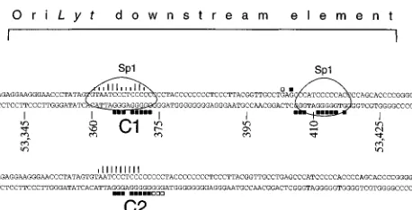
FIG. 9. Summary of the positions for proteins which bind to the oriLyt down-stream component. The nucleotide sequence of this element spans positions 53340 to 53430 of the B95-8 strain of EBV (1). Nucleotides which interfere with the binding of the proteins when methylated are indicated by filled squares at the respective guanine residues. Open squares indicate weaker interferences. Phos-phates which interfere with the binding of the proteins when ethylated are indicated by vertical bars above the sequence for the upper strand. The length of the line is proportional to the intensity of the interference, as determined by scanning of the ethylation interference gels with a PhosphorImager. The circled regions indicate the nucleotides which are found to be protected against DNase I digestion by biochemically purified Sp1.
VOL. 69, 1995 CELLULAR PROTEINS AT oriLyt 1885
on November 9, 2019 by guest
http://jvi.asm.org/
[image:8.612.65.294.516.633.2]Sergeant.1986. Both Epstein-Barr virus (EBV)-encoded trans-acting fac-tors, EB1 and EB2, are required to activate transcription from an EBV early promoter. EMBO J. 5:3243–3249.
4. Countryman, J., and G. Miller. 1985. Activation of expression of latent Epstein-Barr herpesvirus after gene transfer with a small cloned subfragment of heterogeneous viral DNA. Proc. Natl. Acad. Sci. USA 82:4085–4089. 5. Courey, A. J., and R. Tjian. 1988. Analysis of Sp1 in vivo reveals multiple
transcriptional domains, including a novel glutamine-rich activation motif. Cell 55:887–898.
6. Dabrowski, C. E., P. J. Carmillo, and P. A. Schaffer. 1994. Cellular protein interactions with herpes simplex virus type 1 oriS. Mol. Cell. Biol. 14:2545– 2555.
7. Dignam, J. D., R. M. Lebovitz, and R. G. Roeder. 1983. Accurate transcrip-tion initiatranscrip-tion by RNA polymerase II in a soluble extract from isolated mammalian nuclei. Nucleic Acids Res. 11:1475–1488.
8. Farrell, P. J., D. T. Rowe, C. M. Rooney, and T. Kouzarides. 1989. Epstein-Barr virus BZLF1 trans-activator specifically binds to a consensus AP-1 site and is related to c-fos. EMBO J. 8:127–132.
9. Fierer, D. S., and M. D. Challberg. 1992. Purification and characterization of UL9, the herpes simplex virus type 1 origin-binding protein. J. Virol. 66: 3986–3995.
10. Fixman, E. D., G. S. Hayward, and S. D. Hayward. 1992. trans-acting re-quirements for replication of Epstein-Barr virus ori-Lyt. J. Virol. 66:5030– 5039.
11. Glaser, R., and M. Nonoyama. 1974. Host cell regulation of induction of Epstein-Barr virus. J. Virol. 14:174–176.
17. Kadonaga, J. T., K. A. Jones, and R. Tjian. 1986. Promoter-specific activa-tion of RNA polymerase II transcripactiva-tion by Sp1. Trends Biochem. Sci.
11:20–23.
18. Liebowitz, D., and E. Kieff. 1993. The Epstein-Barr virus, p. 107–172. In B. Roizman, R. J. Whitley, and C. Lopez (ed.), The human herpesviruses. Raven Press, New York.
19. Maxam, A. M., and W. Gilbert. 1977. A new method for sequencing DNA. Proc. Natl. Acad. Sci. USA 74:560–564.
20. Reisman, D., J. Yates, and B. Sugden. 1985. A putative origin of replication of plasmids derived from Epstein-Barr virus is composed of two cis-acting components. Mol. Cell. Biol. 5:1822–1832.
21. Sadowski, I., and M. Ptashne. 1989. A vector for expressing GAL4(1-147) fusions in mammalian cells. Nucleic Acids Res. 17:7539.
21a.Schepers, A., and W. Hammerschmidt. Unpublished data.
22. Schepers, A., D. Pich, and W. Hammerschmidt. 1993. A transcription factor with homology to the AP-1 family links RNA transcription and DNA repli-cation in the lytic cycle of Epstein-Barr virus. EMBO J. 12:3921–3929. 23. Schepers, A., D. Pich, J. Mankertz, and W. Hammerschmidt. 1993. cis-acting
elements in the lytic origin of DNA replication of Epstein-Barr virus. J. Virol. 67:4237–4245.
24. Takada, K., N. Shimizu, S. Sakuma, and Y. Ono. 1986. trans activation of the latent Epstein-Barr virus (EBV) genome after transfection of the EBV DNA fragment. J. Virol. 57:1016–1022.
25. Yates, J. L., N. Warren, and B. Sugden. 1985. Stable replication of plasmids derived from Epstein-Barr virus in various mammalian cells. Nature (Lon-don) 313:812–815.