NATIONAL INSTITUTE OF SIDDHA
Tambaram sanatorium
,
Chennai - 47
AFFILIATED TO THE TAMIL NADU DR. M.G.R. MEDICAL UNIVERSITY
CHENNAI - 600 032
A STUDY ON
KALLADAIPPU
(DISSERTATION SUBJECT)
For the partial fulfillment of the
requirements to the Degree of
DOCTOR OF MEDICINE (SIDDHA)
BRANCH I– MARUTHUVAM
ACKNOWLEDGEMENT
I express my sincere thanks to chief siddhar Sivan, for the performs what
is appointed for me.
I express my thanks to The Tamil Nadu Dr. M. G. R. Medical University
Chennai 32
I express my thanks to our director Prof. Dr. V.Arunachalam M.D. (s)
National Institute of Siddha, Tambaram sanatorium – 47.
I would like to express my immense gratitude to our respectable Head of
the department Prof. Dr. K. Manikavasagam, M.D. (s) whose excellent guidance
and valuable suggestion have enabled me to complete this dissertation in good
shape.
I whole heartedly thanks to Dr. M. Logamanian M.D. (s) Asso. Prof. of NIS
for her constant motivation and help in doing this work.
I also express my sincere thanks to Dr. G. Ujjeevanam M.D. (s) for his
I acknowledge my thanks to Prof. Dr. C. Venkataraman, the director of
C.L.Baid medha pharmacy college, Thoraibakkam, Chennai – 96, for his support
for pharmacological and phytochemical study.
I my sincere thanks to Mr. Thirunavakarusu and Mr. Kannathasan and
research student of C.L.Baid medha pharmacy college, Thoraibakkam, Chennai
– 96, for his support for pharmacological and phytochemical study.
My sincere thanks also go to Mr.P.Jayapal MSc Asst. Prof.of Biostatics in
NIS, Tambaram for his guidance in this study.
I fully heartily thanks to my parents, friends and relatives who are behind
CONTENTS
Page No
1. Introduction
1
2. Aim and object
5
3. Literature Review
a) Siddha aspects
6
b) Modern aspects
29
4. Materials and Methods
53
5. Observation and Results
56
6. Discussion
74
7. Summary
79
8. Conclusion
80
9. Annexure
a) Preparation and properties of Trial Drug
81
b) Bio chemical analysis
92
c) Pharmacological evaluations
96
d) Forms
i) Selection proforma
103
ii) Assessment proforma
112
iii) Consent form
116
10. Bibliography
INTRODUCTION
Siddha system of medicine is one of the ancient systems Contemporaneous with those of the submerged lands, Egyptian, Mesopotamian, Chinese and Grecian medicines. The unique nature of the system is its continuous service to humanity fore more then five thousand years in combating diseases, and in maintaining its physical, mental and mortal health.
The Siddha medicine is well founded on the basic principles of nature and its elements after a careful and through study of the human system.
The ancient Tamil in their quest for knowledge for longevity had developed two ways, by which man can achieve mastery over nature; the one is the yogic way and other through medicines. Yogies is known as Siddhars. Hence the system of medicine profounded by them came to be known as siddha system of medicine.
Siddha is a Tamil word that is derived from its root Siddhi – which means perfection in life or heavenly bliss. Siddhars attained eight kinds of supernatural powers. The persons who had attained eight kinds of miraculous power in the life are known as Siddhars.
The earliest medical treatise in Tamil was propounded by Sivanar,
who was the first to preside over the ancient first Tamil academy. It was followed by a number of works of immortal Siddhars.
Siddha system dealing not only science of life, nature of universe, astronomical data, cosmic dance, atomic theory, space travel, alchemy
Agastya siddhar who is the chief of the Siddhars’ school is said to have been a celebrated philosopher and physician who laboured amongst
the Tamil in southern India.
The man is said to be the microcosm and the world the macrocosm; because what exists in the world exist in man; or in other words there is nothing in the macrosom of nature that is not contained in man. So man
must be looked upon as an integral part of universal nature and not as anything separate or different from the latter. In the organism of man, these forces may act in an abnormal manner and cause diseases there by. Similarly, in the great organism of the cosmos, they may act abnormally likewise and bring about disease on earth. Seven planets exercise special power over some part of the body to cause diseases or diseases
accordingly to their influences on the three humours in the system.
The human body is composed ninety six thatwas or constituent principles in nature including elements, bodily and mental organs, faculties matter etc.
The three humours are vadham, pittam and kabham. They are the three fundamental principles and essential factors in the composition and constitution of the human body. Three humours are representing respectively the air, the fire and the water of the five elements. Which form
the connecting link between microcosm or man and macrocosm or world? The external air, heat and water correspond to the internal vatham, pitham and kabam.
Man is thus linked with external world and any change in the elementary condition of the external world has its corresponding changes in
The siddhars materia medica is based on human pathology. It asserts that all substances of the animal, the herbs and the mineral
kingdoms contain one or more of these three humous in their composition. They should plan on important role in the maintenance of these humours the men and women. Siddhars knowledge is on minerals, metals and plants suitable combination of these things on processing and the preparation of medicine. According to the classification of the five elements
of nature was of a very high order as can be learned from the lines of the ancient siddha medicine.
Super intellectual siddhars devised and utilized special technique for diagnosis a disease known as envagai thervugal. These eight factors play a vital role in finding out disease and deranged life elements.
Urinary calculus is known to mankind since antiquity. The earliest recorded example being bladder and kidney stones detected in Egyptian mummies dated to 4800 B C.
Kalladaippu is most common disease of the present society Due to
sedentary life style and abnormal diet habits.
Clinical features of kalladaippu are lower abdomen pain, dysuria, oliguria, burning micturition, nusea, vomiting, haematuria and fullness of
abdomen.
In the contemporary world, till now no internal medicine is found for the renal calculus. In spite of new approach in diagnosis and management like Extra Corporeal Shock-wave Lithotripsy (E. C. S. L) and endoscopy surgery, but it still continues to cause significant morbidity with high
tendency of reoccurrence and this kind of treatment procedure not reach to low socio economic status families, who are mostly affected renal calculus.
medicine is ancient duration system of medicine for centuries has provided proven treatment of disease like kalladaippu without morbidity.
We must praise, safe guard and follow the Siddha system of medicine and all so furnish it to take up with the up to date advancement.
So the author of this dissertation work has selected kalladaippu
AIM AND OBJECTIVES
The aim of this work is to select the case of kalladaippu patients
and administer them with the trial drug, as per the line of treatment and analysis clinically and experimentally to prove efficacy of the drugs. Siddha literature revealed so many drugs for kalladaippu; among them the author has selected kalladaippu thool.
The main objective of the patient study is to create awareness about Siddha medicine and highlight the efficacy of Siddha medicine for the disease of kalladaippu among the public with basic intention in mind. The following specific objectives have been drawn.
To collect and compare varies siddha and modern medicine.
To have an idea about incidence of kalladaippu with reference of age, sex, residence (Nilam), occurrence and duration of diseases.
To have a detailed study of investigation, diagnosis and prognosis of
disease.
To study through investigation by Siddha fundamental and modern investigation during and after treatment in all patients.
To study the urine analysis, hematological study and
ultrasonography for all patients.
To study the biochemical analysis of trial drug.
To study pharmacological diuretic study and acute toxicity study of
trial drug.
lp (
KALLADAIPPU)
The disease kalladaippu is placed under siruneer noi or moothira noi. Which is classified in to,
1. Neerinai arukkal noi (Oligurial diseases)
2. Neerinai perukkal noi (Polyurial diseases)
3. Neerilivu, Venner and Thelimeer.
This has been mentioned by Therayar in his ‘Theran karisal’ as follows
snai i k naii elm
nai p eken nai ken naii !" el#m nard& i e'ns
-"n il
Kalladaippu is one of the Neerinai arukkal noigal
Verupeyar (synonym): Achmari
i
l (
DEFINITION)
According to the text Siddha maruthuvam by Dr. Kuppusamy.
There is gradual or suddenly obstruction to the flow of urine, pain with burning sensation in the urethral tract, low back pain, renal angle pain, and sands like crystal’s deposit in urine. These are characteristic features of
seR +il nanaen
,i"r e-eνm /lp en iie l p
'/ ni1m "nayk " 3en a/i" e/n "m
e'i l#kLn i1m " "en N-ky +ikp
/ +ip nar ""'
"'! µniil eil N&
e- " L d&/p
'! iie RL "
'/ +ip lip -
"'! m" i" nnil
eip "l i" ey9m
na'! RL i,i id!l
na/" L n m"s
-air !m
Agasthiyar says the definition of kalladaippu as sand likes crystals deposited in urine, followed by small size of stones are excreted in urine. Stones are stagnated in kidney, ureter, urinary bladder and urethra. Pain
with burning sensation start in urethral orifice followed to agonizing pain occurs during the stone moving in urethral tract from the bladder, when the stone removed pain also relieved.
Large concentration of minerals in the bladder or kidney produces calculus or gravel. It is altered with difficulty in passing in urine.
- T. S. Sambasivam Pillai.
Kalladaippu is defined as pain in and around the umbilicus, fever,
dysuria and urine smelling like that of goat’s urine.
NOI VARUM VAZHIKAL (AETIOLOGY
)siµ i"m i&d& na"&m
o1i ; e/ il "na
1i /nl ;n" <il
1i µirni&m l !p"/s
-i+r ! im
Stone formed by derangement of humours of vatham and pittam.
se'in"r l !p<p i "'y iinad e!,i" "/n nl 'in"r p i #in "yn
n " k eL= 'in"r in l n e,ly nari ! na'in"r na#ik l !p< naN nad!k ""'s -9i i in/i
It is worth while to mention the poem of Yoogimamunivar who is authority of Siddhars regional and humoral pathology. He has revealed about this disease since 14th century.
Yoogimamunivar says that as blood clotted in urinary bladder due to urinary tract diseases followed by swelling of urinary bladder, urinary stones are formed in urinary tract induced by humour of vatham and pittham.
snad!/yk Rpik ! / ,i
naiNik >!/l kp "i
>d!/yk ! u! / n k
e!/" keN! e& / "rkm d!/y m<m< iin "rkm
/! e&peL ! k
d-" keN& & NΝm
nr l !piR '"s
In this explains that mind plays a major role in causing many diseases and connection between body and mind and soul is established
i.e., the mukkutram deranged by internal factors such as sexual perversion, anger and robbery.
s,i"r Nrn - "rkm le#m< /ir/Nn nnil a,i" n, 'n #m
a1e& µRN! /n#m, /,i"r /pN! /n#m /nil ;m rn n ,i" iniR B #m
Bkykl !p< n "nn "s -9i i in/i
The causes mentioned here,
• Contaminated water
• Stone crystals, bone, hair and sand mixed food.
• Decayed food
• Carbohydrate food
• Humour of vali induced food
snai &l eyin nard& m <Nm i&nj n nil
N< "nam "nai,, mm
naii "nal ey;m ii ;
N< " /n"s
-i /, Bkm
anuria, arthralgia, pain in genital region and deranged of keezh nokkukaal. This leads to the formation of calculus.
The author also explain that ejaculation of semen is one of the 14 visceral reflexes when one suppress this reflex it causes fever, retention of urine which favours urinary calculi, chest pain, arthralgia, and white
discharge.
sBkin !kin Bµ!rk d!m
k/, l n
"nay ii,m /ik/r "na;N!m
/ii&m i"/nn k"r "/in
ii&m ;k >"s
-i/, Bkm
s na!kl i il a-!l
naink l i na!kl
'i"nay /ik µ m o1kµm
!p i-i&l "/µR
ii;l e1/i -p !k
l !p enν, &mii i ';m
'i /i e& /l
n e&, n na
inark i e #m
an le i= e/ns
-"nay i'km
According to noi vilakkam
• Derangement if humour in blood,
• Exessive indulgence in sexual activity or sexual pervertion,
• Trauma on testes,
• Inflammation of bladder
• Syphilis (Mega noi)
• Stagnation of urine in urinary tract.
• Dryness of semen causes the formation of stones.
• Increased intake of food that cause flatulence.
POTHU KURI KUNANGAL
• Gradual or sudden obstruction to flow of urine,
• Unbearable pain(agonizing pain) in the penis
• Excruciating pain and swelling is experienced at tip of penis if the calculus attempts to expel.
• Colicy pain radiating from loin to groin, lower abdomen, urethra and genitalia if the calculus is irregular with sharp projection.
• Burning and scanty micturation and haematuria.
SYNDROMES ASSOCIATED WITHKALLADAIPPU
uk
sµk D in nn
"r y iil µil enanjil aiiil naii /, il
ain /,inn 'rn "/ip /R &k "R p
ienaki +i/, iii ;N!y !, &pe& /i ,i
'rie& /k/y L=n"s
CLASSIFICATION OF KALLADAIPPU
s"ni"r naii! na/, "'y ik in l !p< Eni"r iin l !p< <N!"r "d&/in l !p< ni"r en/, l !p< " Ri" ii /m ni" p ilni in i" i,iR ikn "s
The most experienced of Siddhars, Yoogimamunivar who has studied the disease according to regional and humoral pathology classifies
kalladaippu in to 4types there are
1. l !p<, 2. i l !p<, 3. Jk l !p<, 4. µkR l !p<.
1. lp
(VALI KALLADAIPPU)
sina ik,z Bkyk i /nn /R m/i i"/ i,il i;/i
/i"r eienj Bn d- iii" i!e!p <d! i
"mi" +B/y i µp<m ui"r " urp< /m M,i"r kl !p< "s
• Retention of urine.
• Lower abdomen pain.
• Pain with swelling of penis.
• Agonizing pain.
• Breathlessness.
• Abdomen distention.
2. i lp
(AZHAL KALLADAIPPU)
sa !pi nν /i
a,yi eiR "" Νm < !pp eie,m <1k /ip
Ed&p"l iip <d! m / !pi ui nai /ykl i
niin i,il /d-k eL=m !pik Ryk > ik
d&"/ ikl !p< "s
• Stone blocked in urinary tract followed by oliguria.
• Pain in urethra like an insertion of burning iron bar, followed by sweating all over body.
• Anuria, Agonizing pain.
• Blood stained calculus stagnated in urethra followed by piercing and boring pain with sensitiveness.
3. k lp (
IYYA KALLADAIPPU
)s ep<'i" il# " i/R Bn"/ R" m 3 "& Ln
i&p<n ! ii Νm
" i,in "N / nil
iien" &pi ir m " e=p<kl il
iky ni,nj i"d/n "s
• Abdomen distention like a bow with pain.
• Pain in all joints of upper and lower limb and hip joints.
• Pain in penis, sweating all over body.
4.
µkR lp (
MUKKUTTRA KALLADAIPPU
)
sni,m nar -iR νm / µN!i i;/i enani,i narν /ip ;m
enay i/l"l ena,ik lnj ni,i nariil n 1m
k i, kl ie/nk ni,i iniµ /iin el#m
en/, l !p< Dd-!"s (9i µi)
• Severe pain in urethra.
• Dysuria.
• Oliguria.
• Crystals excreted in urine like a small sands.
• Handful crystals excreted in urine daily.
• It is fatal disease.
CLSSIFICATION IN DHANVANTHIRI VAITHIAM
sini i i"Rm i"l
n B/i n &,leipn
iini&nj i"R B/i iinν
/ini& BkiB/i nan e/ye/n"s -nnii iim
In Thanvanthiri vaithiyam, kalladaippu is classified to four types they are
leipn (
KALLERIPPAN
)
snar na'nniR BkiniR i"Rm
i/ rl lyp ii ! eL=,
• Increased iyyam and azhal kutram dries the urine and semen forming calculi
• Sudden or gradual obstruction in urinary tract
• Dysuria
• Pain in umbilicus
• Fever
• Anorexia.
"i#$% a'%i (
SILETHUMA ACHMARI
)
snar na'nnil nainnar ieN&
"rm ip< eN / Bkim"l1m "re na e/d&p in/yk l# 1m ere i"Rin aB/i en"/s
• Calculus in the Ureter or urethra causes hydronephrosis
• Oliguria,
• Reddish white in colour and falls out like semen
• Stones are expelled as 4 or 8 fragments.
i a'%i
(PITHA ACHMARI
)
sey;mnar na'nnil i eipe1n
ey; P l en ",ed !"l l#n!m
nay" L ey;m nailm innil
eyB/i en"µn naimi iin/ik"r s
• Burning sensation in urethra due to azhal kutram
• Burning micturition
• Formation of stone that appear like semicarpus anacardium seeds.
(ki a'%i
(SUKKILA ACHMARI
)
sBkim ,il miR ikinn
/ikkl i emi i/y nari!/R
• Suppressions of semen during ejaculation, develops on to stones and obstruction in the flow of urine
• Sand like gravels are expelled
• Pallor of the body.
• This is incurable.
CLASSIFICATION ACCORDING TO NOI VILAKAM
s'i µl +niνn "n#m
nar nR "n#m
l ! nal p &e/e/i"s
There are four types of kalladaippu according to noi vilakkam
1. Vali kalladaippu, 2. Analakalladaippu, 3. Iyya kalladaippu,
4. Karuneer kalladaippu
)iklp (
VALI KALLADAIPPU
)
s!r/ip&l RL-l
na&,l uni;m i;m i l
&z 'ie& l a1l
inar 'il en m i m
'iin l !k i" e/i
n inm µ L nm
'iin l - ν e/ns
• Tongue biting , palpitation and shivering,
• Crushing of the lower abdomen and genital organs
• Dribbling of urine.
a*klp (
ANALA KALLADAIPPU
)
sBde! naim /iemi&#m"na#m a k l !ki"
in, /nj'i;m
", -il l "nms
• Burning micturition,
• Dysuria,
• The stones are reddish black or yellow in colour and passing of smalls stones
klp (
IYYA KALLADAIPPU
)snai, l iil, 'irl
eν/i Jk l !k i"
e=m "i/i e/'irnm
e - !m J l !p<s
• Pricking pain, forceful pain with severe intensity when passing urine,
• Fever with rigors,
• White or honey coloured shining or luminant large size stone expelled.
+na-r lp (
KARUNEER KALLADAIPPU
)s narklin 'i ine1n
i 'i &il a &in
nark l ! /i& e/n naim "nal inar !!l
i ,il e#/i i' m
nar l !k ie e/i
nark li 'i µ&i
ii m ei n N&' enaki&m
a inar i e'ip!m
a inai &l naiRm
Ri nai & naiRin
B e!l e'i /p< nar"d
el'i eν/i i 'ni& e/n
irk in l iiin
ii e&l e'i e/n
nark l ! irk il
i ;m uni;m /i p! ,l
inar !p!l "na /il
en il /i ! "nin
i iep "Br <rs
• Sudden or gradual obstruction to flow of urine,
• Swelling of testis,
• Excessive vali kutram breaks the stones into small and large size crystals and expels along with urine.
• Sudden stoppage of urine stream, • Retention of urine,
• Abdominal pain,
• Loss of taste excessive thirst,
• Pricking pain with swelling of abdomen and testis,
• Retention of urine or anuria may leads to renal failure and fatal
Classification
in
Jeevaratchamirtham
and
Anubhava
vaithiya devaragasium:
Fives types
1. Vatha achmari, 2. Pitha achmari, 3. Kabha achmari, 4. Shukila achmari,
Classification of disease in Indian medicine
-By Dr. Juliusjully
Four types; 1. Vatha achmari,
2. Pitha achmari, 3. Kabha achmari, 4. Shukila achmari.
Classification in siddhar aruvai maruthuvam- four types:
1. Vali kalladaippu, 2. Azhal kalladaippu, 3. Iyya kalladaippu, 4. Venneer kalladaippu.
MUKKUTRA VERUPADUKAL (PATHOLOGY)
The imbalance in one’s diet and fluid intake increases the Azhalkutram. This raised kutram dries up the body fluid and urine resulting in concentration of salts; this further affects the keezh nokku kaal. One of the functions of the keezh nokku kaal is to excrete urine. So when this keezh nokkukaal is affected urine will be obstructed within urinary tract.
This favors the deposition of urinary salts to develop in to calculi any where in the kidney or urinary tract.
#nay 0ip µ
(DIAGNOSIS AND PROGNOSIS)
In piniyarium muraikal the following principles are followed in Siddha
system. There are
1.eiR"rl, 2.<il, 3. il.
I, II, PORIYAL ARITHAL AND PULANAL ARITHAL
Poriyal arithal or understanding by the five organs of perception. Pulanal arithal or understanding by the sense objects. There are
1. Mei- Ooru (Somatic sense) 2. Vaai – Suvai (Taste)
3. kan – Oli (Vision) 4. Mookku – Natram (Smell) 5. Sevi – Oosai (Sound)
1.
i*l (
INTEROGATION
)An effective history taking helps one to diagnosis properly. By vinathal the physician should ask the patients native place, mode of living,
food habits, personal habits, complaints and duration of illness etc. If the patient is deaf or dump or if the patient is a child, the particulars should be obtained from his relatives or parents.
Poriyal arithal, pulanal arithal and vinathal are applied through eight special tools of investigation that is envagai thervugal.
ENVAGAI THERVUGAL
sna-pim nanaim e/iii /m +i/i /;ms -air na-
NAADI
The word pulses means the beating of artery felt with the tip of the fingers, its rate and character go to indicate a person’s condition of health. It is also understood as the beating, throbbing or rhythmical dilatation of
Normally the pulse is recorded in the radial artery in the right hand for the male and left hand for the female by keeping the index finger, the middle
finger and the ring finger on it after gently scrubbing the area. It is one unit in vali as felt by index finger and a half unit in azhal as belt by the middle finger and one fourth units in iyya as felt by the ring finger. The different diseases could easily be diagnosed with aid of the pulse.
sna- enl na-l namil "
na/ -kin -ν /l
na- enl i i"Rµ/l
na- e1 in ν/l
na-enl aN!eN!e/lm
na- e1 "RL'y nain
na- yn rl
na-;m eLein na&"s -air na-
lp na1
Aggravation of valinadi produces symptoms of kalladaippu. This is
emphasized in Agathiar nadi, Sathaga nadi and Rathina churukkanadi.
sa n"m "i;!l
a-kN µµm /µm
nai n iiil nar-;m naN! na i! m
nai n µL'y ikn
inar e/i m
u n na,
µ y "µ µN!ys
-air na-
se/νm na- "nil
/ne/& i e/l idi;
µ, ii /"m na /
iL; D i&p<
naµ, i/in/m aN! m
s"/i njey;m imik"=
i i /nn nkl e"nam
"i na i!n inar 1m
i, N"' // kiνms
-ii Bkna-
Aggravation of azhal nadi produces symptoms of kalladaippu,
s3y y i eym i'mk"'y
""l ii in 'inik /ll
" nar enan Bke ni1m
R"/ iien na rnikn"s -ii
si"i eµ!l D!m nai/ µm"nar ii na l µ na µrn ipm
B /nj'y "ni!k N-"!s
Derangement of vali-azhal naadi produces symptoms of kalladaippu.
se' il inj "rn
en ,' µP; i
ei / <'i"pm e/l naiR ip</m i-# nad!ms - na-
Si"m
By sparism the temperature of skin (thatpam-cold or veppam-heat),
smoothness, roughness, sweating, dryness, hard patches, swelling, abnormal growth of organs and tenderness can be felt.
In Kalladaippu patients tenderness over the lower abdomen, renal
Also patient’s temperature is increased in lower abdomen and in ayyakalladaippu, sweating all over the body at the time of colic.
na (
Tongue)
By the examination of the tongue its colour, size, shape, coating,
moisture, movement, ulcer, fissures, crust and condition of teeth and gums can be examined. In kalladaippu if there is constipation (valikalladaippu), the tongue would seem to be coated. Loss of taste in karuneer kalladaippu.
s narklin 'i ine1n B e!l e'i /p< nar"d s
naim
(Colour)
Colour of the skin all over body, a local region of affection in conjuntiva,
tongue, nail bed and hair etc.
Vali udal- Block colour,
Azhal udal- Yellow or red colour,
Iyya udal – White or yellow
In kalladaippu niram of udal depends up on the body constitution,
pallor of the body is observed in sukkila ashmari.
siki nar i/," /li1m e=km"m /ik njBkiB /i i e/n""s
e%4i (
Speech)
By examining mozhi (speech), characters, hoarseness, slurring speech, various disorders of speech such as dysarthria can be noted. In
kalladaippu there is low pitch voice due to agonizing pain in lower
abdomen and burning sensation.
i4i (
Eye)
examine the patients acquits, there may be pallor of eyes due to gross haematuria.
%m (
Stools)
By examining malam, its nature, colour, quantity and presence of blood or pus can be noted. In valikalladaippu constipation and oliguria will be present.
sina ik,z Bkyk i /nn /R m/is
na-ri (
Urine examination)
Urinary examination is good diagnosis method compare to naadi and other Envagai thervugal. Theraiyar mention below as
nar ii p
srk ii '"r
,'iR "rn na-
rk/m na- nil
/k e/n"
uRnarp i yn"
; iR "nay
/Re iin il
/k l"rk"s
-i /, Bkm
Siruneer should be collected in early morning; patient should be eating six tastes of food with regular time and well sleeping over night, urine should be examine with in 3¾hrs. This is quoted as
san/iµm ai"/y
akl arl aUn irnR
R'ni u,i
Q-k ie ey
eµrk kd& nain
naiki enayi naiil !"s
Siruneerin pothugunam:
sn narki e ! /m nu enje
ni#' µ "s
-i /, Bkm
1. Niram (Color), 2. Eadai (Specific gravity), 3. Nurai (Froth), 4. Natram (Smell), 5. Enjal (Deposits).
Above the five parameters by which each urine sample should be
examined.
NIRAM (COLOUR) NIRA THOGAI
sm em / , / eN /en
" , e1 / e na"s
-i /, Bkm
1. Yellow, 2. Red, 3. Green, 4. Block, 5. White.
Urine may be any colour mention above.
lp na-in 0m (
COLOUR INDICATING URINARY
STONES)
The urine colour would look like flesh washing water this is indicated in kidney diseases.
sp<l 1nar eeir N-k
yrp l i na/
rpk µm "i;m eip<p
Rp p in"
EADAI (SPECIFIC GRAVITY
)Urine not thick is considered healthy.
s/i -p<m /i "#m ineil
B m e/y B nar nanes
-i /, Bkm
NURAI (FROTH
)sne/yp i'p&m
nrp E/y ai +iil
mnp&m inu p <"s
Urine may be frothy in nature, if it is reduced in vali, azhal and ayyam are said to be deranged.
NATRAM (SMELL)
%0ik0m
sM/" !" e/i ,m
', m/i "i=k"
iil 1R nii /µin
pna i==, /na '=m
iµN -n"l ey /B/il
i" iN e// n"s
-i /, Bkm
Foul odour with pyuria is observed in patients with urinary lithiasis associated with urinary tract infection and ulcer.
ENJAL (DEPOSITS)
snari narl "
na ;Rn ii; /l
/R µR nai
- /N-k i!n l
in e/1 /,y
Ri li "
R ey en
ei ep "/s
-i /, Bkm
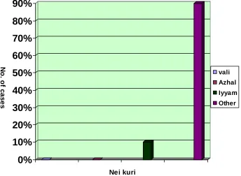
NEI KURI
The urine kept on the kidney tray in sun light, on non wind condition, should be examined by dropping a drop of gingili oil gently with rod. If oil spread
like snake it indicates valineer, a ring indicates azhal neer, and a float like a pearl it indicates iyya neer and sinks in urine indicates mukkutram.
sae naN-k" ms sQi "R in ak" ims
sµe naiRin e/ien"/s
-i /, Bkm
In kalladaippu patients, oil spreading like ring indicates Azhal neer or snake indicates Vali neer.
"im, a"im (
PROGNOSIS
)
sid- i elk "'y B=km in l !p< Ed-d! iin l !p<p
<"d&/i l !p< µd-d! i+nm i/i
µ /n'iR em / m "d-d! en/, l !p<
According to Yoogimamunivar, Vali, Azhal, and Ayya kalladaippu are curable where as Mukkutra kalladaippu is in curable.
%+$m
(LINE OF TREATMENT
)s i el i"/s - i+r 800
The main object of treatment is to bring down the deranged mukkutrams to natural equilibrium by giving pugatives, which cure derangement of vatham, this one of the cause for kalladaippu.
s"il m 1m
nil im 1m
anjl m 1ms
- /m -II
As per the above mention, author gives purgation to all patients as
their body condition.The author of dissertation has selected trial drug
Kalladaippu thool, dose of 5gms two times per day with raddish juice for 2
months period.
In siddha system treatment is not only for removable of disease but
the prevention and improving the body condition after removal of disease. This is said as kappu, neekkam and thiraippu.
DIET
All patients are strictly advised to follow this diet restriction as highly
calcium and oxalate diet likes cabbage, cauliflower, tomato, pees, cashionuts, almonds, grapes, strawberry, pine apple, tea and coffee.
URINARY CALCULUS
PHYSIOLOGICAL ANATOMY OF THE KIDNEY
The kidneys are two bean shaped organs situated retroperitoneal, one on the either side on the posterior abdominal wall. Each kidney weighs
about 120 to 150gms and is enclosed in thin but tough fibrous capsules, and concave medial side of the kidney is the hilum where the renal artery enters and renal vein and ureter leave the kidney. The components of kidney are arranged in three layers.
1. Outer cortex
This is dark and granular appearance. It contain renal capsules and convoluted tubules, at intervals cortical tissues penetrate medulla in this form of columns is called column of Bertini.
2. Inner medulla
This gives radially striated appearance as it contains tubular and vascular structures. Medulla mass is divided in to 8 to 18 medullary or
malpighian pyramids, the base of pyramids and are connect with cortex and apex projects in to minor calyx.
3. Renal sinus
It consist upper expanded part of ureter called renal pelvis, it
subdivided into about 8 minor calyces, and branches of nerves, arteries, and tributary vein and loose connective tissues and fat.
Nephron
Structural and functional unit of the kidney is nephron. Each kidney
has a million nephrons. Each nephron begain in cortex as
• A funal like dilatation called the Bowman’s capsule, which enclose a tuft of capillaries the glomerulus.The Bowman’s capsule together with the glomerulus is called the Malpighian corpuscle or Renal corpuscle.
• It then becomes straight and passes down it the medulla as the descending limb of the loop of the Henle, after varying distances before reaching the end of the papilla, it turns round in the form a U-shaped bend, forming loop of Henle, and passes upward towards the cortex, parallel with its former course as the ascending limb of
the loop of Henle. Each limb has an outer thick and inner thin portion.
• Thick ascending limb approaches its own, glomerulus, and contact with the afferent and efferent arterioles to form the juxtaglomerular apparatus. It then becomes convoluted to form the distal convoluted tubule.
RENAL CIRCULATION
Renal artery arises from the aorta, enters the kidney at the hilum and divided in to an anterior and a posterior branch, which give rise the about five segmental arteries. The segmental artery divided into
interlobararteries, which pass outward in the medulla between the pyramids to reach boundary zone between the medulla and cortex. Here they turn to
take a horizontal course uniting with adjacent arteries to form arterial arches called arcuate arteries. Several straight arteries arise from these arches and run radially outward through the cortex. These are called inter lobular arteries. From each interlobular artery, numerous afferent arteries arise, and enter the Bowman’s capsule forming glomerular capillary tuft. The afferent arterioles divide in to 4 to 5 large capillaries. Each large
The efferent arterioles give rise to renal portal system. The efferent arterioles form a second capillary net work surrounding the tubular portion
of the nephron; the capillaries of second set are called peritubular capillaries.
The tubular portion of juxtamedullary nephron is supplied by some
specialized peritular capillaries called Vasarecta. Vasarecta arise directly from the efferent arteriole of the juxtamedullary nephrons. The peritubular capillaries drain in to the venous system, which include the peritubular venules, interlobular veins, segmental vein and finally the renal vein
URINE FORMATION
Kidney excreted the unwanted substances including metabolic end
products and those substances, which are present in excessive quantities in body, through urine. Normally about 1 to 1.5 litters of urine is formed every day. The mechanism of urine formation includes various processes. First, when blood passes through glomerular capillaries, the plasma filtered in Bowman capsule, when this filtrate passes through the tubular portion of nephron, it undergoes varies changes both in quality and in quantity, many
wanted substances like glucose, aminoacid, water and electrolytes are reabsorbed from the tubules, this process is called tubular reabsorption . And some unwanted substances are secreted in to the tubule from peritubular blood vessels. This process is called tubular secretion. The
urine formation includes three processes. 1. Glomerular filtration
2. Tubular reabsorption 3. Tubular secretion
1. GLOMERULAR FILTRATION
When blood passes through the glomerular capillaries the plasma filtered in to the Bowmans capsule. All the substances of plasma are
1. The endothelium of capillary membrane, 2. Basement membrane and
3. Endothelium of visceral layer of Bowmen capsule.
The glomerular filtration is called ultra filtration because minute particles are filtered, but the plasma proteins are not filtered due to larger
molecular size than size of the slit pores. The composition glomerular filtrate is similar to that of plasma except in the absence of plasma proteins.
Glomerular filtrate rate GFR
The total quantity of filtrate formed in all the nephron of both the kidneys in the given unit of time is called glomerular filtrate rate. The
normal value of glomerular filtrate rate is 125 ml / minute or about 180 litres / day.
Filtration Fraction
The fraction of the renal plasma which becomes the filtrate is called filtration fraction. It is the ratio between renal plasma flow and glomerular filtrate. It is expressed in percentage.
They filtration fraction = GFR × 100 Renal plasma flow
= 125 ml / minute ×100
650 ml / minute
= 19.2%
The normal filtration fraction values from 15 to 20%
Pressure determining filtration:
Glomerular capillary pressure, colloidal osmotic pressure in the
glomeruli and the hydrostatic pressure in the Bowman’s capsule, which are determine the GFR. Among these pressures, the glomerular capillary pressure favours filtration the glomerular capillary pressure is about 60mm Hg and is highest capillary pressure in the body. The colloidal osmotic
concentration of proteins in glomerulus during filtration causes the development of colloidal osmotic pressure. It is about 25mmHg. Hydrostatic
pressure in Bowman’s capsule is exerted by the filtrate in Bowman’s capsule during filtration. It is about 15mmHg.
Net filtration pressure:
The balance between pressure favoring filtration and pressures opposing filtration is called net filtration pressure. This is very essential for
the maintenance of GFR so this is otherwise known as effective filtration pressure of essential filtration pressure.
The net filtration pressure = Glomerular capillary pressure – colloidal osmotic pressure + Hydrostatic pressure in Bowman’s capsule
= 60 – (25+15) = 20mmHg
The normal net filtration pressure is about 20mmhg and it varies
between 15 and 20mmHg
Factors regulating GFR:
Following are the various factors, which regulate or affect the GFR
1. Tubular glomerular feedback mechanism
This is the process in which the GFR is constantly regulated by means of feed back form renal tubule. The macula densa of juxtaglomerular apparatus is responsible for this. When the glomerular
filtrate passes through the end portion of thick ascending segment of renal tubule, the macula densa detects the concentration of sodium chloride and accordingly alters the GFR. It the concentration of sodium chloride is more, macula densa causes constriction of afferent arteriole and filtration rate decreases, the constriction of afferent arteriole may be due to the secretion of thromboxane A2 from macula densa.
2. Glomerular capillary pressure
3. Colloidal osmotic pressure
The GFR is inversely proportional to colloidal osmotic pressure
exerted by protein. During dehydration or increased plasma protein level, colloidal osmotic pressure is more and GFR is reduced.
4. Hydrostatic pressure in Bowman’s capsule
GFR is inversely proportional to this. The hydrostatic pressure in Bowman’s capsule is increased in conditions like obstruction of urethra and oedema of kidney beneath renal capsule.
5. Renal blood flow
This is the most important factor necessary for glomerular filtration. And, the GFR is directly proportional to this.
6. Constriction of afferent arteriole
This reduces the blood flow to the glomerular capillaries and this in turn reduces GFR.
7. Constriction of efferent arteriole
It efferent arteriole is constricted; initially there is an increase in GFR.
8. Systemic arterial pressure
However, increase in mean arteriole pressure up to 180mmHg or reduction up to 60mmhg does not alter renal blood flow or GFR. This is due to auto regulatory mechanism. Variation in pressure above 180mmHg or below 60mmHg affects the renal blood flow and GFR because auto regulating mechanism fails beyond this range.
9. Sympathetic stimulation
The mild or moderate stimulation of sympathetic nerves does not causes any significant change either in renal blood flow or in GFR. This is
due to auto regulation. Strong sympathetic stimulation causes severe constriction of the blood vessels needs to increase filtration initially, but latter decreases.
Tubular reabsorbion
When the glomerular filtrate passes through the tubular portion of
nephron, both quantitative and qualitative changes occur. Large quantity of water, electrolytes and other substances are reabsorbed by the tubular epithelial cells. The substances, which are reabsorbed, pass in to the interstitial fluid of renal medulla, and from here, the substances more in to the blood in peritubular capillaries. As the substances are taken back in to
the blood, the entire process is called tubular reabsorption.
Selective reabsorbtion
The tubular cells of kidney selectively reabsorb the substances present in the glomerular filtrate, according to the needs of the body. So the tubular reabsorption is called the selective absorption. Depending upon the
degree of reabsorption, the various substances are classified into three categories.
1) High threshold substances
The food substances like glucose, amino acid, acetoacetate ions and vitamins are completely reabsorbed do not appear in urine under
normal condition. These substances appear in urine, only if their concentration in plasma is abnormally high or in renal diseases. So these substances are called high threshold substances.
2) Low threshold substances
The substances such of urea, uric acid and phosphate are reabsorbed to little extend. These substances appear in urine even under normal conditions. Such substances are known as low threshold substances.
3) Non threshold substances
The metabolic end products like creatinine are not at all reabsorbed and are excreted in urine irrespective of their plasma level. These substances are called non threshold substances.
The mechanisms involved in tubular reabsorption are of two types
1. Active reabsorption and
2. Passive reabsorption.
1. Active reabsorption
The movement of molecules is against the electrochemical gradient. This needs liberation of energy. And the energy is derived from ATP. The
substances reabsopped actively from the renal tubule are sodium, calcium, potassium, phosphates, sulphates, bicarbonates, glucose, amino acids, ascorbic acid, uric acid and ketone bodies.
2. Passive reabsoption
In this process, the movement of molecules is more along the
electrochemical gradients. This process does not need energy, the substances reabsorped by passive transport are chloride, urea, and water.
Site of reabsorption
Substances reabsorbed from proximal convoluted tubule. Glucose, amino acids, sodium, potassium, calcium, bicarbonates, chlorides, phosphates, uric acid and water are reabsorbed from proximal convoluted tubule. The substances reabsorbed from loop of Henle are sodium chloride.
The substances reabsorbed from distal convoluted tubule are sodium, bicarbonate and water.
Tubular secretion
Some substances secrete in to the lumen from the peritubular
capillaries through the tubular epithelial cells. These known as tubular secretion or tubular excretion.
1. Potassium is secreted actively by sodium – potassium pump in distal convoluted tubule and collecting duct.
3. Hydrogen ions are secreted in the proximal and distal convoluted tubules. Maximum hydrogen ion secreted in proximal tubule.
Thus by the process of glomerular filtration, selective reabsorption and tubular secretion urine is formed in the nephron. It is also concentrated by counter current mechanism and ADH. Finally it passes through ureter into the urinary bladder and is stored there until it is voided
URINARY CALCULUS
Urinary calculus is stone –like body composed of urinary salts bound together by a colloid matrix of organic material, it consist of nucleus around which concentric layers urinary salt are deposited .
Urinary stones have afflected humankind since antiquity, with earliest recorded example being bladder and kidney stones detected in Egyptian mummies dated to 4800 B C.
Until the 1980s, urinary stones were a major problem, with a significant proportion of patients requiring extensive surgical procedures and a sizable losing their kidney. The advent of extra corporeal techniques for stone destruction and refinements in endoscopic surgery, however have greatly decreased the morbidity associated with stone surgery.
EPIDEMOLOGY INTRINSIC FACTORS Hereditary
Several disorders that cause renal stones are hereditary .Familial renal tubular acidosis are associated with nephrolithiosis in almost 70% of patients. Cystinuria, Xanthinuria and Dehydroxyadenuria are disorders cause renal stone
Age and sex
The peak incidence of urinary calculus occurs in the twenties to forties. About three males are affected for every female because increased serum testosterone causes increased endogenous oxalate production in
male, increased urinary citrate in women.
EXTRINSIC FACTORS Geography
Climate and seasonal factors
Price and associates found that the incidence of urinary calculi was higher during the summer months. High temperatures increase perspiration, which may result in concentrated urine. This promotes increased urinary crystallization. Parry and Lister suggest that increased exposure of sun light cause increased production of 1, 25-dihyrdoxyvitamin D3 and increased urinary calcium excretion. This may cause higher
incidence of urolithiosis in summer months.
Water Intake
Urine dilution by increased water intake may increased ion activity
coefficients and hence urinary crystallization, water diuresis reduces the average time of residence of free crystal particles in urine dilutes components of urine that may crystals. Minerals contents of water may contribute to cause stone disease. (E.g. sodium chlorides) Zinc in an inhibitor of calcium crystallization.
Diet
Ingestion of excessive amounts of purines, oxalates, calcium, phosphates, and other elements often results in excessive excretion of these components in urine causes increased incidence of calculi.
Occupation
Lonsdale (1968b.c) indicated that urinary calculi are much more
likely to be found in individuals who have sedentary occupations. The highest were found in cooks and engineering room personnel. The risk of calcium oxalate and uric acid stones formation in Astronauts because of hypercalciuria, hypocitraturia, decreased pH, and lower urinary volumes.
PHYSICAL CHEMISTRY
Urinary stones do not occur unless crystals of the offending substance form in urine. For crystals to occur, the urine should be
for crystals to form or grow: Intermittent super saturations, as is seen during of dehydration or after meals, is sufficient. Crystallizing potential for
calcium oxalate is related not so much to the concentration of calcium or oxalate in urine but to chemical activity of ions in solution. The compounds such as citrate and phosphate form complexes with calcium, and elements such as magnesium and sodium form complexes with oxalate, effectively reducing the free ionic concentration of each.
Urinary crystals can be seen in most urine specimens, particularly after storage, yet most individuals do not form stones. Stone formers as a group excrete larger crystals and crystals aggregates than
healthy individuals. Normal subjects have inhibitors of crystal formation, growth, and aggregation in urine such as low-molicular-weight compounds such as citrate and pyrophate and larger molecules, such as glycosaminoglycans, nephrocalcin, and Tamm – Horsfall protein. Urine from patients with recurrent calcium and oxalate stones tends to have higher calcium and oxalate saturation and lower inhibitors than urine from
patients without stones and a mathematically derived saturation - inhibition index has been reported to discriminate between the groups with better than 90% accuracy.
Free crystals formed within the kidney do not have the ability to grow to a size large enough to occlude a collecting duct and, form a stone in a free – flowing urinary system. Crystal aggregation and retention within the kidney are prerequisites for urinary crystals to be converted to urinary calculi. Crystal aggregation is enhanced in individual who lack inhibitors of
aggregation. The urinary glycoproteins nephrocalcin and Tamm-Horsfall protein are potent inhibitors of crystal aggregation in simple solutions, whereas citrate and magnesium are inhibitors of crystal growth.
proven, bacterial infection may promote calcium oxalate stone formation by increasing urinary matrix, which, in turn, promotes crystal adherence.
Finally, altered transport of calcium and oxalate by renal epithelial cells may result in intracellular or interstitial crystallization. These are retained in the kidney and can become the nidus for stone formation.
TYPES OF CALCULUS
CALCIUM OXALATE STONES
This is most common type of stones. 39% of patients are calcium oxalate type stone. 14% of patients are combined with calcium phosphate. They are irregular in shape and covered with sharp projection, which tend to cause bleeding. The surface of the stone is discoloured by pigments of altered blood. It is very hard and absorbs X-rays well.
PATHOPHYSIOLOGY OF STONE FORMATION
I. HYPERCALCIURIA
Between 30% and 60% of all patients with calcium oxalate kidney stones have increased urinary calcium excretion in the absence of raised serum calcium levels. Hypercalciuria has been defined as the excretion of greater than 4mg calcium per kg body weight per day or greater than 7 mmol in men and 6 mmol in women (parks and Coe, 1986). Final definition
is the excretion of urinary calcium of greater than 0.11 mg/100 ml of glomerular filtrate. Hypercalciuric nephrolithiasis suffer from multiple disturbances in renal tubular function a disturbance in phosphate transport, and accelerated 1, 25 – dihydroxyvitamin D3 synthesis, resulting in increased intestinal calcium absorption.
Hypercalciuria has three types, such as 1. Absorptive hypercalciuria,
2. Renal hypercalciuria
1. Absorptive hypercalciuria
In absorptive hypercalciuria, the primary abnormality is increased
calcium absorption. In absorptive hypercalciuria type 1, intestinal hyper absorption of calcium exists, whether or not the patient is on a calcium-restricted diet. Intestinal magnesium absorption is normal in patients with absorptive hypercalciuria, but oxalate absorption is increased. Vitamin D increases calcium and magnesium, but not oxalate, absorption in both the
jejunum and ileum. Absorptive hypercalciuria type 2 is a variant of this disorder wherein patient’s exhibit increased urinary calcium excretion while on their normal diet but normal calcium excretion on a calcium, low-sodium diet. In the final subcategory, absorptive hypercalciuria type 3, the serum phosphate is low, suggesting that the increased intestinal calcium absorption is the result of stimulation in vitamin D production as the result
of the lowered serum phosphate.
2. Renal hypercalciuria
In this condition, the underlying abnormality is a primary renal
wasting of calcium. The consequent reduction in circulating serum calcium stimulates PTH production. Two facts must be stressed; first, intestinal absorption of calcium is increased in both absorptive and renal hypercalciuria; second, parathyroid function is suppressed in absorptive hypercalciuria but stimulated in renal hypercalciuria. These two criteria
elevated fasting urinary calcium levels and stimulated parathyroid function serve to distinguish renal from absorptive hypercalciuria.
3. Resorptive hypercalciuria
This syndrome is synonymous with subtle hyperparathyroidism. Hypercalciuria results from excessive PTH dependent bone resorption as well as enhanced intestinal absorption of calcium.
4. Idiopathic hypercalciuria
The syndrome of renal phosphate leak, elevated 1, 25
hyper calciuria may be inherited as an autosomal trait, although the pattern can reflect polygenic control of calcium excretion as well.
II. HYPERCALCAEMIA NEPHROLITHIOSIS
CAUSES
1. Primary hyperparathyroidism
These patients are between 39% and 78% with early presented of renal calculi. These patients with higher level of 1, 25-dihydroxyvitamin D3 and greater calcium absorption tend to have renal stones.
2. Malignancy associated hypercalcemia
Malignancy –associated hyper calciuria is an exceedingly rare cause of renal stones. The most common cause of malignancy- associated hypercalcaemia, even in patients with skeletal metastasis, is production by the tumour of a bone resorpting substance called PTH- related polypeptide.
3. Sarcoidosis and other granulomatous diseases
The sarcoid granuloma produces 1, 25-dihydroxyvitamin D3, it causing increased calcium absorption, hypercalcaemia, and hypercalciuria.
4. Hyperthyroidism
About 5% to 10% of patients with hyperthyroidism develop hypercalcimia. Hypercalcaemia and hypercalciuria result from a stimulation
of bone resorption mediated by thyroxine and tri-iodothyronine.
5. Glucocorticoid – induced hypercalcemia
Glucocorticoid excess leads to increased bone resorption, decreased
bone formation, and osteopenia. Glucocorticoid also has direct stimulatory effect on parathyroid gland.
6. Pheochromocytoma
7. Immobilization
Prolonged bed rest can lead to hypercalcaemia, as the result of
increased bone turnover. Hypercalcaemia is seen most often when another condition, such as Paget’s disease- with accelerated bone turnover-primary hyperthyroidism, or malignancy, coexists in an immobilized patient.
III. HYPEROXALURIA CAUSES:
Primary hyperoxaluria is a rare genetic disorder resulting from increased hepatic production of oxalate. Enteric hyperoxaluria occurs in patients with short bowel syndrome or malabsorption. Finally, a group of
patients with recurrent idiopathic calcium oxalate lithiasis exhibits mild hyperoxaluria or increased transport of oxalated by red blood cells.
1. Primary hyperoxaluria
Two types of primary hyperoxaluria exit. Primary hyperoxaluria type1 is an autosomal ressive inborn error of metabolism characterized by nephrocalcinosis and oxalosis. The diseases are characterized by increased urinary excretion of oxalic, glycolic, and glyoxylic acids. Primary hyperoxaluria type I is due to a defect of the enzyme alanine-glyoxylate aminotransferase (AGT) in the liver.
In normal human liver, AGT catalyzes the transamination or detoxification of glyoxylate to glycine, a function that it can perform only if it
is located in the peroxisome. Its deficiency in primary hyperoxaluria results in glyoxylate’s being oxidized to oxalate.
Primary hyperoxaluria type II or L-glyceric aciduria is a much rarer
variant of the disease. Deficiencies of the hepatic enzymes D-glycerate dehydrogenase and glyoxylate reductase lead to increases in urinary oxalate and glycerate excretion.
2. Enteric hyperoxaluria
disease, or jejunoileal bypass, increases the colonic permeability of oxalate as the result of exposure of the colonic epithelium to bile salts.
Furthermore, loss of calcium in the feces results in the presence of less calcium in the intestinal lumen, allowing oxalate to exist in a soluble form. The hyperoxaluria from small bowel malabsorption often exceeds 1 mmol/day and causes recurrent nephrolithiasis, nephrocalcinasis, and renal oxalate deposition.
3. Mild metabolic hyperoxaluria
Mild hyperoxaluria is as least as important a factor in the pathogenesis of idiopathic calcium oxalate stones as hypercalciuria. Baggio
and colleagues are found an increase in oxalate self exchange across red blood cell membrane in 79% of patients with idiopathic calcium oxalate stones. The oxalate absorption was increased with increased calcium absorption. Dietary restriction of oxalate results in decreased oxalate excretion.
IV. HYPERURICOSURIA
Uric acid promotes calcium oxalate crystallization by facilitating the formation nuclei. Sodium acid urate may produce calcium oxalate stone disease by nullifying the effectiveness of naturally occurring inhibitors of
calcium oxalate crystal growth. Excessive dietary intake of purine is main cause of hyperuricosuria. Between 80% and 90% of patients with hyperuricosuria nephrolithiosis are men. Patients with mixed with uric acid and oxalate stones have lower urinary pH than patients with pure calcium oxalate stones.
V. HYPOCITRATURIA
Hypocitraturia has been reported in 15% to 63% of patients with stones. Urinary citrate is normally greater in women than in men.
acidosis are other cause of decreased urinary citrate excretion. A diet rich in animal protein may produce an acid load. Strenuous physical exercise
and sodium intake can likewise produce hypocitraturia. Urinary tract infection with bacteria degrading citrate lowers urinary citrate excretion.
The primary mechanism of action of citrate is as a complexing agent
for calcium. Calcium citrate complexes are considerably more soluble than calcium oxalate. Citrate inhibits the spontaneous nucleation of calcium oxalate, crystal growth and aggregation of calcium oxalate and phosphate.
VI. HYPOMAGNESURIA
Many experimental studies have suggested that administration of magnesium salts prevents stones disease. The most common cause of
hypomagnesuria is inflammatory bowel disease associated with malabsorption. Most patients with hypomagnesuria also have hypocitraturia.
VII. SEX HORMONES AND RENAL STONES
Calcium oxalate renal stones occur much more frequently in men than in women because increased serum testosterone levels resulted in
increased endogenous oxalates production by liver and increased intestinal absorption of calcium in men, increased urinary citrate concentration in urine in women.
CALCIUM PHOSPHATE STONES
Calcium phosphate stones composed predominantly around 10% of stones of renal origin. Although some amount of calcium phosphate is often found in calcium oxalate calculi, pure calcium phosphate stone are rare. It
is usually smooth and dirty white, and easy to see on radiopaqhic film.
Causes:
may be seen with oxalate and stuvite stones. Stone formation typically occurs in papillary tips and in medulla. Stone formation is result of hyper
calciuria, hypocitraturia, and increased urinary pH. Hypercalciuria is result of systemic acidosis on bone deminaralisation and secondary hyperthyroidism. Hypocitraturia result from a primary defect in renal tubular citrate transport, again, the result of metabolic acidosis. Hypocitraturia is probably the most important metabolic factor for stone formation in patient
with renal tubular acidosis. The defect in proximal tubular bicarbonate resorption is associated with this type of stone.
Diagnosis: the patient has hypokalaemia, hyper chloremia, metabolic acidosis and urinary pH of 5.5.
URIC ACID STONE
These are hard, smooth and often multiple. Their colour varies from yellow to reddish brown. The pure uric acid stone are radiolusent most of stone with calcium so they radiolusant shadow.
Causes:
The principal cause of uric acid crystallization is supersaturation of urine with respect to undissociated uric acid. The normal 24-hour urinary uric acid excretion is between 500 and 600mg per litre of urine. Patient with
uric acid stone often have prolonged periods of acidity in urine. The pH of urine in patient with uric acid stones was 5.5 ±0.4 as compared with 6.0± 0.4 in patients who form calcium oxalate stones. Three factors are involved in uric acid urolithiosis. First, patients tend to excrete excessively acid urine at relatively fixed, low urinary pH. Second, they may absorb, produce, or
STRUVITE STONES (INFECTION STONES)
The stone is composed of magnesium, ammonium, and phosphate,
mixed with carbonate.
Pathogenesis
Two conditions must coexist for the crystallization of struvite- a urine
pH of 7.2 or above and the presence of ammonia in the urine. A second mechanism by which bacterial infection may induce stone formation is by increasing crystal adherence. Urease producing bacteria hydrolyze urea to carbondioxide and ammonium molecules. Two molecules of ammonia are produced from one molecule of urea; neutralization of the base is
incomplete. As a result of this, the urinary pH rises.
Clinical presentation
Struvite calculus accounts are the majority of staghorn stones
observed in most countries. Thy can grow quite large and may fill the collecting system. Struvite stones can form on a nidus of calcium oxalate stones and can grow quite rapidly. Most infection stones are radiopaque, but poorly mineralized matrix stones are faintly radiopaque or radiolucent. Women, perhaps because of their increased susceptibility to urinary tract infections, are more commonly affected than men. The presence of a
foreign body in the urinary tract and neurogenic bladder are main causes for struvite calculi.
Patients with struvite stones may present acutely with fever, loin pain, dysuria, frequency, and hematuria. Metabolic abnormalities are present in patients with mixed calciumoxalate and struvite stones but not in patients with pure struvite stones.
CYSTINE STONES
They are uncommon Cystine stones account for about 1% of all urinary calculi in the United States and occur only in patients who have