A STUDY ON
THALASTHAMBAM
Dissertation Submitted To
THE TAMIL NADU DR.M.G.R Medical University
Chennai – 32
For the Partial fulfillment for Awarding the Degree of
DOCTOR OF MEDICINE (SIDDHA)
(Branch – V, NOI NAADAL)
DEPARTMENT OF NOI NAADAL
Government Siddha Medical College
Palayamkottai – 627 002
HIGHLIGHTS OF THE DISSERTATION TOPIC
Thalasthambam comes under Vatha roga nithanam in Yugi Vaithya Sinthamani
800.
For any type of disease the vali humour is first affected. Followed by alteration in
other humour.
In thalasthambam, yugi explains that the altered vali humour is aggravated by the
excessive intake of salt and sour (i.e) these tastes acts as a pre-disposing factor for the
disease.
The disease is characterized by the presence of heaviness of the foot, pain in the
foot, blackening of the foot, spreading upwards from foot, weight loss, Dyspnoea,
fatique, dryness of the body, thirst.
In this disease, the Udal thathukkal Saram, Senneer, Oon, Kozhuppu, Enbu,
Moolai are affected as 6 out of 7 udal thathukkal gets deranged the disease is not –
curable.
The underlying pathogenesis for the Thalasthambam is the peripheral atererial
occlusive disorder resulting in gangrene formation due to pan-arthritis leading to
REVIEW OF LITERATURE
In Segarasasekaram Vaithyam, the following poem is mentioned.
In Sarabanthirar vaithya maraigal and Thanvanthiri Vaithyam. The
thalasthambam is explained with the same poem mentioned by Yugi in Yugi Vaithya
1
INTRODUCTION
Siddha System is the science of right living and as such is intended
to be incorporated in daily life. It works on all aspects of the persons life.
The physical, vital, mental, emotional, pshysic and spiritual.
Siddha Medicines not only cures the diseases but inaddition it aims
at bringing the different bodily functions into perfect co-ordination. So,
that they work for the good of the whole body.
Siddhars, the founder of Siddha System has given the techniques to
purify the body, mind and energy to prepare the ground for higher
practices of meditation and for the ultimate experience of cosmic
consciousness.
To attain all they need is a healthy body. So, they begin to praise
the body.
clw;rpwg;G
“NjfkpUe;jjy;Nyh rpj;njy;yhkhlyhk;
NjfkpUe;jhf;fhw; Nruyhk; G+uzk;
NjfkpUe;jhf;fhw; nranyy;yhk; ghh;f;fyhk;
NjfkpUe;jhf;fhy; NruyhKj;jpNa.”
2
Siddha System of Medicines were not primarly for the sick but for
the healthy also.
Kaayakarpam and Astaanga yogam are needed for a healthy mind
and body. It activates and regulates the life force, to attain a higher state
of vibratory energy.
Kaayakarpam are rejenuvator of our body, they include
Anti-oxidants.
Astanga yoga includes,
1. Iyamam - Self restrains.
2. Niyamam - Self observances.
3. Aasanam - Various exercising posture.
4. Praanaayaamam - Expansion of the dimension of vital
Energy.
5. Prathyagaaram - Disassociation of consciousness from the
outside environment.
6. Dhaaranai - Concentration.
7. Dhyaanam - Meditation.
8. Samaathi - Identification with pure consciousness.
In the past itself Siddhars got clear cut idea on many fields like,
Medicinal preparations, Astrology, etc., They also followed many
3
Naadi - To Diagnose the various diseases and to assess
the prognosis of the diseases and fate of the
diseases.
Manikkadai nool - used as a diagnostic tool
Neikuri - used to observe the prognosis of the patient
and fate of the diseases.
As we are in the twentyfirst century, a spiritual heritage are being
reclaimed of which Siddha System is very much important.
Now, the Modern world wants everything to be based on scientific
proof. So, it is our duty to explore our siddha system by a Science Which
correctly judges our medicine and Siddhars thoughts.
4
SIDDHA PHYSIOLOGY
Man is not merely a mixture of Muscles, bones and Nerves as think
by physiologist. But Siddhars thought clearly says that man, the
Microcosm is having himself all the things within the universe, the
macrocosm.
Each human body is made up of 2 kinds of body.
Paru Udal ( Visible body)
Nun Udal ( Invisible body)
PARU UDAL
It includes, bones, muscles, Blood vessels, Nerves, and all the
functional system of human body like Digestive system, Respiratory
system etc, The Paru Udal is known as the “FUNCTIONAL UNIT OF
HUMAN BEING”
NUN UDAL
It is the basic for the Paru Udal. It makes the Paru Udal to be
active.
The universe is made up of 5 basic elements called Earth, Water,
Fire, Air and Space. As we said before the human being is also made up
5
Each basic elements exists in two forms.
Paru Nilai (Visible form)
Nun Nilai (Invisible form)
PARU NILAI
Those things which are recognized by our senses are called as Paru
Nilai.
NUN NILAI
Those things which are not recognized by our senses but they are
existing in our body are called as Nun Nilai .
Physiology means that the basic process underlying the functioning
of a species.
Basic things for functioning of human beings as explained by
Siddhars include,
96 Thathuvangal
7 Udal katugal
6 Suvaigal.
The factors which influence the functioning of human body are,
Udal Vanmai.
6
96 Thathuvam
According to siddhar’s view, the 96 basic factors are located in the
human body. When the sperm fertilizes the ovum, the human embryo
have the 96 basic factors. They also added that the each and every atom
in universe has this 96 basic factors.
Panchabootham- Five basic elements:
¾ Mann (Earth) - Gives shape to the body and release its energy.
¾ Neer (Water) - It makes the earth supple and helps in
Transmission of energy.
¾ Thee (Fire) - It makes the body steady and gives vigour
stimulation.
¾ Vayu (Air) - Ignite the fire and works as a life carrier and it is
the support of all contact and exchange.
¾ Aagayam(Ether)- It is the creator of life itself in the body.
Pori -5 – Five sense organs
1. Mei - organ of tactile sensation
2. Vaai - Organ of taste
3. Kan - Organ of vision
4. Mooku - organ of smell
7
Pulan-5 – Functions of the five sense organs.
1. Saptham - Hearing.
2. Sparisam - Touch.
3. Roopam - Vision.
4. Rasam - Taste.
5. Gaantham - Smell.
Kanmaenthiriyam -5 – Five Motor organs
1. Vaai - Organ for speech.
2. Kaal - Organ for locomotion.
3. Kai - Organ for performing skilled movements.
4. Eruvaai - Organ for defaecation.
5. Karuvaai - Organ for Reproduction.
Gnanenthiriyam -5 – Functions of five Motor organs
1. Vasanam - Speech.
2. Kamanam - Walk.
3. Dhanam - Movements of the upper limbs (Flexion,
Extension, Supination and pronation).
4. Visarkam - Defecation.
8
Anthakaranam -4 - Four Intellectual
1. Manam - Mind for thinking.
2. Buthi - Power of Discriminating the right from wrong.
3. Sitham - Doing the right thing.
4. Agankaaram - Firm conviction.
Arivu -1
Intellect (or) wisdom
Naadi -10
1. Idakalai - Arises from right big toe and coils round the
suzhumunai and enter the left nostril.
2. Pinkalai - Arises from left big toe and coils round the
suzhumunai and enter the right nostril.
3. Suzhumunai - It flow along the vertebral column upto
medulla oblongata.
4. Siguvai - Present in uvula and help in swalloing.
5. Purudan - Present in right eye and help in right vision.
6. Gaandhaari - Present in left eye and help in left vision.
7. Aththi - Present in right ear and controls its hearing.
8. Alampudai - Present in left ear and controls its hearing.
9. Sangini - Present in external genitalia.
9
Vayu -10 – Ten air forces
1. Praanan
2. Abaanan
3.Viyaanan
4. Uthaanan
5. Samaanan
6. Naagan
7. Koorman
8. Girugharan
9. Thaevathaththan
10. Dhananjeyan - (explained under thodam)
Aasayam -5 – Five visceral cavities
1. Amarvaasayam - Stomach.
2. Pagirvaasayam - Liver, small intestine.
3. Salavaasayam - urinary bladder.
4. Malavaasayam - Rectum, Large Intestine.
5. Sukkilavaasayam - Seminal vesicles (or) ovary.
Kosam -5
1. Annamayakosam - Made up of seven udalthathus.
10
2. Praanamayakosam - Made up of pranan and kanmenthiriyam.
(The Vital energy body)
3. Manomayakosam - Made up of manam and Gnanenthiriyam.
(The mental body)
4. Vingananmayakosam - Made up of puthi and Gnanenthiriyam.
(The psychic (or) vital energy body)
5. Aanandhamayakosam - Made up of pranan and suzhuthi .
(The bliss body)
Aatharam -6
1. Moolaathaaram - It lies between the Anus and
genitalia as kundalini, a vital force.
2. Swaathitaanam - It lies 12 inches above the moolatharam.
3. Manipooragam - It lies 8 inches above the swathitanam.
4. Anaagatham - It lies10 inches above the Manipooragam.
5. Visuthi - It lies 10 inches above the anaagatham.
6. Aackinai - It lies inbetween the two eyebrows.
Malam -3
1. Aanavam - selfishness of all things around him.
2. Kanmam - It is related to both Aanavam and Maayai
it makes good and bad deeds.
3. Maayai - False thinking like others possession is
11
Mandalam -3
1. Gnayirumandalam - It is located in the cardiac region and 4
inches above the stomach.
2. Thingalmandalam - It is located in the head.
3. Agnimandalam - It is situated 2 inches above the
moolatharam and spreads up to umbilical
region.
Thodam-3
1. Vali
2. Azhal
3. Iyam
VALI
Location:
Abaanan, faeces, Idakalai, pelvic bone, Spermatic cord, skin,
nerves, joints, hairs and muscles.
Natural character
In normal condition, vali is responsible for respiration and control
of all movements. It governs the 14 reflexes of our body. It also controls
udal thathukkal. It gives strength to five sensory organs.
Functions When Exaggarated
Body pain like pricking and twitching in nature.
12
Dryness
Loss of body weight
Boring pain
Joint dislocation
Not responding to external stimuli
Thirst
Goose flesh
unable to move upper and lower limbs
Astringent sense of taste in the mouth
Blackish discolouration of skin, stool, urine and conjuctiva.
Types of Vali
Based on its functions and location it has been classified into 10
types.
They are,
1. Uyirkkaal -Praanan
It is responsible for respiration and helps in digestion of ingested
food material.
2. Keelnokkukkaal - Abaanan
It expels urine and faecal mater It constrict the anal sphinchter. It
13
3. Paravukaal – Viyaanan
It is responsible for the movement of various body parts. It percive
tactile sensation. It fills the body with digested food materials and
nourishes the body.
4. Melnokkukkaal – Udhanan
Responsible for all kinds of upward motion
5. Nadukkaal – Samaanan
It controls all other vayus. It is responsible for proper digestion,
assimilation and carries digested Nutrients to each and every organs.
6. Vaanthikkaal - Naagan
It is responsible for learning higher intellectual functions. It causes
opening and closing of eye-lids.
7. Vizhikkaal - Koorman
Responsible for vision and yawning It makes eyes opening and
closing.
8. Thummikkaal - Kirugaran
It is situated in the tongue. Induces appetite, sneezing and cough.
9. Kottavikkaal - Devathaththan
It is situated in Anus and genitalia. It makes laziness while
14
10. Veengukkaal - Dhananheyan
It is situated in craium and produces swelling of the body. It leaves
three days after the death of a person forming way through the skull bone.
AZHAL
Location
Piraanavayu, bladder, Moolakkini, Heart, umbilical region,
abdomen, sweat, saliva, blood, eyes, skin, pingalai and head.
Types of Azhal
1. Aakkanal - Anala pitham
It lies in between stomach and duodenum it is responsible for
digestion of food.
2. Vannaeri - Ranjaka pitham
It lies in intestine. It is responsible for colouring of the blood.
3. Aatralangi - Saathaga pitham
It is situated in the heart and it is responsible for fulfilling a
function.
4. Nokkazhal - Aalosaka pitham
It lies in eye and is responsible for the perception of vision.
5. Ollolithee - Praasaka pitham
It is situated in the skin and is responsible for the complexion of
15
IYAM
Location
Samaanan, suzhumunai, Semen, head, fat, bone marrow, blood,
Nose, Colon, Joints, Chest and tongue.
Types of Iyam
1. Ali Iyam - Avalambagam
It is situated in the lungs. It controls the heart and other four forms
of Iyam.
2. Neerppi Iyam - Kiledhagam
Present in the stomach it makes the food wet and helps in
digestion.
3. Suvaikaan Iyam - Pothogam
It is situated in the tongue. It helps in perception of taste.
4. Niraivu Iyam - Tharpagam
It lies in head and is responsible for the coolingness of the eye.
5. Ondri Iyam - Santhigam
It is located in the joints and it is responsible for the free movement
of the joints.
Edanai -3 Three physical Bindings
1. Porul patru - Material bindings.
2. Puthalvar patru - Relative bindings.
16
Gunam-3
1. Sathuvam - Good characters.
2. Raasadham - Manly characters.
3. Thaamasam - Bad characters.
Vinai -2 – Two types of Actions
1. Nalvinai - Good activities.
2. Theevinai - Bad activities.
Raagam-8
1. Kaamam - Desire.
2. Krotham - Hatred.
3. Ulopam - Stingy.
4. Moham - Lust.
5. Madham - Pride.
6. Marchariyam - Internal conflict.
7. Idumbai - Mockery.
8. Agankaaram - Ego.
Avathai -5 – Five states of consciousness
1. Nanavu - Wakefulness.
2. Kanavu - Dream.
3. Urakkam - Sleep.
4. Paerurakkam - Stupor.
17
7 UDAL KATTUGAL
These are responsible for the formation and maintanence of the
entire structure of the body. They are formed one by one.
Functions
1. Saaram - Plasma
It strengths our body both mentally and physically.
2. Senneer - Blood
It imparts colour to the body. It restores strength, Nourishment and
intellect of an individual.
3. Oon - Muscle
It moulds the shape of the body according to the physical
requirement and helps in bone growth.
4. Koozhuppu - Fat
It lubricates the different organs while doing their function and
maintains oily matter of the body.
5. Enbu - Bone
It give support to the body and protects the internal organs and
acts as a basic for movements of the body.
6. Moolai - Marrow
It fills the bone cavity and gives softness and strength to the bone.
7. Sukkilam / Suronitham - Sperm/ Ovum
18
SUVAIGAL
Combination of two Boothams results in the formation of one taste.
NATURAL CHARACTERS OF SUVAIGAL
1. Inippu – earth + water
This taste gives happiness to mind and body. It gives tastier feeling
to the mouth.
Functions
It gives nourishment to the body, It extends life span, It increases
hair growth, It corrects vitiated Vali and Valiazhal thodams , It increases
milk secretion, It removes poison from the body.
2. Pulippu - earth + fire
It stimulates the secretion of saliva. It produce goose flesh. It clears
waste materials from the mouth.
Functions
It increases appetite and removes tastelessness, It excretes waste
gas from the body. Iyam, Blood and azhal aggravates from its place due
to pulippu.
3. Uppu - water + fire
It increases salivation, It produce inflammation in cheek and throat.
Functions
It removes dryness, constipation, Iyam from our body, It Increases,
19
4. Kaippu - air + ether
It removes waste materials from the mouth. It decreases the
perception of other tastes. It is not tastier to mouth.
Functions
Though it produces tastelessness, It also removes loss of taste,It
removes poison from the body, It kills worms in the body,It normalizes
the excess salivation, It cleanses the throat and mothers milk.
5. Kaarppu - air + fire
It causes burning sensation in tip of the mouth and cheek. It
increases secretion of eye, nose and tongue. It produces hot sensation in
mouth and face.
Functions
It removes the skin diseases, It clears throat problems, It increases
digestion, It clears waste material from the body, It removes the damage
produced by Iyam.
6. Thuvarppu - earth + air
It delays the perception of other tastes.
Functions
It removes the Azhal and Iya thodam, It gives heat sensation to the
body, It clears the blood, It causes nourishment to skin.
20
UDAL VANMAI
The udal vanmai is divided into 3 types. These are,
1. Iyarkai Vanmai - Innate Immunity
The Natural immunity one can gets by birth itself.
2. Kaala Vanmai
Improvement of stamina and Immunity according to age and
seasonal variation.
3. Seyarkai Vanmai - Acquired Immunity
Regulation of healths by taking nutritious food, good activities and
through medicines.
UDAL THEE – FOUR BODY FIRES
The internal fire which keeps body and soul in good condition is
called as body fire. Which is of 4 types.
They are,
1. Samaakkini - Naturally Situated Samaanavayu
2. Vishamaakkini - Altered Samaanavayu from its Natural place
3. Deekshanaakkini - Samaanavayu in the Place of Azhal
21
SIDDHA PATHOLOGY
“ WHERE THERE IS LOVE FOR MANKIND THERE IS
LOVE FOR THE ART OF HEALING ” -Hippocrates.
To heal a patient it is necessary to understand what is the pathology
occurring inside him. So, pathology is very much basic and necessary for
any type of diseases. This has been well explained by our Sidddhars in
the poem,
“kjpj;jplw;fUik tha;e;j
khz;ghpfhu nky;yhk;
Jjpj;jpl Tzh;e;jhNdDe;
Jfswg; gpzpapd;wd;ik
gjpj;jplTzuhdhfpw;
gaDwhdhfhyhNd
tpjpj;jpL gpzpj;jpwj;ij
tpsk;GJ Kjw;fz;kd;Ndh”
- rpfpr;rhuj;d jPgk;.
Before discussing about pathology. It is important to understand
22
HEALTH
Health is a state of complete physical mental, spiritual and social
well being and not merely an absence of diseases (or) infirmity.
DISEASE
The Disease literally means without ease (uneasiness) the opposite
of ease.
Diseases is a condition of the body (or) some part (or) organ of the
body in which its function are deranged (or) disrupted.
DETERMINANTS OF HEALTH
HEALTH Communities
Families
Individuals
Socities
Biological
Behavioural
Environmental
Socio - Economic
Socio Cultural
Aging of People Science and Technology
Information and Communication
Gender Social Justice
23
All these factors contribute a major part on health. In our Siddha
System, the basic factor which are responsible for the health of an
individual are,
Among these, Suvaigal, uyir thathu, udal thathu are related to one
another(i.e) Derangement in one can alter the function of other.
It can be represented as ,
Man or Individual Food (Suvaigal)
Uyir Thaathu
Udal Thaathu
Seasonal Variation
Infection Environmental Changes
Occupation
Congenital Disease
SUVAIGAL UYIR
24
Suvaigal Banchapootham Uyir thathu Udal thathu
Enippu - Sweet Earth+water Iyam
Plasma, oon, fat,
lymph,
Reproduceive
Pulippu -Sour Earth+Fire Azhal Blood
Uppu - Salt Water+Fire Vali
Muscular
tissue(vd;G)
Kaippu - Bitter Air+Ether
Kaarppu - pungent Air+Fire
Thuvarppu -
Astringent
Earth + air
Derangement of Derangement of Derangement of udal
Kaippu, Kaarppu and Vali Thathuesp. Muscular
Thuvarppu tissue
Derangement of Derangement of Derangement of udal
Pulippu, Kaarppu Azhal Thathu especially Blood
25
Derangement of Derangement of Derangement of udal thathu. Enippu,pulippu Iyam
and uppu
Produce Various Signs and symptoms
ALTERED SIX TASTES
Increased intake of taste in food makes a way for various diseases.
They are explained as follows.
1. Enippu
It produces obesity, Excessive fat, increased mucous secretion,
hunger, Indigestion, Diabetes, cervical adenitis, increased Iyam and its
diseases.
2. Pulippu
It produces fatique, dull vision, drowsiness, anemia, dropsy,
dryness of tongue, Acne, blisters, urticaria etc.,
3. Uppu
Greging of hair, hair loss, Aging, herpes, dryness of the tongue,
debility etc.,
26
4. Kaippu and kaarppu
Dryness of tongue, generalized malaise, tremor, back pain, etc.,
5. Thuvarppu
Abdominal discomfort, chest pain, Tiredness, impotence, Vascular
constriction, constipation, dryness of tongue etc.,
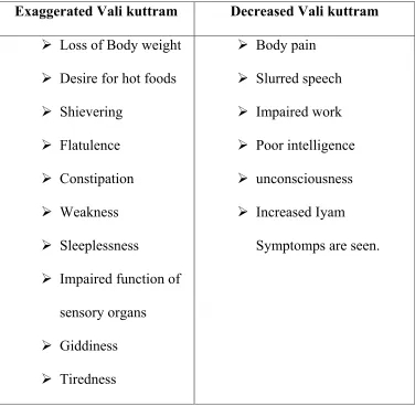
[image:29.612.119.495.301.669.2]INCORRECT TRI – HUMORS
Table 1. Vali kuttram
Exaggerated Vali kuttram Decreased Vali kuttram
¾ Loss of Body weight
¾ Desire for hot foods
¾ Shievering
¾ Flatulence
¾ Constipation
¾ Weakness
¾ Sleeplessness
¾ Impaired function of
sensory organs
¾ Giddiness
¾ Tiredness
¾ Body pain
¾ Slurred speech
¾ Impaired work
¾ Poor intelligence
¾ unconsciousness
¾ Increased Iyam
27
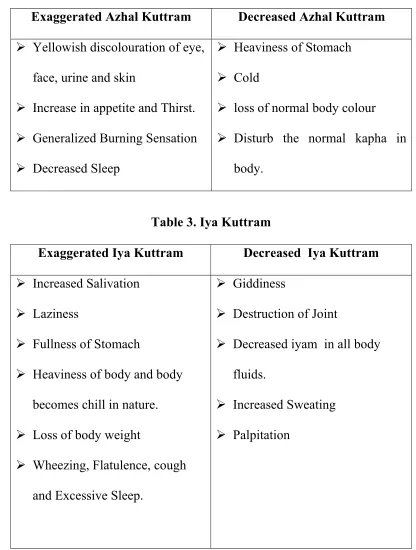
Table 2. Azhal Kuttram
Exaggerated Azhal Kuttram Decreased Azhal Kuttram
¾ Yellowish discolouration of eye,
face, urine and skin
¾ Increase in appetite and Thirst.
¾ Generalized Burning Sensation
¾ Decreased Sleep
¾ Heaviness of Stomach
¾ Cold
¾ loss of normal body colour
¾ Disturb the normal kapha in
[image:30.612.103.510.97.285.2]body.
Table 3. Iya Kuttram
Exaggerated Iya Kuttram Decreased Iya Kuttram
¾ Increased Salivation
¾ Laziness
¾ Fullness of Stomach
¾ Heaviness of body and body
becomes chill in nature.
¾ Loss of body weight
¾ Wheezing, Flatulence, cough
and Excessive Sleep.
¾ Giddiness
¾ Destruction of Joint
¾ Decreased iyam in all body
fluids.
¾ Increased Sweating
28
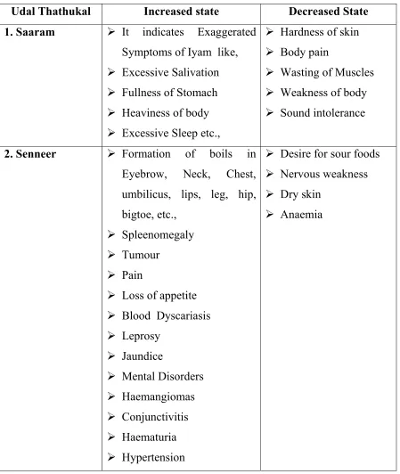
Table 4. DERANGED UDAL THATHUKAL
Udal Thathukal Increased state Decreased State 1. Saaram ¾ It indicates Exaggerated
Symptoms of Iyam like,
¾ Excessive Salivation
¾ Fullness of Stomach
¾ Heaviness of body
¾ Excessive Sleep etc.,
¾ Hardness of skin
¾ Body pain
¾ Wasting of Muscles
¾ Weakness of body
¾ Sound intolerance
2. Senneer ¾ Formation of boils in Eyebrow, Neck, Chest, umbilicus, lips, leg, hip, bigtoe, etc.,
¾ Spleenomegaly
¾ Tumour
¾ Pain
¾ Loss of appetite
¾ Blood Dyscariasis
¾ Leprosy
¾ Jaundice
¾ Mental Disorders
¾ Haemangiomas
¾ Conjunctivitis
¾ Haematuria
¾ Hypertension
¾ Desire for sour foods
¾ Nervous weakness
¾ Dry skin
29
3. Oon ¾ Cervical lymphadenitis
¾ Tumours in cheeks, stomach, male genitalia etc,
¾ Increased musclature in Neck
¾ Poor functioning of Sensory organs
¾ Joint Diseases
¾ loss of musculature in cheeks, gluteal, Male genitalia etc.,
4.Koluppu ¾ Diseases of Increased State of oon.
¾ Tiredness
¾ Dyspnoea on mild Work
¾ Increased Musculature on genitalia,chest, stomach etc.,
¾ Decreased Stability of Hip Joint
¾ Spleenomegaly
¾ Wasting of Muscles
5. Enpu ¾ Hypercalcemia on bones and teeth, leading to hypertrophy of bone and extra teeth formation
¾ Joint pain
¾ Loss of teeth
¾ Breaking of Nails
¾ Falling of hairs 6. Moolai ¾ Obesity
¾ feeling of heaviness in Eyes
¾ Clubbing of fingers and toes
¾ Oliguria
¾ Incurable ulcers
¾ Osteoporosis
¾ Giddiness
¾ Delusion
7.Sukkila thathu ¾ Increased Sexual Desire
¾ Formation of Calculi
¾ Pain in genital organs
¾ Burning sensation in sexual organs
30
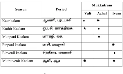
Table 5. SEASONAL VARIATION
Season Period Mukkutram Vali Azhal Iyam
Kaar kalam Mtzp> Gul;lhrp ♦
Kuthir Kaalam Ig;grp> fhh;j;jpif. ♦
Munpani Kaalam khh;fop> ij.
Pinpani kaalam khrp> gq;Fdp
Elavenil kaalam rpj;jpiu> itfhrp ♦
Muthuvenir Kaalam Mdp> Mb
Normal State (jd;dpiyahjy;)
Alteration from Normal State (jd;dpiy tsHr;rp)
♦ Aggrevation and spread to other humour (gpwepiy tsHr;rp)
ENVIRONMENTAL CHANGES
Kurinchi (FwpQ;rp)
Fever that affect blood cells. Ex:- Malaria, Abdominal Mass will
develop, Accumulation of Iyam.
Mullai (Ky;iy)
Azhal diseases, Vali diseases will develop, Liver disorders may
31
Marutham (kUjk;)
Healing of Vali, Azhal and Iyam diseases. It is a good place for
living.
NEITHAL(nea;jy;)
Vali diseases, Liver Enlargement, Flatulence, Obesity may develop
in lean persons.
Paalai(ghiy)
All diseases of Vali, Azhal and Iyam may develop, Not a good
place to live.
INFECTION
It has been not elaborately dealt by siddhars. But the following
poem speaks about some diseases,
‘fpUkpahy;; te;j Njhlk; ngUfTz;L
Nfl;fpyjd; gphpTjidf; fpukkhf
nghUkp tUk; thAnty;;yhq; fpUkpahNy
GOf;fbNghy; fhZkJ fpUkpahNy
nrUkptUk; gTj;jpuq;fs; fpUkpahNy
Njfkjpy; Nrhiff; Fl;lq; fpUkpahNy
JUkptUQ; RNuhzpjq; fpUkpahNy
#l;rKld; fphpifg;ghy; njhopy; nra;tPNu”.
Urticarial rash, Fistula, Anaemia, skin diseases, Sexually transmitted
32
OCCUPATION
In modern World, occupation is also one of the risk factor for
various diseases. Occupation affect our body in 2 conditions. It may
affect,
1. Uyir thathu
2. Udal thathu
By affecting this it results in diseases of the particular thathu as
mentioned before.
CONGENITAL DISEASE
The acquisition of certain disease from parents to offspring leads
to formation of congenital disease.
In our siddha system ,it has been explained as follows,
‘NgW ,sik ,d;gk; gpzp%g;G rhf;fhL
MWk; fUtpyikg;G”
ENNVAGI THERVUGAL
It is otherwise called as piniyari Muraimai diagnosis of the
diseases.
Rules and methods
The diagnosis is based upon three main principles such as,
1. Poriyaalarithal
2. Pulanaalarithal
33
Poriyalarithal and pulanalarithal
Sensory organs - functions
1. Nose - Smell
2. Tongue - Taste
3. Eyes - Vision
4. Skin - Touch
5. Ear - Sound
These two are very much helpful in the diagnosis of the diseases.
Vinaathal - Interrogation
Interrogating with patient or Neighbour (In case, he is not able to
speak or for children) while doing this doctor can use his pori and pulan
for examining patients pori and pulan.
Ennvagai Thervugal
Theraiyar mentions the envagai thervugal as follows.
‘If;Fwp nfhLtl thzpy; mkHe;NjhH
if;fFwp njhpe;j ek; flTis Jjpj;Nj
nka;f;Fwp epwe;njhdp tpop ehtpUkyk;
iff;Fwp KOtJk; fw;whH.”
- NjiuaH
NkYk;>
‘ehbg;ghprk; ehepwk; nkhoptpop
34
1. Naa - Tongue
2. Niram - Colour
3. Mozhi - Speech
4. Vizhi - Eye
5. Malam - Motion
6. Moothiram - Urine
7. Naadi - Pulse
8. Sparisam - By touching (palpation)
Neikkuri
Freshly voided urine in the early morning is taken in a bowl having
smoth surface. A drop of gingely oil is dropped on the upper surface of
urine and watch the mode of spread of gingely oil.
Naadi - pulse
Naadi can be felt at different site. The Important ten sites are
mentioned in our siddha literatures.
such as,
Fjp re;J> fhkpak;> ce;jp> khHG> fhJ> %f;F> fz;lk;> fuk;> GUtk;
cr;rp.
Among this ten places fuk>; lower end of radial artery is
considered to be a best place as it is situated superficial to the radial bone.
35
Vali - Tip of Index finger
Azhal - Tip of Middle finger
Iyam - Tip of ring finger
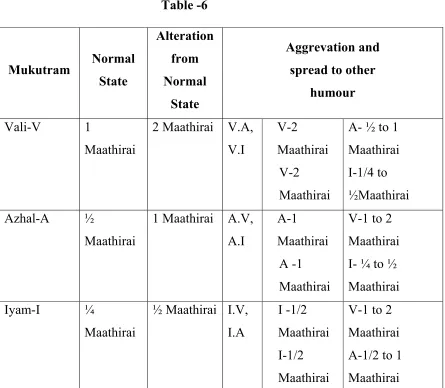
In Normal condition, The ratio of the naadi is
‘nka;asT thjnkhd;W
Nky; gpj;jk; Nkhiuiuahk;
Iaq;fh nyd;Nw mwp.”
[image:38.612.85.530.305.693.2]-fz;Zrhkpak;
Table -6
Mukutram Normal State Alteration from Normal State Aggrevation and spread to other
humour
Vali-V 1 Maathirai
2 Maathirai V.A, V.I
V-2 Maathirai
V-2 Maathirai
A- ½ to 1 Maathirai I-1/4 to ½Maathirai Azhal-A ½
Maathirai
1 Maathirai A.V, A.I
A-1 Maathirai
A -1 Maathirai
V-1 to 2 Maathirai I- ¼ to ½ Maathirai Iyam-I ¼
Maathirai
½ Maathirai I.V, I.A
I -1/2 Maathirai I-1/2 Maathirai
36
AIM AND OBJECTIVES
The author had selected the disease THALASTHAMBAM for
dissertation work because,
The Disease is more common in India and other developing
countries.
The patients are disturbed by both functionally and emotionally.
The suffereings, its prevalence and its major complications made
the author to choose the disease.
AIM
To study the disease on the basis of Siddha physiology and Siddha
Pathology emphasizing more importance to muktram, suvaigal, panja
bootha theory, Udal thathukkal and diagnose the patient on the basis of
Ennvagai thervugal and confirm the prognosis on the basis of “Neikuri”.
OBJECTIVES
To fulfil the aim the following objectives has been drawn.
1. To collect all literary evidences about vadha diseases in detail
2. To study each and every aspect of the diseases
THALASTHAMBAM in the topic of its etiology, signs and
37
3. To concentrate the clinical course of the disease
THALASTHAMBAM by observing carefully its etiology,
pathology, clinical features, Diagnosis and prognosis in patients.
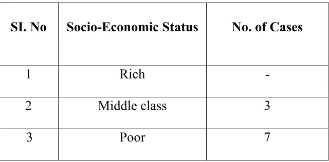
4. To Study in detail about the incidence of the disease with age,
Sex, Socio-economic status, habits and prevalence.
5. To confirm the diagnosis in Siddha System with the help of
38
ELUCIDATION ABOUT THALASTHAMBAM
jy];jk;gk;
‘fUjpNa kpff;fdj;J Ks;sq; fhiyf;
fdf;fNt File;J nehe;J fUfpf; fhZk; tUjpNa tzf;fQ;rw;wpyhk Nyw
typj;JNk NkNdhf;fp tUj;jq; fhZk; fUjpNa rlKyh;e;J Nky;%r; Rz;lha;j;
jhfkhA lk;Gyh;e;J jsh;e;J fhZk; fUjpNa fLTg;G Gspg;G jd;dhw;
fdthjq; Nfhgpj;Nj jy];jk;gq; fhNz.”
(thj Nuhf epjhdk;)
ghly; vz; 257
nghUs;
jyk; - cWg;G, cs;sq;fhy;(Organ,Foot, base)
];jk;gk; - J}z;(Not responding to any stimuli)
fdj;J - Thick
File;J - Pain
nehe;J - Destruction
fUfp - fWj;J(Blackening)
cyHe;J - Nrhk;gy; ( Tiredness)
Nky;%r;R - Dyspnoea
jhfk; - Increased Thirst
cyHe;J - Dryness of the Skin
39
‘fUjpNa kpff;fdj;J Ks;sq; fhiyf;
fdf;fNt File;J nehe;J fUfpf; fhZk;”
Pathology of the disease is described in this first line of poem.
There is thickening of foot, unremitting pain in the foot, there is
destruction of surrounding areas in the foot all of these makes the foot a
blackish appearance.
‘tUjpNa tzf;fQ;rw;wpyhk Nyw
typj;JNk NkNdhf;fp tUj;jq; fhZk;”
Progression of disease is described in this line, the disease is not
responding to treatment and the diseases progresses upwards (i.e) from
foot to upwards.
‘fUjpNa rlKyh;e;J Nky;%r; Rz;lha;j;
jhfkhA lk;Gyh;e;J jsh;e;J fhZk;”
This line clearly says the condition of patient due to chronic illness
the patient becomes fatigue, dyspnoea, there is excessive thirst and loss of
body weight.
‘fUjpNa fLTg;G Gspg;G jd;dhw;
fdthjq; Nfhgpj;Nj jy];jk;gq; fhNz.”
This line point out the aggravating factors of this diseases. The
increased intake of salt and sour in diet aggravates the altered vali
40
In Thalasthambam, Vali humour is altered. The increased intake
of salt and sour in diet aggravates the altered vali humour. As a result of
this,
There is
Thickening of the foot,
Pain in the foot,
Gangrene of the foot,
Diseases not responding to treatment it progress above from
foot,
Excessive thirst,
Dryness of the body,
Dyspnoea,
Fatigue and
Weight loss are occur.
41
DETAILED PATHOLOGICAL VIEW OF
DISSERTATION TOPIC
SIDDHA ASPECT
In the disease THALASTHAMBAM – the author yugi muni
explains about the pathology and prognosis of the diseases. He also
explains the tastes which acts as a pre-disosing factor for the diseases. He
explains these changes in an orderly form.
To explain the pathology of the disease on the basis of Vali - Azhal
-Iyam.
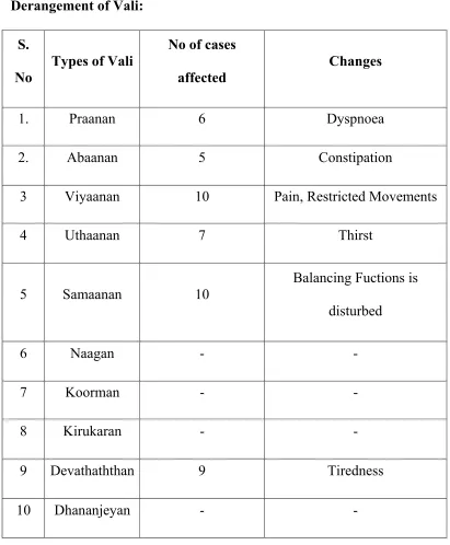
The Derangement of Vali shows the following symptoms,
‘fUjpNa kpff; fdj;J Ks;sq;fhiyf;
fdf;fNt File;J nehe;J fUfp fhZk;”.
The Dwelling place of vali as explained by yugimuni is,
‘ehnkd;w thjj;Jf;Ff; fpUg;gplNk Nfsha;
ehgpf;Ff; fPnod;W etpyyhFk;.”
With the support of above mentioned poem, when vali humour gets
altered it has been reflected in their dwelling place.
‘thjkyhJ Nkdp nflhJ”
- NjiuaH
According to the Therayar, for any type of diseases, the vali
42
In the disease Thalasthambam, the increased vali humour produces
changes in the affected part (i.e) sole of the foot ,producing heaviness of
the foot, making the foot a blackish appearance.
Increased Vali Humour Produces the following symptoms,
¾ Blackening of the affected part.
¾ Weakeness.
¾ Weakness of the sense organs.
¾ Weight loss.
¾ Sleep disturbance.
The increased Vali humour can affect the body in two ways.
i) The altered Vali humour disturbs the Azhal and Iya thaathu.
ii) The changes in Uyirthaathu can be reflected in the
udalthaathu.
Altered Iya Humour
Increased Iya Humour Produces the following symptoms,
¾ Sluggishness.
¾ Dyspnoea.
¾ Weight loss.
43
Altered Udal thathukkal
Udal thaathukkal Decreased Nature
1. Saaram - Dryness of the skin ,Dyspnoea and
Tiredness
2. Seneer - Desire to take sour food, Dryness of the
body
3. Oon - Weakness of sense organs
4. Kozhuppu - Weight Loss
5. Enbu - Falling of hair
6. Moolai - Making hole in the bone
7. Sukkilam - No change.
Thus, decrease in saram will produce decrease in other thathus too.
Based upon the affected part and the duration of the disease it will
produce different symptoms in the body.
In Thalasthambam, the decrease in udal thathukkal will produce
the following symptoms.
‘fUjpNa rlKyHe;J Nky; %r;Rz;lha;j;
jhfkhA lk;GyHe;J jsHe;J fhZk;”
The symptoms produced are,
Dryness of the body. Dyspnoea.
44
‘tUjpNa tzf;fQ; rw;wpyhk Nyw
typj;JNk NkNdhf;fp tUj;jq; fhZk;”
In this disease as there is decrease in 6 Udal thaathukal the disease
is not responding to treatment. The disease progresses to upwards.
The last line depicts that,
‘fUjpNa fLTg;G Gspg;G jd;dhw;
fdthjq; Nfhgpj;Nj jy];jk;gq;fhNZ.”
It clearly says that the increased intake of salt and sour in diet
increases vali and Azhal humour respectively and pre-disposes to
formation of Thalasthambam.
To prove the fact,
‘GspJtH tpQ;Rfwpahw; G+hpf;Fk; thjk;
xspAtH ifg; Ngwpy; gpj;Jk; rPWk”.
- fz;Zrhkpak;
‘khj;jpa Gspg;G kPwpy; te;jpLk; thjkhFk;”
- mfj;jpaH ehb
‘thjNk Gspg;G Ntz;Lk; td;gpj;jq; frg;G Ntz;Lk;”
- ,uj;jpd RUf;fehb
Hence, increased intake of Vali and Azhal again disturbs the Vali
45 ,jidNa>
‘gpj;jNk fjpj;j NghJ ngUj;jpLk; thjKz;lhk;
gpj;jNk fjpj;j NghJ ngUj;jpLk; tapw;wpy; thA
gpj;jNk fjpj;j NghJ; gpjw;wpLk; gpj;Nj NfS
gpj;jNk fjpj;j NghJ gpwe;jpLk; gpzpaNdfk;”
- Fzthfl ehb
Hence Increased intake of both sour and salt in diet Pre disposes to
46
MODERN ASPECT
jy];jk;gk
;The foot which gets affected due to gangrene is cold and
motionless. Due to arterial occlusion, the limb distal to the obstruction
becomes useless and numb, (i.e) having no sensation.
‘fUjpNa kpff;fdj;J Ks;sq;fhiyf;
fdf;fNt File;J nehe;J fUfpf; fhZk;”.
In Chronic heavy smoker due to nicotine poison, there is
inflammation of arteries,which involve all the three layers of arteries. The
injury to endothelial layer of blood vessel initiates haemostatic repair
mechanism or Thrombogenesis.
Thrombus Formation
When endothelial injury occur the sub-endotheliam gets direct
contact with flowing blood,the sub- endothelium consists of Thrombosis
favouring factors like collagen, elastin, fibronectin, laminin and
glycosaminoglycans. which are thrombogenic and thus plays an
important role in initiating haemostasis as well as thrombosis.
Following this the platelet gets aggregate at the site of endothelial
injury, Von-willibrend factor is responsible for the aggregation between
47
Due to sudden increase in platelets at the site of endothelial injury
there is hypercoagulability of blood vessel, which increases the thrombus
size.
When thrombus increase in size it results in arterial occlusion.
Due to arterial occlusion, there is damage to tissue.Alteration in the
micro vasculature (Arterioles, Capillaries and venules) is the earliest
response to tissue injury.
The alteration include,
Haemodynamic changes and
Changes in Vascular permeability
These changes at last leads to local inflammation in the affected
site (fdj;J).
The pan-arteritis of vessel walls leads to thrombosis. When the
lumen is occluded by a thrombus, pain is produced in the affected part
due to,
i. Insufficient blood supply to the affected part.
ii. Due to inadequate blood flow, there is accumulation of
excessive ‘P’ substance which is the cause for pain.
iii. Fibrotic involvement of the nerve accounts for a certain
amount of pain.
iv. The intensity of pain increases with the rate of tissue
48
v. There is accumulation of large amounts of lactic acid in
the tissues, formed as a result of anaerobic metabolism.
vi. Due to increased cell damage, there is production of
chemicals like Bradykinin and proteolytic enzymes, It can
directly attack the nerve endings and excite pain.
vii. Ischaemic neuritis
Pain
Pain is defined as an unpleasant sensation and emotional
experience associated with (or) without actual tissue damage.
The pain has 2 components.
i. Fast pain
ii. Slow pain
Fast pain
This type of pain felt when a needle is stuck into skin (or) when the
skin is cut with a knife. The fast pain is carried by A-delta fibers.
Slow pain
This pain is associated with tissue destruction due to ischaemia. It
can become excruciating and can lead to prolonged, unbearable suffering.
The slow pain is carried by C-type of nerve fibers.
In this diseases, Thalasthambam, the pain receptors in the skin and
arterial walls are all free nerve endings, they act as receptors for both
49
Due to the causes mentioned before, the receptors gets excited and
transmit pain to the first order neuron situated in the posterior root
ganglion.
First order Neuron
These neurons receive impulse of pain sensation from the pain
receptors through their dendrites and their axons reach the spinal cord.
After reaching the spinal cord, the fibre synapse with the second order
neurons in the posterior grey horn.
Second order neuron
The marginal cells and the cells of substantia gelatinosa form the
second order neurons. Fibers from these cells ascend in the form of the
lateral spinothalamic tract.
Fibers of marginal cells for fast pain form the Neo-Spinothalamic
tract - a part of lateral spinothalamic tract.
These nerve fibers terminate in ventral postero-lateral nucleus of
thalamus. Some of the fibers terminate in ascending reticular system of
brainstem.
The fibers of slow pain which arise from substantia gelatinosa
cross the midline and run along with fibers of fast pain as paleo-spino
thalamic fibers in lateral spinothalamic tract.
One fifth of these fibers terminate in ventral postero-lateral
50
reticulate formation in brain stem or in tectum of midbrain (or) in the
grey matter surrounding aqueduct of sylvius.
Third order Neuron
They are the Neurons of thalamic nucleus, reticular formation,
tectum and grey matter around aqueduct of sylvius.
Axons from these neurons reach the sensory area of cerebral
cortex. Some fibers from reticular formation reach hypothalamus.
Center for pain sensation
The center is in the posterior central gyrus of parietal cortex.
Fibers reaching hypothalamus are concerned with arousal
mechanism due to pain stimulus.
The slow pain sensation carrier type C-fibres, when synapsing in
the dorsal horns of the spinal cord, they release substance-P, a synaptic
neuro transmitter.
The substance-P is slow to build up at the synapse and slow to be
destroyed. This is the cause for progressive increase in intensity of slow
chronic pain with time and also the persistence of the slow pain.
(File;J> nehe;J)
Gangrene of the foot:
Massive death of the tissue is the end phase of severe ischaemia.
51
colour changes due to ischemia takes place as follows, at first, there is
pallor,
Later, there may be dusty grey (or) purple discoloration due to
pooling of blood in the part.
Finally, the colour changes to a greenish (or) brownish black, due
to the disintegration of haemogolabin and formation of iron sulphide
(fUfp fhDk;).
Gangrene usually begins in the digits and in arterial obstruction of
the lower limbs, usually on the undersurface of the toes. (cs;sq;fhy;)
‘tUjpNa tzf;fQ;rw; wpyhk Nyw
typj;JNk NkNdhf;fp tUj;jq; fhZk;”
The Patient is unable to flex the affected part , ( tzf;fk; rw;wpyhky;)
The disease has a tendency to progress inspite of treatment. It
progresses from foot to leg. The progression is characterized by
advancing gangrene. The pain is of full and diffuse in nature producing
pain in the small muscle of foot, muscles of calf and muscles of Thigh
and Gluteus maximus.
‘fUjpNa rlKyHe;J Nky;%r;Rz;lha;j;
jhfkha; clk;GyHe;J jsHe;J fhZk;.”
In chronic arterial insufficiency, The Muscles, subcutaneous tissue,
skin and skin appendages shows the effect of long standing impairment of
52
The skin becomes glossy and dry (parch).
Muscle wasting is noted in calf muscles.
Atrophy of several inches of calf muscles is frequent, though part
of it is due to disuse atrophy.
Dyspnoea is as a result of spasm in bronchi.( Nky;%r;R)
If the arterial occlusion affects the main arteries of trunk there is
poor supply of blood and nutrients to the affected parts but there is
continuos venous drainage this will leads to dryness of the affected parts
(clk;G cyh;e;J).
‘fUjpNa fLTg;G Gspg;G jd;dhw;
fdthjk; Nfhgpj;Nj jy];jk;gk; fhNz.”
When Excess amount of salt is taken in the diet, the salt is not
excreted so easily, As it accumulates in the body, salt indirectly
increases the extracellular fluid volume in two ways.
When there is excess salt in the tissues, the osmolality of the body
fluids increases, and this in turn stimulates the thirst centre, lateral
nucleus of hypothalamus, making the person to drink extra amounts of
water to dilute the extracellular salt to a normal concentration. This
increases extra cellular fluid volume (jhfk;)
The increase in osmolality in the extracellular fluid also stimulates
the hypothalamic posterior pituitary gland secretary mechanism to secrete
53
causes the kidney to reabsorb increased quantities of water from the renal
tubular fluid before it is excreted as urine, thereby diminishing the
volume of urine, while increasing the extracellular fluid volume.
The small increases in extra cellular fluid volume can often
increases the arterial pressure greatly due to increase in hydrostatic
pressure. Thus, the accumulation of even a small amount of extra salt in
the body can lead to a considerable elevation of the artrial pressure.
Increased arterial pressure acts as a major risk factor in the
development of Thrombosis.( mjpf cg;G)
During increased intake of tamarind (rich sour food taken daily ) it
produces a chemical substance ,which acts like Bradykinin and excite the
54
REVIEW OF LITERATURE
In Sarabenthirar Vaithya Muraigal The Thalasthambam is
explained with the same poem mentioned by Yugi in Yugi Vaithya
Sinthamani under the vatha roga nithanam. Sarabenthirar also kept the
Poem in vatha roga nithanam. Which is mentioned below,
jy];jk;gk;
‘fUjpNa kpff;fdj;J Ks;sq; fhiyf;
fdf;fNt File;J nehe;J fUfpf; fhZk;
tUjpNa tzf;fQ;rw;wpyhk Nyw
typj;JNk NkNdhf;fp tUj;jq; fhZk;
fUjpNa rlKyh;e;J Nky;%r; Rz;lha;j;
jhfkhA lk;Gyh;e;J jsh;e;J fhZk;
fUjpNa fLTg;G Gspg;G jd;dhw;
fdthjq; Nfhgpj;Nj jy];jk;gq; fhNz.”
Thus clinical signs and symptos mentioned by both Yugi and
55
THEORTICAL VIEW OF DISSERTATION TOPIC IN
MODERN ASPECT
ANATOMY
Bones of The Foot
Each foot is made up of
7 tarsal bone
5 metatarsal bone
14 phalages
Tarsus
The tarsus is made up of seven tarsal bones, arranged in two rows.
In the proximal row, the talus above and the calcaneum below.
In the distal row, From medical to lataral side these are the medial
cuneiform, the intermediate cuneiform, the lateral cuneiform and the
cuboid. Another bone, the navicular is interposed between the talus and
the three cuneiform bones.
Each tarsal bone is roughly cuboidal in shape, having six surfaces.
Metatarsus
They are made up of 5 metatarsal bones, which are numbered from
56
Phalanges
They are 14 in number. Two for the great toe, and 3 for each of the
other toes.
Muscles and fasciae of the foot
The skin and superficial fascia on the dorsum of the foot are thin
and loosely adherent.
The underlying deep fascia, a thin layer is continuous above with
the superior and inferior extensor retinacula on both sides of the foot, it
blends with the plantar aponeurosis, and anteriorly it invests the tendons
on the dorsum of the foot.
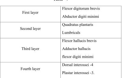
Plantar muscles of the foot
[image:59.612.105.509.431.690.2]Muscles of the sole are conventionally described in four layers,
Table - 7
First layer Flexor digitorum brevis Abductor digiti minimi
Second layer Quadratus plantaris Lumbricals
Third layer
Flexor hallucis brevis
Adductor hallucis
flexor digiti minimi
57
HISTOLOGY OF BLOOD VESSESLS
ARTERIES
All arteries are composed of 3 coats,
1. Tunica intima – Inner coat
2. Tunica media – Intermediate coat
3. Tunica adventitia – external coat
1.TUNICA INTIMA
It consists of endothelial cells. The endothelium is a single layer of
simple squamous cell, polygonal, oval in shape, with rounded nuclei.
The endothelial cells rests on basal lamina.
In large arteries, connective tissue forms a sub-endothelial layer,
that intervenes between endothelium and internal elastic membrane.
The internal elastic membrane have perforations (or) elongated
apertures through which the cytoplasmic processes of endothelial cells
achieve contact with the tunica-media.
The Internal elastic membrane forms the chief thickness of the
tunica intima.
2. TUNICA MEDIA
It is principally composed of thin, cylindrical smooth muscle cells
and elastic tissues.
58
The smooth muscle cells are held together by an abundant amount
of glycoprotein, which contains collagen fibres and reticular and elastic
fibres.
The thickeness of tunica media varies with the size of a vessel.
The external margin of tunica media is made up of elastic tissue
called external elastic membrane.
3.TUNICA ADVENTITIA
It consists of areolar connective tissue that contain fibroblasts
with in a fine meshwork of elastic fibers and bundles of collagen.
Tunica adventitia is not as thick as tunica media.
It consists of vasa vasorum, which are the arteries and veins that
supply the vessel walls. It also contains fine lymphatic vessels and fibers
that supply the arteries.
VEINS
They are made up of 3 layers,
Tunica Adventitia
Tunica adventitia is several times thicker than the tunica media.
Tunica adventitia constitutes a major part of the vessel wall.
It is made up of loose connective tissue with longitudinal elastic
59
Tunica media
It is thinner than accompanying arteries
It is composed of smooth muscle cells
Tunica Intima
It is made up of endothelial cells.
The nuclei of endothelial cells are more oval and less flattened than those in
arteries.
ARTERY
FEMORAL ARTERY
The femoral artery is the main arterial stem of the lower limb.
Commencement
Femoralartery - Commences as the downward continuation of the
external iliac artery. It commences at mid - inguinal point.
Course
The upper half of the artery passes within the femoral triangle and
the lower half of the artery passes within the sub - sartorial canal. It
passes through the adductor opening of the adductor magnus muscle.
Termination
It terminates by becoming the popliteal artery at adductor opening
60
THE POPLITEAL ARTERY
This is the artery of the popliteal fossa.
Commencement
It is the continuation of the femoral artery at the adductor opening
Course
It runs downwards and slightly laterally and then vertically
downwards within the depth of the popliteal fossa.
Termination
It terminates by dividing into anterior and posterior tibial arteries.
ANTERIOR TIBIAL ARTERIES
This is the artery of the anterior compartment of the leg.
Commencement
It is the smaller terminal branch of popliteal artery. It commences
at the lower border of the popliteus muscle, in the back of the leg.
Course
The artery passes forwards between the two heads of tibialis
posterior muscles.
It crosses the upper border of the interosseus membrane, and enters
the anterior compartment of the leg. It runs downwards and slightly
61
Termination
It terminates by becoming the dorsalis Pedis artery.
THE DORSALIS PEDIS ARTERY
This is the artery of the dorsum of the foot.
Commencement
It is the continuation of anterior tibial artery. It commences
midway between the two malleoli.
Course
It passes downwards along the medial aspect of the dorsum of the
foot, towards the first dorsal interosseous muscle. It passes between the
two heads of the first dorsal interosseous muscle and reaches the sole.
Termination
It terminates by anastamosing with the deep branch of the lateral
plantar artery to form the plantar arch. This artery is accompanied by a
pair of venae comitantes.
THE POSTERIOR TIBIAL ARTERY
This is the artery of the posterior compartment of the leg.
Commencement
It commences from the Poplitel artery, at the lower border of the
popliteus muscle. Posterior tibial artery is the large terminal branch of the
62
Course
It passes downwards deep to the origin of soleus muscle and reach
the back of the leg. And then it enters the flexor retinaculam of the ankle.
Termination
It terminates by dividing into medial and lateral plantar arteries
under cover of the flexor retinaculam.
THE MEDIAL PLANTAR ARTERY
Commencement
As one of the terminal branches of the posterior tibial artery. It
commences under the flexor retinaculum of the ankle.
Course
It passes deep to the abductor hallucis and enters the foot. It then
passes between the abductor hallucis and flexor digitorum brevis.
Termination
It terminates by joining the first plantar meta tarsal artery along the
medical border of the big toe.
THE LATERAL PLANTAR ARTERY
Commencement
It is the larger terminal branch of the postaior tibial artery under
63
Course
It passes deep to adductor hallucis and enter the sole.
It passes between flexor digitorum brevis and flexor digitorum
accessories and reaches the base of 5th meta tarsal bone. It lies between
flexor digitorum brevis and abductor digiti minimi. It then turns medially
to the first inter meta tarsal space.
Termination
It terminates by anastamosing with the dorsalis pedis artery to form
the plantar arch.
THE PLANTAR ARCH
Situation
Deeper aspect of the sole.
Extent
From the base of 5th meta tarsal bone to the first inter meta tarsal
64
PHYSIOLOGY
The function of the circulation is to serve the needs of the tissues.
To Transport nutrients to the tissues.
To Transport waste products away from tissues.
To conduct hormones from one part of the body to another.
In general, to maintain an appropriate environment in all the tissue
fluids for optimal survival and function of the cells.
Types of Circulation
The circulation is divided into 2 types as mentioned below,
1. Systemic Circulation
2. Pulmonary Circulation
Systemic Circulation
It supplies blood to all tissues of the body except the lungs, it is
also called the greater circulation (or) peripheral circulation.
Pulmonary circulation
It supplies blood to the lungs
Functional parts of the Circulation
Arteries
The arteries transport blood under high pressures to the tissues.
Hence, the arteries have strong vascular walls and blood flow rapidly in
65
Arterioles
These are the last small branches of the arterial system, and they
act as control valves through which blood is released into the capillaries.
Capillaries
The function of the capillaries is to exchange fluid, nutrients,
electrolytes, hormones and other substances between the blood and
interstitial fluid.
The capillary walls are very thin and permeable to small molecular
substance.
Venules
The venules collect blood from capillaries they gradually coalesce
into progressively larger veins.
Veins
The vein function as a conduits for transport of blood from the
tissues back to the heart.
Blood Flow
Blood flow means the quantity of blood that passes a given point in
66
Velocities of blood flow
Aorta - 2.5 cm2
Small arteries - 20 cm2
Arterioles - 40 cm2
Capillaries - 2500 cm2
Venules - 250 cm2
Small Veins - 80 cm2
Venae cavae - 8 cm2
Blood Pressure
Blood pressure is the force exerted by the blood against any unit
area of the vessel wall.
Effect of Pressure on Blood flow
The Increase in pressure increases the blood flow in two ways.
¾ It increases the force that tends to push blood through the
vessel.
67
GENERAL CHARACTERISTICS OF LEUKOCYTES
Functions of W.B.C
Neutrophils
It play an important role in the defense mechanism of the body.
Along with monocytes, the neutrophils provide the first line of
defense against the invading microorganisms.
They are the free cells in the body and wander freely through the
tissue and no part of the body is spared by these leukocytes.
Neutrophils secrete platelet Activating factor, which accelerate the
aggregation of platelets during injury to the blood vessel.
Eosinophils
It play an important role in the defense of the body.
They are specifically meant for acting against the parasites.
Eosinophil count increases during parasitic infestation and allergic
conditions.
Basophils
It plays an important role in healing processes after inflammation
and in acute hypersensitivity reactions.
The number of basophils is increased during healing process.
Monocytes
It play an important role in defense of the body.
68
It secretes interleukin-1(IL-1), Colony stimulating factor(CSF)and
platelet activating factor (PAF)
They are the precursors of the tissue macrophages.
Lymphocytes
It Play an important role in immunity
They are classified into 2 types,
Namely
i) T- Lymphocytes
ii) B- Lymphocytes
i) T- Lymphocytes
They are responsible for the development of cellular immunity
ii) B- Lymphocytes
They are responsible for the development of humoral immunity.
Platelets
The platelets are inactive and execute their actions only when
activated.
They are responsible for the onset of blood clotting.
Actin, the contractile proteins are responsible for clot retraction.
Platelets have adhesive property.
It secretes 5 Hydroxy tryptamine, which cause the constriction of
blood vessels.
69
PATHOLOGY
THROMBO ANGITIS OBLITERANS
Thrombo Angitis obliterans is an obstructive arterial diseases
caused by segmental Inflammatory and proliferative lesions of the
medium and small arteries and veins of the limbs.
ETIOLOGY
Idiopathic
The cause is not known.
Age
Common between the ages of 25-40 years.
Sex
Formerly considered to be exclusively a disease of male. Recent
reports show that there is an increase in the incidence of the disease in
female, consistent with the increase in their smoking habits.
Race
T.A.O is known to be present throughout the world and no race or
colour is known to be immune.
Heredity
No hereditary basis is established.
Occupation
Has no relation. But it is believed to be more common in farmers
70
Climate
Geographic location and climate are questionable factors.
However cold has a deleterious effect on patients suffering from T.A.O
by causing vasoconstriction superimposed on arterial occlusion.
Tobacco
The great majority suffering from T.A.O are heavy smokers.If the
patient with T.A.O continues to smoke, the disease has a tendency to
progress inspite of treatment. But if the patient discontinues smoking the
disease tends to run a favourable course and exacerbations and new
vascular occlusions are rare.
Pre-disposing factors
Hypertension
Diabetes mellitus
Cigarette smoking
Physical exercise – Lack of physical exercise predisposes to
atherosclerosis
Role of highly saturated fats and cholesterol
The above have been proven to be key factors for assessing risk of
71
Clinical classification of TAO
Allen-Barker-Hynes have classified the disease into 8 groups.
¾ Arterial occlusion causing intermittent claudication as the only
symptom.
¾ Intermittent claudication with cold digits and mild rest pain.
¾ Severe ischaemic neuritis.
¾ Marked colour changes and Raynauds phenomenon.
¾ Minor gangrene with local infection.
¾ Gangrene of digits.
¾ Severe gangrene spreading on to foot or hand.
¾ Thrombophlebitis as major or only complaint.
Clinical Features
¾ Intermittent Pain
¾ Colour Changes
¾ Skin Temperature
¾ Absence of Arterial Pulsation
¾ Nutritional Changes
¾ Gangrene
Intermittent Pain
This type of pain is otherwise known as intermittent claudication.
72
found to be associated with obliteration of the main artery of leg.
Intermittent claudication in man is an indication of obstruction to the free
flow of blood to the tissues of the affected limb. Intermittent claudication
is a symptom and not a disease
The site of claudication is a rough measure of the level of vascular
occlusion. It is more commonly observed in the calf and small muscles
of the foot than in the thigh because in the thigh there is a generous
collateral circulation to compensate for the partial occlusion of the main
vessel.
Colour Changes
Lewis classic monograph (1936) concludes that skin colour is a
good index of the adequacy of peripheral blood flow when the normal
responses to environmental conditions are known.
The colour of skin attributable to circulation depends on two factors (i)
Amount of blood (ii) Colour of blood. The depth of the colour of skin
depends upon the amount of blood contained with in the capillaries of the
skin.
Skin Temperature
The skin temperature of resting limb is dependent upon the balance
between the amount of heat brought to it by the blood and the amount of
heat lost to its surroundings, when the blood flow to a limb is reduced the