A COMPREHENSIVE STUDY OF 80 CASES OF LIVER
ABSCESS
Submitted to
THE TAMIL NADU Dr. M.G.R. MEDICAL UNIVERSITY
For M.S. DEGREE EXAMINATION BRANCH – I (GENERAL SURGERY)
THE TAMIL NADU Dr. M.G.R. MEDICAL UNIVERSITY
CHENNAI, INDIA
CERTIFICATE
This is to certify that “A Comprehensive study of 80 cases of Liver
Abscess” is a bonafide work done by Dr. P.S. GANESH BABU, Post Graduate in department of General Surgery Kilpauk Medical College,
Chennai 600 010. Under my guidance and supervision in fulfillment of
regulations of the Tamil Nadu Dr.M.G.R. Medical University for award of
M.S. degree Branch I, Part II (General Surgery) during the academic period
from March 2007 to March 2010.
Dr. S. Udaya kumar, M.S.,
Prof. & H.O.D. of General Surgery, Kilpauk Medical College, Chennai-10.
Dr. P.K. Baskaran,M.S., Prof. & Chief of General Surgery,
Kilpauk Medical College, Chennai-10.
Dr.Kanagasabai,M.D., The Dean
ACKNOWLEDGEMENT
I am extremely thankful to Prof. Dr. S. Udayakumar, M.S., Professor and Head of the Department of General surgery Kilpauk Medical College for his guidance and help in conducting this study.
I am immensely grateful to my Chief Prof. Dr. P.K. Baskaran, M.S., Department of General Surgery, Government Kilpauk Medical College, for his valuable guidance and encouragement in conducting this study.
I wish to express my sincere thanks to Prof. Dr. Kanagasabai, M.D., Dean. Kilpauk Medical College, Chennai-10, for permitting to conduct this study in Government Kilpauk Medical College Hospital.
My sincere thanks to Prof. Dr. A. Periyasamy, M.S., & Prof. Dr. T. Bhavani Shankar, M.S..
I extend my special thanks to Assistant Professors of our unit
Dr.V.Vijayalakshmi, M.S., Dr. K. Shantha kumar M.S., and Dr. T. Suresh Babu, M.S., for their valuable guidance in initiating this study.
I am thankful to all my post graduate colleagues for their help and encouragement.
CONTENTS
S. No. Page
No.
1. INTRODUCTION 1
2. AIM OF THE STUDY 2
3. HISTROICAL FACTS 3
4. ANATOMY AND FUNCTIONS OF THE LIVER 4
5. REVIEW OF LITERATURE 13
6. MATERIALS AND METHODS 51
7. RESULTS OF STUDY 52
10. DISSCUSSION 57
11. SUMMARY AND CONCLUSION 66
BIBLIOGRAPHY
PROFORMA
INTORDUCTION
Descriptions of Liver abscesses date back to Hippocrates in approximately
4000 BC. But an understanding of their Etiology, Bacteriology, diagnosis and
treatment is a recent event in Twentieth century and is still emerging.
Hepatic abscess often presents a pitfall in diagnosis and challenge to
surgical diagnostic acumen. Early diagnosis and prompt initiation of treatment
almost certainly leads to complete cure.
Of the two types of hepatic abscess, the Amoebic and Pyogenic, the former
seems to be more prevalent in our country. Because studies of Hepatic abscess
especially amoebic abscess have often originated from endemic areas, they offer
little information on the application of modern diagnostic techniques. Diagnosis in
this area mainly depends on the clinical presentation. But since the introduction of
Ultra sonogram and CT Scan as a diagnostic device a more Accurate diagnosis can
be made in every case.
In this study, about 600 cases of Hepatic abscesses admitted in Government
Kilpauk Hospital and Royapettah Government Hospital in the year 2007-2009. Out
AIMS AND OBJECTIVES
Clinically impressed with the great importance of establishing early
diagnosis in patients with liver abscess and this study has been made to obtain
better knowledge of the nature of the disease.
To determine the incidence of Hepatic abscess in our Hospital.
To determine the Age and Sex incidence.
To evaluate the various clinical parameters of both Uncomplicated and
complicated Liver abscess and various modalities of treatment available.
To know about the disease Prevalence occurring as Abdominal Emergencies.
To emphasis the immense use of modern Investigation likes USG and CT Scan
HISTORICAL FACTS
1845 - GROS - Discovery of Amoeba
1875 - LOSCH - Discovered Histolytica
1887 - KOCH - Reported cases of amoebic dysentery with liver
abscess and showed E.Histolytica trophozoites in hepatic capillaries.
1887 - KARTULIS - Described presence of E. Histolytica in Liver
abscess
1891 - COUNCILMAN
AND LAFLEUR - Coined the term Amoebic Abscess of
Liver
1893 - QUINCKE AND
ROOS - Described the cystic form of
E.Histolytica and its life cycle,
differentiated pathogens from other non- pathogenic intestinal amoebae in man.
1904 - KARULIS - Described amoeba in Brain abscess
1927 - CRAUC - Defined the modern concept of
clinical amoebiasis.
Amoebic liver abscess has been known since antiquity in India. In the
Sanskrit Manuscript, BHRIGUSAMHITA (around 3000 B.C.) there is a mention
of germs, which cause diarrhea with stools containing blood and mucus and that
these germs can also give rise to a hard ball like mass in the abdomen, apparently a
ANATOMY AND
FUNCTIONS OF
ANATOMY OF THE LIVER
The liver is the largest metabolic organ in the body. It is situated in the
upper and right parts of an abdominal cavity, occupying almost the whole of the
right hypochondrium, greater part of epigastrium, extending into left
hypochondrium upto left lateral line. In male it weighs about 1400-1800g and
1200-1400g in female.
It has 5 surfaces. (Anterior, posterior, superior, inferior and the right). One
prominent border (Inferior Border). On the posterior surface there lies the “Bare
area of liver” which is contained within the reflection of superior and inferior
coronary ligaments. Liver abscess is commonly situated in the upper part of the
right lobe close to the Bare area. Liver abscess close to the bare area is painless
because they are devoid of peritoneum.
Surface:
Diaphragmatic Surface:
a.Anterior
b.Superior
c.Posterior
Visceral Surface:
Diaphragmatic Surface:
Smooth and doom shape, related to concavity of inferior surface of
diaphragm, separated from diaphragm by sub-phrenic recesses which are separated
by falciform ligament into right and left.
The convex or antero superior surface of liver is in relation with right lung
and the pleura. This explains the erosion of hepatic abscess through the diaphragm
into the pleural cavity and right lung.
PERITONEAL LIGAMENTS OF THE LIVER
a. Falciform ligament
b. Upper (Anterior) coronary ligament
c. Lower (Posterior) coronary ligament
d. Right Triangular ligament
e. Left triangular ligament
f. Lesser omentum
Falciform ligament
It is a sickle-shaped fold which stretches from the liver to the diaphragm
and anterior abdominal wall. It consists of a right and left layer which are attached
between the right and left lobes of the liver on its anterior and superior surfaces.
The two layers are continuous with each other at the lower free border which
Upper (anterior) Coronary Ligament
It is continuous with the right layer of the facliform ligament. It stretches
between the posterosuperior aspect of right lobe of the layer and the diaphragm.
Lower (posterior) Coronary Ligament
It stretches between the postero inferior aspect of right lobe of the liver and
the diaphragm.
Right Triangular Ligament
The upper and lower layers of the coronary ligament, when traced to the
right are continuous with each other at the right triangular ligament which is a
short triangular fold the connects the upper surface of the right lobe of liver with
the diaphragm.
Left triangular ligament:
It is short triangular fold connecting the upper surface of the left-lobe of
liver with the diaphragm. It consists of an anterior and posterior layer. The anterior
layer is continuous with the left layer of the falciform ligament and the posterior
layer is continuous with the anterior layer of the lesser omentum.
PORTA HEPATIS
Is a deep transverse fissure about 2 inch long, situated on the inferior
plexus of Nerves and lets out the right and left Hepatic ducts and few lymphatics.
Relations within the Porta Hepatis are from behind far wards – the portal vein,
hepatic artery, and Bile ducts.
MORPHOLOGICAL ANATOMY
Liver is divided into the right and left lobes by the falciform ligament
anteriorly and superiorly, by the fissure for ligamentum teres inferiorly and by the
fissure for ligamentum venosum posteriorly. Right lobe is much larger and forms
5/6 part. It has 2 additional lobes called the caudate and quadrate lobe.
The caudate lobe, lying between the inferior vena cava and the fissure for
the ligamentum venosum, and the quadrate lobe, lying between the gallbladder
fossa and the fissure for the ligamentum teres, were consequently considered to be
part of the right lobe.
FUNCTIONAL ANATOMY OR SURGICAL ANATOMY OR SEGMENTAL ANATOMY
Functional anatomy was first initiated by cantle in 1898 and was enhanced
by Counseller in 1929 and Couinaud in 1957. Couinaud’s is the most exact and
complete description of liver anatomy.
In essence the liver should be regarded as a paired organ (right and left
livers) that are fused along a line extending from the middle of the gallbladder
Within the liver is this corresponds to a vertical plane (the main portal scissura or
Cantlie’s line) in which lies the middle hepatic vein. The right lobe liver receives
the right portal vein, hepatic artery and bile duct and the left lobe liver the
corresponding left portal vein, hepatic artery and bile duct.
THE RIGHT LOBE LIVER
The right liver is further subdivided into two sectors by the vertical right
portal scissura in which lies the right hepatic vein.
The medial sector is composed of segments V (antero-inferior) and VIII
(postero-superior) and the lateral sector of segment VI (antero-inferior) and VII
(Postero-superior). Thus in the supine patient segments V and VI partially overlap
segments VIII and VII, respectively. Each segment receives a portal pedicle and is
drained by a separate bile duct.
THE LEFT LOBE LIVER
This is divided by the left portal scissura, in which lies the left hepatic vein,
into a medial sector that forms segment IV and a lateral sector that is further
divided into an anterior segment III and a posterior segment II.
Caudate (dorsal, Spigel) lobe
Although this is customarily labeled as segment I, it is really a separate
‘liver’ because it has its own hepatic veins and bile ducts although it receives
Because of this segmental liver anatomy, it is possible to resect a single or
several segments even in the liver that has been distorted by chronic disease.
HEPATIC STRUCTURE
The greater part of the liver is invested with peritoneum, which covers a
thin capsule of connective tissue (Glisson’s capsule). The bulk of the cells within
the liver constituting about 80% are the hepatocyctes or parenchymal cells.
Hepatocytes carry out a multitude of metabolic activities.
Conventional morphology considers that the liver is composed of pyramidal
lobules based on a central vein surrounded on the periphery by portal trunks with
terminal radicles of bile duct, portal vein and hepatic artery. The two vascular
systems of central vein and portal tract lie on planes at right angles to one another
and never interdigitate. Thus the sinusoids are arranged perpendicular to the planes
of the central veins and portal blood passes to the central vein along a pressure
gradient. The walls of the sinusoids are composed of endothelial and phagocytic
cells termed Kupffer cells. Between the hepatocytes and Kupffer cells is the space
of Disse. Bile canaliculi are shown to be channels or grooves in the hepatocyte
surface, lined by microvilli. The network of canaliculi drains the liver lobules into
the terminal bile ducts.
According to Rappaport, there are three zones of liver parenchyma. Zone 1
form. Zone 1 adjacent to the portal triad is the best vascularized and least
susceptible to injury. Zone 3 is adjacent to the central vein and most susceptible to
injury.
BLOOD SUPPLY
The dual afferent blood supply consists of hepatic artery and portal vein.
Liver receives 20% of its blood supply from hepatic artery and 80% from the
portal vein. Before entering the liver the hepatic Artery and portal vein divide into
right and left branches. Within the liver they redivide to form the segmental and
then to interlobular vessels which run in the portal canals. Further ramification of
interlobular branches open into the hepatic sinusoids. Thus hepatic arterial blood
mixes with the portal venous blood in the sinusoids.
VENOUS DRAINAGE
The hepatic venous drainage beings as a central vein of a liver lobule. The
central vein receives sinusoids from all sides and unite with the central vein of
other lobules to form the sub lobular veins which in turn fuse to form collecting
veins which forms 3 major hepatic veins.
LYMPHATIC DRAINAGE
The superficial lymphatics drain into caval, hepatic, paracardial and celiac
lymph nodes. Deep lymphatic end in the nodes around the end of IVC and party
NERVE SUPPLY
Nerves of liver are derived from Hepatic Nerve plexus which are largest
derivative of celiac plexus. Hepatic plexus accompanies branches of Hepatic artery
and portal vein to the liver. It consists of sympathetic fibres from celiac plexus and
para sympathetic fibres from anterior and posterior vagal trunks.
DEVELOPMENT
The liver arises from Hepatic diverticulum from most caudal part of the
foregut. The Hepatic diverticulum extends into septum transversum and expands
the ventral mesentry. It divides into large cranial parts which give rise to
interlacing cords of liver cells and intra epithelial lining of intra Hepatic portion of
biliary apparatus.
FUNCTIONS OF THE LIVER
1. The formation of bile and the metabolism of bilirubin and of bile salts.
2. The synthesis of albumin, fibrinogen and prothrombin.
3. Storage and metabolism of carbohydrates, including the conversion of
monosaccharides (e.g. dextrose) into glycogen, and vice versa.
4. Formation of phospholipids and cholesterol, synthesis of fatty acids from
carbohydrate.
5. Deamination of amino acids with formation of urea. Removal of ammonia
from portal blood.
7. Reticuloendothelial activities
8. Storage of Vitamin B12 and Vitamin A
9. Iron and Copper Storage
10.Destruction of bacteria
REVIEW OF LITERATURE
INCIDENCE
Entamoeba histolytica Infection affects an estimated 10% of the world’s
population. The great majority of such Infections occurring in people living in
Indian sub-continent, sub Saharan Africa, and parts of Central and South America.
In these endemic areas approximately 50% of the population is infected with 90%
or more being Asymptomatic cyst Passers.
Amoebic Liver abscess occurs in less than 10% of individuals infected with
these organisms. Amoebic Liver abscess are 3-5 times more frequent than
Pyogenic Liver abscess. Average age of the patients is between 28-48 years, with
striking male predominance (7:1). Particularly severe Invasive disease occurs in
patients with compromised cellular immunity in young infants, in the
malnourished, in pregnant women and in patients receiving corticosteroid. On the
global scale, Amoebiasis is the third most common Parasitic cause of Death after
malaria and schistosomiasis.
Incidence of Pyogenic Liver abscess being estimated at 8-16 cases
/1,00,000 admissions. Fifty years ago the majority of patients were under the age
of 40 and Appendicitis was the leading cause of the disease. Today the average age
being 43-60. This change corresponds to the finding that appendicitis has been
LIFE CYCLE OF E-HISTOLYTICA
Amoebiasis is defined as the condition of Harboring of entamoeba Histolytica in Humans with or without clinical Manifestation (WHO Tehran in 1968)
E.Histolytica exists in two forms
1. Trophozoite
2. Cyst with a Transitory stage of Precystic Form.
E.Histolytica Passes its life cycle only in one Host the man.
The mature quadrinucleate cysts are the infective forms of the parasite.
When these cysts are swallowed along with contaminated food and drinking water
by a susceptible person, they are capable of further development inside his gut.
The fully developed cysts thus gaining entrance in to the alimentary canal, pass
unaltered through the stomach. The “Excystation” occurs when the cyst reaches
the caecum or lower part of the ileum. Each cyst Liberates a single Amoeba with
four Nuclei, a tetranucleate Amoeba which eventually forms eight Amoebulae
(Metacystic trophozoites) by division of nuclei with successive fission of
cytoplasm. The young Amoebulae being actively motile, invade the tissues and
ultimately lodge in the submucous tissue of the large gut their normal habitat. Here
they grow and multiply by Binary fission.
It is to be noted that the Trophozoite phase of the parasite is responsible for
producing the characteristic lesion of amoebiasis. The Trophozoite of
vein, to be carried away to the liver where their further progress may be arrested.
In the liver the Trophic forms may for a time grow and multiply but encystations
does not occur.
Hence such an invasion is always to be overlooked as an accident on the
part of the Parasite because so far as its biological aspect is concerned it has
reached a dead end. The parasites that remain in the intestinal wall may cause an
attack of acute dysentery (amoebic colitis). A certain number of these
Trophozoites are discharged into the lumen of the Bowel and are transformed into
small precystic forms from which the cysts develop.
The mature quadrinucleate cysts are the most resistant and infective forms
of the parasite. But the cysts produced in an infected individual are unable to
develop in the host in which they are produced. Transfer to another susceptible
host enables them to grow and continue their life cycle.
There are 2 types of E.histolytica namely pathogenic and non pathogenic. It
is now firmly believed that pathogenic and non-pathogenic entamoeba isolates are
distinct species. Pathogenic is called E.Histolytica, Non pathogenic as E.Dispar.
Pathogenic behavior of pathogenic and non pathogenic entamoeba depends on the
quantitative and qualitative difference between molecules (genes/proteins)
E. histolytica
trophozoite
AEITOLOGY AND PATHOGENESIS – AMOEBIC LIVER ABSCESS (Syn TOPICAL OR DYSENTERIC ABSCESS)
The transmission of amoebiasis is clearly related to lack of sanitation and
low socio-economic status rather than to climate. Common modes of transmission
are by food contaminated with cysts or contaminated water through those engaged
in preparation and handling of food. The commonest extra intestinal manifestation
of amoebic infection occurs when organisms escape the colon and reach the liver
via the portal vein.
There is another view that retrograde lymphatic spread of Amoebiasis can
occur from the colon to the liver and from the liver to the lungs.
Lymphangiographic study in case of carcinoma stomach have shown that the
lymphatics from the stomach, colon and liver join the thoracic duct – an evidence
for the retrograde lymphatic spread of amoebiasis. It was favored by the
demonstration of multiple amoebae in the small interlobular branches of the portal
vein. After reaching the liver in large number, the trophozoites set up a thrombus
formation which in turn gives raise to an infarct, the wall of the vessel is destroyed
and amoebae feed on the products of cytolysis. This view was supported by the
demonstration of thrombosed radicals of the portal vein in the walls of the abscess
and amoebae were found entangled in the thrombus.
E-histolytica produces enzymes which cause hydrolytic dissolution of liver
tissue. Various enzymes are produced like hyaluronidase, Glutaminase, amylase,
particularly poor in protein and poor immune status predispose to the development
of amoebic liver abscess.
Adherence to cell by Gal-Gal-Nac (galactose and N-acetyl D-galactosamine)
E. Histolytica lysis of surrounding cells
proteolytic degradation of extra cellular matrix
(Tropical Hepatogastroenterology, Chapter 38 Intestinal and extraintestinal amebiasis-MP Sharma & Vineet Ahuja)
PATHOGENIC CLASSIFICATION OF AMOEBIC LIVER ABSCESS (Gastro Enterology Today Vol-I No.3)
Acute Chronic
a. Benign a. Benign
b. Aggressive b. Acclerated
1. Illness less than 10 days 1. Low Grade symptom for a month or more
2. Fever & Tender Hepatomegaly
present
2. Fever uncommon, mildly Tender
hepatomegaly
3. Low Serological titres
Leukocytosis, increase transminase
3. High Serological titres, severe
anemia ↑ alkaline phosphatase,
normal SGOT
MACROSCOPIC PATHOLOGY
About 83% of liver abscess are located in the right posterosuperior surface
of liver. The propensity for this site reflects the fact that venous return from the
right side of colon (Amoebic infection having a particular impact on the caecum
liver. To the naked eye, the appearance of the abscess area is reddish brown in
color with a semifluid or grumous consistency. The wall of the abscess cavity is
ragged and shaggy in appearance and is formed by the necrotic liver tissue which
gradually merges into the healthy zones with an intervening zone of hyperaemia.
In an old abscess the wall is smooth and is formed by dense connective tissues.
Multiple small abscesses in which the whole organ becomes riddled with scattered
foci of necrosis are probably more common. The liver enlargement is explained on
the basis of oedema and congestion ( lamot and pooler 1958).
MICROSCOPY
If a section is made through the margin of a liver abscess, three zones can
be differentiated from the center to the periphery.
1. A central zone of cytolysed granular material with no amoebae.
2. An intermediate zone consisting of degenerated liver cells, a few
leucocytes, connective tissue cells, red blood cells and an occasional
trophozoite of E.hostolytica.
3. A peripheral zone consisting of congested capillaries with varying degrees
of necrosis of liver cells. The amoebae can be seen to be multiplying in this
area and invading and adjoining healthy liver tissue.
In long standing cases, the third zone may consist of actively proliferating
connectives tissue cells, lymphocytes and monocytes, walling off the abscess
PUS OF LIVER ABCESS
The ‘pus’ is not of suppuration but is a mixture of sloughed liver tissue and
blood. It is chocolate brown in color and thick in consistency (so called
‘Anchovy-sauce’ pus). The smell is rarely offensive. The ‘pus’ is bacteriologically sterile.
Microscopy reveals degenerated liver cells, a few red blood cells and occasional
leucocytes. The trophozoites of E.histolytic are not generally found in freshly
aspirated pus but appear in the escaping ‘pus’ four or five days after the initial
evacuation.
Microscopic examination of the wet mount of pus from the liver or lung
amoebic abscesses is frequently carried out for the demonstration and
identification of amoebic trophozites.
Sensitivity of the method is however very low. Amoebic trophozoites can
be demonstrated only less than 15% cases of amoebic liver abscess. (Text book
medical parasitology 2nd Edition by Subash Chandra Parija)
INTESTINAL LESIONS IN AMOEBIC LIVER ABSCESS
In all cases of hepatic amoebiasis, as also in other metastatic amoebiasis,
the primary lesion is in the large gut. The changes in the intestine observed at
autopsy may be any one of the following.
i) Small superficial ulcers, with thickening of the colon.
iii)Pigmented or non-pigmented scars in the large intestine, representing the
sites of previous ulcers.
iv) No change in the large gut.
v) Extensive ulcers scattered throughout the large intestine are quite
uncommon.
vi) E-Histolytica can produce diffuse inflammation and ulcertation of colon
which may be indistinguishable endoscopically from idiopathic
ulcerative colitis. Further more reappearance of colonic symptoms in
patients with chronic ulcerative colitis has at times found to be due to
infection with E.Histolytica.
Clinical syndromes associated with E. histolytica infection. Intestinal amoebiasis:
¾ Asymptomatic cyst passers
¾ Acute amoebic colitis
¾ Mucosal disease
¾ Transmural disease.
¾ Ulcerative postdysentric colitis,
¾ Appendicitis
¾ Amoeboma,
¾ Amoebic stricture
Extraintestinal amoebiasis:
¾ Perforation and peritonitis
¾ Pleuropulmonary amoebiasis
¾ Amoebic pericarditis
¾ Cutaneous amoebiasis
PYOGENIC LIVER ABSCESS
ETIOLOGY – BACTERIOLOGY – PATHOGENESIS
In the liver the kupffer cell functions as a primary Barrier and filter for the
clearance of micro organisms from arterial, venous, Biliary and local sources.
Pyogenic liver abscess are believed to occur when their normal hepatic clearance
fails or is overwhelmed. Distal infectious sites may seed the liver with pathogenic
Bacteria via portal vein or the Hepatic Artery.
ORGANISMS
Gram Negative Aerobs (50-70%)
E.Coil (35-45%), klebsiella, Proteus, enterobacter.
Gram Positive Aerobs (30%)
Streptococcal Sp, enterococcus Faecalis.
Anaerobes (40-50%)
Bacteroides Sp, Fusobacterium, Pepto Streptococcus.
ETIOLOGY % SOURCE DISTRIBUTION 1° MICRO ORGANISMS
Biliary System 40 Cholangitis
Biliary Obstruction Both Lobes Multiple Single Species Gram-aerobes and anaerobes – E-Coli Portal Circulation 20 Intra abdominal Infection Liver metastasis Right Lobe>Left Multiple or single
Area of Metastasis E.Faecalis, E.Coli, B. Fragilis B. Fragilis Arterial Circulation 12 Bacteremia, Systemic Infection Both lobes-Multiple S. Aureus, S.Pyogenes
Trauma 4 Direct Exposure, Necrosis
Area of injury S. Aureus,
S. Pyogenes Direct Extension 6 Cholecystitis Perforated ulcer
Adjacent area E.Coli
CLINICAL PRESENTATION
The clinical feature often reflects the site, size and the number of abscesses
as well as degree of involvement of adjacent tissues. The onset of symptoms may
be acute or chronic. The clinical features of amoebic abscess are similar to
Pyogenic abscess.
However acute presentations are more common with amoebic abscess. In
amoebic abscess pain in Right Hypochondrium and Right Lower Chest are the
commonest symptom (90%) followed by fever. In Pyogenic abscess, fever is the
most common symptom (80%). Hepatomegaly and Right upper Quadrant
tenderness are the only consistent Physical findings.
Triad of fever, pain in Right Hypochondrium and Tender Hepatomegaly is
strongly suggestive of liver abscess.
Co-morbid diseases associated with Pyogenic liver abscess.
Children Adults
Chronic granulomatous disease Diabetes Mellitus
Complement deficiencies Cirrhosis
Leukemia Chronic Pancreatitis
Malignancy Peptic ulcer disease
Sickle cell anaemia Inflammatory Bowel disease
Polycystic liver disease Jaundice
Congenital hepatic fibrosis Tuberculosis
Post transplant liver failure Malignancy
Necrotizing enterocolitis Leukemia and Lymphoma
Chemotherapy and steroid therapy Chemotherapy and steroid therapy
Acquired Immuno Deficiency Syndrome Acquired Immuno Deficiency
SYMPTOMS SIGNS
Pain Hepatomegaly
Fever Right Upper Quadrant Tenderness
Nausea and Vomiting Guarding and rigidity
Weigh loss Ascites
Malaise Jaundice
Diarrhea Pleural effusion
Pruritus Pleural rub
Cough
AMOEBIC PYOGENIC
Age<50 years >50 years
Male : Female 10:1 Male = Female
Recent Travel to endemic area Malignancy
Pulmonary dysfunction High fevers
Abdominal Pain Pruritus
Diarrhea Jaundice
Abdominal Tenderness Septic shock
It has been noted infrequently at the extremes of age and is seven to nine times
more common in males. Amoebic Liver Abscess (ALA) may present as an acute
process or as a chronic indolent disease. ALA usually occurs in the right lobe of
the liver and is solitary (30% - 70%). Unusual presentations include multiple
abscesses, left lobe abscesses, abscesses presenting as compressive lesion, and
abscesses rupturing into viscera.
Multiple liver abscesses:
Fifteen per cent of patients may have multiple abscesses. They present with
added bacterial infection leading to a more severe disease. E.coli and Klebseilla
are the commonly cultured organisms. These patients present with a clinical
picture indistinguishable from hepatic encephalopathy due to acute hepatocellular
failure. Hepatic encephalopathy in ALA patients possibly results from combination
of right hepatic vein occlusion, pylophlebitis, and occlusion of several portal vein
radicles.
Left lobe abscess :
Thirty-five per cent of patients present with a left lobe abscess. Half of
these have associated lesions in the right lobe while the remaining have solitary
left lobe abscess. These patients have longer duration of symptoms (3-4 weeks)
and fever is less commonly observed as compared to right lobe abscesses. It may
present as a large epigastric mass with minimal movement with respiration. Often,
to the clinician’s despair, it has been confused with pseudocyst of pancreas. These
patients also have weight loss with poor hepatic localisation of symptoms.
Complications like peritonitis and toxaemia are significantly more common in left
lobe abscess. Needle aspiration may be more rewarding in combination with
anti-amoebic drugs.
Compression lesions :
A posteriorly located ALA in the right lobe may present as inferior vena
cava obstruction or hepatic outflow obstruction. This is suggested by bilateral
along with clinical, radiological, and serological features of ALA. These features
disappear after aspiration of the abscess. Leakage of the abscess may occur into the
pleural cavity, with empyema thoracis. Intraabdominal extension following
perforation into the peritoneal cavity is usually associated with shock and
generalised peritonitis and may occur in upto 7% of cases. Rupture into the colon
and biliary tree has also been reported. (Journal, Indian Academy of Clinical
Medicine _ Vol. 4)
COMPLICATIONS OF LIVER ABSCESS
About 40% of patients with Pyogenic liver abscess and 10% of amoebic
liver abscess patients develop complications. Generalized sepsis is the most
common systemic complication in Pyogenic abscess. Rupture into peritoneum and
thorax are the most common complications in amoebic abscess. Posteriorly located
Amoebic Liver Abscess in Right Lobe can present as IVC obstruction or Hepatic
out flow obstruction (Gastro Enterology Today April, June 1997).
Severe Amoebic Liver Abscess (Chau. Sk., Chang, Chiencs, Sheen IS)
Severe Amoebic Liver Abscess was defined as the Rupture of an abscess
that was resistant to 72 hours of medical treatment, complicated by secondary
PLEUROPULMONARY COMPLICATION
It is the most frequent site affected when liver abscess ruptures. It is the
third most common manifestation of amoebiasis after amoebic colitis and amoebic
liver abscess. The reported incidence of pleuropulmoary amoebiasis varied from
14 to 30 percent. Clinical manifestations include chest symptoms with
expectoration of anchovy – sauce sputum. Pleural rub, signs of pleural effusion
and lung abscess may also be present. Typically such lesions originate from right
lobe.
PERITONEAL COMPLICATION
Amoebic peritonitis is the second most common complication of Amoebic
liver abscess and it is the commonest form of rupture into a serous cavity.
Amoebic peritonitis results more often due to rupture of Right Lobe Abscess.
Extension into Peritoneal cavity may form acute generalized peritonitis, chronic
generalized peritonitis or a localized abscess. In sudden rupture of amoebic liver
abscess the clinical picture mimics perforation of a hollow viscus. Mortality may
approach a great as 20%.
PERICARDIAL COMPLICATION
Pericardial complications are relatively rare and are often associated with
left lobe abscess. The reported incidence of this complication varies from 0.6 to 5
per cent. Pericardial extension may cause suppurative pericarditis due to sudden
pericardial effusion. It can also cause constrictive amoebic pericarditis or amoebic
hydro pericardium.
UNCOMMON COMPLICATIONS
Erosion into the intestinal tract, cutaneous fistulation, secondary infection,
obstructive jaundice, subphrenic abscess, hemobilia and vascular complications are
other rare complications. Siddiqui MN, Rizvi SB, Ahmed M, in their case report,
reported a case of Amoebic Liver Abscess complicated by Hepato duodenal fistula
(a finding of air in the liver abscess – on Ultra sonogram – confirmed by
Gastograffin Swallow).
DIAGONSTIC STUDIES
Laboratory Investigations: Even though most of the laboratory
investigations are non specific, they are helpful to some extent in the diagnosis, to
assess the involvement of liver, response to treatment and also as prognostic factor.
BLOOD
• Leucocytosis varying from 15,000-25,000 with a left shift present in
50-60%.
• Anemia is also encounted in liver abscess. It is related to duration and size
of Abscess.
LIVER FUNCTION TESTS
Abnormal liver function test exist more commonly in Pyogenic liver
abscess than Amoebic Liver Abscess.
SERUM BILIRUBIN
• Serum bilirubin is elevated in 10-24% of patients.
ENZYMES
• Serum Alkaline phosphatase is raised in about 50-60% of patients.
• Elevations of SGOT or SGPT and Gamma – Glutamyl Transpeptidase
occur in approximately 40-50% of patients.
SERIUM PROTEINS
• Hypoalbuminemia is quite common with increase in Gamma – Globulin
• Hypoalbuminemia is considered as a poor prognostic marker especially if it
is < 2gm/dl
PROTHROMBIN LEVEL
PARASITOLOGICAL EXAMINATION STOOL EXAMINATION
Cyst & Trophozoites of Entamoeba Histolytica are present in the stools in
<15% of Amoebic liver abscess patients –verlenden et al. This gives information
regarding the persistence of intestinal infection.
E. histolytica cysts may remain viable for some time in unpreserved stools
while trophozoites are labile and remain in stool for about 30 minutes.
ASPIRATE
Aspirate is odourless gram stain and culture are negative in amoebic liver
abscess which is sterile and unless it is secondarily infected, wall scrapings of the
abscess cavity may contain viable tropozoites can increase the sensitivity of
Aspirate. But we can culture the organisms in case of Pyogenic abscess.
SEROLOGY
Serum antibodies to amoebae develop only during E. histolytica infection
and not during E. dispar infection. The absence of serum antibodies to E.
histolytica after 1 week of symptoms is strong evidence against the diagnosis of
invasive amoebiasis of the colon or liver.
Serum antibodies to amoebae are detected in 85-95% of all patients who
present with invasive amoebiasis or liver abscess. Purified native and recombinant
INDIRECT HAEMAGGLUTINATION TEST (IHA)
* 95% Sensitive – pahuchonma, Patterson et al.
Positive if dilutions exceed 1:128
Highly specific for invasive amoebic disease
Remains positive for 20 years.
GEL DIFFUSION PRECIPITIN TEST (GDP)
* 90-95% Sensitive. Detects non-invasive amoebic colitis as well. So it is
sensitive and non specific test. Remains positive for 6 months.
OTHER TESTS USED RARELY
1. Enzyme Linked ImmunoSorbent Assay (ELISA) Amoebic Antibodies in
Titres>1:400 is considered as evidence of Amoebic Liver Abscess.
2. Latex agglutination (LA) Test
3. Immuno Electrophoresis (IE)
4. Counter Current Electrophoresis (CEP)
5. Immuno – Fluorescent Antibody (IF) Test
6. Radio Immuno Assay (RIA) Test
7. Compliment Fixation Test (CF)
8. Intradermal Test
9. Immobilization Test
* Specific DNA probes for pathogenic strain is 145 Base pair multi copy gene.
For non pathogenic strain is a 133 Base Pair multicopy – DNA fragment.
Distinct epitopes present on the 170kDa heavy subunit of the galactose
inhibitable adherence lectin, an important surface adhesin, and a highly
conserved antigen are all found in E.histolytica but not in E. dispar.
NON – INVASIVE METHODS
Radiology
* X RAY –CHEST & ABDOMEN
Radiological changes are found in 60-75% of cases (Wilmot 1962,
Ramachandran et al 1971). There may be no changes when abscess are small
or situated in the lower part of liver.
Radiological signs
1. Elevation of hemi diaphragm
2. Pleural effusion
3. Pulmonary infiltrates
4. Basal Atelectasis
5. Soft tissue mass may be see in Epigastrium causing Displacement of
Non Specific Findings
a. Right upper quadrant gas
b. Air – fluid levels the abscess
c. Ileus
ULTROSONOGRAPHY (USG)
The advent of ultrasonography has opened a new horizon in the diagnosis of
liver abscess. It is non – invasive, less expensive, free from radiation hazards, easy
and rapid diagnostic procedure with high specificity. Ultrasonogram was done in
all cases using 3.5 MHZ sector transducer.
* Advantage – usefulness
1. Sensitivity of 90-95%.
2. Detect lesions as small as 2 cm diameter
3. For detecting the site & number of abscess
4. For guiding the aspiration of abscess
5. For follow up of patients
6. To differentiate from malignant conditions
7. To detect associated Intra abdominal pathology in case of Pyogenic liver
abscess
Boultbee and Ralls demonstrated the appearance of liver abscess in sonogram.
McCarthy and Doust et al also reported the appearance.
1. Rounded lesion abutting the liver capsule without significant rim echoes
interpreted as an abscess wall. The contents of the cavity are usually
hypoechoic and nonhomogeneous.
2. Cyst homogenicity : Uniform distribution of weak echoes or band like
internal echoing, structures representing the walls of loculi or debris.
3. Smooth poorly defined wall. In contrast cyst have well defined walls
4. Distal sonic enhancement
5. A location contiguous with liver capsule.
* Pyogenic abscess hypoechoic mass with thick walls with fluid debris level
posteriorly Abscess with bright echogenic foci may contain air or micro bubbles.
Necrotic liver tumors also have fluid – filled center and may be confused with
abscess but they generally have thicker walls. Nature of Internal echoes correlate
to the thickness of the pus. Coarse internal echoes indicate the presence of thick
pus and fine internal echoes indicated the presence of relatively fluid pus. Fluid
pus noted to be present in early stage of the disease with a short history, while
thick pus was present in a patient who has disease for long duration. Fungal
CT OF LIVER ABSCESS IN THE LEFT LOBE
DRAWBACKS
a. Inaccurate in detecting multiple, small abscess near the dome of diaphragm.
b. Difficult to differentiate liver abscess and tumors with central necrosis.
c. Difficult to detect liver abscess in fatty livers.
• After successful therapy complete resolution of ultrasound abnormality may
be expected in a period of 6 weeks to 23 months (average 7 months)
C.T. SCAN
Abdominal CT scan is probably more sensitive than ultrasound and is
helpful in differentiating amebic from pyogenic abscess, with rim enhancement
noted in the latter. CT scan can also be helpful in identifying simple cysts and
necrotic tumors.
Accuracy 95-100%
Indications
1. Procedure of choice for initial assessment of a suspected pyogenic liver
abscess.
2. Positive Amoebic serological Test exist but Hepatic Sonogram negative.
3. Detects abscess as small as 0.5 cm. More precise anatomical definition than
ultrasound.
4. Better for small abscess near the dome of diaphragm and abscess in fatty
5. For visualizing intra abdominal pathology in case of pyogenic liver abscess
such as appendicitis, pancreatic mass, Diverticulitis, Colonic Cancer and
Intra peritoneal abscess.
MRI (MAGNETIC RESONANCE IMAGING)
• Detects lesions as small as 0.3 cm Superior in defining hepatic venous
anatomy Useful in patients requiring liver resection to treat the abscess
On M R I, Amoebic abscess often have multiple rims of variable signal intensity.
After treatment, the abscess cavity demonstrates concentric rings, Corresponding
to an inner area of inflamed tissue with band of collagen and outer margin of
inflamed liver tissue.
GALLIUM SCANNING OR TECHNETIUM
Technetium 99m liver scans can be helpful in differentiating pyogenic
from amebic abscesses because the latter typically do no contain leukocytes and
therefore do not light up on those scans.
HEPATIC SCINTIGRAPHY
Liver scanning is a valuable aid in the diagnosis and location of liver
abscess with sensitivity of 80-97%. Commonly used scans are 99 m Technetium I
131 Rose Bengal or BSP, Indium – 113 scans. They show abscess as non specific
LIMITATIONS
1. Unable to detect lesions smaller than 2 cm
2. Superficial abscess are frequently missed
3. Unable to differentiate between abscess, cyst or tumor
4. Non specific to the etiology of filling defect
DIAGNOSTIC LAPARASCOPY
Laparoscopy offers much more advantage in that you can see an abscess
with your naked eye. Although it is an invasive investigation, it is one of the best
examinations for visualizing an abscess. Not only that but the differential diagnosis
of an abscess from conditions like malignancy can be made with elegance. One
can also take an open biopsy.
It is worth knowing that superior surface liver abscesses can be better
visualized with laparoscopy than at laparotomy. The reason is that at laparoscopy
the air insufflation exposes the whole superior surface.
INVASIVE Cavitogram
Catiogram were performed by injecting a contrast material viz CONRAY
280 (50 ml) into the abscess cavity through the drainage tube (Malecot’s or foley’s
catheter). Most of these cases reveal a collapse of the abscess cavity with
resorption.
Anti amoebic agents Amoebic abscess
With the introduction of metronidazole in 1960s surgical drainage of most
amoebic liver abscess has become unnecessary. Imidazole antibiotics including
metronidazole, tinidazole and niridazole will eradicate both intestinal and hepatic
amoebic organisms.
Amoebicidal drugs may be classified into 3 groups : luminal, tissue, and
mixed amoebicides. Duodohydroxyquin, diloxanide furoate, and paromomycin are
luminal amoebicides. The amoebicides effective in tissues are emetine and
dehydroemetine, which act in the liver, intestinal wall; along with chloroquine
which acts only in the liver. Emetine and dehydroemetine because of their
cardiotoxicity are currently not used. Amoebicides effective in both tissue and the
intestinal lumen include nitroimidazole derivatives-metronidazole, tinidazole, and
ornidazole. They are the drugs of choice in invasive amoebiasis. Oral or
intravenous metronidazole or tinidazole also leads to rapid clinical improvement of
Pharmacotherapy for E. histolytica infection in adults. Intraluminal
infection
Diloxanide furoate 500 mg tid X 20 days
Paromomycin 30 mg/kg/day X 10 days (in 3 divided doses)
Iodoquinol 650 mg tid X 20 days
Invasive colitis
Metronidazole 800 mg tid X 5 days
Tinidazole 1 gm bd X 3 days
Amoebic liver abscess
Metronidazole 800 mg tid PO X 10 days (500 mg qid IV)
Nitroimidazoles (including metronidazole) are effective in over 90% of
cases. Therapy should continue for at least 10 days. Relapses have been reported
with this duration of therapy and the drug may be administered for upto 3 weeks.
The dose of metronidazole is 40 mg/kg/day in divided dosages. Tinidazole has
been used in a dosage of 1.2 g per day for 7 days , but this dosage has not been
firmly established. Chloroquine, emetine, and dehydroemetine may also be used.
Emetine and deydroemetine are indicated primarily when patients develop
pulmonary complications from amoebic liver abscess.
Chloroquine given in a dose of 600 mg/day for first 2 days then 300 mg/day
– up to 21 days. According to Wilmot A.J. & Powell S.J. addition of chloroquine
is valuable because of its specific concentration in the liver.
• Sumeet Bhatia, Dilip R. Karnead, Jyotsna Loak Department of Medicine –
King Edmond Memorial Hospital has conducted a Randomized double trail
of metronidazole VS Secnidazole in Amoebic Liver Abscess and found
secnidazole 500 mg tds for 5 days or as a single dose of 1.5 gm/day – for 5
days is as effective as metronidazole in Amoebic Liver Abscess. It is safe
FOR PYOGENIC ABSCESS
Although the identification of the specific micro organisms involved in
pyogenic liver abscess requires appropriate culture information, initial empiric
treatment with antibiotics is prudent in most cases. The aetiology of the abscess if
known may suggest appropriate antibiotics regimes; however broad spectrum
coverage with antibiotics effective against gram ‘-‘ ve rods, streptococcal species
and anaerobes should be initiated and continued until definitive organisms are
isolated and their sensitivity established.
Conventionally a prolonged course of antibiotics, lasting 4-6 weeks is
required for patients with a pyogenic abscess. The justification of long term
antibiotic therapy has been, the perceived avascularity of both intact and
decompressed abscess cavities and the concern for concurrent, but undiagnosed
purulent collections within the liver. Prolonged therapy of pyogenic liver abscess
with Antibiotics resulted in lower mortality. (VM DAYAL, AK JAIN, JP
BHATT, VK DIXIT AK AGARVAL Institute of Medical Science BHU /
Varanasi).
Recent studies suggest, however that in adequately drained abscess only 2
weeks of antibiotic administration are necessary. However prolonged antibiotics
therapy does remain necessary in patients with multiple or fungal hepatic abscess.
In most patients antibiotic treatment must be accompanied by adequate drainage of
uncomplicated by concurrent surgical disease are best treated with several weeks
of antibiotic treatment. Finally most fungal abscess are miliary; therefore they are
not amenable to percutaneous or surgical drainage. Prolonged therapy with a
systemic antifungal agent is appropriate.
PERCUTANEOUS TRANSHEPATIC ABSCESS DRAINAGE (PTAD) ASPIRATION
Percutaneous aspiration of abscess with a large bore needle is a safe
procedure if done with meticulous aseptic precaution.
INDICATIONS
1. Patients receiving systemic amoebicidal agents yet with symptoms
persisting for greater than 72 hours after the drug initiation.
2. To rule out secondary bacterial infection of the amoebic abscess
3. If volume of abscess is greater than 250 ml in volume – to reduce the risk of
rupture.
4. Those located in left hepatic lobe
5. Lesions associated with marked tenderness and diaphragmatic elevation
6. When metronidazole is contraindicated during pregnancy
7. In case of pyogenic abscess aspiration provides valuable diagnostic and
bacteriologic data and for young patients and if there is not co-existing intra
abdominal pathology.
With ultra sonogram and CT scan localization & aspiration is easier and
safe. The needle should not be introduced deeper than 10 cm as portal vein
may be entered.
Three usual complications are hemorrhage, tear of the liver and secondary infection.
Aspiration is contraindicated in doubtful cases, suspected cases of hydatid
cysts and in patients with bleeding tendency and also in hepatic malignancy.
Balasegram described the complications of leakage which may follow needle
aspiration namely subphrenic abscess (14%) and peritonitis (9%)
PERCUTANEOUS TRANSHEPATIC ABSCESS DRAINAGE BY CATHETER
Recommended for selected cases of abscess catheter drainage.
INDICATIONS
1. Thick viscous contents of an abscess
2. Failure of repeated aspiration
3. Large abscess bulging through the abdominal wall
4. Most useful for the management of pulmonary, peritoneal and pericardial
The technique of percutaneous catheter drainage of abscess involves the following:
1. The two techniques: (a) Seldinger technique for placement of an 8
French pigtail catheter or 12 french sump drain and (b) A Trocar &
Cannula technique for placement of a 8 French pig tail catheter. Once
in position, the catheters were managed as a surgically placed drain.
2. The abscess was localized by computed tomography or USG and a safe
drainage route planned that avoided the bowel and costophrenic recess
by the shortest pathway.
3. A skin incision was made under lock anesthesia at the planned
cutaneous entry site.
4. A no. 18 sleeve needle was inserted, choosing a pathway that traversed
some parenchymal tissue ahead of the abscess to decrease the risk of
peritoneal spillage of infected material.
5. A guidewire was placed within the abscess.
6. A dilator was passed over the guidewire.
7. An 8.5 French pigtail catheter or a 12 French sump drain was introduced
over the guidewire by the Seldinger technique. Abscesses were
evacuated by manual syringe suction, and the catheters were sutured to
the skin and connected to biliary collection bags. (World Journal of
surgery-14 492-497, 1990.)
* Average length of catheter drainage lasting from 11-19 days. Inadequate
drainage and subsequent treatment failure most commonly results from catheter
abscess. Percutaneous drainage is also frequently unsuccessful in immune
compromised patients
CONTRAINDICATIONS
1. Associated biliary or Intra abdominal pathology requiring simultaneous
operative intervention.
2. Coagulopathy and anatomical inaccessibility preclude percutaneous
catheter placement, owing to the increased risk of life threatening
hemorrhage or violation of hollow viscus.
3. Multiple abscess and generalized ascites are relative contraindications.
COMPLICATIONS
Sepsis, Hemorrhage, contamination of the pleural & peritoneal cavity.
Bowel perforation, Catheter related pain, Catheter displacement.
SURGICAL DRAINAGE
Surgical drainage procedure now are performed less frequently than in the
past.
INDICATIONS FOR OPEN DRAINAGE
1. Most common indication is failure of conservative therapy
2. When amoebic liver abscess erodes into neighboring viscus
4. In case of pyogenic abscess laparotomy is indicated for patients with
co-existing biliary pathology, such as biliary stone or strictures
5. Patients with loculated or multiple abscess, abscess inaccessible to
percutaneous drainage or involving entire lobe.
6. Shock with multi system organ failure
7. Respiratory and Renal failure
8. Weight loss greater than 10 Kgs.
9. Albumin less than 3 gm/dl
APPROACHES:
a. Transperitoneal approach
Transperitoneal approach allows for abscess and abdominal exploration to
identify previously undetected abscess and location of an etiological source.
b. Posterior Transpleural apparoach
For high Posterior lesions, a posterior Transpleural approach is performed.
Although this allows easy access to the abscess, the identification of
multiple lesions or a concurrent abdominal pathology is not possible.
* Transperitoneal operative drainage is now the standard treatment for most
patients.
HEPATIC RESECTION
INDICATIONS
1. Isolated lobar involvement with single or multiple non healing abscess.
2. Patients with infected hepatic malignancies, Hemobilia & chronic
granulomatous disease.
MANAGEMENT OF COMPLICATED LIVER ABSCESS Management of pleuropulmonary complication
Amoebic liver abscess rupturing into pleural cavity is usually managed
conservatively with Intercostal drainage. Most of the patients, resolve with this
treatment. If this is not drained adequately then we have to do decortication. In
case of Broncho Pleural fistula the treatment is also conservative. ICD is enough.
If there is residual abscess in the bronchus not getting resolved with ICD then we
have to resect the particular segment of lung. If there is broncho Pleuro Biliary
fistula then we have to resect the diseased bronchous and suture the Biliary fistula.
Management of peritoneal complications
Acute generalized peritonitis with shock due to sudden rupture of an
Amoebic Liver Abscess requires surgical treatment. Conservative management of
intraperitoneal requires of Amoebic liver abscess was associated with a mortality
of 75% to 100%. Laparotomy with complete toileting of the liver abscess as well
Management of pericardial complications
Pericardial drainage will be enough for abscess rupturing into the
pericardial cavity but if is not doing well and the patients is not improving well
then we have to do pericardiectomy. The prognosis is not good.
PROGNOSIS
• POOR
1. Age > 70 years
2. Diabetes mellitus
3. Associated malignancy
4. Multiple abscess
5. Septicemia
6. WBC > 20,000
7. Biliary etiology
8. Poly microbial bacterimia
9. Increased Bilirubin
10.Albumin Level < 2 gm/dl
11.Pulmonary Complications
12.Rupture
13.Late presentation
14.Increased SGOT
MATERIALS AND METHODS
The material used in the study consisted of 80 cases of liver abscess which
were admitted in the Department of General Surgery Government Kilpauk
Hospital Chennai – 10,and Govt. Royapettah Hospital, Chennai during the year
2007-2009.
CRITERIA FOR INCLUSION
1. Enlarged and tender liver.
2. Presence of Macroscopic and Microscopic features of pus in the liver.
3. Culture and sensitivity of aspirated pus.
4. Radiological evidence of raised and fixed right dome of diaphragm.
5. Ultrasonogram evidence of liver abscess.
• The pathophysiology, Clinical behavior and investigative modalities and
treatment patterns are thoroughly analyzed and presented here.
CRITERIA FOR EXCLUSION
1. Clinical and Radiologically confirmed cases of Liver cyst, Liver
RESULTS OF STUDY
TABLE
AGE DISTRIBUTION IN LIVER ABSCESS
Age in years No. of Cases Percentage
10-20 0 0 21-30 7 9 31-40 20 25 41-50 30 38 >50 23 28
SEX DISTRIBUTION IN LIVER ABSCESS
Sex No. of Cases Percentage
Male 69 87
Female 11 13
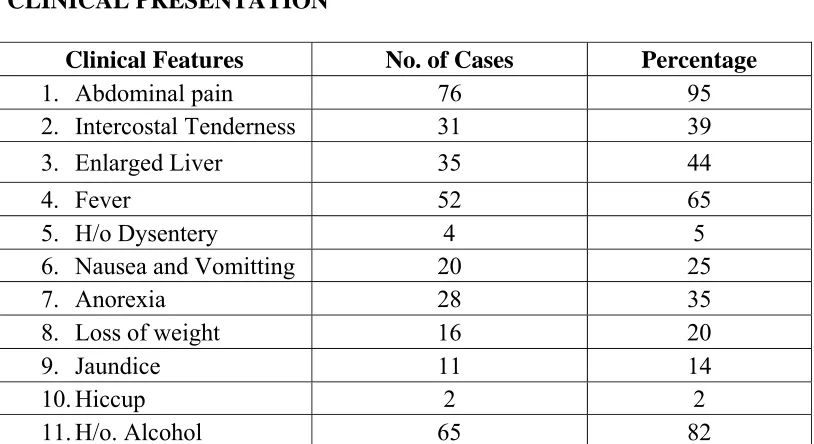
CLINICAL PRESENTATION
Clinical Features No. of Cases Percentage
1. Abdominal pain 76 95
2. Intercostal Tenderness 31 39
3. Enlarged Liver 35 44
4. Fever 52 65
5. H/o Dysentery 4 5
6. Nausea and Vomitting 20 25
7. Anorexia 28 35
8. Loss of weight 16 20
9. Jaundice 11 14
10.Hiccup 2 2
[image:66.612.107.514.440.662.2]MODES OF PRESENTATION
Presentation No. of Cases Percentage
Classical 62 77
Peritonitis 5 7
Atypical 13 16
LABORATORY INVESTIGATIONS HEMATOLOGICAL INVESTIGATION
Investigation Value No. of Cases Percentage
Hemoglobin 7 -10gms./dl. 54 67
10 – 14 gms./dl. 26 33
WBC count 5,000 – 10,000
cells / cu.mm.
33 42
10,000 – 15,000 cells / cu. Mm.
41 51
>15,000 cells / cu.mm.
6 7
ESR <15mm 8 10
>15 mm 72 90
LIVER FUNCTION TESTS
Investigation No. of Cases (80)
Percentage
Increased Bilirubin 42 53
Increased Serum Alkaline Phosphatase
44 55
Decreased Serum Albumin
RADIOLOGICAL INVESTIGATION X-Ray Chest and X-Ray abdomen
Radiological Feature No. of Cases (80)
Percentage
Raised dome of Diaphragm
32 40
Pleural effusion 11 14
Basal atelectasis 8 10
Enlarged Liver 24 30
Ground Glass appearance 5 6
ULTRA SONO GRAM
In our present study all were analysed with 2 dimension ultra sonography.
USG DATA – REGARDING SIZE
Size in Cms. No. of Cases Percentage
<5 15 19
5 – 10 44 55
>10 21 26
DISTRIBUTION OF ABSCESS
Right Lobe Left Lobe Both Lobes
56 (70%) 12 (15%) 12 (15%)
Single Multiple Single Multiple
40 (50%) 16 (20%) 12 (15%) 0
MODES OF TREATMENT
Mode of Treatment No. of Cases Percentage
Conservative 30 37.5
Aspiration 40 50
Percutaneous catheter drainage
5 6.25
Open Drainage 5 6.25
BACTERIOLOICAL CULTURE STUDY OF PUS ASPIRATED No. of persons found to have bacterial
growth
Organisms isolated
24 (30%) E. coli[common]
Klebsiella Pseudomonas
Polymicrobial abscess
Streptococci viridians, Enterococci
TROPHOZOITE AND CYST OF E.HISTOLYTICA RECOVERED
Stools Pus
12 (15%) 8(9%)
QUANTITY OF PUS ASPIRATED
Quantity No. of Cases Percentage
< 100 ml 22 27
100 – 500 ml 36 45
500 – 1000 ml 12 15
TYPES OF ORGANISM REPORTED IN PUS CULTURE
Bacterial Growth is +ve for 24 cases out of those cases.
0 0
7 9
20 25
30 38
23 28
5 10 15 20 25 30 35 40
A g e
dis tribution
in
L iver
A bs c es s
No.P e rc e ntag e of C as e s87% 13%
Sex Distribution in Liver Abscess
Male
50
63
30
37
0
10
20
30
40
50
60
70
T Y P E
OF
AB S C E S S
No.
of
C as es
P erc en tag e
Classical 77% Peritonitis 14% Atypical 9%
MODES OF PRESENTATION
54
67
26
33 33
42 41
51
67 810
72
90
0
20
40
60
80
100
H b 7 ‐10g
ms ./dl. H b 10 –
14 g ms./dl.
WB C 5,00
0 – 10,0
00 c ells
/ c u.mm.
WB C 10,0
00 – 15,0
00 cells
/ c u. Mm.
WB C >15,
000 c ells
/ c u.mE S Rm . < 15mm E S R > 15
mm
H E MAT O L O G IC AL
INVE S T IG AT IO N
No.
of
C as es
53
55
21
42
44
17
0
10
20
30
40
50
60
Inc reas ed B ilirubin
Inc reas ed S erum Alkaline P hos phatas e
D ec reas ed S erum Albumin
L IVE R
F UNC TION
TE S TS
No. of C as es
P erc entag e
64 80 16 20 0 10 20 30 40 50 60 70 80 Mono mic robial P oly mic robial
T Y P E S
O F
O R G ANIS M
R E P O R T E D
IN
P US
C UL T UR E
No. of C as es
32 40 11 14 8 10 24 30 5 6 0 5 10 15 20 25 30 35 40 Raised dome of Diaphragm Pleural effusion Basal atelectasis
Enlarged Liver Ground Glass appearance
No. of Cases (80)
30 37.5
40 50
5 6 5 6
0 5 10 15 20 25 30 35 40 45 50
Conservative Aspiration Percutaneous
catheter drainage
Open Drainage
No. of Cases Percentage
B AC T E R IOL OIC AL
C UL T UR E
S T UD Y
OF
P US
AS P IR AT E D
12%
88%
G rowth
No G rowth
USG DATA – REGARDING
SIZE
<519%
5 >10
22
27
36
45
12
15
10
13
0
5
10
15
20
25
30
35
40
45
<
100
ml 100
–
500
ml
500
–
1000
ml
>
1000
ml
QUANT IT Y
OF
P US
AS P IR AT E D
DISCUSSION
The condition of hepatic abscess and its grave prognosis were known in
ancient time to Hippocrates (160 BC -370BC ) and Celsus (53 BC - &AD)
Hippocrates was able to distinguish liver abscess from cystic liver disease. Celsus
appreciated the poor prognosis of hepatic abscess associated with Jaundice. Not
until 1936 did Bright in his own observation on Jaundice clearly described hepatic
suppuration with true abscess formation.
Oschner, Debakey and a Murray 1938 in their classic article reported
amoebic liver abscess, 75% as very common in the warmer southern climate. Liver
abscess though a well defined clinical entity yet many difficulties were faced in
determining the site, size and number of abscess. Chuttani et al 1963 have
commented that the difficulties in clinical diagnosis of hepatic amoebiasis can be
diverse and real. Those who do not meet the condition frequently are not likely to
appreciate them fully. Untreated liver abscess has higher mortality rates
approaching 100%. Reports of successful medical management with or without
aspiration describe case fatality rates as low as 6%.
In our study out of 22,000 cases admitted in our hospital in 2007 to 2009.
We are reported about 80 cases of liver abscess. Incidence being 2.7%. In our
study peak age incidence was noticed in 4th decade followed by Fifth and Sixth
decade. In our study there is more number of cases in low socio economic status.
alcohol and also in people who live with poor hygienic conditions, contaminated
drinking water, malnutrition, hepatic dysfunction and low host resistance.
Highest incidence of liver abscess in males 87% in our study has been
attributed to acoholism, (Present study H/o alcoholism was present in 82% cases.)
This correlates with the study of Oschner & Debakay which predispose to
hepatitis. Alcohol produces heptaocellular damage and may make it prone to
develop hepatic abscess – Sheila – Sherlock.
Presentation Present Study (n=80)
%
D.S Sing et al (1980)(n=42)
%
Barnas et al (1987( n=96)
%
Pain right Hypochondrium
95 100 67
Fever 65 100 87
H/o.
Diarrhoea/Dysentry
5 85 35
Jaundice 14 24 10
Weight Loss 20 - 10
Appetite 35 - 45
Breathelessness 5 - 24
In our study commonest symptom being abdominal pain and fever.
Commonest sign being Intercostal Tenderness and Tender hepatomegaly.
In present study, anemia was noted in 67% cases and Jaundice in 14%
X-RAY SHOWING THE LIVER ABSCESS WITH
ELEVATION OF THE RIGHT DOME OF DIAPHRAM