0022-538X/97/$04.0010
Copyrightq1997, American Society for Microbiology
Dextran Sulfate Can Act as an Artificial Receptor To Mediate
a Type-Specific Herpes Simplex Virus Infection
via Glycoprotein B
ANGELA P. DYER,1BRUCE W. BANFIELD,1† DIANE MARTINDALE,1
DELL-MARIE SPANNIER,1ANDFRANK TUFARO1,2*
Department of Microbiology and Immunology1and Department of Opthalmology,2University of
British Columbia, Vancouver, British Columbia, Canada V6T 1Z3
Received 25 July 1996/Accepted 17 September 1996
Herpes simplex virus (HSV) adsorption to host cells is mediated, at least in part, by the interaction of viral glycoproteins with cell surface glycosaminoglycans such as heparan sulfate and chondroitin sulfate. To investigate the contribution of various cell surface components in the infection pathway, we isolated a mutant cell line, sog9, which is unable to synthesize glycosaminoglycans (B. W. Banfield, Y. Leduc, L. Esford, K. Schubert, and F. Tufaro, J. Virol. 69:3290–3298, 1995). Although HSV-1 and HSV-2 infection of sog9 cells is diminished, the cells are still infected at about 0.5% efficiency, which suggests that these cells normally express at least one nonglycosaminoglycan receptor. In this report, we used sog9 cells to test whether glycosamino-glycan analogs, such as dextran sulfate (DS), could functionally substitute for cellular glycosaminoglycosamino-glycans to initiate HSV infection. We show that high-molecular-weight DS added either prior to or during inoculation stimulated HSV-1 but not HSV-2 infection by up to 35-fold; DS added after viral adsorption had no effect on infection efficiency. Moreover, DS stimulated HSV-1 infection at 4&C, indicating that this compound impinged on an early, energy-independent step in infection. Using radiolabeled virus, we showed that HSV-1 is more efficient than HSV-2 in adsorbing to DS immobilized on microtiter wells. This raised the possibility that only HSV-1 could engage additional receptors to initiate infection in the presence of DS. To determine which viral component(s) facilitated DS stimulation, a panel of intertypic recombinants and deletion mutant viruses was investigated. These assays showed that DS stimulation of infection is mediated primarily by gB-1. Thus, this study provides direct evidence that a principal role for cell surface glycosaminoglycans in HSV infection is to provide an efficient matrix for virus adsorption. Moreover, by using DS as an alternative adsorption matrix (a transreceptor), we uncovered a functional, type-specific interaction of HSV-1 with a cell surface receptor.
The two serotypes of herpes simplex virus (HSV), HSV-1 and HSV-2, have a broad in vitro and in vivo host range, being capable of infecting a variety of cell types from different animal species. In the human host, however, HSV-1 preferentially infects the oral mucosa, while HSV-2 infection is predominant in the genital mucosa. Several lines of evidence suggest that there are differences in the entry pathways for HSV-1 and HSV-2, which may account, in part, for the different behaviors of these closely related viruses.
HSV entry into permissive cells is a multistep process in-volving interactions between virion envelope glycoproteins and components of the host cell surface. The initial interaction of HSV-1 and HSV-2 with cells is adsorption to highly sulfated cell surface glycosaminoglycans, such as heparan sulfate (10, 14, 16, 32, 43) and dermatan sulfate (3, 42). The evidence for this stems from experiments showing that cells devoid of gly-cosaminoglycans are less susceptible to HSV infection (2, 14, 43), that binding of virus to cells is competitively inhibited by soluble heparin, and that HSV-1 glycoproteins gB-1 and gC-1 (16, 17) can adsorb to heparan sulfate, as can the HSV-2 glycoproteins gB-2 (42) and gC-2 (11). For both viruses, there appear to be additional nonglycosaminoglycan receptors that
facilitate a productive infection. In this regard, it has been shown that sog9 cells, which do not produce any cell surface glycosaminoglycans, retain partial susceptibility to HSV infec-tion (2). It has also been shown that swine testis cells, which comprise a seemingly normal complement of glycosaminogly-cans on their cell surface, are strongly resistant to infection (36), and that polarized epithelial cells display both heparan sulfate-dependent and -independent receptors on their surface (30). Attempts to identify receptors for HSV have yielded several candidate molecules, including mannose-6-phosphate receptors (4, 5), which can be present on the cell surface. In another approach, Montgomery and colleagues have isolated genes that confer HSV susceptibility to CHO cells, which are normally refractory to infection (26).
Although both HSV-1 and HSV-2 bind heparan sulfate, it is apparent that the viruses recognize type-specific cell surface receptors. For example, binding of HSV-1, but not HSV-2, to synaptosomes can be blocked by exposure to HSV-1 (41). Sim-ilarly, a temperature-sensitive HSV-1 mutant, ts1204, which is defective in cell penetration at the nonpermissive temperature, can block entry of wild-type HSV-1, but not HSV-2 (1). In so far as these experiments use whole virions as competitors, it is difficult to draw conclusions regarding the nature of the inter-ference. Using another approach, it has been shown that neo-mycin and polylysine can inhibit HSV-1 but not HSV-2 infec-tion of BHK cells (22–24). Neomycin inhibits HSV-1 infecinfec-tion by competing with gC-1 for binding heparan sulfate glycosami-noglycans, which suggests that gC-1 and gC-2 recognize ent moieties on cell surface heparan sulfate (16). The
differ-* Corresponding author. Mailing address: Department of Microbi-ology and ImmunMicrobi-ology, University of British Columbia, Vancouver, British Columbia, Canada V6T 1Z3. Phone: (604) 822-6914. Fax: (604) 822-6041. E-mail: [email protected].
† Present address: Department of Molecular Biology, Princeton University, Princeton, N.J.
191
on November 9, 2019 by guest
http://jvi.asm.org/
ences in sensitivity to inhibition were shown to reside in the N-terminal 223 amino acids of gC-2, which appears to confer resistance to neomycin inhibition of infection (29). It is also becoming evident that the contributions of gB and gC to viral binding may be different for HSV-1 and HSV-2. HSV-1 gC, although not essential for infection, enhances virus adsorption and, therefore, infection (17). In the absence of gC-1, gB-1 mediates the binding of gC-negative virus to the host cell surface (16). By contrast, HSV-2 glycoprotein C does not ap-pear to play a predominant role in viral adsorption, because it has been shown that gC2-negative virions can bind to the cell surface as efficiently as wild-type HSV-2 (11). Thus, although HSV-1 differs from HSV-2 in its initial interaction with the cell surface, the precise mechanism by which this occurs remains largely unknown.
As part of a broad study to investigate the HSV infection pathway, we previously uncovered a functional difference be-tween HSV-1 and HSV-2 in their ability to engage a nongly-cosaminoglycan cell surface receptor. In sog9 cells, which do not express any cell surface glycosaminoglycans, we were able to enhance HSV-1 but not HSV-2 infection by adding dextran sulfate (DS) to the medium during infection (2). These results were consistent with a model in which DS effectively substi-tutes for heparan sulfate as a matrix to initiate viral adsorption at the cell surface. This then promotes additional interactions between virion and host cell components to stimulate virus entry. In this report, we provide evidence for such a model and show that this ability maps to the presence of gB-1 in the virions, suggesting that gB-1 facilitates virus infection in this system by engaging a type-specific receptor.
MATERIALS AND METHODS
Materials.The parental L cell used for all experiments was the clone 1D line of LMtk2murine fibroblasts. The procedure for the isolation of the mutant sog9 cell line was described previously (2, 14). Cells were grown at 378C in Dulbecco modified Eagle medium (DMEM) supplemented with 10% fetal calf serum (FCS) in a 5% CO2atmosphere. HSV-1 strains F and MP and the HSV-1
intertypic recombinants RS1G25 (in which gC1 is replaced by gC2), R7015 (in which gD1, gE1, and gG1 are replaced by gD2, gE2, and gG2, respectively), and RH1G13 (in which gB1 is replaced by gB2) were generously provided by B. Roizman (University of Chicago, Chicago, Ill.). The HSV-1 strain UL102(which is deficient in gM) was also provided by B. Roizman. The HSV-1 gC2virus, HSV-1(KOS)DgC2-3, was provided by C. Brandt (University of Wisconsin, Mad-ison). HSV-2(G)gC2, a gC2 deletion mutant, was provided by B. Herold (Uni-versity of Chicago). HSV-2 strain G was obtained from S. Sacks (Uni(Uni-versity of British Columbia, Vancouver, British Columbia, Canada). DS with a molecular weight (MW) of 500,000 (MW 500,000 DS) was from Pharmacia (catalog no. 17-0340-01). MW 5,000 (catalog no. D-7037), 15,000 (catalog no. D-3257), and 50,000 (catalog no. D8787) DS were from Sigma. All tissue culture reagents (Gibco) and dishes (Nunc) were from Canadian Life Technologies (Burlington, Ontario, Canada).
DS stimulation assays.Assays for DS stimulation of virus infection were performed on sog9 cells plated in 6-well dishes. The cells were pretreated with DS in DMEM for selected lengths of time prior to infection or inoculated with virus in DMEM previously mixed with DS. The cells preincubated with DS were rinsed three times with phosphate-buffered saline (PBS) prior to infection. After a 60-min viral adsorption period at 378C, the inoculum was removed and the cells were washed once with PBS. The cells were then overlaid with DMEM contain-ing 4% FCS and 0.1% pooled human immunoglobulin G (IgG) (initial protein concentration, 96%; ICN). Plaques were visualized and counted after 3 days by fixation and staining of the cells for 10 min with 5% methylene blue in 70% methanol.
Penetration assays.Confluent monolayers of sog9 cells growing in 6-well dishes were rinsed with PBS and incubated with 1 ml of 100-mg/ml DS in DMEM at 48C for 30 min. The wells were rinsed three times with PBS and inoculated with HSV-1 at 48C for 30 min. The dishes were shifted to 378C to allow pene-tration to proceed. At various times after the temperature shift, the wells were treated for 2 min with 2 ml of low-pH citrate buffer (40 mM sodium citrate, 10 mM KCl, 135 mM NaCl, pH 3.0). The monolayers were washed three times with PBS and overlaid with DMEM containing 4% FCS and 0.1% pooled human IgG. Alternatively, cells were inoculated with virus in the presence of 10mg of DS/ml in DMEM at 378C. At various times, the virus was removed and the monolayers were washed with citrate buffer. Plaques were visualized and counted 96 h postinfection by fixing and staining the cells as described above.
Preparation of radiolabeled virus.Monolayers of L cells were infected with HSV at a multiplicity of infection of 10. At 2 h postinfection, the medium was changed to methionine-free labeling medium (methionine-free DMEM: 0.1 vol-ume of DMEM–10% FBS–4% dialyzed FBS–500mCi of [35
S]methionine [ICN; 3,000 Ci/mmol] per ml). When a generalized cytopathic effect was evident, medium was removed from infected-cell monolayers and subjected to low-speed centrifugation to pellet cell debris. The supernatant was sedimented through a 30% sucrose pad formed in 50 mM NaCl–10 mM Tris (pH 7.8) for 2 h at 39,000 rpm in a Beckman SW41 rotor. Following centrifugation, the sucrose was re-moved by aspiration, and the virus pellet was suspended in PBS at 48C. For determination of viral titers, a sample of virus was diluted serially with medium and used to inoculate monolayers of Vero cells growing in 6-well dishes. Plaques were scored after 3 days.
Binding of HSV to immobilized DS.We used a modified protocol by Leong et al. (25) to examine binding of HSV-1 and HSV-2 particles to immobilized DS. Nunc Maxi-Sorp 96-well microtiter dishes were coated with 50ml of PBS con-taining 5 mg of DS. The dishes were incubated overnight at 48C. The wells were rinsed twice with PBS and then blocked for 2 h in 3.5% bovine serum albumin (BSA) in 50 mM Tris (pH 7.5)–100 mM NaCl–1 mM MgCl2–1 mM MnCl2–1 mM
CaCl2at 208C. 35
S-labeled HSV was diluted in adsorption buffer (PBS, 0.1% glucose). The block was removed, and 50ml of virus was added to each well. The microtiter dishes were centrifuged for 15 min, 1,1003g at 48C, and then rocked for 45 min at 208C. The wells were washed three times with PBS, and the dishes were incubated for 10 min with 100ml of lysis buffer (10 mM Tris-HCl [pH 7.4], 150 mM NaCl, 1% Nonidet P-40, 1% sodium deoxycholate). The dishes were rinsed once more with lysis buffer; all rinses with lysis buffer were added to scintillation vials. The radioactivity associated with the immobilized proteogly-cans was determined by liquid scintillation spectroscopy.
Time course experiment.sog9 cells grown in 35-mm-diameter dishes were inoculated with HSV-1 in DMEM containing 10mg of DS/ml and incubated at 378C. Control dishes were infected with virus in the absence of DS. At different times postinoculation, virus was removed and 2 ml of DMEM with 4% FBS and 0.1% IgG was added to cells after the monolayers were rinsed with PBS. Plaques were counted 3 days postinfection.
RESULTS
HSV infection of the HSV-resistant mutant cell line sog9, which does not synthesize any cell surface glycosaminoglycans, is reduced by nearly three orders of magnitude relative to that of control mouse L cells (2). These cells are still infectible, however, which provides compelling evidence for the existence of a nonglycosaminoglycan receptor for this virus. It was also shown that the susceptibility of sog9 cells to infection by HSV-1, but not HSV-2, could be enhanced by up to 35-fold with the addition of the glycosaminoglycan analog DS to the inoculum. We carried out a series of experiments to elucidate the mechanism by which DS stimulates HSV-1 infection and to identify the viral glycoproteins conferring type-specific en-hancement of infection.
Effect of DS on HSV-1 infection.It has been shown previ-ously that low concentrations of DS (300 ng/ml) in the inocu-lum can stimulate HSV-1 infection of sog9 cells, whereas high concentrations are inhibitory. Inhibition by DS is likely caused by the propensity of soluble DS to bind to virus in solution. To test whether DS-mediated inhibition of HSV-1 infection could be eliminated by treating cells with DS prior to inoculation, cell monolayers were incubated with DS for up to 60 min, rinsed, and incubated with virus for an additional 60 min (Fig. 1A). HSV-1 infection was stimulated by more than 25-fold after only a few minutes of preincubation. Longer incubations up to 1 h did not significantly increase infection efficiency. To estab-lish the optimum DS concentration for stimulation under these conditions, confluent monolayers of sog9 cells were treated with various concentrations of DS either before or during inoculation with HSV-1 (Fig. 1B). Maximum infection effi-ciency (35-fold stimulation) was achieved by preincubating cell monolayers with low concentrations of DS. As expected, less stimulation was achieved with simultaneous DS treatment. We also determined that there was a size requirement for DS (data not shown). MW 5,000 DS and MW 15,000 DS were not active (less than twofold) in stimulating HSV-1 infection, whereas we observed up to 14-fold stimulation with MW 50,000 DS. MW
on November 9, 2019 by guest
http://jvi.asm.org/
500,000 DS possessed the most stimulatory activity (up to 35-fold). It appears from these data that relatively-long-chain DS is required for the efficient stimulation of HSV-1 infection.
To assess the stability of DS on the sog9 cell surface, cell monolayers were treated with two different concentrations of DS for 10 min, incubated in medium for various times (pre-treatment window), and then infected with HSV-1 (Fig. 1C).
With 10mg of DS/ml, stimulation declined rapidly as the length
of the pretreatment window was increased, and by 2 h, stimu-lation was reduced to twofold over that of controls. However,
DS stimulation persisted for up to 60 min when 100 mg of
DS/ml was used. At this concentration, maximal stimulation was achieved when the cells were treated 30 min prior to infection. We concluded that DS rapidly adsorbed to cells in an active form and was then inactivated by either dissociation, degradation, or both. To determine whether DS was toxic to cells, which could account for the loss of DS stimulation when cells were treated hours in advance of infection, cells incubated with DS 3 h prior to infection were treated a second time just before inoculation. DS stimulation was normal in these cells, which showed that DS pretreatment did not reduce cell viabil-ity or susceptibilviabil-ity to infection (data not shown).
Analysis of viral attachment in the presence of DS.On the basis of these results, it appeared that DS stimulated infection by binding stably to the host cell, where it acted as a matrix for subsequent virus adsorption and entry. This possibility was tested directly by experiments in which inoculation of cell monolayers with HSV-1 preceded DS treatment (Table 1, ex-periments A, B, and C) or cells were incubated with DS before inoculation (Table 1, experiment D). These experiments dem-onstrated that DS did not affect virus already bound to the cell surface (experiment C), nor did it influence the internalization of bound virus (experiment A). Experiment B showed that DS did not influence virus once it had been internalized. Only in
experiment D, in which cells were incubated with DS at 48C
prior to inoculation, was infection stimulated. We conclude from these results that DS stimulates HSV-1 infection only when it is present prior to or during viral inoculation and that this activity is energy independent.
We next determined that HSV could bind DS by incubating radiolabelled HSV with DS immobilized on microtiter wells. As a control for adventitious binding, virus was incubated with wells coated with BSA. In these experiments, substantially more HSV-1 bound to DS than to BSA (Fig. 2). By contrast, HSV-2 bound relatively poorly to DS. These data are consis-tent with a model in which HSV-1, but not HSV-2, adsorbs more efficiently to sog9 cells in the presence of DS on the cell surface. This could account in part for the observation that only HSV-1 infection is stimulated by DS, as reported previ-ously (2).
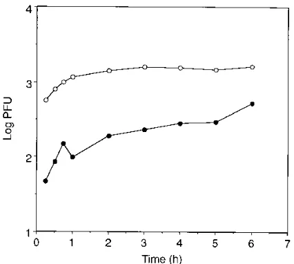
To test whether HSV-1 infection was mediated by a satura-ble site on the host cell surface, sog9 cell monolayers were inoculated with HSV-1 in the presence or in the absence of DS for up to 6 h (Fig. 3). In the absence of DS, virus infection continued to increase during the 6-h incubation, which sug-gested that saturation of cell surface receptors had not yet been achieved. To control for degradation and/or dissociation of DS during this incubation, we also performed an experiment (data not shown) in which fresh virus mixed with DS was added to cells every 2 h throughout the 6-h time course. The results
FIG. 1. Effect of soluble DS on HSV-1 plaque formation. (A) sog9 cell monolayers were incubated with 10mg of DS/ml in DMEM at 378C for various lengths of time prior to inoculation. Following removal of DS, the cells were inoculated with HSV-1 and incubated for 1 h at 378C. Virus was removed, and DMEM containing 0.1% pooled human IgG was added to the monolayers. Plaques were counted after 3 days. Each datum point represents the average of two determinations. Results are ratios of plaque formation on DS-treated mono-layers to that on control monomono-layers not exposed to DS. (B) sog9 cell monomono-layers were either (i) incubated with DS in DMEM for 10 min at 378C prior to infection or (ii) inoculated with virus in the presence of various concentrations of DS. Equivalent concentrations of HSV-1 were used for both experiments. After a 1-h adsorption period at 378C, the inoculum was removed and medium containing 0.1% pooled human IgG was added to the monolayers. Plaques were counted after 3 days. Results from at least two determinations were averaged and are ratios of plaques formed on DS-treated monolayers to plaques formed on un-treated controls. Open circles, preincubation with DS; closed circles, infection with DS in the inoculum. (C) Monolayers of sog9 cells were incubated with 10 or 100mg of DS/ml in DMEM at 378C for 10 min at different times prior to inoculation with HSV-1 (pretreatment window). The monolayers were rinsed three times with PBS after treatment with DS, following which the cells were either (i) incubated with DMEM at 378C until infection or (ii) infected imme-diately after a 10-min pretreatment with DS in DMEM. The cells were rinsed once with PBS before a 1-h adsorption period at 378C, and the inoculum was
removed and replaced with DMEM containing 0.1% pooled human IgG. Plaque numbers were determined after 3 days. Data are ratios of plaque formation on DS-treated monolayers to that on control monolayers not exposed to DS.
on November 9, 2019 by guest
http://jvi.asm.org/
were nearly indistinguishable from the data shown. Our inter-pretation of these experiments is that in the absence of the primary cell surface glycosaminoglycan receptor, HSV-1 could not efficiently engage a downstream receptor. In the presence
of DS, however, saturation was reached by 1 h of incubation
with virus. Moreover, by 6 h, there was only a 3- 60.5-fold
difference between DS-stimulated and control infections in two experiments. These data are consistent with a model in which DS functions by either tethering HSV-1 to the cell sur-face, thereby accelerating its effective interaction with a satu-rable site, or facilitating the presentation of an HSV-1-specific receptor to the virion.
To investigate whether DS exerted any effect on the rate of penetration, which might be suggestive of an alternative entry pathway, several penetration assays were performed. In one instance, virus was adsorbed to the cell surface in the presence
or in the absence of DS at 378C to allow for virus adsorption
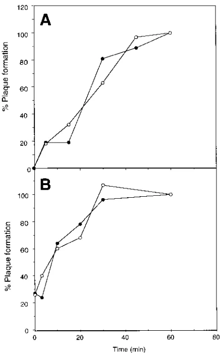
and entry. At various times, monolayers were washed with citrate buffer (pH 3.0) to inactivate extracellular virions, and the resulting plaques were counted after 3 days (Fig. 4A). The adsorption-penetration rates for DS-treated and control cells were similar. In a variation on this assay, cells were pretreated
with DS for 30 min at 48C, rinsed, incubated with virus for 30
min at 48C, and then incubated at 378C. Monolayers were then
treated with citrate buffer (Fig. 4B). Once again, there was no significant difference in virus penetration between DS-treated and untreated controls. These results are consistent with a
FIG. 2. Binding of HSV-1 (A) and HSV-2 (B) to DS. HSV-1 and HSV-2 were labelled with [35S]methionine and purified by centrifugation through a
sucrose gradient. Maxisorp 96-well dishes were coated with 5 mg of DS/ml and incubated with virus for 2 h at room temperature. Unbound material was re-moved by several washes with PBS. Bound virus was harvested with lysis buffer and transferred to scintillation vials for quantitation of radioactivity by liquid scintillation spectroscopy. Two independent experiments were performed with similar results. The results from a single experiment are shown. Open circles, binding to DS; closed circles, binding to BSA control.
[image:4.612.330.543.483.674.2]FIG. 3. Time course of HSV-1 infection of sog9 cells in the presence of DS. sog9 cells were inoculated with HSV-1 previously mixed with 10mg of DS/ml and incubated at 378C. At the indicated times postinfection, the inoculum was re-moved and DMEM with pooled human IgG was added. Each datum point is the average of two determinations. Open circles, infection in the presence of DS; closed circles, infection without DS.
TABLE 1. Effect of DS addition on HSV-1 infection
Expt Experimental conditionsa Infection efficiencyb
A Infect™™3Rinse™™3Add DS in DMEM™™™™™™™™3Rinse™™3Incubate with media1IgG 2
48C 378C
B Infect™™3Rinse™™3Add DMEM™™3Add DS in DMEM™™™™™™™™3Rinse™™3Incubate with media1IgG 1
48C 378C 378C
C Infect™™3Rinse™™3Add DS in DMEM™™3Rinse™™3Add DMEM™™3Rinse™™3Incubate with media1IgG 4
48C 48C 378C
D Add DS in DMEM™™3Rinse™™3Infect™™3Rinse™™3Add DMEM™™3Rinse™™3Incubate with media1IgG 31
48C 48C 378C
a
For experiments A, B, and C, HSV-1(F) was allowed to adsorb to cells for 1 h at 48C, followed by the indicated treatments. In experiment D, cells were incubated with 100mg of DS/ml for 1 h at 48C prior to infection. For all experiments, the cells were rinsed once with PBS following removal of virus and three times after incubation with DS. All incubations were conducted for 1 h.
b
Results are ratios of PFU obtained in the presence of DS to that obtained in the absence of DS. The averages of values from two independent experiments are shown.
on November 9, 2019 by guest
http://jvi.asm.org/
model in which DS-mediated infection occurs via the normal entry pathway.
High-molecular-weight DS is capable of adsorbing specifi-cally to several cell surface receptors, including scavenger re-ceptors. It has also been shown to stimulate endocytosis in certain cell types (38) and to block endocytosis by competing with other molecules for the endocytic machinery (12, 39, 44). In an attempt to inhibit steps in endocytosis, we took advan-tage of observations that a low temperature blocks the move-ment of ligands between compartmove-ments of the endocytic
path-way (13). sog9 cells were treated with 100mg of DS/ml at 48C
for 30 min prior to infection with HSV-1 at 48C for 30 min. The
incubation temperature was then shifted to either 158C, to
inhibit steps in endocytosis, or 378C (control) for 30 min. The
results indicated that HSV-1 infection was stimulated by 20-fold at both temperatures (data not shown), which suggests
that HSV-1 engages the normal entry pathway in the presence of DS.
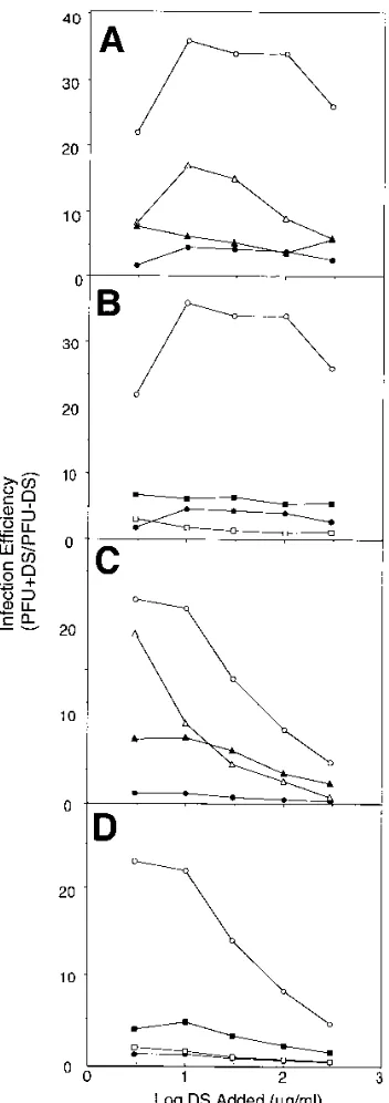
Mapping the DS activation site on the virus.The observa-tion that DS promoted HSV-1 but not HSV-2 infecobserva-tion pro-vided strong evidence for the existence of an HSV-1-specific interaction with the sog9 cell surface. In an attempt to map the viral components that facilitate DS stimulation, sog9 cells were treated with DS either before or during inoculation with a panel of intertypic recombinants and deletion mutants (Table 2; Fig. 5). DS was unable to stimulate (less than twofold) infection of the HSV-1 intertypic recombinant, RH1G13, in which gB-1 is replaced by gB-2, which indicated that DS stim-ulation is mediated, at least in part, by gB-1 (Fig. 5B and D). By contrast, infection with RS1G25, an HSV-1 strain in which gC-1 is replaced by gC-2, was stimulated by DS, although by only five- to sevenfold. This implied a role for gC-1 in DS-mediated infection, which was surprising because HSV-1
(KOS)DgC2-3, a gC-deficient HSV-1 strain, was stimulated by
up to 20-fold (Fig. 5C), similar to the maximal stimulation observed with wild-type HSV-1. One way to account for these results is the possibility that gC-2 was an inhibitor of DS stim-ulation. We tested this, using a gC-2-deficient HSV-2 strain,
HSV-2(G)gC22, and found that it was stimulated by about
eightfold (Fig. 5A and C). This level of stimulation was sub-stantially better than that for HSV-2. On the basis of these results, we conclude that gB-1 facilitates DS stimulation and that gC-2 may be a weak inhibitor of DS-mediated stimulation of infection. This property might account, in part, for the poor DS stimulation observed with RS1G25 and control HSV-2.
DISCUSSION
[image:5.612.68.287.68.417.2]It has recently been reported that HSV-1 and HSV-2 display type-specific differences in their interactions with host cells. These differences include preference for binding to various cell types (40, 41), binding to sulfated glycosaminoglycans (15), and interactions with specific cellular components, such as the C3B receptor (6–9, 31, 34, 37). In this study, we have uncovered an additional type-specific phenotype which involves the interac-tion of virions with host cell surfaces devoid of glycosamino-glycans. Moreover, this interaction is only evident following the exposure of host cells to long chains of the glycosaminoglycan analog DS. Because DS has been shown previously to interact
FIG. 4. Rates of HSV-1 penetration into sog9 cells in the presence and in the absence of DS. (A) Confluent monolayers in 35-mm-diameter dishes were inoc-ulated with HSV-1 in the presence of 10mg of DS/ml at 378C for 1 h. The monolayers were washed at various times with citrate wash buffer (pH 3.0), and the resulting plaques were counted. (B) sog9 monolayers were incubated with 100mg of DS/ml in DMEM for 30 min at 48C. The monolayers were rinsed three times with PBS, and then the inoculum was added. Following a 30-min adsorp-tion period at 48C, the cells were transferred to a 378C environment. At different times after the temperature shift, the monolayers were treated with citrate wash buffer. Results from single experiments are shown. The results are PFU surviving citrate treatment expressed as a percentage of the number of plaques obtained after 60 min of infection, which for each experiment was taken as 100%. Open
circles, infection in the presence of DS; closed circles, infection without DS. TABLE 2. Characterization of HSV infection in the presence of DS
Virus strain Phenotype Stimulation
by DSa
HSV-1(F) Wild type 11
HSV-1(KOS)c Wild type 11
HSV-2(G) Wild type 2
RSIG25 HSV-1; gC-2b 1
RHIG13 HSV-1; gB-2 2
HSV-1(KOS)DgC2-3 HSV-1; gC-1 deficient 11 HSV-2(G)gC22 HSV-2; gC-2 deficient 1
R7015 HSV-1; gD-2, gE-2, gG-2 11
UL102 HSV-1; gM deficient 1
HSV-1(MP) HSV-1; gC-deficient
syncytial strain 1
a2, stimulation of the prototype HSV-2 strain G, which varied from zero- to
fourfold above that of controls in different experiments;11, fold stimulation of the prototype HSV-1 strain F, which varied from 15- to 35-fold above that of controls in different experiments;1, stimulation which lies intermediate to those of the two prototype strains.
bHSV-1 background containing gC-2 in place of gC-1. Other strains of virus
are designated in a similar manner.
cData published by Banfield et al. (2).
on November 9, 2019 by guest
http://jvi.asm.org/
[image:5.612.317.556.532.661.2]with both virions and cells, the simplest model to account for DS-mediated HSV-1 infection is that DS binds to sog9 cells in a saturable, reversible manner and tethers the virion to the cell surface. Our data showing that cells can be treated with DS
prior to infection, that DS stimulates adsorption at 48C, and
that DS interacts with virions all lend support to this model. This then allows HSV-1, but not HSV-2, to infect cells more efficiently. If this model is correct, DS functions essentially as a substitute for cell surface heparan sulfate, albeit one that confers type-specific infection. Heparan sulfate is a naturally occurring, ubiquitous cellular glycosaminoglycan which pro-motes efficient HSV adsorption to the host cell (43). Heparan sulfate may also serve other functions as well, such as promot-ing efficient fusion of the virus with the host cell (33). Because the structure of DS does not resemble that of the highly com-plex heparan sulfate molecule, it is unlikely that DS could provide more than a simple tethering function during infec-tion. This may account, in part, for the inability of DS to completely restore wild-type levels of infection in the sog9 cells. Moreover, DS is not covalently linked to the host cell surface, which is likely to severely compromise its ability to stabilize HSV-1 virions that collide with the cell surface.
What could account for the failure of DS to stimulate HSV-2 infection? There are several possibilities which must be con-sidered. It is clear that DS stimulation of HSV-1 is mediated, at least in part, by gB-1 and that gB-2 is inactive in this capacity when it is present in an otherwise unperturbed HSV-1 virion. The role of gB-1 could be simply to interact with a distinct cellular receptor accessible to the virion at all times. This interaction is promoted when the virion is held in place even transiently by cell surface DS. If RH1G13, which contains gB-2 in an HSV-1 virion, can still interact with DS, then it is likely that access to the gB-2 receptor is not enhanced in this in-stance. gB-1 and gB-2 are highly conserved, with 86% amino acid homology. For the most part, regions that have been shown to have functional significance are conserved, including cysteine residues and predicted glycosylation sites in the exter-nal domain (35). Despite the overall similarity, however, there exist clustered regions of marked divergence between the two proteins. Perhaps most relevant are clustered amino acid sub-stitutions within the N-terminal 85 amino acids of the mature protein and a second region including amino acids 451 to 495. Interestingly, the N-terminal divergence comprises lysine-rich domains that could interact with acidic glycosaminoglycans, and it is known that isolated gB can interact with heparin (16, 17). These differences in the structures of gB-1 and gB-2 may alter the ability to adsorb to sulfated polyanions such as DS or to recognize a second receptor. Differences in gB-1 and gB-2 can also be inferred from studies showing that gC-deficient HSV-2 exhibits no loss in specific binding activity, specific infectivity, or rate of viral penetration (11). This is very differ-ent from studies on gC-deficidiffer-ent HSV-1 which showed a seri-ous impairment in virus adsorption. It may be that gC-1 and gB-2 predominate in their respective viruses to regulate the early interactions that lead to a productive infection.
It was also interesting to discover that gC-2 reduces DS-mediated infection. In this instance, the presence of gC-2 may interfere with the binding of the virion to DS at the cell sur-face, or it may directly impede the interaction of gB-2 with DS.
Our experiments using a gC2virus support the possibility that
[image:6.612.91.267.75.573.2]gC-2 is responsible for at least part of the unresponsiveness of the HSV-2 virion to DS stimulation. It is important to consider that the HSV-2 virion does show some level of DS stimulation in the absence of gC-2, thereby indicating that HSV-2 can engage the DS-mediated pathway. There is no evidence, how-ever, that gB-2 mediates this process in HSV-2.
FIG. 5. Effect of soluble DS on herpes simplex virus infection. (A and B) Fold stimulation of HSV plaque formation observed when sog9 cell monolayers were treated with DS prior to infection. sog9 cells grown in 6-well dishes were incubated at 378C for 10 min with DS diluted in DMEM. The wells were then rinsed three times with PBS, and the viral inoculum was added. After a 1-h adsorption period at 378C, the virus was removed and medium containing 0.1% pooled human IgG was added to facilitate plaque formation. Data are fold stimulation of plaque formation that occurs in the presence of DS. (C and D) Stimulation of HSV infection on sog9 cells when DS is present in the inoculum. Monolayers of sog9 cells were infected with virus diluted in various concentra-tions of DS. Virus was removed following a 1-h incubation at 378C, and medium containing 0.1% pooled human IgG was added to facilitate plaque formation. Data are fold stimulation of infection that is observed when cells are treated with DS compared to untreated controls. Each datum point is the mean of values obtained for at least two determinations. Open circles, HSV-1(F); closed circles, HSV-2(G); open squares, RHIG13; closed squares, RSIG25; open triangles, HSV-1 gC12; closed triangles, HSV-2 gC22.
on November 9, 2019 by guest
http://jvi.asm.org/
It is well established that DS normally inhibits infection of cells by enveloped viruses. DS has been shown to prevent fusion of influenza virus with erythrocytes (21) and the forma-tion of syncytia in human T-cell lymphotropic virus type 1-in-fected cells (18). Infection by human cytomegalovirus (27) and Sendai virus is also inhibited by the polyanion (28). It was therefore surprising that HSV-1 infection of sog9 cells was markedly enhanced by DS. Our data also indicate that this effect was mediated specifically by DS; soluble heparan sulfate and chondroitin sulfate neither enhanced nor inhibited HSV infection. This is interesting, because heparan sulfate is a nat-ural cell surface receptor for the virus. Several cell types, in-cluding macrophages and endothelial cells, express scavenger receptors capable of binding various polyanionic ligands (19, 20). In particular, scavenger receptors present on the surface of murine macrophages have broad ligand binding specificity and can bind DS and fucoidan but not heparin or chondroitin sulfate. Although the ligands for these receptors are all polya-nions, not all polyanions can function as ligands. It is possible, therefore, that sog9 cells possess a receptor with a binding specificity similar to that of macrophage scavenger receptors, and this may account for our observations that cells pretreated with DS retain enhanced susceptibility to HSV-1 even after the soluble DS is removed. It may be the case that the effects of DS are simply not detectable in cells that display heparan sulfate on the cell surface. Additional experiments will be needed to test this hypothesis directly.
We have likely uncovered type-specific interactions in cell culture because sog9 cells are devoid of glycosaminoglycans. This in effect reduces the adsorption of HSV to the cell surface in a manner that allows for a more sensitive readout of the virus-host interactions that ensue during the infection process. The striking differences in the behavior of HSV-1 and HSV-2 in this system are most likely defined by differences in the propensity of principally gB to interact with sulfated polyan-ions, as well as perhaps additional cell surface receptors. The significance of this study is that DS stimulates HSV-1 infection of sog9 cells, thereby acting as a trans receptor to initiate a productive infection. By contrast with previous experiments in which HSV-1 and HSV-2 were differentiated by their suscep-tibility to inhibitors, we have identified an interaction that promotes infection. We think this distinction is significant; whereas inhibition of infection could be caused by relatively nonspecific blocking, stimulation of infection requires that spe-cific host-virus interactions be maintained. It will be interesting to take advantage of the differences in susceptibility conferred by DS to identify and characterize the domains of gB that mediate this effect. These domains are likely to be important in conferring type-specific properties on the respective virions. It may be that differences in several stages of adsorption and entry, such as those found for the interaction with heparan sulfate, and those described herein, result in the profound differences in the epidemiology of these two viruses.
ACKNOWLEDGMENTS
We thank Pat Spear, Betsy Herold, and Bernard Roizman for help-ful comments throughout the course of this study.
A.P.D. is supported by a scholarship from the Natural Science and Engineering Research Council of Canada. D.M. is supported by a scholarship from the Neuroscience Network of Centers of Excellence of Canada. F.T. is supported by a grant from the Medical Research Council of Canada.
REFERENCES
1. Addison, C., F. J. Rixon, J. W. Palfreyman, M. O’Hara, and V. G. Preston. 1984. Characterisation of a herpes simplex virus type 1 mutant which has a
temperature-sensitive defect in penetration of cells and assembly of capsids. Virology 138:246–259.
2. Banfield, B. W., Y. Leduc, L. Esford, K. Schubert, and F. Tufaro. 1995. Sequential isolation of proteoglycan synthesis mutants by using herpes sim-plex virus as a selective agent: evidence for a proteoglycan-independent virus entry pathway. J. Virol. 69:3290–3298.
3. Banfield, B. W., Y. Leduc, L. Esford, R. J. Visalli, C. R. Brandt, and F. Tufaro.1995. Evidence for an interaction of herpes simplex virus with chon-droitin sulfate proteoglycans during infection. Virology 208:531–539. 4. Brunetti, C. R., R. L. Burke, B. Hoflack, T. Ludwig, K. S. Dingwell, and D. C.
Johnson.1995. Role of mannose-6-phosphate receptors in herpes simplex virus entry into cells and cell-to-cell transmission. J. Virol. 69:3517–3528. 5. Brunetti, C. R., R. L. Burke, S. Kornfeld, W. Gregory, F. R. Masiarz, K. S.
Dingwell, and D. C. Johnson.1994. Herpes simplex virus glycoprotein D acquires mannose 6-phosphate residues and binds to mannose 6-phosphate receptors. J. Biol. Chem. 269:17067–17074.
6. Eisenberg, R. J., M. Ponce de Leon, H. M. Friedman, L. F. Fries, M. M. Frank, J. C. Hastings, and G. H. Cohen.1987. Complement component C3b binds directly to purified glycoprotein C of herpes simplex virus types 1 and 2. Microb. Pathog. 3:423–435.
7. Friedman, H. M., G. H. Cohen, R. J. Eisenberg, C. A. Seidel, and D. B. Cines. 1984. Glycoprotein C of herpes simplex virus 1 acts as a receptor for the C3b complement component on infected cells. Nature 309:633–635.
8. Friedman, H. M., J. C. Glorioso, G. H. Cohen, J. C. Hastings, S. L. Harris, and R. J. Eisenberg.1986. Binding of complement component C3b to gly-coprotein gC of herpes simplex virus type 1: mapping of gC-binding sites and demonstration of conserved C3b binding in low-passage clinical isolates. J. Virol. 60:470–475.
9. Fries, L. F., H. M. Friedman, G. H. Cohen, R. J. Eisenberg, C. H. Hammer, and M. M. Frank.1986. Glycoprotein C of herpes simplex virus 1 is an inhibitor of the complement cascade. J. Immunol. 137:1636–1641. 10. Fuller, A. O., and W. C. Lee. 1992. Herpes simplex virus type 1 entry through
a cascade of virus-cell interactions requires different roles of gD and gH in penetration. J. Virol. 66:5002–5012.
11. Gerber, S. I., B. J. Belva, and B. C. Herold. 1995. Differences in the role of glycoprotein C of HSV-1 and HSV-2 in viral binding may contribute to serotype differences in cell tropism. Virology 214:29–39.
12. Greenspan, P., and R. L. Gutman. 1994. Endocytosis of sulfatides by mac-rophages: relationship to the cellular uptake of phosphatidylserine. J. Leu-kocyte Biol. 55:99–104.
13. Gruenberg, J., and K. E. Howell. 1989. Membrane traffic in endocytosis: insights from cell-free assays. Annu. Rev. Cell Biol. 5:453–481.
14. Gruenheid, S., L. Gatzke, H. Meadows, and F. Tufaro. 1993. Herpes simplex virus infection and propagation in a mouse L-cell mutant lacking heparan sulfate proteoglycans. J. Virol. 67:93–100.
15. Herold, B. C., S. I. Gerber, B. I. Belval, A. M. Siston, and N. Shulman. 1996. Differences in the susceptibility of herpes simplex virus types 1 and 2 to modified heparin compounds suggest serotype differences in viral entry. J. Virol. 70:3461–3469.
16. Herold, B. C., R. J. Visalli, N. Susmarski, C. R. Brandt, and P. G. Spear. 1994. Glycoprotein C-independent binding of herpes simplex virus to cells requires cell surface heparan sulphate and glycoprotein B. J. Gen. Virol. 75:1211–1222.
17. Herold, B. C., D. WuDunn, N. Soltys, and P. G. Spear. 1991. Glycoprotein C of herpes simplex virus type 1 plays a principal role in the adsorption of virus to cells and in infectivity. J. Virol. 65:1090–1098.
18. Ida, H., A. Kurata, K. Eguchi, I. Yamashita, M. Nakashima, M. Sakai, Y. Kawabe, T. Nakamura, and S. Nagataki.1994. Mechanism of inhibitory effect of dextran sulfate and heparin on human T-cell lymphotropic virus type I (HTLV-I)-induced syncytium formation in vitro: role of cell-to-cell contact. Antiviral Res. 23:143–159.
19. Krieger, M. 1992. Molecular flypaper and atherosclerosis: structure of the macrophage scavenger receptor. Trends Biochem. Sci. 17:141–146. 20. Krieger, M., S. Acton, J. Ashkenas, A. Pearson, M. Penman, and D. Resnich.
1993. Molecular flypaper, host defense and atherosclerosis. J. Biol. Chem. 268:4569–4572.
21. Krumbiegel, M., D. S. Dimitrov, A. Puri, and R. Blumenthal. 1992. Dextran sulfate inhibits fusion of influenza virus and cells expressing influenza hem-agglutinin with red blood cells. Biochim. Biophys. Acta 1110:158–164. 22. Langeland, N., H. Holmsen, J. R. Lillehaug, and L. Haarr. 1987. Evidence
that neomycin inhibits binding of herpes simplex virus type 1 to the cellular receptor. J. Virol. 61:3388–3393.
23. Langeland, N., L. J. Moore, H. Holmsen, and L. Haarr. 1988. Interaction of polylysine with the cellular receptor for herpes simplex virus type 1. J. Gen. Virol. 69:1137–1145.
24. Langeland, N., A. M. Oyan, H. S. Marsden, A. Cross, J. C. Glorioso, L. J. Moore, and L. Haarr.1990. Localization on the herpes simplex virus type 1 genome of a region encoding proteins involved in adsorption to the cellular receptor. J. Virol. 64:1271–1277.
25. Leong, J. M., P. E. Morrissey, E. Ortega-Barria, M. E. A. Pereira, and J. Coburn.1995. Hemagglutination and proteoglycan binding by the Lyme disease spirochete, Borrelia burgdorferi. Infect. Immun. 63:874–883.
on November 9, 2019 by guest
http://jvi.asm.org/
26. Montgomery, R. I., M. S. Warner, B. J. Lum, and P. G. Spear. 1996. Herpes simplex virus 1 entry into cells mediated by a novel member of the TNF/NGF receptor family. Cell 87:427–436.
27. Neyts, J., R. Snoeck, D. Schols, J. Balzarini, J. D. Esko, A. Van-Schepdael, and E. De-Clercq.1992. Sulfated polymers inhibit the interaction of human cytomegalovirus with cell surface heparan sulfate. Virology 189:48–58. 28. Ohki, S., K. Arnold, N. Srinivasakumar, and T. D. Flanagan. 1992. Effect of
anionic polymers on fusion of Sendai virus with human erythrocyte ghosts. Antiviral Res. 18:163–177.
29. Oyan, A. M., K. E. Dolter, N. Langeland, W. F. Goins, J. C. Glorioso, L. Haarr, and C. S. Crumpacker.1993. Resistance of herpes simplex virus type 2 to neomycin maps to the N-terminal portion of glycoprotein C. J. Virol. 67:2434–2441.
30. Sears, A. E., B. S. McGwire, and B. Roizman. 1991. Infection of polarized MDCK cells with herpes simplex virus 1: two asymmetrically distributed cell receptors interact with different viral proteins. Proc. Natl. Acad. Sci. USA 88:5087–5091.
31. Seidel, D. C., M. Ponce de Leon, H. M. Friedman, L. F. Fries, M. M. Frank, G. H. Cohen, and R. J. Eisenberg.1988. C3b receptor activity on transfected cells expressing glycoprotein C of herpes simplex virus types 1 and 2. J. Virol. 62:4027–4036.
32. Shieh, M.-T., D. WuDunn, R. I. Montgomery, J. D. Esko, and P. G. Spear. 1992. Cell surface receptors for herpes simplex virus are heparan sulfate proteoglycans. J. Cell Biol. 116:1273–1281.
33. Shieh, M. T., and P. G. Spear. 1994. Herpesvirus-induced cell fusion that is dependent on cell surface heparan sulfate or soluble heparin. J. Virol. 68: 1224–1228.
34. Smiley, M. L., and H. M. Friedman. 1985. Binding of complement compo-nent C3b to glycoprotein C is modulated by sialic acid on herpes simplex
virus type 1-infected cells. J. Virol. 55:857–861.
35. Stuve, L. L., S. Brown-Shimer, C. Pachl, R. Najarian, D. Dina, and R. L. Burke.1987. Structure and expression of the herpes simplex virus type 2 glycoprotein gB gene. J. Virol. 61:326–335.
36. Subramanian, G., D. S. McClain, A. Perez, and A. O. Fuller. 1994. Swine testis cells contain functional heparan sulfate but are defective in entry of herpes simplex virus. J. Virol. 68:5667–5676.
37. Tal-Singer, R., C. Seidel-Dugan, L. Fries, H. P. Huemer, R. J. Eisenberg, G. H. Cohen, and H. M. Friedman.1991. Herpes simplex virus glycoprotein C is a receptor for complement component iC3b. J. Infect. Dis. 164:750–753. 38. Thiele, B., and F. Steinbach. 1994. Dextran sulphate induces a PKC and actin
independent internalisation. Immunol. Lett. 42:105–110.
39. Tokuda, H., S. Masuda, Y. Takakura, H. Sezaki, and M. Hashida. 1993. Specific uptake of succinylated proteins via a scavenger receptor-mediated mechanism in cultured brain microvessel endothelial cells. Biochem. Bio-phys. Res. Commun. 196:18–24.
40. Vahlne, A., B. Svennerholm, and E. Lycke. 1979. Evidence for herpes simplex virus type-selective receptors on cellular plasma membranes. J. Gen. Virol. 44:217–225.
41. Vahlne, A., B. Svennerholm, M. Sandberg, A. Hamberger, and E. Lycke. 1980. Differences in attachment between herpes simplex type 1 and type 2 viruses to neurons and glial cells. Infect. Immun. 28:675–680.
42. Williams, R. K., and S. E. Straus. Specificity and affinity of binding of herpes simplex virus type 2 glycoprotein B to glycosaminoglycans. J. Virol., in press. 43. WuDunn, D., and P. G. Spear. 1989. Initial interaction of herpes simplex
virus with cells is binding to heparan sulfate. J. Virol. 63:52–58.
44. Xu, X., H. G. Remold, and J. P. Caulfield. 1993. Potential role for scavenger receptors of human monocytes in the killing of Schistosoma mansoni. Am. J. Pathol. 142:685–689.