ACKNOWLEDGEMENT
First of all, the author extremely grateful to the Lord Almighty who empowered the author with his blessings and grace to complete this dissertation work successfully.
It is a great pleasure for the author to acknowledge the deep sense of gratitude to the vice chancellor, Tamilnadu, Dr.M.G.R. Medical University, Chennai and Special commissioner, Director of Indian Medicine and Homeopathy, Chennai.
The author wishes to express grateful thanks to Dr. M. Thinakaran, M.D(S)., Principal, Government Siddha Medical College, Palayamkottai .
The author is thankful to Dr. R. Devarajan, M.D (S)., Vice Principal, Head of the Noi Naadal Department (PG), Government Siddha Medical College, Palayamkottai for rendering his valuable suggestion, guidance and encouragement in all aspects from time to time.
The author expresses her heart felt gratitude to
Dr. I. Sornamariammal M.D(S)., Retired Joint Director of Indian Medicine and Homeopathy, Chennai, for her valuable guidance and encouragement.
The author expresses her whole-hearted thanks to Dr. S.K.Sasi M.D(S)., assistant Lecturer, for her encouragement and most valuable guidance to undertake this dissertation work.
The author likes to express her profound gratitude to Dr. A. Vasuki M.D(s)., assistant Lecturer, for her suggestion helping me in my dissertation.
The author expresses her sincere thanks to Dr. Paramasivam M.D (Patho), M.D (FM), H.O.D.of Pathology Department, Tirunelveli Medical College, Tirunelveli for his guidance in the modern approach.
The author expresses her special thanks to Dr. Mohan M.D.,
Professor, Department of modern medicine, Government Siddha Medical College, Palayamkottai for his information regarding her dissertation work.
The author places her special thanks to Dr.P. Udhaya singh M.B.B.S.,D.PHYS.MED., Physiatrist, Tirunelveli Medical College, Tirunelveli for his clinical guidance.
The author is thankful to Dr. Padma M.B.B.S, DMRD.,
Radiologist, Government Siddha Medical College, Palayamkottai for her guidance in doing my dissertation topic.
The author is also thankful to Dr. S. Bagirathy M.B.B.S.,
The author expresses her special thanks to P. Arumugam M.A.,M.P.S.,P.G.D.C.A., Part time Professor in Bio-Statistics, Government Siddha Medical College, Palayamkottai for his statistical analysis doing my dissertation topic
The author would like to thank the librarian Tmt.T.Poongodi M.A., Mhil,MLIS., for her co-operating in referring the books.
INTRODUCTION
Siddha system is such a great system of medicine, by taking care of both physique body and psychic mind (nun udal and paru udal). In olden days, it was called as Tamil Maruthuvam, ‘Tamil’ means ‘Amirtham’, which bring bless to oneself.
Siddha system of medicine which was founded by the Siddhars is considered to be one of the ancient medical systems in the world. The word ‘siddha’ comes from the word ‘Siddhi’ which means an object to be attained or perfection or heavenly bliss. Siddhi generally refers to Astama siddhi (ie) eight great supernatural powers.
The ancient Siddhars have developed the siddha system of medicine which based on Panchaboothangal and Mukkutrangal.
The body is made up of the five basic principles (i.e.) sky, wind, fire, water and earth.
The universe is made up of the same. It is well known by the version.
“அ ட தி ளேத ப ட
ப ட தி ளேத அ ட
அ ட ப ட ஒ ேற
அறி தா பா ேபாேத”
- ச ட ன ஞான
According to siddhar philosophy, man is considered as a microcosm, universe is considered as the macrocosm. It means whatever
occurs in man, occurs in universe and whatever occurs in universe and
The physical body functions on the basis of the three humours like Vali, Azhal and Iyam. These three humours are nothing but combination of five elements. Any increase or decrease in ratio of the three humours causes disease in human body.
All living creatures need energy to lead their life. The energy is attained by what they consume. So regulating the dietary habits, one can make their help and mind moral. It is learnt by the following quotation.
“மா பா ய லாத உ ம ண
ஊ பா ைல உய ”
- தி ற
Importance of diagnosis is stated in as,
“ வன நாயகன ெகௗரவ
பா டவ ப ேபானெத ன வ வரெமன
ேநா கவ த ெம பத றி ம தி வ ைன ேடா
கவைல ேநாயாள னா ேவதிய ப சா சிேபானா
வரவ ப ேபால க ரவ
ேநா கைள ெகா ல ெதாட கினாேன”
-ேதர ம வ பாரத
Treatment in Siddha system is arrived at keeping the three thodam in equilibrium and maintenance of the seven udal thathus. So proper diet medicine adjuvant regimens of life are advised for a healthy living and restore equilibrium of humours in diseased condition.
Disease affects an individual based on the immunity, dietary and personal habits, ultimate and environmental factors etc. Treatment is fruitful only of the basic pathology behind is well diagnosed.
The disease can be diagnosed based on eight entities.
“ெம றி நிற ெதான வ ழிநாவ மல ைக றி”
- ேதர ந றி ெந றி
To lead a disease in free healthy life, one has to follow the ‘Pini anuga vithi muraigal’ i.e. preventive measures throughout the life time.
“தி ண மிர ல சி கவட காம
ெப ண பாெலா ைற ெப ககாம -உ கா
ந கி ேமா ெப கி ெந கி பவ த
ேப ைர கி ேபாேம ப ண”
- ேதர ேநாய லா ெநறி
SIDDHA PHYSIOLOGY
Physiology is the most fascinating and ancient branch of science. It is fascinating because, it unfolds the mystery of complicated functional aspects of individual organ in the body. It is ancient because, it exits ever since the origin of life. Even before knowing the language, culture and society, man knows about the hunger, thirst, pain and fear which are the basics of physiology.
The udal thathuvam (Physiology) of siddha system composed of
Thathuvangal - 96 basic elements
Udal Kattukkal - 7 physical constituent
Vegangal - 14 reflex function
Suvaigal - 6 tastes
Udal Thee - 4 body fires
Udal Vanmai - 3 immunities
96 THATHUVAM
The human body composed of 96 basic thathuvangal or constituent principles. The basic thathuvangal are responsible for the creation, protection and destruction of life, which is mediated through the Panchabootha and Mukkuttra theory.
1. The five basic elements – Bootham
2. The five organs of sense – Pori
3. The five objects of sense – Pulan
4. The five organs of action – Kanmenthiriyam
5. The five organs of perception – Kanmenthirya vidayam
6. The four intellectual faculties – Anthakaranam
8. The ten naadi – Dasa naadi
9. The ten vital airs – Dasa vaayu
10. The five visceral cavities – Aasayam
11. The five cases of sheaths
of the soul – Kosam
12. The six station of the soul – Aatharam
13. The three regions – Mandalam
14. The three principle of moral evil – Malam
15. The three humours – Dhodam
16. The three physical binding – Edanai
17. The three cosmic qualities – Gunam
18. The eight prominent passions – Raagam
19. The two deeds – Vinaigal
20. The five status of soul – Avaththai
DOSHAM 3
“மிகி ைறய ேநா ெச ேலா வள தலா எ ண ய .”
-தி ற
The three humours are the fundamental principles and essential factors in the composition and constitution of the human body. The three humours Vali, Azhal, Iyam represent wind, bile and phlegm respectively.
RELATION BETWEEN BOOTHAS AND MUKKUTTRAM
Vali – Air
Azhal – Fire
FORMATION OF THREE HUMOURS
“வ த கைல றி வா வா மபான ட
த த ப ராண சமான - ச த ற
றவ ேரசி த வாத ப த
நா கபேமயா நா .”
- க சாமிய
Vali – Edakalai + Abaanan
Azhal – Pinkalai + Praanan
Iyam – Suzhumunai + Samaanan”
“வாதமா பைட
ப த வ ன யா கா
ேச ம சீதமா ைட ”
- ேதைரய ம வ பாரத
Vali → Creation
Azhal → Protection
Iyam → Destruction
“வழ கிய வாத மா திைர ெயா றாகி
தழ கிய ப த த ன லைரவாசி
அழ கப தானட கிேய காேலா
ப ற கிய சீவ ப செகா மி ைலேய”
- ணவாகட நா
Vali → 1 maathirai
Azhal → ½ maathirai
Iyam → ¼ maathirai
VALI
Dwelling places of Vali
Abaanan
Hip region
Idakalai
Bones
Kaamakodi
Joints
Unthiyin keezh moolam
Skin
Hair follicles
Stools
Nerves
Muscles
Nature properties of Vali
Giving briskness
Respiration
Optimal functioning of the mind, thoughts and body
Regulation of the fourteen physiological reflexes
Uniform functioning of the seven udal thathukkal
Strengthening the five sensory organs
Types of Vali
1. Praanan
Lies in the chest
Regulates the respiration and digestion
2. Abaanan
Constrict the anal sphincter
Expels the faeces and urine
Spreads the nutrient of the digested food all over the
body for its utilizations
It expels the sperm, ova and menstrual outflow.
Its derangement leads to bowel disturbances and
reproductive system disease.
3. Viyaanan
It spreads all over the body.
It controls voluntary and involuntary movement.
It responsibles for movement and sensory perception.
It causes theflow of fluids, sweat and Opening and
Closing the Eye.
It distributes the energy of the assimilated to various parts
of the body.
Its derangement leads to Neurological disturbances and
locomotor problems.
4. Udaanan
It regulates the Speech.
Induces the physiological reflexes such as vomiting,
hiccough, cough, sneezing.
Derangement leads to Gastro intestinal tract disturbances.
5. Samaanan
It Controls the digestion.
It acts as an activity factor for the other Vayus.
Its derangement leads to Gastro intestinal, Respiratory
6. Naagan
Intelligence quotient of an individual.
Open and Closes the eye lids.
Derangement leads to impaired memory and lack of
comprehensing the thinking
7. Koorman
It makes the closure of the eyes, lacrimation and yawning.
It is responsible for vision.
It supplies energy to build up the body.
8. Kirukaran
It helps to salivary secretion, nasal secretion, hunger,
sneeze, cough and mind concentration.
9. Devathaththan
It causes the laziness, angry, arguing and occular movement.
10. Dhananjayan
It produces swelling all over the body.
Leaves body breaking up the cranium after three days of
death.
It is responsible for destruction of bodily elements.
AZHAL
It is the representation of the Thee Bootham.
Location
Types of Azhal- 5
1. Analam – It controls the appetite and help in digestion
2. Ranjagam – It gives colour to the blood.
3. Saathagam – It has the property of fulfillment and controls
the body.
4. Aalosagam – It is located in the eyes and responsible for
visual perception.
5. Prasagam – It gives complexion to the skin.
IYAM
It is representation of Neer and Mann Bootham
Properties of Iyam
It gives stability, lubrication, holding together of the joints, power endurance for hunger, thirst, sorrow, disturbed mind and heart
Types of Iyam
1. Avalambagam
It is present in the lungs and is responsible for the basic function of the heart and other four types of Iyam.
2. Kilethagam
It is present in the stomach. It makes the food wet and helps for digestion
3. Pothagam
It is present in tongue and is responsible for the sense of taste.
4. Tharpagam
It is located in the head and keeps the eye cool.
5. Santhigam
SUVAIGAL 6
Taste denotes the enrichment of the food. This is very much related with the udal thathukkal (physical constituents) as well as the uyir thathukkal (humours).
The tastes are six in number. Each taste is formed by combination of two boothas.
Suvaigal Bootham Humour
1. Inippu(sweet) Mann + Neer Iyam
2. Pulippu (sour) Mann + Thee Azhal
3. Uppu (salt) Neer + Thee
4. Kaippu (bitter) Vaayu + Aakayam Vali
5. Karppu (pungent) Vaayu + Thee
6. Thuvarppu (astringent) Mann + Vaayu
UDAL THATHUKKAL 7 (Seven physical constituents)
The seven Udal Thathukkal are responsible for the entire structure of the body. These are
“இரச உதிர இைற சி ேதா ேமைத
ம வ ய வ தி ெய ெபா ம ைச பரவ ய கில பாழா உபாதி
உ பம லா ட ஒ ெறனலாேம”
- அக திய ஆ ேவத 1500
1. Saaram (chyle)
2. Senneer (blood)
Blood is a complex fluid which contains both organic and inorganic constituents suspended in a colloidal medium called as plasma. It is responsible for knowledge.
3. Oon (muscle)
It forms the shape of the body.
4. Kozhuppu (fat)
It maintains lubrication and oiliness of all the tissues and gives energy to the body.
5. Enbu (bone)
It forms the frames and structure of the body.
6. Moolai (bone morrow)
It nourishes the bones
7. Sukkilam and Suronitham
It is responsible for reproduction.
VEGANGAL 14 (The fourteen reflexes)
Reflex is generally understood as a psycho neuro muscular function of the body. The natural reflex, excretions, protective and preventive mechanisms are called fourteen vegangal.
1. Vatham – flatus
2. Thummal – sneezing
3. Siruneer – urine
4. Malam – stool
5. Kottavi – yawning
6. Pasi – hunger
7. Neervetgai – thirst
9. Elaippu – fatigue / exhaustion
10. Nithirai – sleep
11. Vaanthi – vomit
12. Kanneer – tear
13. Sukkilam / Suronitham – genital secretion
14. Suvasam – respiration
All the above mentioned reflexes are closely linked between the neurological functions and muscular activity.
UDAL VANMAI 3 (Three immune status)
1. Iyarkkai vanmai – It is inherited vitality
2. Kaala vanmai – Vitality that is generally found in
different age and periods.
3. Seyarkkai vanmai – Improvement of vitality obtained by good
habits, physical exercise and proper diet.
UDAL THEE 4 (Four body fires)
The normal digestive fire is called sadaraakkini and it is a combination of samaanavayu, analapitham and kilethagam.
1. Samanaakkini
When the sadaraakkini normal with the proper balance of the above three constitutent it is called samanaakkini. The balanced diet of an individual is properly digested in time.
2. Mandaakkini
3. Dheekshaakini
An increased analapitham with the defieciency of kilethagam leads this condition, causing excessive digestive fire burning larger quantum of food in a lesser duration of time.
4. Vishamaakkini
SIDDHA PATHOLOGY
The word pathology is derived from two Greek words – ‘pathos’
meaning suffering and ‘logos’ meaning study. Pathology is scientific
study of structure and function of the body in disease; it deals with causes, effects, mechanism and nature of disease. The knowledge and understanding of pathology be is essential for all would be doctors as well as general practitioners. And specialists since unless they know the cause and mechanism of disease and understood the language spoken by the pathologist in the form of laboratory reports, they would not be able to institute appropriate treatment or suggest prevention measure to the patient.
“உ றேதா உடலி
உ ட வ ரவ நி
ேம ேநா க எ லா
தலதன ேல ேதா ேபா
ப ேம வாத ப த
சிேல பன தன த ன ஒ ைற
ப றிேய ேதா ெம
பக தன ன வ தாேம”
- அக திய நா
ேநா தலி வள, அழ , ஐய றி ஏதாவெதா ைற
ப வதா தா ேதா எ அக திய கிறா .இதைனேய
வ வ ,
“மிகி ைறய ேநா ெச ேலா
வள தலா எ ண ய ”
“மதி திட க ைம வா த
மா ப காரெம லா
தி திட ண தாேன
களற ப ண ய ற ைம
பதி திட ணரானாகி
பய றானாகலாேன
வ தி தி ப ண திற ைத
வ ள த க ம ேனா”
- சிகி சா ர ன தப
ப கார ைறக யா ெத தி தா ேநா நாட ேநா
த நாடெல ப ண ய த ைம அறி கைல ெத யாதி ப
சிறி பயன ைல.
“ேநா நா ேநா த நா அ தண
வா நா வா ப ெசய ”
- தி ற
The knowledge and understanding of pathology is essential for all would be doctors as well as general practitioners
MUKKUTRAM
The changes of the three uyir thathus are called mukkutram.
The mukkutram is the basic principle of all disease.
The changes take place in the uyir thathu caused by
1. Variation in the intake of diet.
3. Environmental changes,
a. Seasonal variation in humours.
b. Regional variation in humours.
4. Self suppression of fourteen Reflexes.
When the normal maathirai proportions of the uyir thathus are disturbed, it leads to mukkutram. This condition is called us disease.
“மிகி ைறய ேநா ெச ேலா
வள தலா எ ண ய ”
- தி ற
The three humours changed and causes disease by self exaggeration and combining with other humours, and thus the disease are classified under 9 major groups of naadi nadai.
1. Vali naadi - self exaggeration of vali
2. Vali azhal naadi - maximum exaggeration of vali combined
with exaggerations of azhal
3. Vali iya naadi - maximum exagerretion of vali combined
with exaggeration of iyam
4. Azhal naadi - self exaggeration of azhal
5. Ahal vali naadi - maximum exaggeration of azhal
combined with exaggeration of vali
6. Azhal iya naadi - maximum exaggeration of azhal
combined with exaggeration of iyam
7. Iya naadi - self exaggeration of iyam
8. Iya vali naadi - maximum exaggeration of iyam
combined with exaggeration of vali
9. Iya azhal naadi - maximum exaggeration of iyam
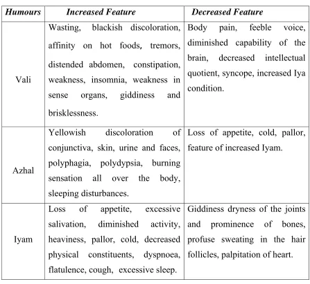
Table-1
Thannilai valarchi and vettrunilai valarchi of the three humours causes the symptoms of increasing and decreasing properties of the uyir thathukkal.
Humours Increased Feature Decreased Feature
Vali
Wasting, blackish discoloration,
affinity on hot foods, tremors,
distended abdomen, constipation,
weakness, insomnia, weakness in sense organs, giddiness and
brisklessness.
Body pain, feeble voice, diminished capability of the brain, decreased intellectual quotient, syncope, increased Iya condition.
Azhal
Yellowish discoloration of conjunctiva, skin, urine and faces, polyphagia, polydypsia, burning sensation all over the body, sleeping disturbances.
Loss of appetite, cold, pallor, feature of increased Iyam.
Iyam
Loss of appetite, excessive salivation, diminished activity, heaviness, pallor, cold, decreased physical constituents, dyspnoea,
flatulence, cough, excessive sleep.
Giddiness dryness of the joints and prominence of bones, profuse sweating in the hair follicles, palpitation of heart.
1. Variation in the intake of diet
“உணேவ ம
ம ேத உண ”
So, any alteration in the normal, regular diet will produce changes in the proportion of the suvaigal resulting disease.
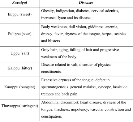
Table-2
Excessive intake of a particular suvai may produce hyperactivities and develops some clinical manifestation. These are given below,
Suvaigal Diseases
Inippu (sweet) Obesity, indigestion, diabetes, cervical adenitis,
increased Iyam and its disease.
Pulippu (sour)
Body weakness, dull vision, giddiness, anemia, dropsy, fever, dryness of the tongue, herpes, scabies and blisters.
Uppu (salt) Grey hair, aging, falling of hair and progressive
weakness of the body.
Kaippu (bitter) Disease related to vali, disorder of physical
constituents.
Kaarppu (pungent)
Excessive dryness of the tongue, defect in
spermatogenesis, general malaise, syncope, lassitude, tremors and back pain.
Thuvarppu(astringent) Abdominal discomfort, heart disease, dryness of the
Table-3
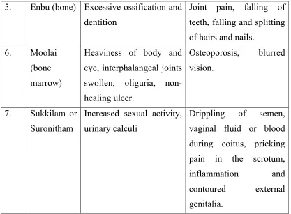
Alterations in Udal Kattukkal
S.No
Udal
Kattukkal
Increase Feature Decreased Feature
1. Saaram (Chyle)
Loss of appetite, excessive salivation, diminished activity, pallor cold, decreased physical constituents, dyspnoea, flatulence, cough, excessive sleep.
Dryness of skin, tiredness, loss of weight, lassitude, less ability in hearing.
2. Senneer (Blood)
Boils in different parts of the body, splenomegaly, tumours, pricking pain, loss of appetite, haematuria, hypertension, reddish eye and skin, leprosy, jaundice
Affinity to sour and cold food, nervous debility, dryness and pallor.
3. Oon (muscle)
Tubercular adenitis, venereal diseases, extra growth around neck, cheeks, abdomen, thigh and genitalia.
Lethargic sense organ, pain in the joints, muscle wasting in chin, gluteal region, penis and thigh.
4. Kozhuppu (fat)
Identical features of increased oon, tiredness, dyspnea on exertion, extra musculature in the genital region, external genitalia, chest, abdomen and
thighs.
5. Enbu (bone) Excessive ossification and dentition
Joint pain, falling of teeth, falling and splitting of hairs and nails.
6. Moolai
(bone marrow)
Heaviness of body and eye, interphalangeal joints swollen, oliguria, non-healing ulcer.
Osteoporosis, blurred vision.
7. Sukkilam or
Suronitham
Increased sexual activity, urinary calculi
Drippling of semen, vaginal fluid or blood during coitus, pricking pain in the scrotum, inflammation and contoured external genitalia.
Environmental changes
The environmental changes consist of two factors. (a). Seasonal changes of humours
[image:24.612.104.521.66.373.2](b). Regional changes of humours
Table-4
(a) Seasonal changes of humours
Humour Thannilaivalarchi Vetrunilaivalarchi Thannilai adaithal
Vali Mudhuvenil kaalam Kaar kaalam Koodir kaalam
Azhal Kaarkaalam Koodir kaalam Munpani kaalam
(b)Regional Changes of Humours:
Kurunji – Iya diseases occur
Mullai – Azhal diseases occur
Marutham – Diseases will not occur
Neydhal – Vali diseases occur
Paalai – Mukkutra diseases occur
Table-5
Effects on self suppression of fourteen vegangal
Reflexes are essential for the normal physiology when there is any self suppression to those reflexes that will lead to the pathologic state.
S.No Vegangal Diseases
1. Vatham Heart disease, gastritis, umbilical hernia, body
pain, liver disorder, constipation, oliguria, loss of appetite.
2. Thummal Head ache, defect of the special sensory organ and
its activities, pain over the face, hip joint pain
3. Siruneer Anuria, urethral ulcer, pain in the joints, pain in
the penis, gas formation in the abdomen.
4 Malam Diarrohoea caused by increased abaanan, cold,
knee pain, head ache, flatulence, weakness and its leads to many diseases.
5 Pasi All organs are affected, pricking pain all over the
body, schizophrenia, emaciation, apathetic face, pain in the joints.
6 Kottavi Lethargic face, exhaustion, indigestion, urinary
7 Neer Vetkai All organs are affected, pricking pain all over the body, schizophrenia, emaciation, apathetic face, pain in the joints.
8 Erumal Increased cough, bad breath, heart disease
9 Elaippu Urinary disorder, peptic ulcer, syncope, rigor,
identical features of suppression of sneezing.
10 Thookkam Heaviness of head, pain in the eyes, deafness,
unclear speech
11 Vaanthi Urticarial rashes, itching, anemia, eye diseases,
disease of increased azhal, asthma, fever, cough
12 Kanneer Heart diseases, upper respiratory disorders, eye
diseases, wound in the scalp, peptic ulcer.
13 Sukkilam or
Suronitham
Fever, anuria, joint diseases of the upper and lower limbs, acute chest pain, increased urinary diseases.
14. Suvasam Cough, abdominal discomfort, tastelessness,
epigastric pain, fever, venereal diseases
PINIYARI MURAMAI
Diagnosis of the disease is an essential factor before commencing
the treatment whatever may be the system of medicine.
“ேநா நா ேநா த நா அ தண
வா நா வா ப ெசய .”
- தி ற
1. Poriyaal arithal
2. Pulanaal arithal
3. Vinaadhal
1. Poriyal Arithal
Poriyaal arithal means the art of perception of five organs viz.
1. Mei – skin
2. Vaai – mouth
3. Kan – eye
4. Mookku – nose
5. Sevi – ear
2. Pulanaal Arithal
Pulanaal arithal means art of knowing objective senses viz.
1. Sparisam – sensation
2. Rasam – taste
3. Roobam – vision
4. Kantham – smell
5. Saptham – sound
3. Vinaadhal
It is a method of history taking. History taking is an art of diagnosing a disease very interestingly.
Envagai Thervuvugal:
The diagnosis is also made by the eight tools of diagnosis as mentioned below.
“ெம றி நிற ெதான வ ழிநா வ மல ைக றி.”
- ேதர ந றி ெந றி
1. Meikuri – signs in the body
2. Niram – colour
3. Thoni – sound and speech variation
4. Vizhi – eye
5. Naa – tongue
6. Malam – faeces
7. Moothiram – urine
8. Kaikuri – signs in hand pulse
In the above verse, inspection, palpation, percussion and interrogation are mentioned first. The pulse is mentioned in the last. This order is suitable for the diagnosis.
1. Meikuri
By meikuri, the temperature of skin (heat or cold), smoothness, roughness, softness, sweat, dryness, tenderness, ulcers, hard patches, swellings, abnormal growth and nourishment can be examined by the following examination.
Palpation – feeling
Percussion – tapping
Auscultation – hearing with stethoscope.
The following should be noted,
Condition of the skin
Organ not mentioned in the Envagi thervugal
Nail
Enlargement of visceras
Tenderness
Touch, pain, and temperature sensation
2. Niram
Diagnosis made with the help of colour of the skin, nails, hairs, conjunctiva, teeth and mucous membrane etc.
3. Thoni
The quality of the sound is assessed in the examination of them. The following also should be examined under speech.
Pitch
Fluency
Articulation
Intelligence
Breathlessness
Aphasia
4. Vizhi
As, both the physiological and pathological conditions are reflected in the eyes, the examination of eyes are important in the diagnosis of disease.
Size and shape
Colour
Conjuctiva and cornea
Colour of vision
Field of vision
Acuity of vision
Light reflex
Inflammation
Condition of eye lids and eye lashes.
5. Naa
In the examination of the tongue,
Colour of the tongue
Coating
Dryness, increased salivation, any deviation, movement of
the tongue
Taste sensation of tongue
Ulceration
Macroglossia
Microglossia
Teeth and gum
6. Malam
Malam examined under the qualities of,
Colour
Froth
Solid
Semi solid or liquid
Quantity
Odour
Consistency
7. Moothiram
The diagnostic value of urine is observed by two peculiar studies.
Neerkuri
Neikuri
Neerkuri
“வ த ந க எைட மண ைர எ செல
ைற திய ளவைவ யைற ைறேய”
-ேதர ந றி ெந றி
From the above quotation, the neerkuri consists of the following characters.
Niram - It indicates the colour of the urine.
Manam - It indicates the smell of the urine.
Edai - It indicates the specific gravity of the urine.
Nurai - It indicates the frothy of the urine.
Enjal - It indicates the quantity of the urine.
Neikuri
The patient is advised to take a balanced diet and should have a good sleep prior to the day of urine examination.
After early morning wake up, the first urine voided by the patient is collected in a glass container. The analysis should be performed within one and a half hour.
A drop of gingerly oil is dropped at the top of the urine without shaking, the spreading mode of oil noted.
“அரெவன ந அ0ேத வாத
ஆழிேபா பரவ அ0ேத ப த
Oil spreading like snake indicated Vali.
Oil spreading like ring indicated Azhal. !
Oil remains floating as a pearl indicated Iyam.
Mixed reaction of any two of the above indicates Thontham.
The siddhars relied on these methods for prognosis of the disease and classify the disease as curable and incurable.
NAADI
The rhythmic expansion of an artery which may be felt by the finger which represents the state of function of the heart. Naadi is nothing but, the vital energy that sustains the life in our body.
Naadi plays the important role in envagai thervu and it has been considered to be the most important for assessing the prognosis and diagnosis of the disease. Any variation that occurs in the three humour is reflected in the naadi. These three humour organize, regularize and integrate the functions of the human body. So, naadi serves as a good indicator of all ill health. Naadi can be perceived by feeling it at the appropriate site, suitable places for pulse reading
“தா ைற ேக தன த தி ச ேதா
ஓ காமிய தி ெந மா
கா ெந க ட கர வ
ேபா சி க ப பா திேட.”
- தி ல நா
Naadi is felt as
Vali – tip of index finger
Azhal – tip of the middle finger
In normal condition the ratio of naadi is
Vali – 1
Azhal – ½
Iyam – ¼
The gait of the naadi compared to the various animals, reptiles and birds
“வாகின ல ன ேகாழி மய ெலன நட வாத
ஏகிய வாைமய ைட ய ைவெயன நட ப த
ேபாகிய தவைள பா ேபாலவா ேச ம தா .”
Vali – movement of swan and peacock
Azhal – movement of tortoise and leech
Iyam – movement of frog and serpent
Other than this the following should be noted.
Rate of the pulse
Rhythm
Volume
Character
Whether felt in all peripheral areas and Condition of the
AIM AND OBJECTIVES
“வாதமலா ேமன ெகடா ”As Vali is the main cause of all sorts of ailments the author chosen
Vali disease.
“Face is the index of the mind”
From the above proverb one’s feeling and thoughts can be reflected in the face. That’s why the face is considered to be very important in the world of beauty. If any flaw or blemish or inability has occurred in face it leads to profound stress.
The main aim of present study of Uragan Vatham with clinical study is to evaluate the Mukkutra verupadugal, changes of Udal kattugal in this disease.
Collection of various literatures dealing with definition, etiology,
classification, signs and symptoms of the Uragan Vatham.
To expose the Siddhar’s diagnostic methods.
To have a better understanding, regarding the incidence of this
disease with disease with reference to Age, Sex, and Paruvakaalam.
To study under the topics of Mukkutram, Pori, Pulangal, Udal
thathukkal, Envagai thervugal, Naadi, Neerkkuri, Neikuri and Manikkadai Nool. The changes brought about by this disease under normal condition.
The pathogenesis of the disease ruled out on the basis of etiology.
To use modern parameters in the investigation of the disease that
ELUCIDATION ABOUT DISSERTATION TOPIC
In Yugi vaithiya Chinthamani Vatha roga nithanam is mentioned in Chapter 7. The ‘Uragan Vatham’ is mentioned in poem 282.
“அ தமா ேநாவாகி வ கா
அ க தி பாதிதா வலி ெத ேபா
த கமா ேகாண ேய தைல வா
தாவேவ மிகந கி ள
வ தமா வ ழிக தா ய ைம றா
ெமலிவாகி உட ெப லா வ ய ைவயா
தமா வா ந மிகேவ
க ேமா ரக வாத தி ேபேர”
- B,gq!juk<kqb!sqf<kil{q!
[
உரக - கா , வா
ேநா - ேநா - Disease
அ க - உட ப பாக - Part of body
வலி த - இ த - Pulling
த க - இய நிைலைம - Normal side
ந க - பத த - Involuntary
movement
“அ தமா ேநாவாகி வ கா
அ க தி பாதிதா வலி ெத ேபா ”
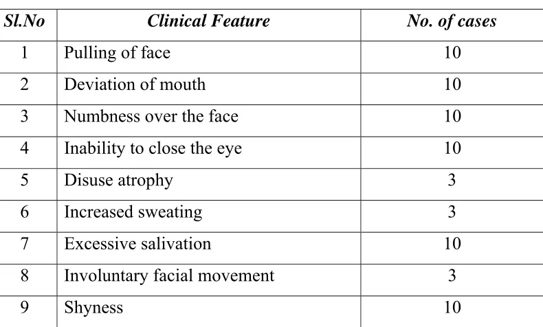
These lines describe the disease which affects the ear and the eye brows. They are partially affected: only half of the face is affected, that is pulling of face in one side.
“தாவேவ மிகந கி ள ”
Involuntary facial movement called ‘Tics’. Some times the involuntary facial movements are present in Facial Nerve Paralyzis. Heaviness and numbness over the face. But no sensory loss is demonstrable.
“வ தமா வ ழிக தா இைம றா ”
In ability to close the affected eye: St. Yugi mentioned the word
‘வ ழிக தா ’ , to give more importance to the special senses (i.e.) eye. To prove this fact St. Yugi already mentioned in the above line,
‘அ க தி பாதிதா ’.
“ெமலிவாகி உட ெப லா வ ய ைவயா ”
Disuse atrophy and excessive sweating
‘ தமா வா ந மிகேவ ’
Excessive Salivation
Summary
The clinical features of the Uragan Vatham are, Pulling of face,
DETAILED PATHOLOGICAL VIEW OF THE
DISSERTATIONTOPIC
SIDDHA ASPECT
“வாதமலா ேமன ெகடா ”
-ேதர ேசகர பா
ேதக தி ஓள எ அழ , வ ைம ெக வத
கியமான த காரண வள றமா .
வள வா மிட
அபான , மல , இடகைல, ேதா , நர , ஊ , கீ க ,
மய கா க .
வள உடலி ெச ெதாழி
நர தலியன ற , ந க , உ தள சி, உ க
மர ேபா கிட த , உட ேநாத , த , வற சி ம பல.
உரக வாத தி வள ற மி தியைட , வள வா
இடமாகிய நர ட தி உ ள க நர ட வதா க தி
உ ள தைசகள ெதாழிைல ெச யவ டாம மர ேபா கிட க
ெச கிற . ேதைரய தம வாகட தி ப வ மா கிறா ,
“த க வா ேகாப தா ச ைள ைலேநாவா
மி க ெகா டாவ வ ட ெக மல க
ஒ க நர தா ட மல வா ந றிவ
மி க ள ந கமா ேமன ளறி வ காேன.”
- ேதைரய வாகட
வள ற மி தியைடவதா ச உைளத , ைல,ெகா டாவ,
மல க , நர டஙக , வா ந த , ள , ந க , உடலி
Altered thirithodam in Uragan Vatham
The following types of Vali humour actions are increased in Uragan Vatham
Praanan
Dyspnoea present
Abaanan
Constipation present
Viyaanan
Diminished facial muscle action, Inability to close the eye
Udhaanan
Mild dysarthria present
Samaanan
Increased appetite present
Naagan
Inability to close the eye present
Koorman
Inability to close the eye and tear present
Kirukaran
Increased salivation present
Devathathan
Azhal
The above increased Vali humour is also increases the Azhal humour. The following types of Azhal humour actions are increased.
Anarpitham
Increased appetite present
Ranjagapitham
Pallorness present
Saathaga pitham
Diminished facial muscles action.
Praasagapitham
Dryness is present.
Iyam
The Iya humour action is decreased. Following types of Iya humour action is decreased.
Avalambagam
Dyspnoea present.
Pothagam
Loss of taste.
Altered Udal Thathukkal in Uragan Vatham
Saaram
Depression and anxiety.
Senneer
Nerve paralyzis.
Oon
Diminished facial muscle action.
Kozhuluppu
MODERN ASPECT
“அ க தி பாதிதா வலி ெத ேபா
த கமா ேகாண ேய தைல வா ”
Muscles of facial expressions are,
Orbicularis oris .
Levator labii superioris alaeque nasi
Levator labii superioris
Zygomaticus major
Zygomaticus minor
Levator anguli oris
Depressor labii inferioris
Depressor anguli oris
Mentalis
Buccinator
These facial expression muscles are sub cutaneous muscles and
“வ தமா வ ழிக தா இைம றா ”
Opening of the palpebral fissure
Sphincter - Orbicularis oculi
Dilator - Levator palpebrae superioris
Frontalis
The above muscles are supplied by Facial Nerve. If the nerve is paralyzed the above muscle actions are weakened. So the Eye lid cannot be closed.
“ தமா வா ந மிகேவ ”
Nerve supply to Salivary Gland
Salivary glands are under the control of Autonomic Nervous system.
Parasympathetic Nerve fibers supplying the salivary glands arise from superior and inferior Salivatory Nuclei of Pons and Medulla.
Parasympathetic Nerve fibers of Facial Nerve supply to sub mandibular and sub lingual gland
Schematic representation of Nerve supply to Salivary Gland
Superior salivatory Nucleus
Nervous intermedius
Geniculate ganglion
Chorda Tympani
Lingual Nerve
SubMaxillary ganglion
Paralytic secretion of Saliva
When the parasympatic Nerve to salivary gland is cut, salivary secretion increase for three weeks and diminishes and then stop at about
6th week. This is because of release of more amount of Adrenaline from
DIFFERENTIAL DIAGNOSIS
mw;Gj thjk;
!
“jPh;f;fkha; ];jphp rq;fk; gz;Zk; NghJk; jpLf;nfdth h;j;ijNfh gpj;j NghJk; Cf;fkh Awj;Jjhd; ghLk; NghJk;
cz;Zkty; fr;rhak; ghf;Fj;jhDk; Mh;f;fkha;j; jl;bNa fbj;j NghJk;
mofhd Kfe;jd;dpy; thANfh gpj;Jj; jhh;f;fkha; kpfr;rpjwp thAq; NfhZk;
rq;fkha; w;Gjth je;jhdhNk”
.!A+fp itj;jpa rpe;jhkzp
!!!
REVIEW OF LITERATURE
The same clinical features of Uragan Vatham are found in various literatures under various headings. They are,
கவாத
“ வ மதர க ெபா வ ெசவ க மிக தி
உைறேச நா த மாறி ெயா க வலி திமி டா
தன தன ேய ழி வா கவாத
உைறெச ப க வாதெமன ெச ண கேளா வா
க ண தைலய வ தி காதி ெசவ டா
ப ண பதி க தைன ப க ப றி தி கிவ
எ ண ெசா கவாத மி சா தியமாெம
ந ண ெசா ேனா ெமா பாலினா க தி வாதமிேத.”
- பரராச ேசகர
Ear pain
Dysarthria
Deviation of face and mouth
அ தித வாத ண க
“ ெகா க வா க ெச பாகி ேகா
நா க ள ெச ன ந கி காதைட
க பய டா மறி ந
ஆ க மழ அ தித வாதமாேம
- த வ த ைவ திய Part I
Deviation of face
Dysarthria
கவள
“ க தி ேலா பா ேகா ட மாத
மிட ேகாட தைலந கி த
ேப ெசழாைம வ ழிக வ
க ேபாவா ப லிைவ ேநாத
எ மிைவ கவள றிெயன ெமாழிய”
- மா கிய
Difficulty in swallowing
Dysarthria
Pain over the face
அ த கவாத
“ப ன வ அ த கவாத ேந ைம
ப வைகயதைன ெசா ேவ பாரா
உ னதமா க தி ேநா க
உ தி ள கைணைய ெக ேபா
நிக ேவ க ண ைமக டா ஐயா
நி சயமா வ தமடா இ ம பா
மக வமா க னம தள ெதா கி
மான ட வாேயார கீழிற கி ெகா ”
- அக திய ணவாகட
Loss of naso labial fold
Inability to close the eye
Drooling of face
கவாத ச ன ண
“வ டமா க ேகா வா ப தா ேகா
க டவ ேச ம க கா ேய மிெழா ணா
எ டாம ட ந கெள ெகா பள ெகா ணா
ேய ய சீ கவாத ச ன யாேம”
- அக திய ஆ ேவத 1500
Deviation of mouth and face
THEORETICAL VIEW OF DISSERTATION TOPIC IN
MODERN ASPECT
ANATOMY OF THEFACIAL NERVE
Facial nerve is a seventh cranial nerve. It is a mixed cranial nerve.
Development
The cartilage of second pharyngeal arch or hyoid arch (Reichert’s Cartilage) gives rise to the stapes, styloid process of the temporal bone, stylohyoid ligament, the lesser horn and upper part of the body of the hyoid bone. Muscles of the hyoid arch are the stapedius, stylohyoid, posterior belly of the digastric, auricular muscles and muscles of facial expression.
The facial nerve, the nerve of the second pharyngeal arch, supplies all of these muscles.
Nuclei of the facial nerve
Nuclei are situated within the dorsal part of the pons. The nuclei are
Motor nucleus
Sensory nucleus – nucleus of the tractus
solitarius
Para sympathetic nucleus – superior salivatory nucleus.
Upper part of nucleus of the spinal tract of trigeminal.
Motor nucleus
The lateral portion of the nucleus mainly supplies muscles around the mouth including buccinator. The intermediate portion mainly supplies muscles of the upper face including orbicularis oculi.The medial portion mainly supplies auricular muscles, platysma and occipitofrontails.
Nucleus of the tractus solitarius
This is the sensory nucleus of the facial nerve, situated in the upper part of the tractus solitarius. To this nucleus, the sensory part of the facial nerve or the nervous intermedius brings sensation from the anterior 2/3 of tongue and palate.
Superior salivatory nucleus
It is the parasympathetic nucleus to supply submandibular and sublingual salivary glands.
Upper part of the nucleus of the spinal tract of trigeminal
This nucleus receives auricular sensation via the auricular branch of vagus nerve. The geniculate ganglion of the facial nerve contains cell bodies of these fibres.
Connections
Course within the pons
The motor and sensory roots winds round the abducent nerve nucleus to form the facial colliculus. They pass forward and leave the pons. They emerge between the lower border of pons and upper border of the olive of the medulla.
Course
After emerging from the pons, two roots of facial nerve pass laterally and enter the internal auditory meatus. This part of the nerve is accompanied by the stato acoustic nerve. Laterally, the two roots unite to form the geniculate ganglion and from the trunk of the facial nerve. Now
the nerve is passing through facial canal or canal of Fallopei.
On reaching medial wall of middle ear it runs posteriorly. It is situated superior to the promontory of the middle ear. It reaches the medial wall of mastoid antrum and passes behind the posterior wall of the middle ear cavity, it runs vertically downwards to the stylomastoid foramen.
Extra cranial course
After emerging through the stylo mastoic foramen it runs forward and it crosses styloid process of the temporal bone. It enters the posterio medial surface the parotid gland. Within the gland it crosses the retro mandibular and external carotid artery.
Termination
It terminates by dividing into temporo facial and cervico facial branches.
The temporo facial branch passes upwards and divides into
Temporal nerve
The cervico branch passes downwards and divides into
Buccal nerves
Marginal mandibular nerve
Cervical nerve
From the anterior border of the parotid gland five terminal branches of facial nerve are emerging.
Branches of facial nerve
1. Branches in the facial nerve canal
a. Nerve to stapedius to supply the stapedius muscle of the
middle ear.
b. Chorda tympani nerve. It commences about 0.5cm. above
the stylomastoid foramen.
2. Branches immediately below the stylomastoid foramen.
a. Posterior auricular nerve to supply posterior muscles of
the pinna and occipitalis in the scalp.
b. Nerve to posterior belly of the digastric. This nerve
supplies
Posterior belly of the digastric
Stylohyoid muscle
3. Branches in the face
a. Temporal branch
b. Zygomatic branch
This nerve runs along the zygomatic arch and supplies lower half of orbicularis oculi.
c. Buccal branch
This nerve divides into superficial and deep branches. The superficial branch supplies procerus muscle. The deep branch divides into superior and inferior divisions. The superior divisions supplies zygomaticus major, zygomaticus minor, levator labii superioris, levator labii superioris alaeque nasi and levator angularis. The inferior division of deep buccal nerve supplies buccinator and orbicularis oris muscle.
d. Marginal mandibular
This nerve passes downwards and enters the neck below the angle of the mandible. It then passes along the lower border of the mandible. It supplies depressor anguli , orbicularis oris, risorius, depressor labii inferioris, mentalis.
Marginal mandibular nerve near the angle of mandible is related to a lymph gland. This lymph gland may be infected and form an abscess. During draining this abscess the incision may endanger this nerve; incisions along the lower border of the mandible may injure this nerve.
e. Cervical branch
Communications of the facial nerve
1. Within the internal acoustic meatus it communicates with the vestibulocochlear nerve.
2. At geniculate ganglion
a. It communicates with the external petrosal nerve. (middle meningeal plexus)
b. It communicates with the lesser superficial petrosal nerve. (Otic ganglion)
c. It communicates with greater superficial petrosal nerve. (Pterygo palatine ganglion).
3. At facial nerve canal, it communicates with vagus (auricular branch)
4. Just below the stylomastoid foramen it communicates with the
glossopharyngeal, vagus, auriculo temporal and great auricular
nerves.
5. Behind the pinna, it communicates with the lesser occipital nerve.
6. In the face, it communicates with branches of the trigeminal nerve.
7. In the neck, it communicates with the transverse cutaneous nerve of the neck.
Muscles of the facial expression
These are sub cutaneous muscles and they are attached to the skin.
They are developed from the second pharyngeal arch. Hence these muscles are supplied by the facial nerve. These muscles are arranged around the openings of the face. The main functions of these muscles will be either to open or close these openings. While doing these movements the facial expression results as a biproduct.
Muscles of the nose
Nasalis
Depressor septi nasi
Muscles of the mouth
Orbicularis oris
Levator labii superioris alaeque nasi
Levator labii superioris
Zygomaticus major
Zygomaticus minor
Levator anguli oris
Depressor labii inferioris
Depressor anguli oris
Mentalis
Buccinator
Muscles of the eye lid
Orbicularis oculi
Corrugator supercilli
Orbicularis oris
This is the sphincter of the mouth.
This muscle encircles the oral fissure. It is partly formed by
other muscles inserted in to the lips and partly formed by proper lip muscles.
It is made in to many layers.
Nerve supply
Buccal nerve
Marginal mandibular branch of the facial nerve.
Actions
It compresses the lips against the teeth.
It helps in mastication and speech.
It produces the lips.
The superficial layer helps in opening of the lips.
Buccinator muscle (trumphet muscle)
This is the muscle of the cheek.
Origin
Lateral surface of the maxilla and mandible at the
level of the third molar tooth.
Pterygomandibular raphe
Insertion
All the fibres converge at the angle of the mouth.
Upper fibres go to the upper lip.
Lower fibres go to the lower lip.
Middle fibres decussate near the angle of the mouth.
The lower part of the middle fibres reaches the upper lip. The upper part of the middle fibres reaches the lower lip.
Nerve supply:
Buccal Nerve of the facial nerve.
Action
It compresses the cheek against the teeth. This action is
acquired during the process of chewing, when the food accumulates within the vestibule of the mouth.
It helps in sucking by compressing the cheek. Its action is
required to blow the cheek. Hence it is called as the trumphet muscle.
The levator labii superioris alaeque nasi
Origin
Frontal process of the maxilla.
Insertion
It divides into nasal and labial parts. The nasal part is
medially situated and inserted to the skin and cartilage of the ala of the nose.
The labial part is laterally situated and it is inserted into the
orbicularis oris.
Nerve supply
Buccal branch of the facial nerve.
Actions
The medial part dilates the nose by lifting the ala of the nose. The lateral part elevates the upper lip.
The levator labii superioris
Origin
Infra orbital margin of the maxilla, above the infra orbital
foramen.
Insertion
Orbicularis oris
Nerve supply
Buccal branch of the facial nerve.
Actions
It elevates and everts the upper lip. Its action is required for the formation of the nasolabial furrow.
Zygomaticus minor
Origin
Outer surface of the zygomatic bone behind the zygomatico maxillary suture.
Insertion
Orbicularis oris
Nerve supply
Buccal branch of the facial nerve.
Actions
It pulls the upper lip upwards. Its action is required for the formation of the naso labial furrow.
Zygomaticus major(Smiling muscle)
Origin
Zygomatic bone infront of the zygomatico temporal suture.
Insertion
Orbicularis oris.
Nerve supply
Buccal branch of facial nerve.
Action
The Depressor anguli oris
Origin
Oblique line of the mandible.
Insertion
Blends with the orbicularis oris near the angle of the mouth.
Nerve supply
Marginal mandibular branch of the facial nerve.
Action
Draws the angle of the mouth downwards and laterally.
The Depressor labii inferioris
Origin
Oblique line of mandible near symphysis menti.
Insertion
Orbicularis oris
Skin of the lower lip
Nerve supply
Marginal mandibular nerve
Action
Depressor of the lower lip.
The mentalis
Origin
Incisive of the lower lip
Nerve supply
Marginal mandibular nerve.
Action
The Risorius
Origin
Fascia covering the parotid gland.
Insertion
Angle of the mouth
Nerve supply
Buccal branch of facial nerve.
Action
Its action expresses grining. It retracts the angle of the mouth.
Orbicularis oculi - muscles around the orbit
It is found within the eyelid, forehead, temporal region and upper part of the cheek. It encircles the orbital margin.
Parts of orbicularis oculi
Orbital part
Palpebral part
Lacrimal part
Orbital part
Origin
Medial palpebral ligament
Frontal process of the maxilla
Nasal process of the frontal bone.
Insertion
Palpebral part
Origin
Medial palpebral ligament
Medial border of the orbit above and below the medial
palpebral ligament.
Insertion
Lateral palpebral raphe
Lacrimal part
Origin
Lacrimal fascia
Lacrimal crest and lateral surface of the lacrimal bone.
Insertion
It divides into upper and lower portions. They enter the eyelids. They are attached to the tarsalplate and lacrimal canaliculi. Most fibres of this muscle extend laterally and decussate at lateral palpebral raphe.
Nerve supply
Temporal and zygomatic branch of the facial nerve.
Actions
1. Orbital part
It locks the eyelids. Thus it acts as a strong sphincter of the
orbit.
It opposes the action of the frontalis muscle.
2. Palpebral part
Shuts the eyelids tightly during blinking and sleeping.
3. Lacrimal part
The corrugator supercilli (Muscle of frowning)
Origin
Medial part of super ciliary arch.
Insertion
Skin and fascia above the supra orbital margin.
Nerve supply
Temporal branch of facial nerve.
Action
PHYSIOLOGY
The Facial Nerve has four component with distinct function. These are,
1. Branchial motor (Special visceral efferent)
Branchial motor supplies the muscles of Facial expression, Posterior belly of Digastric, Stylo hyoid and Stapedius.
2. Visceral motor (General visceral efferent)
Visceral motor supplies the Parasympathetic innervations of the Lacrimal, Sub mandibular and Sub lingual glands, as well as mucous membrane of Naso pharynx, Hard palate and soft palate.
3. Special sensory (Special afferent)
Taste sensation from the anterior 2/3 of tongue, hard palate and soft
palate.
4. General sensory (General somatic efferent)
General sensation from the skin of the concha of auricle and from a small area behind the ear.
PATHOLOGY
The symptoms explained in the poem are, Inability to closing the eye, Deviation of mouth and face in to normal side, numbness over the face, excessive salivation, increased sweating.
Bell’s Palsy is caused by an inflammation within a small bony tube called the fallopian canal. The canal is an extremely narrow area. An inflammation within it is likely to exert pressure on the nerve, compressing it. Likewise, if the nerve itself becomes inflamed within this small canal, it can encounter pressure, with the same result of compression.
The main Pathology behind these symptoms are paralysis of Facial Nerve.
Bell’s palsy is a one type of Idiopathic acute Facial Nerve paralysis. This is accurately described as a multiple cranial nerve ganglionitis that involves the Facial Nerve.
Pathogenesis
FACIAL PARALYSIS
Facial Paralysis is due to paralysis of the VII Cranial Nerve.
Classification of facial palsy
It may be broadly classified into two types. They are:
Upper motor neuron type or supra nuclear type
Lower motor neuron type
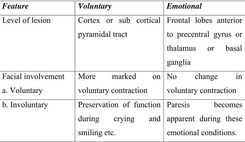
1. UPPER MOTOR NEURON TYPE OR SUPRA NUCLEAR TYPE
In this type, the cortico spinal tract supplying the facial neuron muscles are affected and the lesion is always above the level of pons.
It is again classified into two types. They are:
Voluntary
[image:63.612.107.506.477.708.2] Emotional
Table -6
Difference Between Voluntary And Emotional Palsy
Feature Voluntary Emotional
Level of lesion Cortex or sub cortical
pyramidal tract
Frontal lobes anterior to precentral gyrus or thalamus or basal ganglia
Facial involvement a. Voluntary
More marked on voluntary contraction
No change in voluntary contraction
b. Involuntary Preservation of function
during crying and smiling etc.
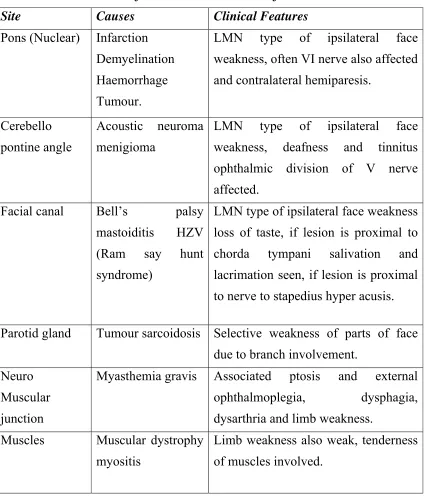
LOWER MOTOR NEURON TYPE
[image:64.612.110.534.192.688.2]The lower motor neuron is the common pathway. Hence a lesion at this site produces measures of entire half of the face on the ipsilateral side.
Table -7.
The exact site and level of lesions are tabulated as follows.
Site Causes Clinical Features
Pons (Nuclear) Infarction Demyelination Haemorrhage Tumour.
LMN type of ipsilateral face weakness, often VI nerve also affected and contralateral hemiparesis.
Cerebello pontine angle
Acoustic neuroma menigioma
LMN type of ipsilateral face weakness, deafness and tinnitus ophthalmic division of V nerve affected.
Facial canal Bell’s palsy
mastoiditis HZV (Ram say hunt syndrome)
LMN type of ipsilateral face weakness loss of taste, if lesion is proximal to chorda tympani salivation and lacrimation seen, if lesion is proximal to nerve to stapedius hyper acusis.
Parotid gland Tumour sarcoidosis Selective weakness of parts of face
due to branch involvement. Neuro
Muscular junction
Myasthemia gravis Associated ptosis and external
ophthalmoplegia, dysphagia, dysarthria and limb weakness.
Muscles Muscular dystrophy
myositis
Since the Uragan Vatham may correlate with Bell’s palsy, it is
discussed here in detail.
BELL’S PALSY
Definition
Bell’s palsy is a form of facial paralysis resulting from damage to the VII cranial nerve.
The condition is named for Charles Bell, a Scottish surgeon in Edinburgh who studied the nerve and its innervations of the facial muscle in 1821.
Since the function of the facial nerve is so complex, many symptoms may occur when the fibres of the facial nerve are disrupted.
Bell’s palsy temporarily prevents the nerve from transmitting signals to the muscles causing weakness or paralysis.
Age incidence
It may occur at any age group, but it is slightly more common in the age group from 20-50 years.
Incidence rate
It is about 23 per 1,00,000 annually, or about one in 60 or 70 persons in a life time.
Sexual preponderance
This disorder affects men and women more or less equally and occurs at all ages and at all times of the year.
Aetiology
The specific cause of Bell’s palsy is unknown. A number of things can damage the facial nerve. Several factors predispose this disease. The predisposing factors are,
Diabetic mellitus.
Hyper tension.
Respiratory infection.
Chronic otitis media.
Parotitis.
Exposure to cold.
Stress.
Dental treatment.
Poor nutrition.
Leprosy.
Brain stem injuries.
Trauma to facial nerve.
Surgical wounds.
Temporal bone fracture.
Tumours.
Clinical features
Onset
The onset of Bell’s palsy in fairly abrupt, maximum measures being attained by 48 hours as a general rule.