Involved in Cell Surface Targeting as Well as Postattachment
Processing
Christina Raupp,a* Matthias Naumer,aOliver J. Müller,bBrittney L. Gurda,c* Mavis Agbandje-McKenna,cand Jürgen A. Kleinschmidta
German Cancer Research Center, Research Program Infection and Cancer, Heidelberg, Germanya
; Internal Medicine III, University Hospital Heidelberg, Heidelberg, Germanyb
; and Department of Biochemistry and Molecular Biology, University of Florida, Gainesville, Florida, USAc
Adeno-associated virus (AAV) has attracted considerable interest as a vector for gene therapy owing its lack of pathogenicity and
the wealth of available serotypes with distinct tissue tropisms. One of the most promising isolates for vector development, based
on its superior gene transfer efficiency to the liver in small animals compared to AAV type 2 (AAV2), is AAV8. Comparison of the
in vivo
gene transduction of rAAV2 and rAAV8 in mice showed that single amino acid exchanges in the 3-fold protrusions of
AAV8 in the surface loops comprised of residues 581 to 584 and 589 to 592 to the corresponding amino acids of AAV2 and vice
versa had a strong influence on transduction efficiency and tissue tropism. Surprisingly, not only did conversion of AAV8 to
AAV2
cap
sequences increase the transduction efficiency and change tissue tropism but so did the reciprocal conversion of AAV2
to AAV8. Insertion of new peptide motifs at position 590 in AAV8 also enabled retargeting of AAV8 capsids to specific tissues,
suggesting that these sequences can interact with receptors on the cell surface. However, a neutralizing monoclonal antibody
that binds to amino acids
588QQNTA
592of AAV8 does not prevent cell binding and virus uptake, indicating that this region is not
necessary for receptor binding but rather that the antibody interferes with an essential step of postattachment processing in
which the 3-fold protrusion is also involved. This study supports a multifunctional role of the 3-fold region of AAV capsids in
the infection process.
A
deno-associated virus (AAV) vectors belong to the most
fre-quently used viral vectors in current gene therapy
applica-tions. They combine several advantageous features, including a
good safety profile, stable long-term gene expression in several
tissues, the ability to transduce dividing and nondividing cells, and
physicochemical stability (
11
,
66
). AAV vectors show generally a
low innate immunity, as well as low efficiency to transduce
pro-fessional antigen-presenting cells (
87
), although recent studies
warrant a more differentiated view of the immune response to
AAV-mediated gene transfer (
8
,
22
,
23
,
26
,
29
,
32
,
33
,
37
,
50
,
65
,
84
,
88
). Humoral immune responses are generated and memory
CD8
⫹T-cell responses have been observed in clinical trials (
36
,
40
,
58
). To circumvent these issues, many different AAV capsid
variants have been isolated from nonhuman primates (
6
,
14
,
16
,
41
,
61
,
63
,
82
), which exhibit enhanced transduction of certain
tissues and potentially a lower seroprevalence and diminished
preexisting capsid immune responses. In addition, different types
of capsid modifications have been explored for improving tissue
targeting and transduction efficiency (
10
,
39
,
59
,
70
).
Among the AAV serotypes characterized thus far, AAV type 8
(AAV8) vectors showed an outstanding liver tropism in mice
compared to AAV2, the most extensively studied serotype (
12
,
15
,
27
,
45
). This trend is also true in skeletal muscle (
35
,
60
,
76
),
cardiac tissue (
53
,
68
,
76
), pancreas (
72
), and glioblastoma (
21
),
and specialized cells in brain and retina are preferentially
trans-duced by AAV8 (
5
,
9
). In many animal models, AAV8 has already
proven to provide an enormous therapeutic potential (
7
,
18
,
38
,
47
,
54
,
55
,
62
), although translation of the exceptional
perfor-mance of AAV8 to higher primates has been challenging (
25
,
74
,
75
). Application of AAV8 vectors in a clinical trial has recently
provided successful proof of concept for expression of the factor
IX gene in human liver (
48
). These observations have stimulated
an interest in understanding the different vector performances of
AAV2 and AAV8 at a mechanistic level and relating it to structural
features of the AAV capsids. The crystal structure of the AAV8
capsid revealed significant differences to AAV2 within the BC and
GH loops between the

-strands of the core eight-stranded (

B-
I)

-barrel (
46
). Of particular interest, these regions play a
cru-cial role in AAV2-mediated gene transduction and antibody
rec-ognition (
28
,
34
,
52
,
80
,
81
). A reduced amount of basic residues
was observed for AAV8 at the mapped AAV2 heparan sulfate
pro-teoglycan (HSPG) binding region, which is consistent with the
non-heparin-binding phenotype of AAV8 (
46
). A primary
attach-ment receptor for AAV8 has not yet been reported. The 37/67-kDa
laminin receptor (LamR) has been suggested to act as a coreceptor
of AAV8, with the binding site mapped to two protein domains
including amino acids (aa) 491 to 547 and aa 593 to 623 on the
AAV capsid exterior (
1
). However, AAV2 can also use this
recep-tor for cell entry (
1
), although with lower affinity. Therefore,
bind-ing to LamR may only partially explain the different transduction
efficiencies of AAV8 and AAV2.
In vitro
experiments showed that
serine proteases cathepsin B and L bind and cleave AAV8 and
Received25 January 2012 Accepted13 June 2012 Published ahead of print20 June 2012
Address correspondence to Jürgen A. Kleinschmidt, [email protected]. * Present address: Christina Raupp, ICON Clinical Research GmbH, Langen, Germany, and Brittney L. Gurda, Gene Therapy Program, Department of Pathology and Laboratory Medicine, University of Pennsylvania, Philadelphia, Pennsylvania, USA.
Copyright © 2012, American Society for Microbiology. All Rights Reserved. doi:10.1128/JVI.00209-12
on November 7, 2019 by guest
http://jvi.asm.org/
AAV2 in slightly different patterns to prime the AAV capsids for
subsequent nuclear uncoating (
2
). AAV8 capsids appear to be
cleaved more efficiently and quicker than AAV2 capsids, a finding
that is consistent with a study demonstrating a faster uncoating
rate of AAV8 (
69
). Altogether, the slower rate of uncoating vector
genomes is likely a limiting step of AAV2 transduction compared
to AAV8.
Recently, other studies have reported differences between
AAV8 and AAV2 that may contribute to the different gene
trans-duction
in vivo
(
43
,
64
). In a protein microarray-based protein
binding assay, AAV8 capsids bound a number of kinases that were
not bound by AAV2 (
43
). However, the influence of these
differ-ent interaction partners on gene transduction was not assayed. In
another report, domain swap experiments showed that crucial
differences between the liver transduction efficiencies of AAV2
and AAV8 are located in the GH loop, between aa 432 and 639 of
AAV8, which form the surface of the 3-fold protrusions (
64
).
An-other study identified a peptide within this region, aa 585 to 590 of
AAV8, as conferring its muscle transduction properties to AAV2
(
3
). These observations suggest that residues within the 3-fold
protrusions or adjacent to it are critical structures involved in
AAV transduction, given that they also contain an HS binding site
and an integrin binding site in AAV2 (
4
,
28
,
52
).
The aim of the present study was to map the capsid protein
sequences of AAV8 involved in tissue tropism and superior gene
transduction compared to AAV2. Knowledge of such capsid
pro-tein positions should provide the basis for the insertion of vector
targeting peptide sequences. By conversion of specific sequence
motifs in the GH loop of AAV8 to AAV2 and vice versa, we have
identified two very closely located sequences that strongly
influ-ence gene transfer specificity and efficiency of AAV8 and AAV2.
Modification of this region by single amino acid substitutions not
only affected the transduction efficiency and tissue tropism of
AAV8 but also that of AAV2. The AAV8 capsid proteins tolerate
peptide insertions at one of these sites for retargeting AAV8 gene
transduction
in vivo
. Most surprisingly, however, specific
block-ing of one of the motifs by a neutralizblock-ing antibody did not prevent
vector binding to the host cell but inhibited gene transduction at a
postbinding step. This strongly suggests that this region of the
AAV8 3-fold protrusion determines receptor binding and is also
involved in gene transduction at a postattachment processing
step.
MATERIALS AND METHODS
Animals, cell lines, and cell culture.Six- to nine-week-old, female NMRI mice and male B6 mice were used for the experiments. Mice were pur-chased from Charles River Wiga (Sulzfeld, Germany) and kept according to the guidelines of the German Cancer Research Center. HEK293T (56) and HepG2 cell lines were maintained in Dulbecco modified Eagle
me-dium supplemented with 10% heat-inactivated fetal calf serum (FCS), 100 U of penicillin/ml, and 100g of streptomycin/ml at 37°C in 5% CO2.
HEK293T cells were transfected at a confluence of 70%. Primary murine hepatocytes were isolated by liver perfusion as previously described (73). About 4⫻106primary murine hepatocytes were added to a
collagen-coated 10-cm dish (collagen I-collagen-coated plates; Nunc, Langenselbold, Ger-many); after 4 h, adhesion medium (Williams E medium [Sigma-Aldrich, Munich, Germany], 10% FCS, 1% penicillin-streptomycin, 2 mM glu-tamine, insulin [0.01 mg/ml], 100 nM dexamethasone) was exchanged with the culture medium (Williams E medium [Sigma-Aldrich], 10% FCS, 1% penicillin-streptomycin, 2 mM glutamine). After 24 h, the pri-mary hepatocytes were used forin vitroselection.
Plasmids and site-directed mutagenesis.Vector plasmid pTRUF2CMV-Luc is a recombinant AAV2 plasmid expressing a luciferase reporter gene under the control of a cytomegalovirus (CMV) promoter and is flanked by inverted terminal repeats (90). Plasmid pDG⌬VP expresses all essential Ad helper proteins and the AAV Rep proteins but not the VP proteins (13). The p5E18-VD2/8 helper construct (16) supports the synthesis of AAV2 Rep proteins and AAV8 VP proteins, and pBS⌬TR18 provides AAV2 Rep and VP proteins (78). Both plasmids served as templates for site-directed mutagenesis reactions. Mutagenesis was performed using a QuikChange site-directed mutagenesis kit (Stratagene, Amsterdam, Netherlands) according to the manufacturer’s instructions. For domain swap exchanges between p5E18-VD2/8 and pBS⌬TR18, three sets of mu-tations had to be produced. For the Eco47III insertion into pBS⌬TR18, the primers I-f-AAG (5=-GGAAAACAGCAAGCGCTGGAATCCCG-3=) and I-r-CTT (5=-CGGGATTCCAGCGCTTGCTGTTTTCC-3=) were de-signed. For the MluI insertion, the primers III-f-ACGC (5=-GCCAGCAA CGCGTATCAAAGACATCTGC-3=) and III-r-GCGT (5=-GCAGATGTC TTTGATACGCGTTGCTGGC-3=) were used. Finally, for the removal of one HindIII site, the primers II-f-CGA (5=-CGATATCGAGCTTATCGA TACCG-3=) and II-r-TCG (5=-CGGTATCGAGCTTATCGATACCG-3=) were used. Subsequently, five domain swaps between AAV8 and AAV2 could be performed.
Single amino acid exchanges between AAV2 and AAV8 were gener-ated using the primer combinations listed inTables 1and 2. The complete fragments with domain or single residue swaps were sequenced to exclude additional mutations.
[image:2.585.38.549.77.166.2]Plasmids and peptide insertions.For the insertion of peptides into the AAV8 VP proteins, a SfiI binding site was generated (42) by synthesis of a 743-bp fragment (GeneArt, Regensburg, Germany). It was cloned as a XcmI1277-EcoRV2020fragment into plasmid p5E18-VD2/8. Four pep-tides were chosen for insertion into the SfiI binding site: PSVSPRP and VNSTRLP (85), ASSLNIA (86), and GQHPRPG (Y. Ying, unpublished data). The oligonucleotides encoding the respective peptides were de-signed and synthesized by MWG Biotech (Ebersberg, Germany) with a SfiI linker for vector plasmid insertion. To convert oligonucleotides into double-stranded DNA (dsDNA), 2g of each forward and reverse oligo-nucleotide was mixed with 40l of annealing buffer (10 mM Tris-HCl [pH 8.5],150 mM NaCl) and annealed under one cycle of 5 min at 95°C, 20 min at 76°C, and 20 min at 37°C in a PCR thermal cycler. The backbone plasmid, p5E18-VD2/8⫹SfiI, was digested with SfiI to insert the annealed dsDNA in the presence of 1 U of T4 DNA ligase (Roche, Mannheim, TABLE 1Single amino acid exchanges from AAV2 into thecapgene of AAV8 (p5E18-VD2/8)
Region Primer Sequence (5=–3=)
1 QQR-for/rev GGCACGGCAAATCAGCAGCGTCTGGGCTTCAGCC/GGCTGAAGCCCAGACGCTGCTGATTTGCCGTGCC
2 DMR-for/rev GGTGGGCCTAATGATATGAGAAATCAGGCAAAGAAC/GTTCTTTGCCTGATTTCTCATATCATTAGGCCCACC
3 ERT-for/rev GGCAAACAAAATGCTGAGAGAACCAATGCGGATTACAGC/GCTGTAATCCGCATTGGTTCTCTCAGCATTTTGTTTGCC
4 I-for/rev CAGAGACAATGCGGATATCAGCGATGTCATGCTC/GAGCATGACATCGCTGATATCCGCATTGTCTCTG
5 SVAT-for/rev GAGGAATACGGTTCCGTGGCAACTAACTTGCAGCAGC/GCTGCTGCAAGTTAGTTGCCACGGAACCGTATTCCTC
6 GNRQ-for/rev GATAACTTGCAGCAGGGAAACCGGCAACCTCAAATTGG/CCAATTTGAGGTTGCCGGTTTCCCTGCTGCAAGTTATC
7 ATGD-for/rev CACGGCTCCTGCAACTGGAGATGTCAACAGCCAGGGG/CCCCTGGCTGTTGACATCTCCAGTTGCAGGAGCCGTG
on November 7, 2019 by guest
http://jvi.asm.org/
Germany) and 10⫻ligation buffer (Roche). A final volume of 20 to 30l was kept overnight at 12°C. Plasmids were sequenced (GATC Biotech, Constance, Germany) to verify the insertion of the correct oligonucleo-tides.
Generation of an AAV8 random peptide display library.To produce an rAAV8 library backbone plasmid, the plasmid pE18-VD2/8⫹SfiI was restricted by EcoRV and HindIII and cloned into pMT187-XX2 (85) to obtain pMT182-XX2/8. After ligation had been checked in a sequence analysis, pMT182-XX2/8 was restricted with XbaI to ligaterepandcap
gene (plus stuffer) into pSSV9 (received from J. Samulski, Chapel Hill, NC). The pLib588-92⫹ITRs plasmid was used for production of the AAV8 peptide display library, which was carried out as described previ-ously (42,77).
In vitroselection by the AAV8 peptide display library.Forin vitro
selection, 2⫻106primary murine hepatocytes, were seeded out in culture
dishes 1 day prior to infection. At 70% confluence, the cells were infected with the random AAV8 display peptide library at a multiplicity of infec-tion (MOI) of 10,000 viral genomes (vg) per cell for the first screening round. After 4 to 6 h of incubation at 37°C, the cells were washed with phosphate-buffered saline (PBS; 18.4 mM Na2HPO4, 10.9 mM KH2PO4,
125 mM NaCl), superinfected with Ad5 at 50 PFU/cell, and incubated for 3 to 5 days at 37°C until 50% of the cells showed a cytopathic effect. The cells were then harvested, pelleted by centrifugation at 1,000⫻gfor 10 min, and resuspended in 1 ml of PBS. A 200-l aliquot of cell suspension was removed to extract viral DNA for further analysis. The remaining suspension was pelleted by additional centrifugation and resuspended in 1 ml of lysis buffer. Replicated rAAV particles were harvested from cell lysates after three freeze-thaw cycles, and the numbers of viral genomes were determined. For each subsequent selection round, preselected vi-ruses recovered from the preceding screening round were added to target cells at reduced MOIs.
PCR amplification and sequencing of selected clones.Isolated viral DNA from harvested cells of selection rounds 3 and 4 served as templates for PCR amplification. AAV genomic DNA comprising the oligonucleo-tide library insert region of thecapgene was amplified by PCR using the TopoLib8-f primer (5=-CTGGCATCGCTATGGCAACACAC-3=) and the TopoLib8-r primer (5=-GGATCTGAGGCGGAGGATGTTTC-3=). The PCR product was purified and subcloned into the plasmid pCR2.1 using a TOPO-TA cloning kit (Invitrogen, Karlsruhe, Germany). After 24 h, white colonies were selected, and 32 clones were sent for sequencing for every third and fourth selection round (GATC Biotech) using the M13 primer 5=-AGGAAACAGCTATGACCATG-3=. According to the isolated peptides, primers were designed and inserted into the AAV8 backbone plasmid p5E18-VD2/8⫹SfiI as described above.
Cell transfection and vector production.A triple transfection (1:1:1) was carried out using calcium phosphate precipitation. For serotypes, as well as the vector mutant, HEK293T cells were seeded out and transfected 24 h later. Per plate, 50g of DNA was resuspended in 1.125 ml of sterile Braun H2O, mixed with 125l of CaCl2(Sigma, St. Louis, MO), and
added slowly to 1.25 ml of 2⫻Hanks balanced salt solution (280 mM
NaCl, 50 mM HEPES, 1.5 mM Na2HPO4, 10 mM KCl, 12 mM glucose
[pH 7.05]), with constant shaking. After 1 min of incubation, the mix was added to 7.5 ml of medium and added to the cells. After 48 h at 37°C, 5% CO2, the cells were harvested, washed with PBS, centrifuged at 200⫻gfor
15 min, and stored at⫺80°C until additional purification.
Purification and titration of rAAV vector stocks.A 15-ml portion of lysis buffer (150 mM NaCl, 50 mM Tris-HCl [pH 8.5]) was added to the cell pellets, followed by five rounds of freeze-thawing, 30 min of Benzo-nase (Sigma) treatment (50 U/ml) at 37°C, and centrifugation for 15 min at 3,000⫻g. By an iodixanol step gradient, rAAV particles were purified from the crude cell lysate (89) by a centrifugation step at 50,000⫻gat 4°C for 2 h. Viral particles were aspirated from the 40% iodixanol phase and deep-frozen at 20°C. Quantization of rAAV genomic particles was assayed by quantitative real-time PCR adapted from previously described meth-ods (71). After alkaline lysis of the rAAV particles, genomes were sub-jected to the TaqMan Universal Master mix, including the primers for-5=-TGCCCAGTACATGACCTTATGG -3=and rev-5=-GAAATCCC CGTGAGTCAAACC-3=and the probe 6-FAM-AGTCATCGCTATTAC CATGG-MGB, and analyzed under standard quantitative real-time PCR conditions (Applied Biosystems, Inc., Foster City, CA).
Heparin binding analysis.The heparin binding of rAAV8 and rAAV2 vectors was analyzed by chromatography of 1011genome-containing
vec-tor particles on heparin agarose (Sigma-Aldrich). After loading of the vector, the columns were washed twice with PBS, and heparin-bound vector particles were eluted with PBS containing 1 M sodium chloride. Input, flowthrough, wash, and elution fractions were collected for the purification of viral DNA using a DNeasy Blood & Tissue kit (Qiagen, Hilden, Germany) according to the manufacturer’s protocol. Isolated vi-ral DNA from each fraction was immobilized onto a GeneScreen Plus nylon membrane (Perkin-Elmer, Rodgau, Germany) and analyzed by DNA dot blotting with a CMV-specific radioactive32P-labeled probe.
AAVin vivoapplication and analysis.NMRI mice were injected in-travenously (i.v.) with 1011vg-containing particles. Groups of at least
three mice were injected per vector. One month after injection, animals were intraperitoneally injected withD-luciferin (Synchem, Felsberg, Ger-many) and imaged with an IVIS imager system (IVIS 100; Xenogen, Illi-nois) for 5 min, 10 min after injection. Mice were sacrificed, and organs were extracted.
[image:3.585.40.546.80.193.2]Luciferase expression analysis.For thein vivoexpression analysis, excised organs of an injected animal were shock frozen in liquid nitrogen and stored at⫺80°C until further analysis. Before expression analysis could be performed, 1l of reporter lysis buffer (Promega GmbH, Mann-heim, Germany) was added per mg of organ, and the organ samples were homogenized and left for 10 min at room temperature. Samples were centrifuged at 10,000⫻gfor 10 min, and the supernatant was transferred into a new tube. Expression analysis with a luminometer was performed using 50l of sample plus 100l of luciferin, and emitted photons were measured for 20 s to determine the relative light units (RLU) per organ. Protein amounts were determined by the standard protocol of the Nano-Orange technology (Invitrogen).
TABLE 2Single amino acid exchanges from AAV8 into thecapgene of AAV2 (pBS⌬TR18)
Region Primer Sequence (5=–3=)
1 TQTL-for/rev CCAAGTGGAACCACCAATACGCAAACTCTTGGGTTTTCTCAGGC/GCCTGAGAAAACCCAAGAGTCTGCGTATTGGTG
GTTCCACTTGG
2 NTMAN-for/rev CTCAGGCCGGAGCGAATACAATGGCCAATCAGTCTAGGAACTG/CCAGTTCCTAGACTGATTGGCCATTGTATTCGCT
CCGGCCTGAG
4 DYSD-for/rev CTCAGAGAAAACAAATGTGGACTACAGCGATGTCATGATTACAG/CTGTAATGATGATCGCTGTAGTCCACATTTGTTT TCTCTGAGC
5 IVAD-for/rev GGAGCAGTATGGTATTGTAGCTGACAACCTCCAGAGAGG/CCTCTCTGGAGGTTGTCAGCTACAATACCATACTGCTCC
6 QQNTA-for/rev CCAACCTCCAGCAACAAAACACAGCGGCAGCTACCGC/GCGGTAGCTGCCGCTGTGTTTTGTTGCTGGAGGTTGG
7 QIGTV-for/rev GGCAACAGACAAGCACAGATCGGAACTGTCAACACACAAGGC/GCCTTTGTGTTACAGTTCCGATCTGTGCTTGT
CTGTTGCC
on November 7, 2019 by guest
http://jvi.asm.org/
Genomic DNA extraction and analysis.The peqGOLD tissue DNA minikit (Peqlab Biotechnology, Erlangen, Germany) was a rapid method to isolate up to 30g of genomic DNA from mouse tissue. Tissue samples were cut into small pieces (30 mg in all), and genomic DNA was extracted according to the manufacturer’s protocols. Quantitative reverse tran-scription-PCR was performed to determine viral vector genomes and complete genome amounts. Probes against CMV and GAPDH (glyceral-dehyde-3-phosphate dehydrogenase; TaqMan, Applied Biosystems), re-spectively, were used.
Analysis of viral protein expression.Western blot analysis was per-formed with equal amounts of genome containing particles according to standard methods (Harlow and Lane 1988). Monoclonal antibodies were applied as previously described (79).
Molecular modeling.The three-dimensional illustration of the AAV8 capsid trimer was visualized by Visual Molecular Dynamics (VMD 1.8.7.) (24) based on the coordinates of the AAV8 structure (Protein Data Bank [PDB] accession no. 2QAO).
Statistical data analysis.Statistical analysis was obtained by using an unpaired Studentttest. The presented values are means of at least three independent measurements plus the corresponding standard deviations.
Pvalues of⬍0.05 were considered to be significant.
Analysis of the influence of antibody ADK8 on rAAV8 gene trans-duction.The influence of ADK8 on rAAV8-mediated cell transduction was analyzed by incubation of the vector with HepG2 cells for 4 h, fol-lowed by analysis of the luciferase transgene expression. On the day before the experiment, 5⫻103cells were seeded per well of a 96-well plate,
followed by incubation at 37°C under appropriate conditions. Per well, 1.25⫻108rAAV-8 vector particles (representing an MOI of 25,000 vector
genomes per cell) were preincubated at 37°C with or without the addition of 10 ng of ADK8 antibody for 1 h at 37°C, before the vector solution was added to the cells, followed by incubation for 4 h. Thereafter, vector so-lution was replaced by medium. At 24 h postinfection, the cells were harvested, and the luciferase transgene expression was analyzed using a luciferase assay system (Promega).
The influence of ADK8 on the cell binding of rAAV8 was analyzed by incubation of the vector with HepG2 cells at 4°C, followed by quantifica-tion by real-time PCR. One day before the experiment, 5⫻105cells were
seeded per well of a six-well plate, followed by incubation at 37°C under appropriate conditions. Per well, 1.25⫻1010rAAV8 vector particles
(rep-resenting an MOI of 25,000 vector genomes per cell) were preincubated at 37°C for 1 h with or without the addition of 100 ng of ADK8 antibody. For the binding experiment, the cells were cooled down to 4°C for 20 min to stop all endocytic processes prior to the addition of the precooled rAAV8 vector solution in a total volume of 500l of culture medium containing no FCS. After incubation for 1 h at 4°C under minimal agitation (⬃10 rpm), the rAAV vectors present either in the supernatant or attached to the cells were recovered, and the viral DNA was purified using a Blood & Tissue kit (Qiagen). Purified DNA was then quantified by real-time PCR using a probe directed against the CMV promoter.
RESULTS
VP protein domain exchange between AAV2 and AAV8
identi-fies transduction determinant.
Even though AAV2 and AAV8
share an 82% capsid amino acid sequence identity, the VP protein
sequences responsible for their unequal
in vivo
gene transduction
efficiency have not been identified. A first comparative study
be-tween AAV2 and AAV8 demonstrated that within the VP1
encod-ing sequence, amino acids within the GH loop (between the

G
and

H strands of the core

-barrel) play a critical role for mouse
liver gene transduction efficiency (
64
). In a comparable approach
five different VP domains were transferred from AAV2 into AAV8
to generate chimeric AAV packaging vectors (
Fig. 1A
). All domain
swap (DS) mutant vectors showed strongly reduced transduction
efficiency compared to rAAV8, although the packaging efficiency
and vector yields were equivalent for these vectors (data not
shown). The
in vivo
performance of several domain swap mutant
vectors was even lower than that of wild-type (wt) rAAV2,
espe-cially in liver, lung, and muscle tissues, indicating that AAV2 and
AAV8 VP protein domains influence each other. The mutant
vec-tor, containing a complete swap of the GH loop of AAV2 inserted
in AAV8, demonstrated by far the lowest transduction efficiencies
(DS IV in
Fig. 1B
), confirming the observations of Shen et al. (
64
).
These observations provided a basis for a more detailed sequence
analysis of this region.
Comparison of amino acid sequences of the GH loops of AAV2
and AAV8 shows seven regions containing nonconserved amino
acids (
Fig. 2A
). In a trimer of AAV8 VPs (
Fig. 2B
), region 1 is
located close to the top of the protrusions near a previously
de-fined AAV2/AAV8 variable region IV (VRIV) (
46
). Region 2 is
located in a surface loop at the base of the protrusions
surround-ing the icosahedral 3-fold axes and is adjacent to VRI in a
symme-try related VP monomer. Regions 3 and 4 are located in VRVII on
the outer surface of the protrusions facing the 2-fold axes. Regions
5 to 7 are located on the surface of the protrusions facing the 3-fold
axis with region 6 on the wall and regions 5 and 7 at the base, with
all three located in AAV VRVIII. Based upon this information,
FIG 1AAV2 capsid protein domain insertions into the AAV8 capsid. (A) Schematic overview of the cloning strategy for domain swaps cloned into pack-aging helper plasmid P5E18-VD2/8 (16). Therep2gene andcap8gene are indicated. Domain swaps I to V were produced by insertion of parts of the AAV2 genome (dark gray boxes) into AAV8. Restriction sites for MluI and Eco47III were introduced in the AAV2 genome by site-directed mutagenesis. Amino acids of the VP proteins of the AAV2 and AAV8 at the boundaries are indicated within the boxes. (B) Reporter gene expression analysis of domain swap capsid mutants (DS I to V) in comparison to rAAV2 and rAAV8 in different mouse tissues. Seven groups of NMRI mice (n⫽5) were i.v. injected with 1011vg-containing particles and dissected 1 month after injection. Lucif-erase transgene expression per mg of protein was determined for heart, liver, lung, spleen, kidney, and muscle tissues. Bars indicate the standard deviations.on November 7, 2019 by guest
http://jvi.asm.org/
[image:4.585.301.541.76.337.2]FIG 2Sequence and structural analysis of AAV2 and AAV8 capsid protein sequences comprising loop IV. (A) Sequence alignment indicating identical residues (*), conserved substitutions (:), and semiconserved substitutions (.). Gray boxes illustrate the seven regions (regions 1 to 7) containing nonconserved amino acids that were mutated. (B) Top-down and side views (90° angle rotated) of an AAV8 capsid VP trimer (black, dark gray, and light gray, generated from the coordinates of its crystal structure (PDB accession no. 2QA0 [46]), shown as a ribbon diagram with the positions of nonconserved amino acids in regions 1 to 7 indicated by spheres colored according to the key on the right-hand side of panel C. The approximate positions of the icosahedral 2-, 3-, and 5-fold axes are depicted by the filled ovals, triangles, and pentagons, respectively, in the image on the left. (C) The capsid surface of AAV2 shown in gray and viewed down an icosahedral 3-fold axis with the positions of the seven regions of nonconserved aa colored as in panel B. The HI loop residues (between the-H and-I strands of the core eight-stranded-barrel) which lie on the floor of the depression surrounding the icosahedral 5-fold axes are colored white; the 5-fold symmetry related DE loops (between the-D and-E strands), which form the channel at the icosahedral 5-fold axes, are colored brown.
on November 7, 2019 by guest
http://jvi.asm.org/
[image:5.585.81.502.60.628.2]vectors were generated in which the nonconserved amino acids in
each region were converted from AAV8 to AAV2 (in the AAV8
capsid) and vice versa. Vector stocks were produced and analyzed
for luciferase reporter gene expression in different tissues at 4
weeks after tail vein injection of mice. Vector yields of wt rAAV2,
wt rAAV8, and mutants were equivalent.
Single amino acid residue swaps of nonconserved amino
ac-ids within the GH loop of AAV2 and AAV8 pinpoints tissue
spe-cific transduction regions.
Figure 3A
and
C
show the capsid
amino acid sequences of AAV8 (aa 460 to 597) and AAV2 (aa 458
to 595) with the amino acid substitutions in regions 1 to 7
indi-cated by capital letters and arrowheads. Transgene expression was
measured for six different tissue types of mutants (8
¡
2 and 2
¡
8)
in comparison to nonmutated rAAV8 and rAAV2 (
Fig. 3B
and
D
).
The results demonstrated that the mutations have an impact on
transduction efficiency and tissue specificity. Most surprisingly,
the chimera 8
¡
2 GnRQ (region 6), which partially reconstituted
HS binding on the AAV8 capsid (
Fig. 3E
), resulted in a strong
increase (
⬎
10-fold) in the transduction of heart tissue and a slight
increase in the transduction of liver tissue compared to rAAV8
(
Fig. 3B
). The transduction of lung, spleen, and muscle tissue was
equivalent to wt rAAV8. Similarly, the very closely located mutant
8
¡
2 SvaT (region 5) also transduced heart and liver tissue very
robustly and better (
⬎
10-fold) than wt AAV8 in heart tissue;
how-ever, its transduction of the other tissues was reduced compared to
wt AAV8, indicating that modification of this site influenced
tis-sue tropism in a different manner. Most of the other mutants (in
regions 1, 3, 4, and 7) showed intermediary transduction
efficien-cies between AAV8 and AAV2 without selective effects on tissue
tropism and transduction efficiency, suggesting a nonspecific
re-duction of transre-duction performance in total. The region 2
ex-change (8
¡
2 DmR; region 2) resulted in a vector that transduced
all tested tissues very similarly to AAV2. This result shows that
conversion of these two amino acids of AAV8 to those of AAV2
completely reversed the higher transduction efficiency of AAV8
without affecting the tissue tropism.
Reverse mutants of regions 1, 2 and 4 (2
¡
8) (mutant region 3
showed no difference to wt AAV2 and is not discussed further)
showed a heterogeneous picture, including conservation of
trans-duction efficiency in heart (regions 1 and 2) and lung (region 1)
tissues, but a reduction of gene transgene expression in the other
tissues compared to wt rAAV2 (
Fig. 3C
and
D
). Reversion of
re-gion 2, which decreased transgene expression of rAAV8 to the
level of wt rAAV2 vectors in all tissues when the AAV2 amino acids
were introduced (
Fig. 3B
), did not improve transgene expression
of rAAV2. This indicates that the phenotype of this sequence in
AAV8 requires the adjacent residues in the AAV8 capsid to lead to
improved gene transduction. Mutant 2
¡
8 QIGTv (region 7)
showed also decreased gene expression in most tissue types and
was not able to improve AAV2 performance. Unexpectedly, the
amino acids exchanged in region 5 (2
¡
8 IvAD) increased rAAV2
transduction efficiency in heart (
⬎
10-fold) and skeletal muscle
(
⬃
3-fold) tissues and kept it at the level of wt rAAV2 in the lungs,
kidneys, and spleen. This means that transduction rates obtained
with amino acids swaps at this position in both directions, AAV8
to AAV2 (8
¡
2 SvaT vector) or AAV2 to AAV8 (2
¡
8 IvAD),
improved gene transduction. Conversion of region 6 (2
¡
8
QQnTA) slightly increased transduction of heart tissue (
⬃
4-fold),
was neutral with respect to the transduction of spleen and skeletal
muscle tissues, but significantly decreased the transduction of
liver and lung tissues (
⬎
10-fold). This observation is in line with a
recent report by Asokan et al. (
3
) for a nearly identical AAV2
mutant that showed improved heart tropism associated with
re-duced liver tropism. This mutation destroyed the HSPG binding
motif in AAV2, which has been previously shown to decrease liver
but not heart tissue transduction (
28
). Taken together, analysis of
exchanges of nonconserved amino acids in the GH loop of AAV8
and AAV2 point to a selective and, in some instances, a strong
influence of amino acids between positions 581 and 592 (regions 5
and 6) on gene transduction in heart and liver tissues.
Unexpect-edly, exchange in both directions induced in some tissues an
im-provement of transduction. All other mutations led to general
reduction of gene transduction without selectivity and were not
able to restore the overall high transduction efficiency of AAV8 in
AAV2. We next sought to determine whether peptide insertions in
region 6 of AAV8 would allow further manipulation of efficiency
and specificity of transgene expression in different tissues.
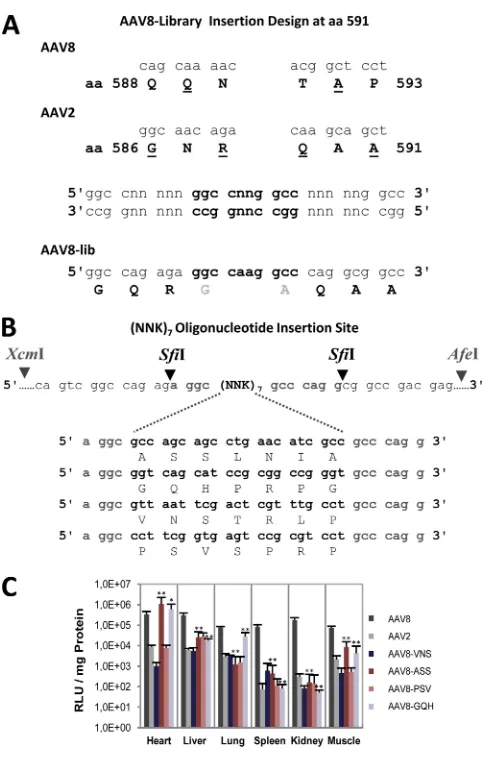
Validation of a peptide insertion site within the AAV8
cap-sid.
An insertion site for displaying new peptides on the AAV8
capsid was designed at position 590 in the 3-fold protrusion with
a SfiI restriction site inserted into the packaging plasmid
p5E18-VD2/8 (
16
) (
Fig. 4A
). Two new amino acids (G and A) flanking
the peptide insertions were introduced. In addition, several amino
acids between positions 588 and 593 were mutated. Without
pep-tide insertion, the modified helper plasmid could not generate
functional AAV8 capsids, while with peptide insertions the vector
yields were similar to wt rAAV8 vectors (data not shown). Four
peptide sequences from previously derived AAV2 library
selec-tions (
85
,
86
) were inserted into the AAV8 based capsids
(VNSTRLP, ASSLNIA, PSVSPRP, and
AAV8-GQHPRPG) (
Fig. 4B
). Reporter gene expression in different
tis-sues, 1 month after i.v.-injected particles (10
11vg/animal) showed
that the insertion of two peptide sequences, ASSLNIA and
GQHPRPG, resulted in a change in the transduction profile
com-pared to rAAV8 (
Fig. 4C
). The transduction efficiency was slightly
increased in heart tissue (4-fold, respectively 2-fold), while it was
largely reduced in other tissues (10- to 100-fold), except for
GQHPRPG, which showed nearly unchanged transgene
expres-sion in lung tissue. The other two peptides had only a negative
effect on gene transduction. The results show that insertion of
peptide sequences at this site can influence rAAV8 tissue targeting.
Consequently, a random peptide display library was inserted into
the AAV8 capsid at this position.
Generation, selection, and validation of an AAV8 random
peptide display library.
A preparation of an AAV8 random
pep-tide display library inserted at position 590 consisting of 7
⫻
10
12vg was generated.
In vitro
selections were performed on primary
murine hepatocytes. After four selection rounds, several peptide
sequences were enriched (
Fig. 5A
). The most highly enriched
pep-tide sequence, SEGLKNL, was inserted into the AAV8 packaging
helper plasmid after position 590, and chimeric vectors were
pro-duced by a triple transfection of HEK293T cells. The chimeras
could efficiently be packaged and purified by iodixanol step
gra-dients for gene transduction analysis.
In vivo
imaging showed that the insertion of peptide sequence
SEGLKNL triggered detargeting away from nonhepatic tissue
(
Fig. 5B
). Quantification of reporter gene expression for the vector
displaying peptide SEGLKNL in several tissues revealed only a
7-fold-lower transgene expression in the liver compared to AAV8
but a dramatic reduction in most other organs (
⬎
100-fold in the
on November 7, 2019 by guest
http://jvi.asm.org/
on November 7, 2019 by guest
http://jvi.asm.org/
lungs, spleen, and kidneys), close to the detection level. In heart
and skeletal muscle tissue, an
⬃
50-fold-reduced gene expression
could be detected. In conclusion, the
in vitro
selection of an AAV8
peptide display library on primary hepatocytes gave rise to a
pep-tide sequence SEGLKNL, which provided significant liver tropism
in the mouse with only a weak reduction of liver transduction
efficiency compared to rAAV8. This implies detargeting from
na-tive tropism by peptide insertions at the 3-fold protrusions of the
AAV8 capsid. Thus, the question arises as to whether AAV8 VP
protein sequences in this structurally prominent domain are
in-volved in cellular receptor binding.
Binding of a monoclonal antibody to the 3-fold protrusions
of AAV8 neutralizes infection at the postbinding level.
We have
recently isolated and characterized an AAV8-specific monoclonal
antibody, ADK8 (
67
), which efficiently neutralizes AAV8 gene
transduction
in vitro
(
Fig. 6A
) and also
in vivo
in mice (
20a
).
ADK8 binds to the sequence
588QQNTA
592located at the 3-fold
protrusions of the AAV8 capsid as shown by cryo-electron
mi-croscopy and image reconstruction of the AAV8-ADK8 complex
structure and analysis of capsid mutants (
20a
). The sequence
rep-resents region 6 and comprises the position where selected
pep-tides and the peptide display library were inserted (see
Fig. 4A
and
B
). Mutation of this sequence or insertion of peptides within this
sequence had a strong impact on
in vivo
gene transfer efficiency
and tissue tropism as shown above and prevented binding of
ADK8 to AAV8 capsids (
20a
). To test whether the binding of
ADK8 prevents cell attachment, we analyzed rAAV8 cell binding
by quantitative real-time PCR in the presence or absence of ADK8.
As shown in
Fig. 6B
, preincubation of AAV8 vectors with ADK8
did not prevent binding of rAAV8 to HepG2 cells. This result was
confirmed by an indirect immune fluorescence assay using HeLa
cells (
20a
). Virus uptake, measured by shifting the temperature
after binding at 4 to 37°C for 2 h, followed by trypsin treatment,
indicated a weak reduction in the amount of internalized vector in
the presence of the ADK8 antibody and a strong impairment of
intracellular trafficking toward the nucleus (
20a
). This means that
the binding of neutralizing antibody ADK8 to the sequence
588
QQNTA
592(region 6) located at the inner shoulder of the
3-fold protrusions of AAV8 interfered with a postattachment
pro-cess in AAV8 gene transduction.
Ratios of transgene expression to vector genome uptake level
correlate with the transduction efficiency.
It has been reported
that superior transduction of liver tissue by rAAV8 vectors
com-pared to rAAV2 vectors is mainly due to more efficient uncoating
(
69
). The contribution of postentry processing in gene
transduc-tion should be reflected by the ratio of transgene expression to the
amount of accumulated genomes in a given tissue. To calculate
FIG 3Transduction analysis of AAV vectors with amino acid exchanges from AAV8 to AAV2 and from AAV2 to AAV8. (A) Part of the AAV8 capsid protein amino acid sequence with the seven regions containing nonconserved amino acids between AAV2 and AAV8 demarked by large letters. Amino acids converted from AAV8 into AAV2 are marked by arrowheads. (B)In vivoreporter gene expression analysis of AAV8 capsid mutants in comparison to rAAV2 and rAAV8. Nine groups of mice (n⫽3) were i.v. injected with 1011vg-containing particles. Mice experiments were performed twice with two independent vector productions. Animals were dissected 1 month after injection. Luciferase transgene expression per mg of protein was determined in heart, liver, lung, spleen, kidney, and muscle tissue. **,P⬍0.01 (significantly increased or decreased transgene expression compared to rAAV8). (C) Excerpt of the AAV2 capsid protein amino acid sequence with regions containing nonconserved amino acids between AAV2 and AAV8 demarked by large letters. Amino acids converted from AAV2 into AAV8 are marked by arrowheads. (D)In vivoreporter gene expression analysis of AAV2 capsid mutants in comparison to rAAV2 and rAAV8. Eight groups of mice (n⫽4) were i.v. injected with 1011vg-containing particles. Mouse experiments were performed twice, with two independent vector productions. Animals were dissected 1 month after injection. Luciferase transgene expression per mg of protein was determined for heart, liver, lung, spleen, kidney, and muscle tissues. *,P⬍0.05; **,P⬍0.01 (significantly increased or decreased transgene expression compared to rAAV2). ns, not significant. (E) The heparin binding of rAAV2, rAAV8, and the domain swap mutants rAAV8/region 2 and rAAV8/region 6 was analyzed by chromatography on heparin agarose. Isolated viral DNA from input, flowthrough (FT), wash, and elution fractions was analyzed by DNA dot blotting with a CMV-specific radioactive probe.
FIG 4AAV8 capsid retargeting by peptide insertions. (A) Concept of an AAV8 insertion site to insert AAV2 library selected peptides into the AAV8 capsid se-quence. Stuffer sequences with SfiI restriction sites were generated and inserted into AAV8. In addition, aa 588 and 590 were exchanged from AAV8 to AAV2 because of previous mutant evaluations. (B) Helper plasmid p5E18-VD2/8 into which four different oligonucleotides (coding for 7 aa in length) were inserted into the SfiI restriction site to produce peptide-displaying AAV8 capsid mutants. (C)In vivoreporter gene expression analysis of four peptide-displaying AAV8 capsid mutants in comparison to rAAV2 and rAAV8 was performed. Six groups of mice (n⫽3) were i.v. injected with 1011vg-containing particles. Mouse experiments were performed twice, with two independent vector productions. Animals were dissected 1 month after injection. Luciferase transgene expression per mg of pro-tein was determined for heart, liver, lung, spleen, kidney, and muscle tissues. *,P⬍
0.05; **,P⬍0.01 (significantly increased or decreased transgene expression com-pared to rAAV8).
on November 7, 2019 by guest
http://jvi.asm.org/
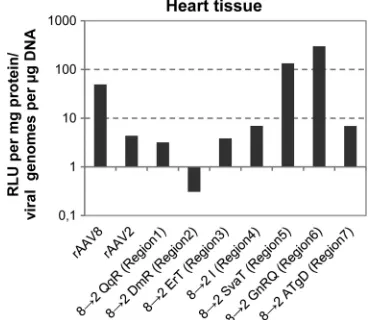
[image:8.585.42.283.61.440.2]such ratios, we determined the amount of vector genomes in heart
tissue after tail vein injection of wt and mutant vectors and related
the information obtained to the respective reporter gene
expres-sion. Heart tissue transduction with rAAV8 gave a 10-fold-higher
expression-to-genome ratio than transduction with rAAV2
(
Fig. 7
), confirming the observation made for liver transduction
that rAAV8 is more efficiently processed after cell uptake (
69
).
Introduction of AAV2 amino acids into AAV8 at regions 1, 3, 4,
and 7 (8
¡
2 QqR; 8
¡
2 ErT; 8
¡
2 I; 8
¡
2 ATgD) showed a similar
expression/genome ratio as did rAAV2, indicating that these
mu-tants are impaired in intracellular processing compared to wt
rAAV8 (
Fig. 7
). Exchange of AAV8 region 2 (8
¡
2 DmR) by AAV2
amino acids, which are located at the base of the 3-fold protrusion
facing the 2-fold axes at a VP-VP interface, resulted in a very low
ratio of transgene expression to genome uptake, suggesting that
this region is most likely critical for intracellular conformational
changes of the capsid leading to genome release and gene
expres-sion (
Fig. 7
). The two mutants that improved heart transduction
of rAAV8 vectors after incorporation of AAV2 sequences of
re-gions 5 or 6 (8
¡
2 SvaT; 8
¡
2 GnRQ) showed a clearly increased
expression/genome ratio (
Fig. 7
). This suggests that their
im-FIG 5In vivoreporter gene expression analysis of AAV8 capsid mutants con-taining peptides enriched by selection of an AAV8 random peptide display library on primary hepatocytes. (A) Three peptide sequences enriched byin vitroselection of an AAV8 random peptide display library on primary hepato-cytes. (B) IVIS images of mice analyzed 1 month after i.v. injection with rAAV8 or with the AAV8 capsid mutant displaying the most enriched peptide SEGLKNL (1011vg). Images were taken 15 min after intraperitoneal substrate injection. (C)In vivoreporter gene expression analysis of the SEGLKNL pep-tide displaying AAV8 capsid mutant in comparison to rAAV8. Two groups of mice (n⫽3) were i.v. injected with 1011vg-containing particles. Mouse ex-periments were performed twice, with two independent vector productions. Animals were dissected 1 month after injection. Luciferase transgene expres-sion per mg of protein was determined for heart, liver, lung, spleen, kidney, and muscle tissues.
FIG 6Neutralization of AAV8 gene transduction with monoclonal antibody ADK8. (A) Reporter gene transduction of HepG2 cells infected with an MOI of 25⫻103vg/cell of a luciferase expressing AAV8 vector and inhibition of transduction by application of 10 ng of ADK8 antibody per 5⫻103cells. (B) Detection of bound and unbound vector genomes after incubation of HepG2 cells with an MOI of 25⫻103vg/cell in the absence or presence of 100 ng of ADK8 antibody per 5⫻103cells. Shown are the mean values of three inde-pendent experiments. Bars indicate standard deviations.
FIG 7Correlation of viral vector genomes and transgene expression in heart tissue. By quantitative real-time PCR, the numbers of viral genomes perg of total DNA of capsid mutants with amino acid substitutions from AAV8 to AAV2, as well as of AAV2 and AAV8, were determined in heart tissue after i.v. injection. Organs of six animals were analyzed in the experiment. The total amount of DNA was determined by using GAPDH. The ratios between gene expression (RLU/mg of protein) and viral genomes (viral genomes/g of DNA) for heart tissue were determined.
on November 7, 2019 by guest
http://jvi.asm.org/
[image:9.585.344.501.65.235.2] [image:9.585.42.286.66.462.2] [image:9.585.328.512.486.646.2]proved transduction efficiency is, at least partially, due to
im-proved postentry processing.
DISCUSSION
Viral capsids are multifunctional protein assemblies that
encapsi-date, protect, and deliver the viral genome to the host cell. Relating
structural elements of capsids to functions in the infection
path-way is important for understanding AAV biology. Recombinant
AAV8 and AAV2 show striking differences in the efficiency of
transducing mouse tissues after systemic application. Domain
swap experiments indicated that amino acid residues within the
GH loop, which forms a significant component of the 3-fold
pro-trusions, is mostly responsible for the transduction differences
(
64
) (
Fig. 1
). This loop contains five structurally variable regions
VRIV, VRV, VPII, VRVII, and VRVIII (as defined in reference
20
)
and seven clusters of nonconserved amino acids between AAV8
and AAV2 (designated regions 1 to 7 here). Single amino acid
exchanges in these clusters, which did not affect capsid assembly
or genome packaging, resulted in mutants with different gene
transduction phenotypes. They are grouped according to their
efficiency and selectivity of transduction.
Exchange of regions 1, 2, 3, 4, and 7 did not lead to
transduc-tion improvement or enhanced selectivity, nor did any of the
mu-tants convert the low transduction efficiency of AAV2 to the high
efficiency of AAV8. Replacement of region 2 in AAV8 by AAV2
amino acids converted the high transduction efficiency of AAV8
to the low efficiency of AAV2, but the reciprocal substitution did
not restore the high transduction phenotype of AAV8. Strong
in-hibition of transduction of some tissues by AAV2, chimeric in
regions 2 and 4, was not combined with improved transduction in
other tissues. We therefore conclude that this part of the capsid
structure does not harbor structural elements which provide the
superior transduction properties of AAV8 compared to AAV2 and
do not determine tissue tropism in a positive way. Interestingly,
with the exception of region 7, these regions are all located on the
outer wall of the 3-fold protrusions facing the 2-fold and 5-fold
symmetry axes. The negative influence on gene transduction of
substituting these regions may indicate that they perturb the
in-terplay of chimeric VP amino acids and capsid regions with each
other and/or with cellular proteins.
Surprisingly, introduction of amino acids of AAV2 into
re-gions 5 and 6 of AAV8 improved the already high gene
trans-duction efficiency of AAV8 in the heart and liver (
Fig. 3A
and
B
). Mutation of region 6 (QNTA to GNRQ) also maintained
the level of transduction by rAAV8 in all other tissues.
Muta-tion of region 5 (IVAD to SVAT), however, enhanced gene
transduction only in the heart and reduced the transduction of
lung, spleen, kidney, and skeletal muscle, indicating tissue
se-lectivity and a mechanistically different influence on rAAV8
transduction. Both sites, region 5 and region 6, are located at
the base and close to the top, respectively, of the protrusions
facing the 3-fold axis. Region 6 contains in AAV2 R588 and
R585, which have been shown to be involved in primary
recep-tor binding and tissue tropism (
28
,
30
,
31
,
51
,
52
,
83
). It is not
known whether this region is also involved in cell binding and
tissue tropism in AAV8. The laminin receptor-binding
do-mains mapped to aa 492 to 557 and aa 593 to 623 of AAV8 do
not overlap with this region (
1
), although the second stretch of
residues is adjacent to the region 6 residues. There is a nearly
symmetrical increase in heart transduction when regions 5 and
6 of AAV2 were replaced by the sequences of AAV8. With
re-spect to region 6, this means that not only did conversion of
QNTA in AAV8 to GNRQ enhance the transduction of the
heart and liver, but also conversion of GNRQ to QNTA in
AAV2 enhanced AAV2 transduction of the heart. This result
excludes the interpretation that the corresponding sequences
in AAV8 and AAV2 permit transduction by direct binding to
the same receptor. The chimeric capsids could, however,
rec-ognize different receptors on the same cells or on different cell
types within the same tissue. The same conclusion must be
drawn for region 5, and in both cases the binding of different
receptors may, by chance, enhance transduction to similar
lev-els. On the other side, both results can also be interpreted by
assuming that region 5 and 6 sequences cooperate with other
receptor binding sites in the AAV capsids, which then permit
cell entry.
By converting QNTA to GNRQ (region 6), a weak HS binding
motif is created in AAV8 (
Fig. 3E
), which may enhance the
reten-tion period of the viral particle on the cell surface, thereby
increas-ing the likelihood for interaction with a secondary receptor
re-quired for cell entry. This may explain why the transduction of all
tissues tested is high. In the reciprocal conversion of GNRQ to
QNTA in AAV2, the HS binding motif is partially destroyed. A
previous report showed that disruption of the HS binding motif in
AAV2 impaired transduction of most mouse tissues but still
al-lowed good transduction of heart tissue (
28
), suggesting a second
receptor binding site on the AAV2 capsid, which is different from
the HS binding domain. Replacement of part of the HS binding
domain, region 6, of AAV2 by sequences of different AAV
sero-types also identified QNTA from AAV8 as a promising
substitu-tion (
3
). It showed that replacement of region 6 by sequences of
other AAV serotypes did not increase AAV2 transduction
imply-ing that the sequence QNTA contains information which is either
recognized by a receptor or used in another capsid protein
inter-action process. Influences of these sequences on postentry
pro-cessing can also not be excluded and are even suggested by the
ratio of transgene expression to genomes accumulated in heart
tissue. An unexpected aspect of this detailed sequence
replace-ment analysis is the observation that modifications of the very
closely located sequences in the capsid (regions 5, 6, and 7) have
strikingly different influences on gene transduction in different
tissues. In particular, regions 5 and 7 are located at nearly
overlap-ping positions, but chimeras in region 7 did not show any
im-proved gene transduction in contrast to region 5. Thus far, we do
not have an explanation for this observation.
Due to the observation that domain swaps in region 5 and 6
resulted in improved gene transduction by rAAV8 and rAAV2,
these regions represented potential insertion sites for peptide
se-quences in AAV8 for tissue targeting, as has been demonstrated
for AAV2 (
19
,
39
,
42
,
44
,
57
). Insertion of targeting peptides in
region 6 clearly influenced transduction efficiency and tissue
tro-pism of rAAV8 (
Fig. 4
). Although several peptides inserted in
AAV8 capsids generally reduced transduction in all tissues, other
peptides clearly induced selective transduction of heart or liver.
These different transduction profiles indicate that there is specific
sequence information required at this position, which was also
concluded from the AAV2 to AAV8 sequence replacement
exper-iments discussed above. The most obvious interpretation is that
the peptides provide binding to an uptake receptor, either directly
on November 7, 2019 by guest
http://jvi.asm.org/
or by enhancing the interaction with a secondary
receptor-bind-ing domain located on another part of the capsid.
Antibodies can be used to characterize receptor-ligand
inter-actions by blocking the interaction through competitive binding
to the ligand or the receptor. Monoclonal antibody ADK8 binds to
the sequence
588QQNTA
592on the AAV8 capsid corresponding
exactly to region 6 in AAV8 (
20a
). If this sequence is the only one
involved in receptor binding by AAV8, the antibody should
com-pete for binding of the virus to the cell. Although ADK8
neutral-izes rAAV8 gene transduction, it does not block the binding of
AAV8 to cells (
Fig. 6
). This result argues for the interpretation that
region 6 is not (or not alone) involved in receptor binding in
AAV8 and that antibody binding inhibits a postbinding
process-ing step. Also expression/genome ratios suggest an influence of
regions 5 and 6 on postbinding processes. The mechanism by
which the 3-fold protrusions could influence postbinding events
remains a matter of speculation. In this regard it should be
men-tioned that a stem-like connection between packaged DNA and
the inner capsid surface below the 3-fold symmetry axes has been
observed (
17
,
49
). Such a connection could mediate interactions
with the 3-fold protrusions occurring outside the capsid to
ge-nome-capsid interactions inside the capsid and influence genome
release. Alternatively, selection of an entry pathway mediated by
the 3-fold protrusions may indirectly promote efficiency of
postentry processing.
Overall, the data presented here suggest that modification of
regions 5 and 6, which strongly influence efficiency and selectivity
of AAV gene transduction
in vivo
, requires specific sequence
in-formation. There are several levels at which gene transduction
in
vivo
could be influenced. These include blood clearance,
penetra-tion of the endothelial cell layer, sequestrapenetra-tion at the extracellular
matrix, cell binding and entry, and intracellular processing. It is
unlikely that sequence-specific information is required for escape
from trapping in a nonproductive state, e.g., at the extracellular
matrix or opsonization by blood components. The need for
spe-cific sequence information for transduction improvement and
re-targeting rather suggests a gain of function. A gain-of-function
mutation could lead to improved endothelial cell layer
penetra-tion, cell binding, cell entry, or postentry processing. The different
assays conducted here point to a role of the 3-fold protrusions in
both cell surface interaction and postentry processing; however,
other activities cannot be excluded. Future studies are required to
elucidate the role of this prominent structural domain of the viral
capsid in AAV gene transduction.
ACKNOWLEDGMENTS
We thank A. Sacher and R. Popa-Wagner for valuable help.
This project was funded in part by grant KL516/7-1 from the Deutsche Forschungsgemeinschaft (to J.A.K.) and by National Institutes of Health grants R21 AI072341 and R01 GM082946 (to M.A.-M.).
REFERENCES
1.Akache B, et al.2006. The 37/67-kilodalton laminin receptor is a receptor for adeno-associated virus serotypes 8, 2, 3, and 9. J. Virol.80:9831–9836. 2.Akache B, et al.2007. A two-hybrid screen identifies cathepsins B and L as uncoating factors for adeno-associated viruses 2 and 8. Mol. Ther.15: 330 –339.
3.Asokan A, et al.Reengineering a receptor footprint of adeno-associated virus enables selective and systemic gene transfer to muscle. Nat. Biotech-nol.28:79 – 82.
4.Asokan A, Hamra JB, Govindasamy L, Agbandje-McKenna M,
Samul-ski RJ.2006. Adeno-associated virus type 2 contains an integrin␣51 binding domain essential for viral cell entry. J. Virol.80:8961– 8969. 5.Auricchio A.2011. Fighting blindness with adeno-associated virus
sero-type 8. Hum. Gene Ther.22:1169 –1170.
6.Bantel-Schaal U, Delius H, Schmidt R, and Hzur Hausen.1999. Human adeno-associated virus type 5 is only distantly related to other known primate helper-dependent parvoviruses. J. Virol.73:939 –947.
7.Barbon CM, et al.2005. AAV8-mediated hepatic expression of acid sph-ingomyelinase corrects the metabolic defect in the visceral organs of a mouse model of Niemann-Pick disease. Mol. Ther.12:431– 440. 8.Breous E, Somanathan S, Bell P, Wilson JM.2011. Inflammation
pro-motes the loss of adeno-associated virus-mediated transgene expression in mouse liver. Gastroenterology141:348 –355.
9.Broekman ML, Comer LA, Hyman BT, Sena-Esteves M.2006. Adeno-associated virus vectors serotyped with AAV8 capsid are more efficient than AAV-1 or -2 serotypes for widespread gene delivery to the neonatal mouse brain. Neuroscience138:501–510.
10. Choi VW, McCarty DM, Samulski RJ. 2005. AAV hybrid serotypes: improved vectors for gene delivery. Curr. Gene Ther.5:299 –310. 11. Daya S, Berns KI.2008. Gene therapy using adeno-associated virus
vec-tors. Clin. Microbiol. Rev.21:583–593.
12. Denby L, Nicklin SA, Baker AH.2005. Adeno-associated virus (AAV)-7 and -8 poorly transduce vascular endothelial cells and are sensitive to proteasomal degradation. Gene Ther.12:1534 –1538.
13. Dubielzig R, King JA, Weger S, Kern A, Kleinschmidt JA.1999. Adeno-associated virus type 2 protein interactions: formation of pre- encapsida-tion complexes. J. Virol.73:8989 – 8998.
14. Gao G, et al.2004. Clades of adeno-associated viruses are widely dissem-inated in human tissues. J. Virol.78:6381– 6388.
15. Gao G, Vandenberghe LH, Wilson JM.2005. New recombinant sero-types of AAV vectors. Curr. Gene Ther.5:285–297.
16. Gao GP, et al.2002. Novel adeno-associated viruses from rhesus monkeys as vectors for human gene therapy. Proc. Natl. Acad. Sci. U. S. A.99: 11854 –11859.
17. Gerlach B, Kleinschmidt JA, Bottcher B.2011. Conformational changes in adeno-associated virus type 1 induced by genome packaging. J. Mol. Biol.409:427– 438.
18. Ghosh A, et al.2006. Long-term correction of murine glycogen storage disease type Ia by recombinant adeno-associated virus-1-mediated gene transfer. Gene Ther.13:321–329.
19. Girod A, et al. 1999. Genetic capsid modifications allow efficient re-targeting of adeno-associated virus type 2. Nat. Med.5:1052–1056. 20. Govindasamy L, et al.2006. Structurally mapping the diverse phenotype
of adeno-associated virus serotype 4. J. Virol.80:11556 –11570. 20a.Gurda BL, Raupp C, Popa-Wagner R, Naumer M, Olson NH, Ng R,
McKenna R, Baker TS, Kleinschmidt JA, Agbandje-McKenna M.16 May 2012. Mapping a neutralizing epitope onto the capsid of adeno-associated virus serotype 8. J. Virol. doi:10.1128/JVI.00218-12. 21. Harding TC, et al.2006. Enhanced gene transfer efficiency in the murine
striatum and an orthotopic glioblastoma tumor model, using AAV-7- and AAV-8-pseudotyped vectors. Hum. Gene Ther.17:807– 820.
22. Hoffman BE, et al.2011. Nonredundant roles of IL-10 and TGF-beta in suppression of immune responses to hepatic AAV-factor IX gene transfer. Mol. Ther.19:1263–1272.
23. Hosel M, et al.2012. Toll-like receptor 2-mediated innate immune re-sponse in human nonparenchymal liver cells toward adeno-associated viral vectors. Hepatology55:287–297.
24. Humphrey W, Dalke A, Schulten K.1996. VMD: visual molecular dy-namics. J. Mol. Graph.8:27–28.
25. Hurlbut GD, et al.2010. Preexisting immunity and low expression in primates highlight translational challenges for liver-directed AAV8-mediated gene therapy. Mol. Ther.18:1983–1994.
26. Jayandharan GR, et al.2011. Activation of the NF-B pathway by adeno-associated virus (AAV) vectors and its implications in immune response and gene therapy. Proc. Natl. Acad. Sci. U. S. A.108:3743–3748. 27. Jiang H, et al.2006. Multiyear therapeutic benefit of AAV serotypes 2, 6,
and 8 delivering factor VIII to hemophilia A mice and dogs. Blood108: 107–115.
28. Kern A, et al.2003. Identification of a heparin-binding motif on adeno-associated virus type 2 capsids. J. Virol.77:11072–11081.
29. Laredj LN, Beard P.2011. Adeno-associated virus activates an innate immune response in normal human cells but not in osteosarcoma cells. J. Virol.85:13133–13143.
on November 7, 2019 by guest
http://jvi.asm.org/
30. Lerch TF, Chapman MS.2012. Identification of the heparin binding site on adeno-associated virus serotype 3B (AAV-3B). Virology423:6 –13. 31. Levy HC, et al.2009. Heparin binding induces conformational changes in
adeno-associated virus serotype 2. J. Struct. Biol.165:146 –156. 32. Li H, et al.2011. Capsid-specific T-cell responses to natural infections
with adeno-associated viruses in humans differ from those of nonhuman primates. Mol. Ther.19:2021–2030.
33. Li H, et al.2011. Adeno-associated virus vectors serotype 2 induce pro-longed proliferation of capsid-specific CD8⫹T cells in mice. Mol. Ther. 19:536 –546.
34. Lochrie MA, et al.2006. Mutations on the external surfaces of adeno-associated virus type 2 capsids that affect transduction and neutralization. J. Virol.80:821– 834.
35. Louboutin JP, Wang L, Wilson JM.2005. Gene transfer into skeletal muscle using novel AAV serotypes. J. Gene Med.7:442– 451.
36. Manno CS, et al.2006. Successful transduction of liver in hemophilia by AAV-Factor IX and limitations imposed by the host immune response. Nat. Med.12:342–347.
37. Martino AT, et al.2011. The genome of self-complementary adeno-associated viral vectors increases Toll-like receptor 9-dependent innate immune responses in the liver. Blood117:6459 – 6468.
38. McEachern KA, et al.2006. AAV8-mediated expression of glucocerebro-sidase ameliorates the storage pathology in the visceral organs of a mouse model of Gaucher disease. J. Gene Med.8:719 –729.
39. Michelfelder S, Trepel M.2009. Adeno-associated viral vectors and their redirection to cell-type specific receptors. Adv. Genet.67:29 – 60. 40. Mingozzi F, et al.2007. CD8⫹T-cell responses to adeno-associated virus
capsid in humans. Nat. Med.13:419 – 422.
41.Mori S, Wang L, Takeuchi T, Kanda T. 2004. Two novel adeno-associated viruses from cynomolgus monkey: pseudotyping characteriza-tion of capsid protein. Virology330:375–383.
42. Muller OJ, et al.2003. Random peptide libraries displayed on adeno-associated virus to select for targeted gene therapy vectors. Nat. Biotech-nol.21:1040 –1046.
43. Murphy SL, Bhagwat A, Edmonson S, Zhou S, High KA.2008. High-throughput screening and biophysical interrogation of hepatotropic AAV. Mol. Ther.16:1960 –1967.
44. Muzyczka N, and Warrington KH, Jr.2005. Custom adeno-associated virus capsids: the next generation of recombinant vectors with novel tro-pism. Hum. Gene Ther.16:408 – 416.
45. Nakai H, et al.2005. Large-scale molecular characterization of adeno-associated virus vector integration in mouse liver. J. Virol.79:3606 –3614. 46. Nam HJ, et al.2007. Structure of adeno-associated virus serotype 8, a
gene therapy vector. J. Virol.81:12260 –12271.
47. Nathwani AC, et al.2011. Long-term safety and efficacy following sys-temic administration of a self-complementary AAV vector encoding hu-man FIX pseudotyped with serotype 5 and 8 capsid proteins. Mol. Ther. 19:876 – 885.
48. Nathwani AC, et al.2011. Adenovirus-associated virus vector-mediated gene transfer in hemophilia B. N. Engl. J. Med.365:2357–2365. 49. Ng R, et al.Structural characterization of the dual glycan binding
adeno-associated virus serotype 6. J. Virol.84:12945–12957.
50. Nietupski JB, et al. 2011. Systemic administration of AAV8-alpha-galactosidase A induces humoral tolerance in nonhuman primates despite low hepatic expression. Mol. Ther.19:1999 –2011.
51. O’Donnell J, Taylor KA, Chapman MS.2009. Adeno-associated virus-2 and its primary cellular receptor– cryo-EM structure of a heparin com-plex. Virology385:434 – 443.
52. Opie SR, Warrington KH, Jr, Agbandje-McKenna M, Zolotukhin S, Muzyczka N.2003. Identification of amino acid residues in the capsid proteins of adeno-associated virus type 2 that contribute to heparan sul-fate proteoglycan binding. J. Virol.77:6995–7006.
53. Pacak CA, et al.2006. Recombinant adeno-associated virus serotype 9 leads to preferential cardiac transduction in vivo. Circ. Res.99:e3– e9. 54. Passini MA, et al.CNS-targeted gene therapy improves survival and
motor function in a mouse model of spinal muscular atrophy. J. Clin. Invest.120:1253–1264.
55. Paulk NK, et al.Adeno-associated virus gene repair corrects a mouse model of hereditary tyrosinemia in vivo. Hepatology51:1200 –1208. 56. Pear WS, Nolan GP, Scott ML, Baltimore D. 1993. Production of
high-titer helper-free retroviruses by transient transfection. Proc. Natl. Acad. Sci. U. S. A.90:8392– 8396.
57. Perabo L, et al.2003. In vitro selection of viral vectors with modified tropism: the adeno-associated virus display. Mol. Ther.8:151–157. 58. Pien GC, et al.2009. Capsid antigen presentation flags human
hepato-cytes for destruction after transduction by adeno-associated viral vectors. J. Clin. Invest.119:1688 –1695.
59. Pulicherla N, et al.2011. Engineering liver-detargeted AAV9 vectors for cardiac and musculoskeletal gene transfer. Mol. Ther.19:1070 –1078. 60. Qiao C, et al.2009. Hydrodynamic limb vein injection of
adeno-associated virus serotype 8 vector carrying canine myostatin propep-tide gene into normal dogs enhances muscle growth. Hum. Gene Ther. 20:1–10.
61. Rutledge EA, Halbert CL, Russell DW. 1998. Infectious clones and vectors derived from adeno-associated virus (AAV) serotypes other than AAV type 2. J. Virol.72:309 –319.
62. Sarkar R, et al.2006. Long-term efficacy of adeno-associated virus sero-types 8 and 9 in hemophilia a dogs and mice. Hum. Gene Ther.17:427– 439.
63. Schmidt M, et al.2008. Adeno-associated virus type 12 (AAV12): a novel AAV serotype with sialic acid- and heparan sulfate proteoglycan-independent transduction activity. J. Virol.82:1399 –1406.
64. Shen X, Storm T, Kay MA.2007. Characterization of the relationship of AAV capsid domain swapping to liver transduction efficiency. Mol. Ther. 15:1955–1962.
65. Shin O, Kim SJ, Lee WI, Kim JY, Lee H.2008. Effective transduction by self-complementary adeno-associated viruses of human dendritic cells with no alteration of their natural characteristics. J. Gene Med.10:762– 769.
66. Snyder RO, Francis J.2005. Adeno-associated viral vectors for clinical gene transfer studies. Curr. Gene Ther.5:311–321.
67. Sonntag F, et al.2011. The assembly-activating protein (AAP) promotes capsid assembly of different AAV serotypes. J. Virol.85:12686 –12697. 68. Sun B, et al.2005. Efficacy of an adeno-associated virus 8-pseudotyped
vector in glycogen storage disease type II. Mol. Ther.11:57– 65. 69. Thomas CE, Storm TA, Huang Z, Kay MA.2004. Rapid uncoating of
vector genomes is the key to efficient liver transduction with pseudotyped adeno-associated virus vectors. J. Virol.78:3110 –3122.
70. Vandenberghe LH, Wilson JM, Gao G.2009. Tailoring the AAV vector capsid for gene therapy. Gene Ther.16:311–319.
71. Veldwijk MR, et al.2002. Development and optimization of a real-time quantitative PCR-based method for the titration of AAV-2 vector stocks. Mol. Ther.6:272–278.
72. Wang AY, Peng PD, Ehrhardt A, Storm TA, Kay MA.2004. Comparison of adenoviral and adeno-associated viral vectors for pancreatic gene de-livery in vivo. Hum. Gene Ther.15:405– 413.
73. Wang H, et al.1998. Post-isolation inducible nitric oxide synthase gene expression due to collagenase buffer perfusion and characterization of the gene regulation in primary cultured murine hepatocytes. J. Biochem.124: 892– 899.
74. Wang L, et al.2011. AAV8-mediated hep