The MS Risk Allele of CD40 Is Associated with
Reduced Cell-Membrane Bound Expression in
Antigen Presenting Cells: Implications for
Gene Function
Judith Field1*, Fernando Shahijanian2, Stephen Schibeci2, Australia and New Zealand MS Genetics Consortium (ANZgene)¶,Laura Johnson1, Melissa Gresle3,4, Louise Laverick3,4,
Grant Parnell2, Graeme Stewart2, Fiona McKay2, Trevor Kilpatrick1,3,5, Helmut Butzkueven3,4, David Booth2
1Multiple Sclerosis Division, Florey Institute of Neuroscience and Mental Health, University of Melbourne, Melbourne, Victoria., Australia,2Centre for Immunology and Allergy Research, Westmead Millennium Institute, University of Sydney, Sydney, New South Wales, Australia,3Melbourne Brain Centre at the Royal Melbourne Hospital, University of Melbourne, Parkville, Australia,4Department of Medicine, University of Melbourne, Melbourne, Victoria, Australia,5Melbourne Neuroscience Institute, University of Melbourne, Melbourne, Victoria, Australia
¶ Membership of the ANZgene Consortium appears in the Acknowledgments. *[email protected]
Abstract
Human genetic and animal studies have implicated the costimulatory molecule CD40 in the development of multiple sclerosis (MS). We investigated the cell specific gene and protein expression variation controlled by the CD40 genetic variant(s) associated with MS, i.e. the T-allele at rs1883832. Previously we had shown that the risk allele is expressed at a lower level in whole blood, especially in people with MS. Here, we have defined the immune cell subsets responsible for genotype and disease effects on CD40 expression at the mRNA and protein level. In cell subsets in which CD40 is most highly expressed, B lymphocytes and dendritic cells, the MS-associated risk variant is associated with reduced CD40 cell-sur-face protein expression. In monocytes and dendritic cells, the risk allele additionally reduces the ratio of expression of full-length versus truncated CD40 mRNA, the latter encoding se-creted CD40. We additionally show that MS patients, regardless of genotype, express sig-nificantly lower levels of CD40 cell-surface protein compared to unaffected controls in B lymphocytes. Thus, both genotype-dependent and independent down-regulation of cell-sur-face CD40 is a feature of MS. Lower expression of a co-stimulator of T cell activation, CD40, is therefore associated with increased MS risk despite the same CD40 variant being associated with reduced risk of other inflammatory autoimmune diseases. Our results high-light the complexity and likely individuality of autoimmune pathogenesis, and could be con-sistent with antiviral and/or immunoregulatory functions of CD40 playing an important role in protection from MS.
a11111
OPEN ACCESS
Citation:Field J, Shahijanian F, Schibeci S, Australia and New Zealand MS Genetics Consortium (ANZgene), Johnson L, Gresle M, et al. (2015) The MS Risk Allele of CD40 Is Associated with Reduced Cell-Membrane Bound Expression in Antigen Presenting Cells: Implications for Gene Function. PLoS ONE 10(6): e0127080. doi:10.1371/journal. pone.0127080
Academic Editor:Alain Haziot, INSERM, FRANCE
Received:September 8, 2014
Accepted:April 10, 2015
Published:June 11, 2015
Copyright:© 2015 Field et al. This is an open access article distributed under the terms of the Creative Commons Attribution License, which permits unrestricted use, distribution, and reproduction in any medium, provided the original author and source are credited.
Data Availability Statement:All relevant data are within the paper.
Funding:This work was supported by the National Health and Medical Research Council of Australia (NHMRC) (grant numbers 633275 and 1065157).
Introduction
The CD40 gene has been previously identified as a risk gene for multiple sclerosis (MS) [1–4]
and other autoimmune diseases, including Graves’disease (GD) [5–8], rheumatoid arthritis
(RA) [9–12], systemic lupus erythematosus (SLE) [13] and Crohn’s disease (CD) [3]. CD40 is
an important co-stimulatory molecule expressed on the surface of a variety of antigen present-ing cells (APCs) includpresent-ing dendritic cells (DCs) and B-lymphocytes, as well as cells of the in-nate immune system such as macrophages and microglia. CD40 has previously been shown to play a role in the development of animal models of autoimmune demyelinating disease.
Deple-tion by antagonistic antibodies [14–16] or ablation (gene knock-out) [17] of CD40 expression
results in amelioration of disease, highlighting the importance of the secondary activation sig-nal in these inflammatory models. More recently, over-expression of CD40 in the thyroid has been shown to lead to spontaneous induction of hyperthyroidism in a murine model [18].
While GD and RA are associated with the major allele at rs1883832 (C) associated with in-creased CD40 expression [5,6] and therefore might be predicted to enhance a pro-inflammatory environment/response [19], the risk allele for MS at rs1883832 (T, minor allele) is associated with reduced CD40 expression [1,20]. Although there are many SNPs in linkage disequilibrium (LD) with rs1883832, it is possible that rs1883832 itself mediates the functional effects of this LD block. It is located at -1bp of the transcription start site (TSS) within the Kozak consensus se-quence, in which the major C allele has been shown to lead to enhanced efficiency of translation of the corresponding gene transcript [5,6]. However it is entirely possible that other SNPs in the LD block may be contributing to or causing the functional effect driving association with disease susceptibility. In addition, the effects of individual SNPs on expression of CD40 may, as for other immune cell genes, be highly dependent on context (i.e. inflammation) and cell subset.
Previous studies have suggested that CD40 expression is increased at the mRNA level in pe-ripheral blood mononuclear cells (PBMC) in MS compared to healthy/non-MS controls [21], but is not different in cultured B lymphocytes or monocytes at the protein level [22]. However, these studies involved small cohorts of varying disease duration and disease course (including secondary progressive MS and primary progressive MS), and/or subjects concurrently treated with disease-modifying therapies, all of which could potentially effect CD40 expression.
In this study, we used a relatively large cohort of untreated MS patients and unaffected con-trols to investigate the effect of genotype on expression of peripheral blood mononuclear cell types that produce the highest levels of CD40: B lymphocytes and monocytes. As other antigen presenting cells (APCs) are rare in blood, but the APCs from secondary lymphoid organs and tissues have the highest expression of CD40 of all subsets analysed in published databases
(www.immgen.org,www.biogps.org), we also usedin vitrodifferentiation of monocytes to
pro-duce dendritic cells representative of these cell types. Further, we examined the effect of disease on CD40 expression in B-lymphocytes and monocytes freshly isolated from the peripheral blood of MS patients with relapsing-remitting MS (RRMS) compared to age- and sex-matched healthy controls. Our findings implicate lowered cell-surface CD40 levels in the development of MS, and should lead to further mechanistic investigations with potential
therapeutic implications.
Materials and Methods
Subject recruitment and demographics
MS patients were recruited according to the following criteria:—definite relapsing-remitting MS (RRMS) according to McDonald criteria or Clinically Isolated Syndrome (CIS), aged
be-tween and inclusive of 18–65 years, not currently on immunomodulatory therapy for MS, or
none within in the last three months and no other concurrent autoimmune disease. In our co-hort, MS patients were between the ages of 21 and 54 years (38.4± 7.7). The mean number of years from diagnosis at the time of sample collection was 6.0 ± 7.0. Neurological outcome was assessed using the Kurtzke Expanded Disability Status Scale (EDSS). The median EDSS was 2.0 (mean EDSS 1.9 ± 1.6). There were 18 females and 3 males within our MS cohort. Unaffected controls were aged between and inclusive of 18–65 years of age and had no personal history of neurological disease or autoimmune disease. Two independent healthy control cohorts were used. Cohort 1 was composed of 56 females and 28 males, aged 37.46 (± 10.1) and cohort 2: 32 females and 17 males (entirety or subsection of cohort 2 used in figures as described). All MS patients and healthy controls recruited for this study were Caucasian.
Ethics Statement
Ethical approval for this project was obtained from the Eastern Health Research Ethics Com-mittee (Melbourne) and the Western Sydney Local Health District Human Ethics ComCom-mittee. All participants provided written consent.
Cell subset purification
Immune cell subsets were purified from PBMCs isolated from MS patients and controls by ei-ther Ficoll (GE Healthcare) or Histopaque (Invitrogen) density gradient separation. CD4, CD8, CD4+CD45RO+ and CD4+CD45RA+ T cells, T regulatory cells (Tregs) (CD4+CD25hi), B lymphocytes (CD19+) NK cells (CD3-CD56+), monocytes (CD14+), plasmacytoid dendritic cells (CD303+CD304+CD123+CD11c-) and myeloid dendritic cells (CD19+CD1c-hi) were purified using magnetic bead separation as previously described [23]. For monocyte subsets, a monocyte enrichment kit (StemCells) was used prior to CD14 purification as per the
manufac-turers’instructions (Human CD14 Microbeads, Miltenyi Biotec, Germany). Th1, Th2 and
Th17 subsets were differentiatedin vitrofrom CD4+CD45RA+ as previously described [24].
DC1 and DC2s were generatedin vitrofrom monocytes by sequential incubation with IL-4
and GMCSF, LPS and either IFNγ(DC1) or IFNβ(DC2) as previously described [23]. Purified
subsets were stored in RLT buffer (Qiagen) or Cells-to-signal Buffer (Ambion) for subsequent RNA extraction. Purity of the cellular subsets was determined by flow cytometry.
Flow cytometric analysis
Fresh PBMC from MS patients and healthy controls were analysed by flow cytometry using flow cytometry antibodies purchased from Miltenyi Biotec (Germany), including CD14-PE (TUK4) and CD16-FITC (VEP13) for monocytes and CD19-FITC (LT19), IgG-PE (IS11-3B2.2.3), IgD-PE (IgD26), CD27-PECy5 (M-T271), CD38-PE (IB6), and CD24-biotin (32D12/ anti-biotin-PerCP for B lymphocyte subsets. Regulatory B cells were identified as
CD19+CD38hiCD24hi using CD19-FITC, CD38-PE and CD24-biotin/antibiotin-PerCp anti-bodies as described by the Mauri laboratory [25,26]. CD40 expression was determined in PBMCs or purified/cultured subsets by comparison of CD40-APC (HB14) or CD40-PE
(HB14) to an isotype control (Mouse IgG1clone IS5-21F5). Labelled cells were analysed using
Genotyping and gene expression of CD40
DNA was extracted from whole blood as previously described [1]. DNA was genotyped for rs1883832 using the Taqman SNP genotyping assay C_11655119 (Applied Biosystems), or by restriction fragment length polymorphism: PCR amplification using oligonucleotides
5’- ACAGCAAGATGCGTCCCTAAAC- 3’and5’- CTTCCCTTTCCTTCTCATTCCC- 3’
followed by enzyme digestion withNco1(Promega), generating products of 114 bp and 226 bp
for the C allele and 340 bp for the T allele at rs1883832.
RNA was extracted from purified cells using the RNeasy Mini Kit (Qiagen) including DNAse treatment. RNA from whole blood was collected using PAXgene blood RNA tubes (PreAnalytiX, Switzerland) and total mRNA extracted using the PAXgene Blood RNA Kit (Qiagen, Germany). Total expression of CD40 was determined by SYBR green qRT-PCR
using the following primers- forward:5’-GCAGGGGAGTCAGCAGA-3’; reverse:
5’-TTCCTTCCCTTT CCTTCTCA-3’; and the housekeeping gene GAPDH as previously scribed [20], or by next generation mRNA sequencing (RNA-Seq) analysis as previously de-scribed [27]. The percentage of CD40 transcript encoding the full-length protein (FL) was determined by PCR amplification of a cDNA region spanning CD40 exon 4 to exon 10
using primers:—forward5’-CAGACACCATCTGCACCTGT-3’and reverse,
5’-AATTGATCTCCTGGGGT TCC-3’. Molarity of the largest splice form (~400bp, encoding the full-length isoform) as a proportion of all isoforms expressed was determined by electro-phoresis and UV detection (Bioanalyzer, Agilent Technologies) as previously described [24]. The expression level of CD40 isoforms was determined for a series of immune cell subsets using RNAseq (annotated using Ensembl build GRCh37.62) as previously described [23].
Statistical Analysis
Statistical analysis was performed using GraphPad Prism 5. Comparisons between groups were made by unpaired t-test or Mann-Whitney U test as appropriate. Box and whisker plots depict maximum and minimum values, median and interquartile range.
Results
In peripheral blood immune cells, B-lymphocytes express the highest
level of CD40 mRNA and protein
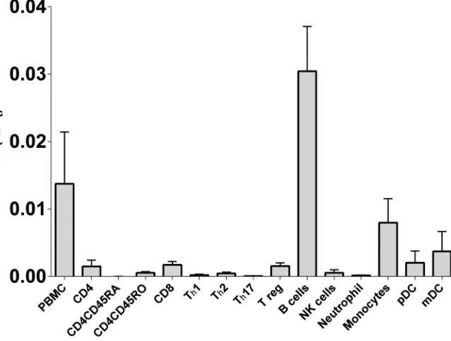
In our earlier work [20] we demonstrated that CD40 mRNA expression was genotype depen-dent in whole blood. Here we compared expression in cell subsets purified from blood to con-firm the likely source of these differences in mRNA expression. As expected, of the common cell subsets found in blood, B-lymphocytes have the highest level of CD40 mRNA, with mono-cytes and dendritic cells also contributing (Fig 1). In an RNAseq analysis of immune cell sub-sets we previously conducted [28], several mRNA isoforms were identified in key subsub-sets of immune cells, including transcript encoding the full length protein (dominant) and those lack-ing exon 5 and/or exon 6 that encode for the transmembrane region, resultlack-ing in the transla-tion of soluble CD40 protein.
Expression of the CD40 MS risk allele correlates with reduced CD40
levels on B-cells
B-lymphocytes from healthy controls and MS patients were analysedex vivofor expression of
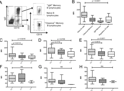
These were analysed for CD40 expression compared to an isotype control (Fig 2A). Naïve B cells expressed significantly more CD40 on the cell surface compared to classical memory, IgM memory B and regulatory B lymphocyte subsets (Fig 2B). Total B-lymphocytes showed a geno-type-dependent reduction in the surface expression of CD40 (Fig 2C); with homozygous CC individuals (n = 49) expressing 30% more total CD40 on the cell surface compared to CT (n = 27; p = 0.0113) and TT (n = 10; p = 0.0216) individuals. The surface expression of CD40 on naïve B cells (CD19+IgD+CD27-) was not significantly associated with genotype (Fig 2D; CC vs. CT p = 0.1715, CC vs. TT p = 0.0706), while classical memory B cells (Fig 2E, CD19+-IgD-CD27+) demonstrated a trend towards a genotype-dependent CD40 expression profile (CC vs. CT p = 0.0515; CC vs. TT p = 0.0571). No significant genotype-dependent expression effects were observed in total B cells (Fig 2F; CC vs. CT p = 0.2511; CC vs. TT p = 0.3924)), naïve B cells (Fig 2G; CC vs. CT p = 0.5701, CC vs. TT p = 0.1271)) or classical memory B cells (Fig 2H; CC vs. CT p = 0.2511, CC vs. TT p = 0.3924) isolated from MS patients. In this study, the genotype effect on CD40 mRNA expression measured in whole blood by RNA-Seq did not reach significance (data not shown).
CD40 is under expressed on MS patient B-lymphocytes independent of
the CD40 risk allele effect
[image:5.612.209.528.80.321.2]Comparison of B lymphocyte expression of CD40 in MS patients failed to show a significant ef-fect of genotype on surface CD40 expression levels in total B lymphocytes (Fig 2F), naïve B lymphocytes (Fig 2G) or classical memory B lymphocytes (Fig 2H). However, comparison of cell surface CD40 expression on B-lymphocytes between healthy controls and MS patients (Fig 3) showed that CD40 expression was significantly lower in MS patients compared to healthy
Fig 1. CD40 mRNA expression in peripheral blood immune cell subsets.CD40 mRNA expression was determined by RT-PCR in freshly purified immune cell subsets orin vitrodifferentiated subsets (Th1, Th2, Th17; differentiated from fresh CD4CD45RA) from healthy controls (n = 3, or n = 2 for pDC).
controls in all CD19+ B-lymphocytes (Fig 3A; p<0.0001), as well as in the naïve B lympho-cytes\ (Fig 3C; p<0.0001), classical memory B-lymphocyte (Fig 3D; p = 0.0001) and IgM memory B lymphocyte subsets (Fig 3E; p = 0.0004). A subset comparison of patients and unaf-fected controls homozygous for the rs1883832 C allele (CC) also demonstrated a significant
de-crease in CD40 expression on the total B—lymphocytes of MS patients compared to controls
(Fig 3B; p<0.0001) The relative proportions of total B-lymphocytes and subsets as a percent-age of total white cells were not affected by genotype or phenotype (data not shown).
CD40 is expressed at significantly lower levels in
“
classical
”
monocytes
compared to
“
intermediate
”
and
“
non-classical
”
monocytes
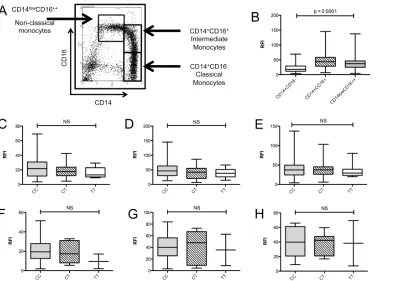
[image:6.612.40.455.79.382.2]Monocytes from MS patients and healthy controls defined by FSC/SSC profiles and CD14 posi-tivity were analysed for expression of CD40. Classical monocytes were defined as CD14 +CD16-, intermediate monocytes as CD14+CD16+, and non-classical monocytes as CD14low, CD16++ (Fig 4A). The proportion of monocytes and the individual monocyte subtypes were not affected by risk-genotype or phenotype (data not shown). CD14+ CD16- classical
Fig 2. Genotype dependent CD40 protein expression in peripheral B-lymphocyte subsets of MS patients and healthy controls.B lymphocyte subsets from healthy controls (n = 86) and MS patients (n = 21) were identified by flow cytometry (A) and CD40 expression determined relative to an isotype control (relative fluorescence intensity; RFI). Regulatory B cells were identified as CD19+CD38hiCD24hi (data not shown) (B). Association of rs1883832 genotype with CD40 expression in healthy controls (CC = 49, CT = 27, TT = 10) was examined in total B lymphocytes (C), naïve B-lymphocytes (D) and classical memory B-lymphocytes (E), and in total B lymphocytes (F), naïve B lymphocytes (G) and classical B lymphocytes (H) of MS patients (CC = 12, CT = 7, TT = 2). p values were determined by Mann-Whitney U test comparison of each group.
Fig 3. CD40 protein is under- expressed in B lymphocytes of MS patients.B lymphocyte subsets from healthy controls (n = 86) and MS patients (n = 24) were identified by flow cytometry and CD40 expression determined relative to an isotype control (relative fluorescence intensity; RFI). Surface levels of CD40 were compared in total B-lymphocytes (A), B lymphocytes from rs1883832 CC individuals (B; n = 49 healthy controls, n = 12 MS patients), naïve B
lymphocytes (C), classical memory B lymphocytes (D) and IgM memory B lymphocytes (E). P-values were determined using Mann—Whitney test.
doi:10.1371/journal.pone.0127080.g003
Fig 4. Genotype dependent CD40 protein expression in peripheral monocyte subsets of MS patients and healthy controls.Monocyte subsets were identified by flow cytometry (A) and CD40 expression determined relative to an isotype control (relative fluorescence intensity; RFI) (B). Association of rs1883832 genotype with CD40 expression was examined in classical CD14+CD16- (C), intermediate CD14+CD16+ (D) and non-classical CD14lowCD16+ + monocytes (E) from healthy controls (CC = 49, CT = 27, TT = 10), and classical CD14+CD16- (F), intermediate CD14+CD16+ (G) and non-classical CD14lowCD16++ monocytes (H) from MS patients (CC = 12, CT = 7, TT = 2). P—values were determined by Mann-Whitney test.
[image:7.612.39.435.366.647.2]monocytes expressed significantly lower levels of CD40 on the cell surface compared to the in-termediate and non-classical monocyte subtypes in both healthy controls and MS (Fig 4B).
CD40 expression is not significantly affected by genotype or phenotype
in monocyte subsets
There was no significant difference in the level of CD40 expression in monocytes between MS and controls (Fig 4C). In addition, there were no genotype-dependent effects on CD40 expres-sion in healthy controls (Fig 4D).
The CD40 MS risk-allele is under expressed in dendritic cell subsets
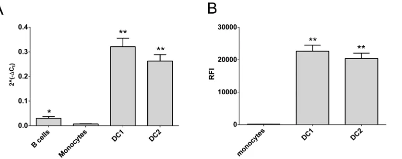
Dendritic cells are the major antigen presenting cells. However, DCs from whole blood are not typical of those in the secondary lymphoid organs and tissues, which are thought to drive T cell activation in autoimmune diseases [29]. Fortunately DCs representative of tissue DCs can be differentiated from monocytes, and have been verified as inflammatory (DC1) or tolerogenic (DC2) on the basis morphology and IL12p40 and IL10 mRNA and protein expression [23]. These DCs express much higher levels of CD40 mRNA and protein than monocytes and B cells (Fig 5). In these cells, CD40 expression was genotype dependent, with reduced expression of the risk allele at the mRNA level in DC2s (Fig 6A, p<0.011) and at the protein level in both DC phenotypes (Fig 6B; p<0.0047, DC1s; p<0.0043, DC2s).
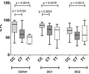
Lower proportions of the full-length isoform from the CD40 MS risk-allele
[image:8.612.44.437.77.233.2]Greater splicing of the CD40 risk allele was evident with a lower percentage of the full length mRNA isoform expressed in DCs and monocytes compared to expression levels in DC carrying at least one protective allele (Fig 7; p<0.0020, monocytes; p<0.0014, DC1s; p<0.0026, DC2s). A similar trend was evident in whole blood in healthy controls (CC>CT; p<0.13) and MS (CT>TT; p<0.056;Fig 8). As CD40 isoform usage was affected by the MS risk geno-type, we sought common SNPs located between exon 4 and exon 8 that might affect splicing. All SNPs identified as inherited in strong LD with rs6074022 in the 1000 genome project and located between exon 4 and exon 8 (rs73115010, rs66815221, rs73622651, rs6074028,
Fig 5. CD40 expression is higher in differentiated dendritic cell subsets.CD40 expression was determined in freshly purified immune cell subsets (B cells, monocytes) orin vitrodifferentiated dendritic cells (DC1, DC2) from healthy controls. Gene expression by RT-PCR (A; n = 49) and relative fluorescence intensity (RFI) by flow cytometry (B; n = 41) are shown;*significantly different from monocytes and DCs;**significantly different from B cells and monocytes (A) or from monocytes (B); p<0.05 by Mann-Whitney test.
Fig 6. The CD40 risk allele is under expressed in dendritic cell subsets.Association of rs1883832 genotype with CD40 expression was determined inin vitrodifferentiated dendritic cells (DC1, DC2) from healthy controls (non-carriers, CC, or carriers, CT/TT, of the risk allele). Gene expression by RT-PCR (A; n = 49) and relative fluorescence intensity (RFI) by flow cytometry (B; n = 41) are shown; p values by Mann-Whitney test.
doi:10.1371/journal.pone.0127080.g006
Fig 7. Lower proportions of the full-length isoform expressed from the CD40 risk allele in monocytes and dendritic cells.Association of rs1883832 genotype (CC, TC, TT) with the proportion of full-length isoform of CD40 (%FL) expressed inin vitrodifferentiated dendritic cells (DC1, DC2) from healthy controls (n = 49). Molar ratios of isoforms were quantitated by RT-PCR and amplification of a region spanning CD40 exon 4–10, followed by electrophoretic separation and fluorescent detection (Bioanalyzer, Agilent); p values by Mann-Whitney test.
[image:9.612.205.571.320.623.2]rs3746821, rs11569333) were intronic and in regions unlikely to affect splicing, as assessed with the Human Splicing Finder tool [30]. The minor allele frequency of the exonic SNPs from exon 4 to exon 8 was less than 4%, so unlikely to be driving the genotype association. This in-cluded 3 SNPS (rs369901991, rs371997367, rs144600981) calculated in Ensembl to potentially affect splicing.
Discussion
In this study we show an MS risk genotype-dependent reduction of CD40 cell-surface protein in B-lymphocytes and polarised dendritic cells. This is paralleled by lower levels of CD40 mRNA production from the risk genotype in these cells, and an increased relative proportion of isoforms encoding the secreted form of CD40. In addition, and for the first time, we show that the level of CD40 protein expression is significantly reduced in B-lymphocytes isolated from MS patients compared to healthy controls, independent of risk genotype. This is consis-tent with our previous findings that whole blood CD40 mRNA was reduced in carriers of the risk genotype, and that the effect of genotype on expression was enhanced in MS [20]. These results also point towards additional factors leading to the down-regulation of CD40 protein expression in MS patients besides CD40 genotype, and thus implicate lower cell surface CD40 protein expression in the complex pathogenesis of MS.
However, our findings are in contrast to previous studies that have shown no difference in either the mRNA or protein levels of CD40 expression between MS patients and controls [21,22]. These previous cohorts were of varying disease phenotype, including patients in the progressive phase of disease, varying relapse status and with a wider range of disease duration [21,22] possibly capturing, at least for those with relapse, a more inflammatory state with con-comitantly higher CD40 expression, thus masking the reduced CD40 expression we observed in our cohort of patients with stable relapsing-remitting disease.
[image:10.612.62.471.77.236.2]The opposite genetic association of CD40 rs1883832 with increased susceptibility to MS, but with protection from RA and GD are intriguing and point to distinct roles for CD40 in the pathogenic processes in these autoimmune diseases. Our studies here and previously [20], as well as studies in RA and GD [5,6] have consistently demonstrated an association of the T allele with reduced CD40 expression. The association of protection from RA and GD with lower
Fig 8. Proportions of the full-length isoform expressed from the CD40 risk allele in whole blood.Association of rs1883832 genotype (CC, TC, TT) with the proportion of full-length isoform of CD40 (%FL) expressed in whole blood from healthy controls (A; n = 38) and MS (B; n = 32). Molar ratios of isoforms were quantitated by RT-PCR and amplification of a region spanning CD40 exon 4–10, followed by electrophoretic separation and fluorescent detection (Bioanalyzer, Agilent). Trends were observed for CC>CT in controls (p<0.13) and for CT>TT in MS, (p<0.056); p values by Mann-Whitney test.
expression of a T cell activation gene fits with a primarily costimulatory role of CD40 support-ing the autoimmune inflammatory process, and supported by animal models in which CD40 inhibition reduces inflammation. In contrast, the association of protection from MS with higher expression of CD40 may suggest that these inflammatory processes are protective in MS, and/or that thymic tolerogenic processes mediated by CD40 are of greater importance in protection from MS than CD40-mediated autoimmune inflammatory processes are in disease initiation or propagation.
It is intriguing that Epstein Bar virus (EBV), long implicated in MS pathogenesis [31], en-codes a homologue of human CD40, This protein is expressed in infected B cells, and constitu-tively signals, promoting B cell proliferation [32]. Relative susceptibility to EBV may be dependent on competition between human and EBV CD40/CD40L signaling. Notably though, a genome wide association study of SNPs with EBNA-1 antibody levels did not implicate CD40 [33]. This would suggest any CD40 genotype associations with EBV susceptibility, or EBV con-tribution to MS, may be independent of EBNA-1 antibody levels.
Tolerogenic responses mediated by CD40 stimulation include naïve B lymphocyte- mediat-ed stimulation of T cells leading to the expansion of regulatory T cells [34]. In addition, naïve B lymphocytes (and regulatory B cells found within the naïve B lymphocyte pool) have been shown to produce regulatory cytokines such as IL-10 upon stimulation with CD40L [35]. In addition to these peripheral immunoregulatory mechanisms, cortical and medullary thymic epithelial cells (cTECS and mTECs) express functional CD40 [36], which has been shown to be essential for the establishment of the mTEC microenvironment leading to tolerance to self-an-tigens [37]. The role of Vitamin D/UVR driven immunomodulation [38,39], mediated by APCs, may also provide an additional link between reduced CD40 expression and increased MS risk, with under-expression of CD40 by DCs leading to the failure of the protective effects of increased Vitamin D/UVR.
A decrease in CD40 expression by these cell types could plausibly lead to a failure of toler-ance/immunomodulatory mechanisms mediated by CD40 stimulation, and by extension, a failure of protection from the development of MS in subsequent years.
While CD40 expression is known to be increased in thyroid tissue in GD [40], no examina-tion has been made of either the genotype-dependent effects of CD40 expression in GD pa-tients, or CD40 expression by peripheral immune cells in the context of disease. In SLE, a risk-genotype dependent correlation in CD40 expression level in B lymphocytes has been identified, present in both patients and healthy controls [13], however there is no apparent genotype-in-dependent effect in CD40 expression levels between SLE patients and healthy controls. Cer-tainly, the genotype-independent decrease in CD40 expression by peripheral immune cells in MS is a unique and novel finding, and leads to many questions as to the reasons for this de-crease, as well as the subsequent effects. Is this decrease a result of“exhaustion”of CD40 ex-pression in MS similar to that of CD8“exhaustion”in chronic viral infection [41], or is it a result of homeostatic down-regulation in the context of inflammation?
Further work is needed to understand the consequences of decreased CD40 expression on B lymphocyte and dendritic cells in antiviral/immunoregulatory functions in demyelinating dis-ease, and what drives reduced expression of CD40 in B lymphocytes in established MS. Defin-ing the genotype-independent but disease-dependent changes in B lymphocyte and dendritic cell CD40 levels identified in this study could uncover protective roles for CD40 in MS that fundamentally distinguish its pathogenesis from that of other autoimmune diseases.
Acknowledgments
The members of the ANZgene Consortium are:Alan Baxter (James Cook University, Towns-ville, Australia), Allan G Kermode (Sir Charles Gairdner Hospital, Perth, Australia), Bruce Taylor (Menzies Research Institute Tasmania, University of Tasmania, Hobart, Australia), David R Booth (ANZgene Consortium Chair) (Westmead Millennium Institute, University of
Sydney, Sydney, Australia)[email protected], Deborah Mason (Canterbury District
Health Board, Christchurch, New Zealand), Graeme J Stewart (Westmead Millennium Insti-tute, University of Sydney, Sydney, Australia), Helmut Butzkueven (University of Melbourne, Melbourne, Australia), Jac Charlesworth (Menzies Research Institute Tasmania, University of Tasmania, Hobart, Australia), James Wiley (Florey Institute of Neuroscience and Mental Health, University of Melbourne, Melbourne, Australia), Jeannette Lechner- Scott (Hunter Medical Research Institute, Newcastle, Australia), Judith Field (Florey Institute of Neurosci-ence and Mental Health, University of Melbourne, Melbourne, Australia), Lotti Tajouri (Bond University, Gold Coast, Australia), Lyn Griffiths (Griffith Institute of Health and Medical Re-search, Griffith University, Gold Coast, Australia), Mark Slee (School of Medicine, Flinders University of South Australia, Adelaide, Australia), Matthew A Brown (University of Queens-land Diamantina Institute, Translational Research Institute, Brisbane, Australia), Pablo Moscato (Hunter Medical Re- search Institute, Newcastle, Australia), Rodney J Scott (Hunter Medical Research Institute, Newcastle, Australia), Simon Broadley (School of Medicine, Grif-fith University, Gold Coast, Australia), Steve Vucic (Westmead Millennium Institute, Universi-ty of Sydney, Sydney, Australia), Trevor Kilpatrick (UniversiUniversi-ty of Melbourne, Melbourne, Australia), William M Carroll (Sir Charles Gairdner Hospital, Perth, Australia).
We thank individuals with MS in Australia for supporting this research and all investigators of the study who have contributed to the recruitment of MS patients.
Author Contributions
Conceived and designed the experiments: JF GS GP TK HB DB. Performed the experiments: JF FS SS LJ MG LL FM. Analyzed the data: JF FS LJ FM. Contributed reagents/materials/analysis tools: JF SS GP GS TK HB DB. Wrote the paper: JF FM TK HB DB.
References
1. ANZgene. (2009) Genome-wide association study identifies new multiple sclerosis susceptibility loci on chromosomes 12 and 20. Nat Genet 41: 824–828. doi:10.1038/ng.396PMID:19525955
2. Sawcer S, Hellenthal G, Pirinen M, Spencer CC, Patsopoulos NA, Moutsianas L, et al. (2011) Genetic risk and a primary role for cell-mediated immune mechanisms in multiple sclerosis. Nature 476: 214–219. doi:10.1038/nature10251PMID:21833088
3. Blanco-Kelly F, Matesanz F, Alcina A, Teruel M, Diaz-Gallo LM, Gomez-Garcia M, et al. (2010) CD40: novel association with Crohn's disease and replication in multiple sclerosis susceptibility. PLoS ONE 5: e11520. doi:10.1371/journal.pone.0011520PMID:20634952
5. Jacobson EM, Huber AK, Akeno N, Sivak M, Li CW, Concepcion E, et al. (2007) A CD40 Kozak se-quence polymorphism and susceptibility to antibody-mediated autoimmune conditions: the role of CD40 tissue-specific expression. Genes Immun 8: 205–214. PMID:17344890
6. Jacobson EM, Concepcion E, Oashi T, Tomer Y (2005) A Graves' disease-associated Kozak sequence single-nucleotide polymorphism enhances the efficiency of CD40 gene translation: a case for transla-tional pathophysiology. Endocrinology 146: 2684–2691. PMID:15731360
7. Yang J, Qin Q, Yan N, Zhu YF, Li C, Yang XJ, et al. (2012) CD40 C/T(-1) and CTLA-4 A/G(49) SNPs are associated with autoimmune thyroid diseases in the Chinese population. Endocrine 41: 111–115. doi:10.1007/s12020-011-9510-1PMID:21866398
8. Li M, Sun H, Liu S, Yu J, Li Q, Liu P, et al. (2012) CD40 C/T-1 polymorphism plays different roles in Graves' disease and Hashimoto's thyroiditis: a meta-analysis. Endocr J 59: 1041–1050. PMID: 22863718
9. Raychaudhuri S, Remmers EF, Lee AT, Hackett R, Guiducci C, Burtt NP, et al. (2008) Common vari-ants at CD40 and other loci confer risk of rheumatoid arthritis. Nat Genet 40: 1216–1223. doi:10.1038/ ng.233PMID:18794853
10. Orozco G, Eyre S, Hinks A, Ke X, Wilson AG, Bax DE, et al. (2010) Association of CD40 with rheuma-toid arthritis confirmed in a large UK case-control study. Ann Rheum Dis 69: 813–816. doi:10.1136/ ard.2009.109579PMID:19435719
11. van der Linden MP, Feitsma AL, le Cessie S, Kern M, Olsson LM, Raychaudhuri S, et al. (2009) Associ-ation of a single-nucleotide polymorphism in CD40 with the rate of joint destruction in rheumatoid arthri-tis. Arthritis Rheum 60: 2242–2247. doi:10.1002/art.24721PMID:19644859
12. Li G, Diogo D, Wu D, Spoonamore J, Dancik V, Franke L, et al. (2013) Human Genetics in Rheumatoid Arthritis Guides a High-Throughput Drug Screen of the CD40 Signaling Pathway. PLoS Genet 9: e1003487. doi:10.1371/journal.pgen.1003487PMID:23696745
13. Vazgiourakis VM, Zervou MI, Choulaki C, Bertsias G, Melissourgaki M, Yilmaz N, et al. (2011) A com-mon SNP in the CD40 region is associated with systemic lupus erythematosus and correlates with al-tered CD40 expression: implications for the pathogenesis. Ann Rheum Dis 70: 2184–2190. doi:10. 1136/ard.2010.146530PMID:21914625
14. Gerritse K, Laman JD, Noelle RJ, Aruffo A, Ledbetter JA, Boersma WJ, et al. (1996) CD40-CD40 ligand interactions in experimental allergic encephalomyelitis and multiple sclerosis. Proc Natl Acad Sci U S A 93: 2499–2504. PMID:8637903
15. Boon L, Brok HP, Bauer J, Ortiz-Buijsse A, Schellekens MM, Ramdien-Murli S, et al. (2001) Prevention of experimental autoimmune encephalomyelitis in the common marmoset (Callithrix jacchus) using a chimeric antagonist monoclonal antibody against human CD40 is associated with altered B cell re-sponses. J Immunol 167: 2942–2949. PMID:11509643
16. Kim DY, Hong GU, Ro JY (2011) Signal pathways in astrocytes activated by cross-talk between of as-trocytes and mast cells through CD40-CD40L. J Neuroinflammation 8: 25. doi: 10.1186/1742-2094-8-25PMID:21410936
17. Becher B, Durell BG, Miga AV, Hickey WF, Noelle RJ (2001) The clinical course of experimental auto-immune encephalomyelitis and inflammation is controlled by the expression of CD40 within the central nervous system. J Exp Med 193: 967–974. PMID:11304557
18. Huber AK, Finkelman FD, Li CW, Concepcion E, Smith E, Jacobson E, et al. (2012) Genetically driven target tissue overexpression of CD40: a novel mechanism in autoimmune disease. J Immunol 189: 3043–3053. doi:10.4049/jimmunol.1200311PMID:22888137
19. Barbe-Tuana FM, Klein D, Ichii H, Berman DM, Coffey L, Kenyon NS, et al. (2006) CD40-CD40 ligand interaction activates proinflammatory pathways in pancreatic islets. Diabetes 55: 2437–2445. PMID: 16936191
20. Gandhi KS, McKay FC, Cox M, Riveros C, Armstrong N, Heard RN, et al. (2010) The multiple sclerosis whole blood mRNA transcriptome and genetic associations indicate dysregulation of specific T cell pathways in pathogenesis. Hum Mol Genet 19: 2134–2143. doi:10.1093/hmg/ddq090PMID: 20190274
21. Huang WX, Huang P, Hillert J (2000) Systemic upregulation of CD40 and CD40 ligand mRNA expres-sion in multiple sclerosis. Mult Scler 6: 61–65. PMID:10773848
22. Buck D, Kroner A, Rieckmann P, Maurer M, Wiendl H (2006) Analysis of the C/T(-1) single nucleotide polymorphism in the CD40 gene in multiple sclerosis. Tissue Antigens 68: 335–338. PMID:17026470 23. Shahijanian F, Parnell GP, McKay FC, Gatt PN, Shojoei M, O'Connor KS, et al. (2014) The CYP27B1
24. Hoe E, McKay FC, Schibeci SD, Gandhi K, Heard RN, Stewart GJ, et al. (2010) Functionally significant differences in expression of disease-associated IL-7 receptor alpha haplotypes in CD4 T cells and den-dritic cells. J Immunol 184: 2512–2517. doi:10.4049/jimmunol.0902900PMID:20097866
25. Blair PA, Norena LY, Flores-Borja F, Rawlings DJ, Isenberg DA, Ehrenstein MR, et al. (2010) CD19(+) CD24(hi)CD38(hi) B cells exhibit regulatory capacity in healthy individuals but are functionally impaired in systemic Lupus Erythematosus patients. Immunity 32: 129–140. doi:10.1016/j.immuni.2009.11.009 PMID:20079667
26. Flores-Borja F, Bosma A, Ng D, Reddy V, Ehrenstein MR, Isenberg DA, et al. (2013) CD19 +CD24hiCD38hi B cells maintain regulatory T cells while limiting TH1 and TH17 differentiation. Sci Transl Med 5: 173ra123.
27. Parnell GP, Gatt PN, McKay FC, Schibeci S, Krupa M, Powell JE, et al. (2014) Ribosomal protein S6 mRNA is a biomarker upregulated in multiple sclerosis, downregulated by interferon treatment, and af-fected by season. Mult Scler 20: 675–685. doi:10.1177/1352458513507819PMID:24126065 28. Parnell G, McLean A, Booth D, Huang S, Nalos M, Tang B (2011) Aberrant cell cycle and apoptotic
changes characterise severe influenza A infection–a meta-analysis of genomic signatures in circulating leukocytes. PLoS ONE 6: e17186. doi:10.1371/journal.pone.0017186PMID:21408152
29. Compston A, Coles A (2008) Multiple sclerosis. Lancet 372: 1502–1517. doi:10.1016/S0140-6736(08) 61620-7PMID:18970977
30. Desmet FO, Hamroun D, Lalande M, Collod-Beroud G, Claustres M, Beroud C (2009) Human Splicing Finder: an online bioinformatics tool to predict splicing signals. Nucleic Acids Res 37: e67. doi:10. 1093/nar/gkp215PMID:19339519
31. Pender MP (2012) CD8+ T-Cell Deficiency, Epstein-Barr Virus Infection, Vitamin D Deficiency, and Steps to Autoimmunity: A Unifying Hypothesis. Autoimmune Dis 2012: 189096. doi:10.1155/2012/ 189096PMID:22312480
32. Graham JP, Arcipowski KM, Bishop GA (2010) Differential B-lymphocyte regulation by CD40 and its viral mimic, latent membrane protein 1. Immunol Rev 237: 226–248. doi:10.1111/j.1600-065X.2010. 00932.xPMID:20727039
33. Rubicz R, Yolken R, Drigalenko E, Carless MA, Dyer TD, Bauman L, et al. (2013) A genome-wide integra-tive genomic study localizes genetic factors influencing antibodies against Epstein-Barr virus nuclear anti-gen 1 (EBNA-1). PLoS Genet 9: e1003147. doi:10.1371/journal.pgen.1003147PMID:23326239 34. Reichardt P, Dornbach B, Rong S, Beissert S, Gueler F, Loser K, et al. (2007) Naive B cells generate
regulatory T cells in the presence of a mature immunologic synapse. Blood 110: 1519–1529. PMID: 17392507
35. Duddy ME, Alter A, Bar-Or A (2004) Distinct profiles of human B cell effector cytokines: a role in im-mune regulation? J Immunol 172: 3422–3427. PMID:15004141
36. Galy AH, Spits H (1992) CD40 is functionally expressed on human thymic epithelial cells. J Immunol 149: 775–782. PMID:1378865
37. Akiyama T, Shimo Y, Yanai H, Qin J, Ohshima D, Maruyama Y, et al. (2008) The tumor necrosis factor family receptors RANK and CD40 cooperatively establish the thymic medullary microenvironment and self-tolerance. Immunity 29: 423–437. doi:10.1016/j.immuni.2008.06.015PMID:18799149
38. Adorini L, Penna G, Giarratana N, Uskokovic M (2003) Tolerogenic dendritic cells induced by vitamin D receptor ligands enhance regulatory T cells inhibiting allograft rejection and autoimmune diseases. J Cell Biochem 88: 227–233. PMID:12520519
39. Griffin MD, Lutz W, Phan VA, Bachman LA, McKean DJ, Kumar R (2001) Dendritic cell modulation by 1alpha,25 dihydroxyvitamin D3 and its analogs: a vitamin D receptor-dependent pathway that promotes a persistent state of immaturity in vitro and in vivo. Proc Natl Acad Sci U S A 98: 6800–6805. PMID: 11371626
40. Smith TJ, Sciaky D, Phipps RP, Jennings TA (1999) CD40 expression in human thyroid tissue: evi-dence for involvement of multiple cell types in autoimmune and neoplastic diseases. Thyroid 9: 749– 755. PMID:10482365
41. Wherry EJ, Ha SJ, Kaech SM, Haining WN, Sarkar S, Kalia V, et al. (2007) Molecular signature of CD8 + T cell exhaustion during chronic viral infection. Immunity 27: 670–684. PMID:17950003
42. Witten JT, Ule J (2011) Understanding splicing regulation through RNA splicing maps. Trends Genet 27: 89–97. doi:10.1016/j.tig.2010.12.001PMID:21232811