Structure-based identification and characterisation of structurally novel
human P2X7 receptor antagonists
Emily A. Caseley
a, Stephen P. Muench
a, Colin W. Fishwick
b, Lin-Hua Jiang
a,⇑ aSchool of Biomedical Sciences, Faculty of Biological Sciences, University of Leeds, UK b
School of Chemistry, University of Leeds, UK
a r t i c l e i n f o
Article history: Received 10 June 2016 Accepted 28 July 2016 Available online 29 July 2016
Keywords: hP2X7R antagonists Virtual screening Ca2+
imaging Patch-clamp recording YO-PRO-1 dye uptake
a b s t r a c t
The P2X7 receptor (P2X7R) plays an important role in diverse conditions associated with tissue damage and inflammation, meaning that the human P2X7R (hP2X7R) is an attractive therapeutic target. The crys-tal structures of the zebrafish P2X4R in the closed and ATP-bound open states provide an unprecedented opportunity for structure-guided identification of new ligands. The present study performed virtual screening of100,000 structurally diverse compounds against the ATP-binding pocket in the hP2X7R. This identified three compounds (C23, C40 and C60) out of 73 top-ranked compounds by testing against hP2X7R-mediated Ca2+ responses. These compounds were further characterised using Ca2+imaging, patch-clamp current recording, YO-PRO-1 uptake and propidium iodide cell death assays. All three com-pounds inhibited BzATP-induced Ca2+responses concentration-dependently with IC
50s of 5.1 ± 0.3lM, 4.8 ± 0.8lM and 3.2 ± 0.2lM, respectively. C23 and C40 inhibited BzATP-induced currents in a reversible and concentration-dependent manner, with IC50s of 0.35 ± 0.3lM and 1.2 ± 0.1lM, respectively, but sur-prisingly C60 did not affect BzATP-induced currents up to 100lM. They suppressed BzATP-induced YO-PRO-1 uptake with IC50s of 1.8 ± 0.9lM, 1.0 ± 0.1lM and 0.8 ± 0.2lM, respectively. Furthermore, these three compounds strongly protected against ATP-induced cell death. Among them, C40 and C60 exhibited strong specificity towards the hP2X7R over the hP2X4R and rP2X3R. In conclusion, our study reports the identification of three novel hP2X7R antagonists with micromolar potency for the first time using a structure-based approach, including the first P2X7R antagonist with preferential inhibition of large pore formation.
Ó2016 The Authors. Published by Elsevier Inc. This is an open access article under the CC BY license (http:// creativecommons.org/licenses/by/4.0/).
1. Introduction
P2X receptors are a family of trimeric protein complexes that function as ATP-gated calcium-permeable, nonselective cationic channels[32]. The P2X7 receptor (P2X7R), the last identified mem-ber of the P2X receptor family, acts as an ion channel during brief stimulation. However, prolonged receptor activation can induce the formation of large pores which allow the passage of molecules of up to 900 Da in size which ultimately results in cell death. Because of this, the P2X7R is known as a cytolytic receptor[42]. The P2X7R is found in almost all tissues of the body, being
expressed in particular in cells of haematopoietic origin[22]. It plays an important role in a diversity of conditions associated with tissue damage or inflammation including chronic pain, rheumatoid arthritis and age-related macular degeneration, making the P2X7R an attractive therapeutic target [33,38,1]. Huge drug discovery efforts over the past few years have led to the discovery of numer-ous P2X7R antagonists[17,41,27]. Whilst clinical trials of the first two P2X7R antagonists against rheumatoid arthritis showed disap-pointing outcomes[26,39], a more recent clinical trial shows pro-mise in the therapeutic use of a P2X7R antagonist (AZD9056) in the treatment of moderate-to-severe Crohn’s disease[10].
In order to exploit the full potential of P2X7R antagonists as therapeutics, further efforts are inevitably required to develop more cost-effective and efficient drug discovery approaches. These will help to expand the structural diversity of P2X7R antagonists as well as to develop tools which will help us better understand the role of the P2X7R in disease mechanisms, particularly its large pore formation [37,43,14]. None of the known antagonists can
http://dx.doi.org/10.1016/j.bcp.2016.07.020
0006-2952/Ó2016 The Authors. Published by Elsevier Inc.
This is an open access article under the CC BY license (http://creativecommons.org/licenses/by/4.0/).
Abbreviations: zfP2X4R, zebrafish P2X4 receptor; P2X7R, P2X7 receptor;
hP2X7R, human P2X7R; rP2X7R, rat P2X7R; BzATP, 2,3-O-(4-benzoylbenzoyl)-ATP; PI, propidium iodide; BBG, brilliant blue G; TNP-ATP, trinitrophenyl-2,3-O-(4-benzoylbenzoyl)-ATP; HEK293 cells, human embryonic kidney 293 cells.
⇑Corresponding author at: School of Biomedical Sciences, Faculty of Biological Sciences, University of Leeds, Woodhouse Lane, Leeds LS2 9JT, UK.
E-mail address:[email protected](L.-H. Jiang).
Contents lists available atScienceDirect
Biochemical Pharmacology
definitively discriminate between the ion channel and large pore-forming functionalities of the P2X7R. Furthermore, all the currently available P2X7R antagonists have been discovered by high-throughput screening which involves the random testing of large compound libraries against the receptor; a process that is often time-consuming, costly and inefficient. A relatively recent break-through in the study of P2X receptors is the determination of the atomic structures of the zebrafish P2X4R (zfP2X4R) in both the apo, closed, and ATP-bound, open, states[18]. Homology models of the P2X7R have been useful in providing structural insights into the effects of disease-associated mutations on P2X7R function
[7,35,2,24]and the striking species difference of P2X7R antagonists
[6]. The key residues coordinating ATP binding identified in the ATP-bound zfP2X4R structure are almost completely conserved through the mammalian P2X receptors[4]but not all the residues participating in the formation of the ATP-binding pocket are iden-tical[24]. The ATP binding pocket in itself is unconventional, not consisting of a well-known ATP binding motif such as the Walker motif. As such, this region is a favourable target for therapeutic compounds due to its reduced likelihood of interactions with other vital ATP-binding proteins. Such structural features offer a feasible opportunity permitting the structure-based identification of small molecules which bind to this ATP-binding pocket with receptor-subtype specificity. In the present study, we used a sphere 10 Å in diameter encompassing the ATP-binding pocket in the hP2X7R homology model in the virtual screening of a structurally diverse compound database. In combination with functional assays, we have identified three structurally novel hP2X7R antagonists, including the first hP2X7R antagonist which displays selective inhibition of the large pore formation without having an effect on the ion channel function.
2. Materials and methods
2.1. Chemicals
General chemicals used in the study, ATP,
2,3-O-(4-benzoylbenzoyl)-ATP (BzATP), trinitrophenyl-ATP (TNP-ATP), bril-liant blue G (BBG), YO-PRO-1 iodide, propidium iodide (PI) and G418 were purchased from Sigma (Dorset, UK). AZ11645373 and 5-BDBD were from Tocris Bioscience (Bristol, UK). All 73 com-pounds tested in the study (C1-C73) were sourced from Enamine (Ukraine) with a purity ofP93% determined by high performance liquid chromatography analysis.
2.2. Homology modelling
Structural models of the human and rat P2X7R were produced based on the atomic structures of the zfP2X4R in the apo, closed state and ATP-bound open state (Protein Data Bank code 4DW0 and 4DW1, respectively) using Modeller version 9.12 [11], as described in our previous studies[21,6]. In brief, one hundred ver-sions of the P2X7R were generated for each model and the five with the lowest energy were analysed using MolProbity[8]. Those with the greatest percentage of residues in allowed regions of the Ramachandran plot were selected for use in further investigations. The non-conserved loop region between theb2 andb3 strands was modelled de novo using the ModLoop server[13].
2.3. Virtual screening
Virtual screening was carried out using eHiTS version 12[45]. The ATP-binding pocket file used in the screening was produced in SPROUT[16]and consisted of a sphere 10 Å in diameter centred on the bound ATP molecule. The ZINC12 database, containing
approximately 100,000 structurally diverse compounds[20], were docked to this ATP-binding pocket. 500 of the best eHiTS scoring compounds were further scored using SPROUT. 42 of the 50 com-pounds (C1–C42) ranked with the highest predicted energy bind-ing scores were commercially available and were tested in the initial functional assay. Further screening of the ZINC12 database was performed based on structural similarity using the ZINC12 website search function in order to carry out ‘structure-activity relationships by catalogue’ against the common shapes and func-tional groups of the two hits (C23 and C40). The top 31 compounds (C43–C73) with >80% structural similarity were tested in func-tional assays. PyMOL [9] was used for the visual inspection of ligand-receptor interactions and later graphical representations.
2.4. Cell culture
Human embryonic kidney (HEK) 293 cells stably expressing C-terminally EE-tagged human or rat P2X7R[42,34], human P2X4R
[15] and rat P2X3R [28] were generated in previous studies. A stable HEK293 cell line expressing C-terminally His-tagged human P2X7R was generated in this study and there was no difference in the experimental results using EE-tagged and His-tagged human P2X7R. These cells were maintained in Dulbecco’s Modified Eagle Medium (Life Technologies) supplemented with 10% foetal bovine serum and 200 ng/ml G418 in a tissue culture incubator at 37°C and 5% CO2 under humidified conditions. Cells were cultured in T25 flasks and split (every 3–4 days) once confluent.
2.5. Calcium measurements
FlexStation II and III (Molecular Devices) were used to measure intracellular Ca2+ concentrations ([Ca2+]
i). Human or rat P2X7R-expressing HEK293 cells were plated in poly-D-lysine-coated 96-well plates (Molecular Devices) with 50,000 cells per 96-well 24 h prior to use. Cells were rinsed with standard buffer solution (SBS) containing: 134 mM NaCl, 5 mM KCl, 1.2 mM MgCl2 and 1.5 mM CaCl2, 8 mM glucose, 2.4 mM HEPES, pH 7.4. After 100
l
l of loading buffer consisting of SBS containing 1l
M Fura-2/AM (Life Technologies) and 0.01% (v:v) pluronic acid (ThermoFisher) was added to each well, the plate was incubated at 37°C for 45 min. Cells were rinsed once with SBS, after which 160l
l of SBS contain-ing either 0.01–0.02% dimethyl sulphoxide (DMSO) used to prepare compound stock solutions or test compound were added to each well. The plate was incubated at 37°C for 30 min. Cells were excited at 340 nm and 380 nm alternatively and the emission at 510 nm recorded using a FlexStation. The ratio of fluorescence intensity (F340/F380) was used to indicate the [Ca2+]i. The basal [Ca2+]
iwas recorded for 60 s prior to the exposure to agonist and recording was continued for a further 120 s. BzATP at 300
l
M was used to evoke maximal activation of human and rat P2X7R (e.g. [3]), and ATP at 100l
M at the hP2X4R and rP2X3R[28,15]. The maximal changes in F340/F380 (DF340/380) were used for quantitative analysis.2.6. Patch-clamp recording
Precision Instruments). Cells were kept at a holding potential of 80 mV. Agonists and antagonists were applied using a rapid solu-tion changer (RSC-160, Biologic Science Instruments). The standard extracellular solution contained: 147 mM NaCl, 2 mM KCl, 1 mM MgCl2, 2 mM CaCl2, 10 mM HEPES, and 13 mM glucose. The intra-cellular solution contained: 145 mM NaCl, 10 mM EDTA and 10 mM HEPES. Divalent cations are known to strongly inhibit the P2X7R [42] and thus in a majority of experiments agonist-induced currents were recorded in low divalent extracellular solu-tion containing: 147 mM NaCl, 2 mM KCl, 0.3 mM CaCl2, 10 mM HEPES and 22 mM glucose. All solutions were adjusted to pH 7.3 with 5 M NaOH. The currents were evoked by 4 s application of 300
l
M BzATP with an interval of 2–4 min and the peak currents were used for quantitative analysis as described in our previous studies[3].2.7. YO-PRO-1 uptake assay
YO-PRO-1 uptake was recorded mainly using FlexStation as described in a previous study [5]. HEK293 cells expressing the hP2X7R were seeded in poly-D-lysine coated 96-well plates as described above for Ca2+ measurements. The assay buffer com-prised: 147 mM NaCl, 2 mM KCl, 0.3 mM CaCl2, 10 mM HEPES, and 22 mM glucose, pH 7.3. Cells were washed with assay buffer and incubated at 37°C for 20 min with 80
l
l of assay buffer con-taining 1l
M YO-PRO-1 with either DMSO or the test compound. Cells were excited at 485 nm and the emission recorded at 530 nm. Recordings lasted 120 s and BzATP was added at 20 s with a final concentration of 300l
M which is known to induce maximal P2X7 receptor-dependent dye uptake[25,3]. The maximal changes in F530 were used for quantitative analysis. Single cell imaging was also used to examine the effect of C60 on ATP-induced hP2X7R-dependent dye uptake. Cells were plated in 24-well plates with 20,000 cells per well 24 h prior to use. 1 mM ATP alone or with 5l
M C60 and 10l
M C10 as well as YO-PRO-1 at a final concentra-tion of 1l
M were added into the cell culture media and incubated at 37°C for 30 min. Cells were imaged using an EVOS FL cell imag-ing system (Life Technologies). For each well, YO-PRO-1 stained cells were counted in one randomly selected field and presented as the percentage of the total number of cells identified by DAPI staining in the same field. Cell counting was carried out using ImageJ.2.8. PI staining cell death assay
PI staining was used to assess cell death as described in our pre-vious study[29]in cells treated with ATP, a condition that is sim-ilar to P2X7 receptor-mediated cell death under pathological conditions such as tissue damage and inflammation. Cells were plated in six well-plates with 40,000 cells per well 24 h prior to use. ATP at 3 mM alone or together with the test compounds at the indicated concentrations were added to the culture media and cells were incubated at 37°C and 5% CO2for 6 h. PI was added with a final concentration of 5
l
g/ml and cells were incubated for a further 30 min. Cells were counterstained with DAPI. Images were captured using an EVOS FL cell imaging system (Life Technologies). For each well, PI-stained cells were counted in one randomly selected field and presented as the percentage of the total number of cells identified by DAPI staining in the same field. Cell counting was carried out using ImageJ.2.9. Data presentation and analysis
All results, where appropriate, are presented as mean ± stan-dard error of the mean (SEM). The antagonist concentrations evok-ing half inhibition (IC50) were derived using Origin to fit the data
from each independent experiment (Ca2+response and YO-PRO-1 uptake) or cells (current) to the Hill equation:I= 100/(1 + ([B]/ IC50)n), where I is the BzATP-induced responses (Ca2+ response, current or YO-PRO-1 uptake) following exposure to identified con-centrations of antagonist ([B]) and expressed as the percentage of the control responses andnis Hill coefficient. The solid line shown in the figures represent the fitting of mean data. Statistical analysis was carried out using Origin. Student’st-test was used for two groups and one-way analysis of variance test and Tukey’s post hoctest for more than two groups, and the difference was consid-ered to be significant atp< 0.05.
3. Results
3.1. Identification of novel hP2X7R antagonists by structure-based virtual screening
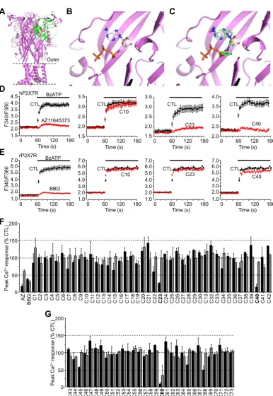
Comparison of the ATP-bound zfP2X4R structure and the hP2X7R homology model reveals 62% amino acid sequence identity between these receptors within the ATP-binding pocket in a 10 Å sphere centred on the bound ATP molecule (Fig. 1A–B). The essen-tial nature of the ATP binding pocket and its non-traditional fold in the hP2X7R made it a suitable target to perform virtual screening of the ZINC12 database, which contains approximately 100,000 structurally diverse compounds[20]. Each compound was docked individually into the ATP-binding pocket using eHiTS (e.g.,
Fig. 1C) with a corresponding predicted energy binding score being calculated. For the 50 compounds with the highest scores, 42 were commercially available and had predicted energy binding scores ranging from 4 to 10.5 kcal/mol. These values are comparable to those predicted for known hP2X7R antagonists including AZ11645373 ( 10.8 kcal/mol), SB203580 ( 8.75 kcal/mol) and KN-62 ( 5.2 kcal/mol) as reported in our recent study[6]. To ini-tially test the top 42 compounds we applied the compounds at 10
l
M to determine their effects on Ca2+responses in HEK293 cells expressing hP2X7R induced by 300l
M BzATP, a structural ana-logue of ATP which is more potent than ATP at the P2X7R and is predicted to bind to the ATP-binding site (data not shown). None of the compounds showed detectable agonist activity. Twocom-pounds, ZINC67825876 (C23 from here onward) and
ZINC58368839 (C40), inhibited BzATP-induced Ca2+responses by 73.2 ± 2% and 84.3 ± 7% respectively, whilst all other compounds had no or modest effect, as illustrated by ZINC19868610 (C10) (Fig. 1D and F). The inhibition by C23 and C40 was similar to that by BBG (71.5 ± 5%) and AZ11645373 (81.9 ± 5%) (Fig. 1D and F). These 42 compounds were also tested against BzATP-induced Ca2+ responses in HEK293 cells expressing the rP2X7R (Fig. 1E and G). BBG was used as a positive control and strongly inhibited BzATP-induced Ca2+responses, whereas AZ11645373 was far less effective (Fig. 1F). None of the compounds caused significant inhi-bition of the rP2X7R, including C23 and C40 (Fig. 1E and F). Exam-ination of C23 and C40 reveals noticeable similarities as well as substantial differences in their structures (Table 1). A number of additional compounds with a high level of structural similarity (P80%) were identified from the ZINC12 database using the ZINC12 website search function. The top 31 compounds from this new search were tested at 10
l
M against the human and rat P2X7R using FlexStation measurements of BzATP-induced Ca2+responses. ZINC09315614 (C60) almost completely ablated
A C
0 60 120 180 1.5
2.0 2.5 3.0 3.5
Time (s)
2.0 2.5 3.0 3.5 4.0
1.5 2.0 2.5 3.0 3.5
1.5 2.0 2.5 3.0 3.5 4.0 4.5
0 60 120 180 Time (s)
F340/F380
0 60 120 180
Time (s) 0 60Time (s) 120 180 CTL
C23 C40
AZ11645373
C10
D
BzATP
CTL CTL CTL
F340/F380
CTL
BBG
1.0 2.0 3.0 4.0 5.0 6.0 7.0
Time (s) 0 60 120 180
C10 CTL
1.0 2.0 3.0 4.0 5.0 6.0 7.0
Time (s) 0 60 120 180
C23 CTL
1.0 2.0 3.0 4.0 5.0 6.0 7.0
Time (s) 0 60 120 180
C40 CTL
1.0 2.0 3.0 4.0 5.0 6.0 7.0
Time (s) 0 60 120 180
E
hP2X7R
rP2X7R BzATP
B
Outer
F
C43 C44 C45 C46 C47 C48 C49 C50 C51 C52 C53 C54 C55 C56 C57 C58 C59 C60 C61 C62 C63 C64 C65 C66 C67 C68 C69 C70 C71 C72 C73 0
50 100 150 200
Peak
Ca
2+ response
(%
CT
L)
G
0 50 100 150 200
Peak
Ca
2+ response
(%
CT
L)
AZ C1 C2 C3 C4 C5 C6 C7 C8 C9 C10 1 C1 C12 C13 C14 C15 C16 C17 C18 C19 C20 C21 C22 C23 C24 C25 C26 C27 C28 C29 C30 C13 C32 C33 C34 C35 C36 C37 C38 C39 C40 C41 C42
[image:4.595.109.496.58.619.2]BBG
Fig. 1.Three compounds identified from virtual screening of the ZINC12 database that inhibit the hP2X7R.A.The trimeric hP2X7R homology model in the closed state, with an ATP molecule docked to one of the three inter-subunit ATP-binding pockets. The 10 Å sphere ATP-binding pocket centred on the bound ATP molecule is highlighted in green.B.The predicted ATP binding conformation within the ATP-binding pocket in the hP2X7R.C.Docking of compound C23 (green) from the ZINC12 compound library in the ATP-binding pocket in the hP2X7R and ATP (silver) shown for comparison.D.The predicted binding BzATP conformation within the ATP-binding pocket in the hP2X7R.E–
F. Representative Flex-Station recordings of Ca2+
responses induced by 300lM BzATP in HEK293 cells expressing the hP2X7R (E) and rP2X7R (F), with the control responses shown in black and the responses in cells treated with compound C10, C23 or C40 at 10lM in red. 200 nM AZ11645373 and 10lM BBG were used as the positive control inhibiting the human and rat P2X7R respectively.G.Summary of the effects of 42 compounds on BzATP-induced Ca2+
responses mediated by the hP2X7R shown in black and the rP2X7R in grey. Results were from 8 to 12 wells of cells from 3 independent experiments.H. Summary of the effects of 31 compounds with structural similarity to C23 and C40 on BzATP-induced Ca2+
of 73 compounds tested, and cause strong inhibition of the hP2X7R.
3.2. Further pharmacological characterisation of C23, C40 and C60
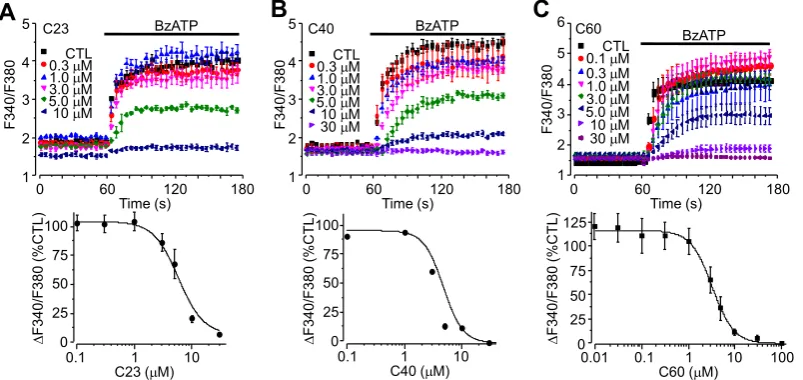
The potency of C23, C40 and C60 when inhibiting the hP2X7R was further characterised by recording BzATP-induced Ca2+ responses and currents in HEK293 cells expressing the hP2X7R. The FlexStation and patch-clamp recording were used respectively, as these methods represent the most commonly used techniques in the study of ion channel pharmacology. As illustrated inFig. 2, these three compounds inhibited Ca2+ responses induced by
300
l
M BzATP in a concentration-dependent manner, withcomplete inhibition at the highest concentrations applied (10–30
l
M). Fitting the concentration-response relationship curves to the Hill equation yielded IC50values of 5.1l
M ± 0.3l
M (n = 6), 4.8 ± 0.8l
M (n = 6) and 3.2 ± 0.2l
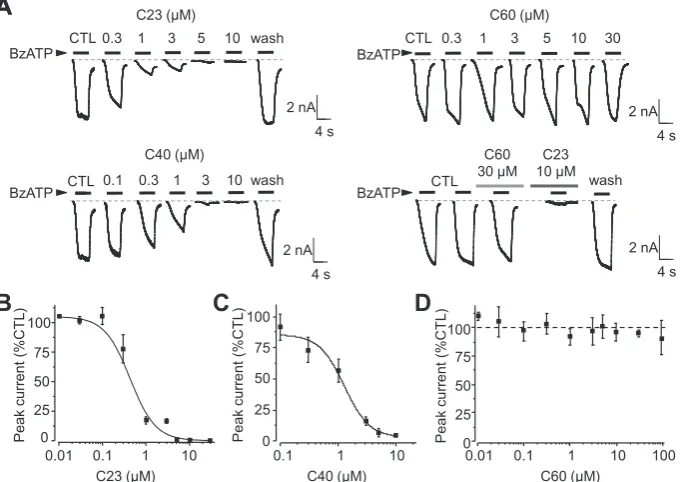
M (n = 6) for C23, C40 and C60, respectively (Fig. 2A–C). Similarly, C23 and C40 concentration-dependently inhibited BzATP-induced currents, with IC50values of 0.35 ± 0.3l
M (n = 6) and 1.2 ± 0.1l
M (n = 6) respectively (Fig. 3A–C). Current inhibition was complete and read-ily reversed upon washing (Fig. 3A). Surprisingly, despite exhibit-ing similar potency as C23 and C40 in inhibitexhibit-ing BzATP-inducedCa2+ responses, C60 caused no inhibition of BzATP-induced cur-rents even when applied at 30–100
l
M (Fig. 3A and D). BzATP-induced currents in the same current recordings were abolished by the subsequent application of C23 (Fig. 3A). [image:5.595.30.562.85.229.2]As introduced above, prolonged P2X7R activation can induce large pore formation. This can be visualised under physiological conditions by monitoring the uptake of fluorescent dyes with large molecular weights such as YO-PRO-1 (375 Da)[44]. C23, C40 and C60 were further investigated to see if they inhibited P2X7R-dependent large pore formation using FlexStation to measure YO-PRO-1 uptake in hP2X7R-expressing HEK293 cells. C23, C40 and C60 concentration-dependently inhibited YO-PRO-1 uptake induced by 300
l
M BzATP, with IC50 values of 1.8 ± 0.9l
M (n = 12), 1.0 ± 0.1l
M (n = 12) and 0.8 ± 0.2l
M (n = 16), respec-tively (Fig. 4A–C). We also used single cell imaging to examine the effect of C60 on ATP-induced hP2X7R-dependent dye uptake. The percentage of YO-PRO-1 positive cells was increased by >4 times upon treatment with 1 mM ATP for 30 min (14.7 ± 4.0% and 63.0 ± 11.7% in control cells and ATP-treated cells, n = 3, p < 0.01). The percentage of YO-PRO-1 positive cells in ATP-treated cells was significantly reduced by treatment with 5l
M C60 (33.8 ± 5.6%, n = 3, p < 0.001) but not affected by treatment with 10l
M C10 (69.2 ± 3.2%, n = 3; p > 0.05). These results provideFig. 2.Concentration-dependent effects of C23, C40 and C60 on BzATP-induced Ca2+
[image:5.595.93.495.523.713.2]responses in hP2X7R-expressing HEK293 cells.Top, representative Ca2+ responses induced by 300lM BzATP in control cells or cells treated with C23 (A), C40 (B) and C60 (C) at the indicated concentrations.Bottom, concentration-Ca2+response relationship curves with the solid lines representing the fit of the mean data to Hill equation. Each data point represents the mean from 8 to 12 wells of cells from 3 to 5 independent experiments.
Table 1
Summary of chemical and pharmacological properties of novel hP2X7R antagonists.
Compound ID ZINC12 database ID Chemical structure IC50(lM) (Ca2+
response)
IC50(lM) (current)
IC50(lM) (dye uptake)
C23 ZINC 67825876 5.1 ± 0.3 0.35 ± 0.3 1.8 ± 0.9
C40 ZINC 58368839 4.8 ± 0.8 1.2 ± 0.1 1.0 ± 0.1
further evidence to support selective inhibition by C60 of hP2X7R-dependent large pore formation.
Taken together, these results provide clear evidence to show that C23 and C40 inhibit both hP2X7R ion channel activation and large pore formation but C60 preferentially targets large pore for-mation without effect on the ion channel function.
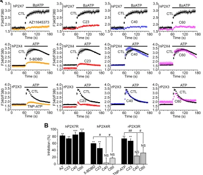
3.3. Effects of C23, C40 and C60 on other P2XRs
C23, C40 and C60 were examined next for their specificity by determining their effects on the other members of the P2X receptor
family, particularly the hP2X4R. The P2X4R has the highest level of sequence identity with the P2X7R over the whole receptor and also at the ATP-binding pocket used in virtual screening and, further-more, the two receptors co-exist in many cell types. C23 applied at 10
l
M strongly reduced 100l
M ATP-induced Ca2+ responses in hP2X4R-expressing HEK293 cells in a manner comparable to the P2X4R antagonist 5-BDBD (Fig. 5A and B). However, both C40 and C60 also applied at 10l
M resulted in no significant effect (Fig. 5A and B). The three compounds were further tested in rP2X3R-expressing HEK293 cells, in which 100l
M ATP induced Ca2+responses with transient kinetics (Fig. 5A) due to the strongD
0.01 0.1 1 10 0 25 50 75 100 Peak curr ent (%CT L ) 0 25 50 75 100
0.1 1 10
--Peak curr ent (%C T L ) 0 25 50 75 100
0.01 0.1 1 10 100
Peak curr ent (%CT L )
A
B C
C23 (µM) C40 (µM) C60 (µM)
A
BzATP wash C23 (µM) C40 (µM) C60 (µM)CTL 0.3 1 3 5 10 30 BzATP
BzATP BzATP
CTL 0.3 1 3 5 10 wash
[image:6.595.133.475.71.312.2]CTL 0.1 0.3 1 3 10 wash CTL C60 30 µM C23 10 µM 2 nA 4 s 2 nA 4 s 2 nA 4 s 2 nA 4 s
Fig. 3.Concentration-dependent effects of C23, C40 and C60 on BzATP-induced currents in hP2X7R-expressing HEK293 cells.A.Representative recordings of currents induced by 4 s application of 300lM BzATP in cells before (CTL) and after exposure to C23, C40 and C60 at the indicated concentrations. Note that the current inhibition by C23 and C40 was reversed upon washing, and the current was not inhibited by C60 but abolished by subsequent application of C23.B–D.Concentration-current response relationship curves for C23 (B), C40 (C) and C60 (D). The solid lines in B and C represent the fit of the mean data to the Hill equation. Each data point represents mean from 4 cells from 3 to 4 independent experiments.
C
A B
0 60 120
4
Time (s)
0.3 M µ µ µ µ µ µ µ µ µ µ µ µ µ µ µ µ µ µ 1.0 M 3.0 M 5.0 M 10 M CTL 30 M 10 14 BzATP F490/F510 6 8 12
0 60 120
4 Time (s) 0.3 M 1.0 M 3.0 M 5.0 M 10 M CTL 30 M 10 14 BzATP F490/F510 6 8 12
0 60 120
4 Time (s) 0.3 M 1.0 M 3.0 M 5.0 M 10 M CTL 30 M 10 14 BzATP F490/F510 6 8 12 Y O -PRO-1 uptake (% CT L) Y O -PRO-1 uptake (% CT L) 0 25 50 75 100 125
0.1 1 10
C23 (µM) Y O -PRO-1 uptake (% CT L) 0 25 50 75 100 125
0.1 1 10
C40 (µM) 0 25 50 75 100 125
0.1 1 10
C60 (µM)
[image:6.595.99.506.383.583.2]desensitisation characteristic of this receptor [32]. C23 was also effective at 10
l
M in suppressing rP2X3R-mediated ATP-induced Ca2+responses in a manner similar to the P2X3R antagonist TNP-ATP (Fig. 3A and B). Both C40 and C60 caused smaller but statisti-cally insignificant inhibition (Fig. 5A and B). Therefore, whilst C23 is largely indiscriminate, both C40 and C60 exhibit much greater specificity towards the hP2X7R.3.4. Inhibition of P2X7R-mediated cell death
The P2X7R is a well-known cytolytic receptor which plays a critical role in mediating ATP-induced cell death in vivo under pathological conditions as well as under in vitro experimental con-ditions, a unique functional role which distinguishes the P2X7R from all the other P2X receptors [22]. As such, inhibition of the P2X7R offers protection against ATP-mediated cell death in a num-ber of cells expressing the P2X7R[12,30,36]. Therefore, C23, C40 and C60 compounds were finally tested for their effectiveness in preventing ATP-induced cell death in hP2X7R-expressing HEK293 cells to provide further supporting evidence for their action on the hP2X7R. Exposure of cells to 3 mM ATP for 6 h caused cell death by 74 ± 7% (n = 12) as determined by PI staining assays (Fig. 6A and B). Pre-treatment with C23 at 3 and 10
l
M, C40 at 3 and 10l
M or C60 at 10 and 30l
M strongly reducedATP-induced cell death by approximately or >50% (Fig. 6A and B). These results provide further evidence to support the notion that com-pounds identified in this study inhibit the hP2X7R.
4. Discussion
Studies over the past decade have revealed a crucial role for the P2X7R in a range of debilitating disease conditions[23]. Emerging evidence supports the hP2X7R as a druggable target[10]. In order to exploit the full potential of hP2X7R antagonists as therapeutics, more efforts are required to increase the structural diversity of hP2X7R antagonists as well as to better understand the underlying disease mechanisms. All currently known P2X7R antagonists have been identified through the random functional screening of large compound libraries, which can be time-consuming, costly and inefficient. Using virtual screening of a large set of compounds against the hP2X7R ATP-binding pocket followed by functional assays, the present study identified two hits (C23 and C40) (Fig. 1D and F). A further 31 compounds were generated based on structural similarities to C23 and C40 which identified a third compound, C60 (Fig. 1G). Detailed functional characterisations, by determining their effects on hP2X7R-mediated Ca2+responses, currents and YO-PRO-1 uptake, consistently indicate micromolar potency at the hP2X7R, albeit with some variations that are likely
0 60 120 180
1.5 2.0 2.5 3.0 3.5
Time (s) 0 60 120 180
1.5 2.0 2.5 3.0 3.5
Time (s) 0 60 120 180
1.5 2.0 2.5 3.0 3.5 Time (s) 1.5 2.0 2.5 3.0 3.5 1.0 4.0 1.5 2.0 2.5 3.0 3.5 1.0 4.0 1.5 2.0 2.5 3.0 3.5 1.0 4.0 1.5 2.0 2.5 3.0 3.5 1.0 4.0 1.5 2.0 2.5 3.0 3.5 1.0 4.0 1.5 2.0 2.5 3.0 3.5 1.0 4.0
0 60 120 180
Time (s) 0 60Time (s) 120 180 0 60Time (s) 120 180
0 60 120 180
Time (s) 0 60Time (s) 120 180 0 60Time (s) 120 180 C23 CTL hP2X7 C23 CTL C23 CTL C40 CTL C40 CTL C40 CTL C60 CTL C60 CTL C60 CTL hP2X7
BzATP BzATP hP2X7 BzATP
hP2X4 hP2X4 ATP ATP hP2X4 ATP
rP2X3 rP2X3 ATP ATP rP2X3 ATP
F340/F380
F340/F380
F340/F380
A
0 60 120 180
1.5 2.0 2.5 3.0 3.5 Time (s) F340/F380
hP2X7 BzATP
AZ11645373 CTL 1.5 2.0 2.5 3.0 3.5 1.0 4.0
0 60 120 180
Time (s) 5-BDBD CTL hP2X4 ATP
F340/F380 1.5 2.0 2.5 3.0 3.5 1.0 4.0
0 60 120 180
Time (s) CTL rP2X3 ATP
F340/F380 TNP-ATP
B
0 20 40 60 80 100 Inhibition (%)hP2X7R hP2X4R rP2X3R
** ** *** *** ** ** ** ** # # ## #
NS NS NS
[image:7.595.92.501.73.426.2]NS
Fig. 5.Effects of C23, C40 and C60 on agonist-induced Ca2+responses in HEK293 cells expressing different P2X receptors.A.Representative recording of Ca2+responses in HEK293 cells expressing hP2X7R, hP2X4R and rP2X3R evoked by 100lM BzATP and 100lM ATP respectively in the absence and presence of 10lM C23, C40 C60, AZ116435373, 5-BDBD and TNP-ATP. Note the distinct kinetics of the Ca2+
related to the functional assays used (Figs. 2–4). Moreover, all three compounds effectively protected against ATP-induced cell death (Fig. 6), providing further evidence to support that they antagonise the hP2X7R. Subtype specificity is of great importance when using antagonists in the study of the role of the hP2X7R in ATP-induced physiological functions and diseases as well as for the development of therapeutic compounds. Of these three com-pounds, C40 and C60 exhibited significant specificity towards the hP2X7R over the other P2X receptors examined (Fig. 5), including the hP2X4R which shares the highest level of sequence identity with the hP2X7R. Thus, it is unlikely but remains to be confirmed that C40 and C60 cause potent inhibition at the other P2X recep-tors with less structural similarity to the hP2X7R. Evidently, structure-activity relationship studies are required for structural optimisation of these compounds in order to further improve their potency and specificity. Nonetheless, the present study is the first to identify structurally novel P2X7R antagonists using a structure-based approach, which can be more time-saving and cost-effective than the conventional screening approach.
It has been well-documented that prolonged or repeated activa-tion of the P2X7R induces large pore formaactiva-tion, but it still remains a subject of debate if this is through P2X7R itself or via the activa-tion of a distinctive protein which forms the large pore[32,44,21]. Regardless of the underlying mechanisms, recent studies show that P2X7R-dependent large pore functionality is of high relevance to the pathogenesis of diseases such as chronic pain[37], osteoporo-sis[43], and aged-related macular degeneration responsible for the loss of vision in elderly people[14]. Emerging evidence supports that blocking this pore is a valuable therapeutic intervention
[14]. None of the currently available P2X7R antagonists effectively discriminate between the ion channel and large pore functions. In this regard, C60 is of interest as it is the first reported P2X7R antag-onist that selectively prevents large pore formation (Fig. 4) without affecting the ion channel function (Fig. 3). C60 will be useful as a pharmacological tool to better understand large pore formation and its role in P2X7R-mediated physiological functions and
dis-eases. It may also act as a potential lead compound for development of therapeutics targeting this pivotal pore-forming function.
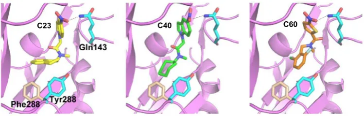
C23, C40 and C60 all contain an N-methyl amide group in their structure, with a prominent NO2group at one end and a bulky ring structure at the opposite end (Table 1), thus suggesting that they represent a class of new hP2X7R antagonists with a structurally conserved pharmacophore. Preliminary studies show that replace-ment of the NO2group in C40 with NH2retains activity, whereas substitution with CH3 reduces inhibition of hP2X7R-mediated Ca2+responses (data not shown). This suggests that nitrogen is a vital component of the compound structure which is possibly required for hydrogen bonding with residues such as Gln143 in the ATP-binding pocket (Fig. 7). A compound containing a benzene ring structure in replacement of the bulky ring structure in C23 and C40 is ineffective in inhibiting hP2X7R-mediated Ca2+ responses (data not shown), implying that the bulky ring structure is pivotal in interacting with the hP2X7R, particularly Tyr288. Furthermore, assessment of the compound structures predicts the central com-mon moiety is a relatively flexible linker, allowing antagonists to adopt a bent configuration to facilitate interactions with the recep-tor (Fig. 7). Finally, all three compounds identified in this study preferentially inhibit the hP2X7R over the rP2X7R (Fig. 1D–G). Such striking species specificity has been repeatedly reported for known hP2X7R antagonists such as KN62[19], AZ11645373[40]
and SB203580 [31]. Examination of ligand-receptor interactions has led us to hypothesise that the species specificity of C23, C40 and C60 arises from structural differences in the ATP-binding pocket such as Tyr288. This residue in the hP2X7R flips further into the ATP-binding pocket and its orientation is expected to facilitate, whereas Phe288 in the rP2X7R is positioned in a way that is pre-dicted to disfavour interactions with the antagonists, specifically the bulky ring structure discussed above (Fig. 7). Further site-directed mutagenesis studies will inform the importance of these parts in the receptor including Gln143 and Tyr288 in determining antagonist binding.
A
CTL 3 mM ATP ATP+10 µM C23 ATP+10 µM C40 ATP+10 µM C60
B
0 20 40 60 80 100
Cell death
(%)
** ++ ++ ++ ++ ++ ++
In summary, the present study identifies for the first time struc-turally novel hP2X7R antagonists using a structure-based approach combining virtual screening and functional testing. It reports the first P2X7R antagonist with preferential inhibition of the large pore formation without effect on the ion channel function.
Author contributions
E.A.C., S.P.M. and L.H.J. conceived and designed the research. E. A.C. performed the experiments, E.A.C. and L.H.J. analysed the data. S.P.M. and C.W.F. contributed to intellectual inputs, E.A.C. S.P.M. and L.H.J. wrote and revised the manuscript.
Conflicts of interest
The authors state no conflict of interest.
Acknowledgements
This work was supported by the Wellcome Trust. We are grate-ful to Mrs Jocelyn Baldwin for instruction of homology modelling, and to Dr Katie Simmons and Dr Martin McPhillie for valuable assistance with virtual screening and analysis of compound-receptor interactions.
References
[1]R. Bartlett, L. Stokes, R. Sluyter, The P2X7 receptor channel: recent
developments and the use of P2X7 antagonists in models of disease,
Pharmacol. Rev. 66 (2014) 638–675.
[2]H.J. Bradley, JM. Baldwin, G.R. Goli, B. Johnson, J. Zou, A. Sivaprasadarao, S.A.
Baldwin, L.-H. Jiang, Residues 155 and 348 contribute to the determination of P2X7 receptor function via distinct mechanisms revealed by single-nucleotide
polymorphisms, J. Biol. Chem. 286 (2011) 8176–8187.
[3]H.J. Bradley, L.E. Browne, W. Yang, L.H. Jiang, Pharmacological properties of the
rhesus macaque monkey P2X7 receptor, Br. J. Pharmacol. 164 (2011) 743–754.
[4]L.E. Browne, L.-H. Jiang, R.A. North, New structure enlivens interest in P2X
receptors, Trends Pharmacol. Sci. 31 (2010) 229–237.
[5]S. Cankurtaran-Sayar, K. Sayar, M. Ugur, P2X7 receptor activates multiple
selective dye-permeation pathways in RAW 264.7 and human embryonic
kidney 293 cells, Mol. Pharmacol. 76 (2009) 1323–1332.
[6]E.A. Caseley, S.P. Muench, S.A. Baldwin, K. Simmons, C.W. Fishwick, L.-H. Jiang,
Docking of competitive inhibitors to the P2X7 receptor family reveals key differences responsible for changes in response between rat and human,
Bioorg. Med. Chem. Lett. 25 (2015) 3164–3167.
[7]E.A. Caseley, S.P. Muench, S. Roger, H.-J. Mao, S.A. Baldwin, L.-H. Jiang,
Non-synonymous single nucleotide polymorphisms in the P2X receptor genes: association with diseases, impact on receptor functions and potential use as
diagnosis biomarkers, Int. J. Mol. Sci. 15 (2014) 13344–13371.
[8]I.W. Davis, A. Leaver-Fay, V.B. Chen, J.N. Block, G.J. Kapral, X. Wang, L.W.
Murray, W.B. Arendall, J. Snoeyink, J.S. Richardson, MolProbity: all-atom contacts and structure validation for proteins and nucleic acids, Nucleic Acids
Res. 35 (2007) W375–W383.
[9]W.L. Delano, The PyMOL molecular graphics system, 2002.
[10]A. Eser, J.-F. Colombel, P. Rutgeerts, S. Vermeire, H. Vogelsang, M. Braddock, T.
Persson, W. Reinisch, Safety and efficacy of an oral inhibitor of the purinergic receptor P2X7 in adult patients with moderately to severely active Crohn’s disease: a randomized placebo-controlled, double-blind, phase IIa study,
Inflamm. Bowel Dis. 21 (2015) 2247–2253.
[11]N. Eswar, B. Webb, M.A. Marti-renom, M. Madhusudhan, D. Eramian, M.Y.
Shen, U. Pieper, A. Sali, Comparative protein structure modeling using
Modeller, Current Protocols in Bioinformatics (2006) 5.6.1–5.6.30.
[12]U.B. Eyo, S.A. Miner, K.E. Ahlers, L.-J. Wu, M.E. Dailey, P2X7 receptor activation
regulates microglial cell death during oxygen-glucose deprivation,
Neuropharmacology 73 (2013) 311–319.
[13]A. Fiser, R.K.G. Do, A. Šali, Modeling of loops in protein structures, Protein Sci. 9
(2000) 1753–1773.
[14]B.J. Fowler, B.D. Gelfand, Y. Kim, N. Kerur, V. Tarallo, Y. Hirano, S. Amarnath, D.
H. Fowler, M. Radwan, M.T. Young, Nucleoside reverse transcriptase inhibitors
possess intrinsic anti-inflammatory activity, Science 346 (2014) 1000–1003.
[15]M. Garcia-Guzman, F. Soto, J.M. Gomez-Hernandez, P.-E. Lund, W. Stühmer,
Characterization of recombinant human P2X4 receptor reveals pharmacological
differences to the rat homologue, Mol. Pharmacol. 51 (1997) 109–118.
[16]V.J. Gillet, G. Myatt, Z. Zsoldos, A.P. Johnson, SPROUT, HIPPO and CAESA: tools
for de novo structure generation and estimation of synthetic accessibility,
Perspect. Drug Discovery Des. 3 (1995) 34–50.
[17]S.D. Guile, L. Alcaraz, T.N. Birkinshaw, K.C. Bowers, M.R. Ebden, M. Furber, M.J.
Stocks, Antagonists of the P2X7 receptor. From lead identification to drug
development, J. Med. Chem. 52 (2009) 3123–3141.
[18]M. Hattori, E. Gouaux, Molecular mechanism of ATP binding and ion channel
activation in P2X receptors, Nature 485 (2012) 207–212.
[19]B.D. Humphreys, C. Virginio, A. Surprenant, J. Rice, G.R. Dubyak, Isoquinolines
as antagonists of the P2X7 nucleotide receptor: high selectivity for the human
versus rat receptor homologues, Mol. Pharmacol. 54 (1998) 22–32.
[20]J.J. Irwin, T. Sterling, M.M. Mysinger, E.S. Bolstad, R.G. Coleman, ZINC: a free tool
to discover chemistry for biology, J. Chem. Inf. Model. 52 (2012) 1757–1768.
[21]B. Jelassi, A. Chantome, F. Alcaraz-Perez, A. Baroja-Mazo, M. Cayuela, P.
Pelegrin, A. Surprenant, S. Roger, P2X7 receptor activation enhances SK3
channels-and cystein cathepsin-dependent cancer cells invasiveness,
Oncogene 30 (2011) 2108–2122.
[22]L.-H. Jiang, Inhibition of P2X7 receptors by divalent cations: old action and
new insight, Eur. Biophys. J. 38 (2009) 339–346.
[23]L.-H. Jiang, P2X receptormediated ATP purinergic signalling in health and
disease, Cell Health Cytoskelet 4 (2012) 83–101.
[24]L.-H. Jiang, J.M. Baldwin, S. Roger, S.A. Baldwin, Insights into the molecular
mechanisms underlying mammalian P2X7 receptor functions and
contributions in diseases, revealed by structural modeling and single
nucleotide polymorphisms, Front. Pharmacol. 4 (2013).
[25]L.-H. Jiang, F. Rassendren, A. Mackenzie, Y.-H. Zhang, A. Surprenant, R.A. North,
N-methyl-D-glucamine and propidium dyes utilize different permeation pathways at rat P2X7 receptors, Am. J. Physiol. Cell Physiol. 289 (2005)
C1295–C1302.
[26]E.C. Keystone, M.M. Wang, M. Layton, S. Hollis, I.B. Mcinnes, Clinical evaluation
of the efficacy of the P2X7 purinergic receptor antagonist AZD9056 on the signs and symptoms of rheumatoid arthritis in patients with active disease despite treatment with methotrexate or sulphasalazine, Ann. Rheum. Dis.
(2011) 143578.
[27]C. Lambertucci, D. dal Ben, M. Buccioni, G. Marucci, A. Thomas, R. Volpini,
Medicinal chemistry of P2X receptors: agonists and orthosteric antagonists,
Curr. Med. Chem. 22 (2015) 915–928.
[28]C. Lewis, S. Neidhart, C. Holy, R. North, G. Buell, A. Surprenant, Coexpression of
P2X2 and P2X3 receptor subunits can account for ATP-gated currents in
sensory neurons, Nature 377 (1995) 432–435.
[29]L. Liu, J. Zou, X. Liu, L.-H. Jiang, J. Li, Inhibition of ATP-induced macrophage
death by emodin via antagonizing P2X 7 receptor, Eur. J. Pharmacol. 640
(2010) 15–19.
[image:9.595.107.482.67.187.2]C23 C40 C60
[30]J. Luo, S. Lee, D. Wu, J. Yeh, H. Ellamushi, A. Wheeler, G. Warnes, Y. Zhang, X. Bo, P2X7 purinoceptors contribute to the death of Schwann cells transplanted into
the spinal cord, Cell Death Disease 4 (2013) e829.
[31]A. Michel, K. Thompson, J. Simon, I. Boyfield, E. Fonfria, P. Humphrey, Species
and response dependent differences in the effects of MAPK inhibitors on P2X7
receptor function, Br. J. Pharmacol. 149 (2006) 948–957.
[32]R.A. North, Molecular physiology of P2X receptors, Physiol. Rev. 82 (2002)
1013–1067.
[33]R.A. North, M.F. Jarvis, P2X receptors as drug targets, Mol. Pharmacol. 83
(2013) 759–769.
[34]F. Rassendren, G.N. Buell, C. Virginio, G. Collo, R.A. North, A. Surprenant, The
permeabilizing ATP receptor, P2X7 cloning and expression of a human cDNA, J.
Biol. Chem. 272 (1997) 5482–5486.
[35]S. Roger, Z.-Z. Mei, J.M. Baldwin, L. Dong, H. Bradley, S.A. Baldwin, A.
Surprenant, L.-H. Jiang, Single nucleotide polymorphisms that were
identified in affective mood disorders affect ATP-activated P2X 7 receptor
functions, J. Psychiatr. Res. 44 (2010) 347–355.
[36]E. Salas, L.M.G. Carrasquero, L.A. Olivos-Oré, D. Bustillo, A.R. Artalejo, M.T.
Miras-Portugal, E.G. Delicado, Purinergic P2X7 receptors mediate cell death in mouse cerebellar astrocytes in culture, J. Pharmacol. Exp. Ther. 347 (2013)
802–815.
[37]R.E. Sorge, T. Trang, R. Dorfman, S.B. Smith, S. Beggs, J. Ritchie, J.-S. Austin, D.V.
Zaykin, H. Vander meulen, M. Costigan, Genetically determined P2X7 receptor pore formation regulates variability in chronic pain sensitivity, Nat. Med. 18
(2012) 595–599.
[38]B. Sperlágh, P. Illes, P2X7 receptor: an emerging target in central nervous
system diseases, Trends Pharmacol. Sci. 35 (2014) 537–547.
[39]T.C. Stock, B.J. Bloom, N. Wei, S. Ishaq, W. Park, X. Wang, P. Gupta, C.A. Mebus,
Efficacy and safety of CE-224,535, an antagonist of P2X7 receptor, in treatment
of patients with rheumatoid arthritis inadequately controlled by
methotrexate, J. Rheumatol. 39 (2012) 720–727.
[40] L. Stokes, L.H. Jiang, L. Alcaraz, J. Bent, K. Bowers, M. Fagura, M. Furber, M.
Mortimore, M. Lawson, J. Theaker, Characterization of a selective and potent antagonist of human P2X7 receptors, AZ11645373, Br. J. Pharmacol. 149
(2006) 880–887.
[41]C. Subramanyam, A.J. Duplantier, M.A. Dombroski, S.-P. Chang, C.A. Gabel, C.
Whitney-Pickett, D.G. Perregaux, J.M. Labasi, K. Yoon, R.M. Shepard, Discovery, synthesis and SAR of azinyl-and azolylbenzamides antagonists of the P2X 7
receptor, Bioorg. Med. Chem. Lett. 21 (2011) 5475–5479.
[42]A. Surprenant, F. Rassendren, E. Kawashima, R. North, G. Buell, The cytolytic
P2Z receptor for extracellular ATP identified as a P2X receptor (P2X7), Science
272 (1996) 735–738.
[43]S. Syberg, P. Schwarz, S. Petersen, T.H. Steinberg, J.-E.B. Jensen, J. Teilmann, N.R.
Jørgensen, Association between P2X7 receptor polymorphisms and bone
status in mice, J. Osteoporosis 2012 (2012).
[44]L. Wei, E. Caseley, D. Li, L.-H. Jiang, ATP-induced P2X receptor-dependent large
pore formation: How Much Do We Know? Front. Pharmacol. 7 (2016).
[45]Z. Zsoldos, D. Reid, A. Simon, S.B. Sadjad, A.P. Johnson, EHiTS: a new fast,
exhaustive flexible ligand docking system, J. Mol. Graph. Model. 26 (2007)