R E V I E W
Open Access
Tick-borne pathogens and the vector potential of
ticks in China
Zhijun Yu, Hui Wang, Tianhong Wang, Wenying Sun, Xiaolong Yang and Jingze Liu
*Abstract
Ticks, as obligate blood-sucking ectoparasites, attack a broad range of vertebrates and transmit a great diversity of
pathogenic microorganisms. They are considered second only to mosquitoes as vectors of human disease, and the
most important vector of pathogens of domestic and wild animals. Of the 117 described species in the Chinese tick
fauna, 60 are known to transmit one or more diseases: 36 species isolated within China and 24 species isolated
outside China. Moreover, 38 of these species carry multiple pathogens, indicating the potentially vast role of these
vectors in transmitting pathogens. Spotted fever is the most common tick-borne disease, and is carried by at least
27 tick species, with Lyme disease and human granulocytic anaplasmosis ranked as the second and third most
widespread tick-borne diseases, carried by 13 and 10 species, respectively. Such knowledge provides us with clues
for the identification of tick-associated pathogens and suggests ideas for the control of tick-borne diseases in China.
However, the numbers of tick-associated pathogens and tick-borne diseases in China are probably underestimated
because of the complex distribution and great diversity of tick species in this country.
Keywords:
Ticks, Tick-borne pathogens, Vector potential, China
Review
Ticks, as obligate blood-sucking ectoparasites, attack a
broad range of vertebrates, including humans, and they
are considered second only to mosquitoes as vectors of
human disease, and the most important vector of
patho-gens of domestic and wild animals [1]. They transmit a
variety of pathogens of medical and veterinary interest,
including viruses, bacteria, rickettsiae, helminthes, and
protozoans, all of which are able to cause damage to
live-stock production and human health. The global threat of
tick-borne diseases is increasing, with new pathogens
identified continuously [2]. There are an estimated 899
species of ticks belonging to three families: Argasidae,
Ixodidae, and Nuttalliellidae (represented by a
mono-typic species restricted to South Africa) [3].
In China, 117 species of the following genera have
been identified:
Argas
(seven species),
Carios
(four species),
and
Ornithodoros
(two species) in the family Argasidae;
and
Amblyomma
(eight species),
Anomalohimalaya
(two
species),
Dermacentor
(twelve species),
Haemaphysalis
(forty four species),
Hyalomma
(six species),
Ixodes
(twenty four species), and
Rhipicephalus
(eight species)
in the family Ixodidae [4]. Some of these species carry
or transmit one or more infectious pathogens, resulting
in severe zoonotic diseases. The most commonly observed
human tick-borne diseases in China are reportedly
Lyme disease, tick-borne encephalitis, Crimean-Congo
hemorrhagic fever, Q fever, tularemia, and North-Asia
tick-borne spotted fever [5]. Epidemiologically
import-ant tick-borne diseases, such as Human Granulocytic
Anaplasmosis (HGA) and severe Fever with
Thrombocy-topenia Syndrome (FLTS), have also emerged in recent
years. The characterization of a new bunyavirus
(associ-ated with fever, thrombocytopenia, and leukopenia
syn-drome) in 2010 has prompted greater attention to ticks
and borne diseases throughout China. However,
tick-associated pathogens and diseases are still underestimated
because of the complex distribution and the large diversity
of tick species in China.
Although the rapid development of molecular
tech-niques has greatly advanced the identification of
emer-ging tick pathogens, continuous research is required to
fully comprehend the diversity of tick-borne pathogens
and to completely identify the vector roles of ticks in
* Correspondence:[email protected]Key Laboratory of Animal Physiology, Biochemistry and Molecular Biology of Hebei Province, College of Life Sciences, Hebei Normal University, Shijiazhuang 050024, China
© 2015 Yu et al.; licensee BioMed Central. This is an Open Access article distributed under the terms of the Creative Commons Attribution License (http://creativecommons.org/licenses/by/4.0), which permits unrestricted use, distribution, and
China. In this study, with regard to the Chinese tick
fauna, we reviewed the tick-associated pathogenic
mi-croorganisms that have been identified world-wide, and
evaluated the potential roles of the ticks as vectors
throughout China. This will extend the identification of
tick-associated pathogens and suggest better strategies
for the control of tick-borne diseases in China.
Role of argasid ticks as vectors in China and their
associated tick-borne pathogens
In China, there are 13 species of argasid ticks, belonging
to three genera:
Argas
(seven species),
Carios
(four
spe-cies), and
Ornithodoros
(two species) [4]. The majority
of these are nidicolous, usually residing in the burrows,
caves, or nests of their hosts. Among all the argasids
found in China, four
Argas
species, two
Carios
species,
and two
Ornithodoros
species are competent to transmit
or cause human disease (Table 1) [6-17]. Among these
eight tick species, four (A. japonicas,
A. persicus,
O.
tarta-kovskyi, and
O. tholozani) have been confirmed as causing
host illnesses in China. A case of human dermatitis was
recorded in 1986 after a bite by
A. japonicas, but no
pathogen has been identified from this tick species in
China [6]. The tick
A. persicus
mainly infests poultry and
carries the most diverse array of pathogens in the family
Agarsidae, including
Borrelia anserine, Kyasanur Forest
disease virus, and
Wolbachia persica
n. sp. However, only
B. anserine, known to cause avian spirochetosis, has been
confirmed in China [7]. Lake Clarendon virus was isolated
from
A. robertsi; Quaranfil virus and Gissar virus were
identified in
A. vulgaris; and
“
Issyk-Kul
”
virus has been
identified in
C. vespertilionis. No virus has been detected
in ticks collected in China. The symptoms or diseases
caused by these viruses are still unclear [11-13,16], and
the vector roles of these ticks in China remain unknown.
Carios capensis
can be coinfected by pathogen DNA from
Borrelia,
Coxiella, and
Rickettsia, as well as West Nile
virus [14,15], although no pathogens have been reported
in this tick species collected in China.
Ornithodoros
tarta-kovskyi
and
O. tholozani
both cause tick-borne relapsing
fever in China, but carry different pathogens,
B. latyshevyi
and
B. persica, respectively [17].
Ixodid ticks in China, their roles as vectors, and
associated tick-borne pathogens
There are 104 species of ixodid ticks in China in seven
genera:
Amblyomma
(eight species),
Anomalohimalaya
(two species),
Dermacentor
(twelve species),
Haemaphy-salis
(forty four species),
Hyalomma
(six species),
Ixodes
(twenty four species), and
Rhipicephalus
(eight species)
[4]. Of these, 52 species from six genera have been
shown to carry or transmit pathogenic microorganisms:
Ixodes
(seven species),
Amblyomma
(three species),
Der-macentor
(nine species),
Haemaphysalis
(twenty one
spe-cies),
Hyalomma
(five species), and
Rhipicephalus
(seven
species) (Table 2) [18-114]. Of these 52 species, 32 occur
in China (Table 2). Tick-borne spotted fever is the most
commonly detected disease, carried by at least 27 tick
spe-cies. Lyme disease and human granulocytic anaplasmosis
are the second and third most widespread tick-borne
diseases, transmitted by at least 13 and 10 tick species,
respectively (Table 2). Eight tick species are vectors for
human granulocytic ehrlichiosis, seven tick species carry
tick-borne encephalitis and babesiosis, and six species
transmit hemorrhagic fever. The ixodid ticks that act as
vectors of
Babesia
are usually coinfected with more than
one
Babesia
species. These ticks include
I. persulcatus,
D. nuttalli,
Rh. microplus, and
Rh. haemaphysaloides,
which are often infected by
Babesia bigemina
and
Ba.
[image:2.595.56.540.539.718.2]bovis
(Table 2).
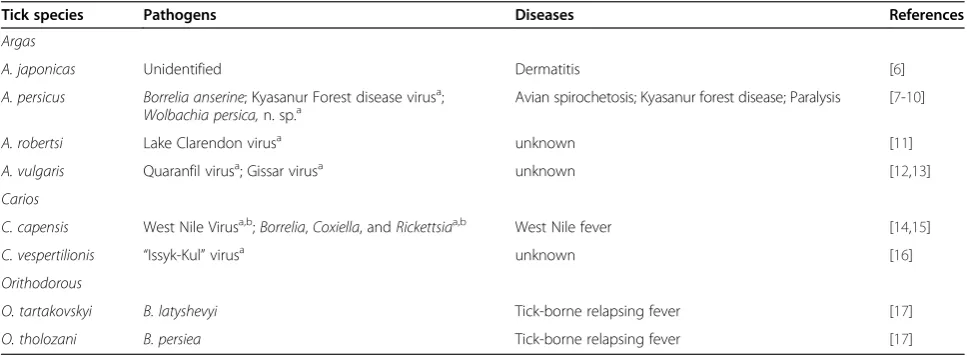
Table 1 Tick-borne pathogens and the vector role of argasid ticks distributed in China
Tick species Pathogens Diseases References
Argas
A. japonicas Unidentified Dermatitis [6]
A. persicus Borrelia anserine; Kyasanur Forest disease virusa;
Wolbachia persica,n. sp.a Avian spirochetosis; Kyasanur forest disease; Paralysis [7-10]
A. robertsi Lake Clarendon virusa unknown [11]
A. vulgaris Quaranfil virusa; Gissar virusa unknown [12,13]
Carios
C. capensis West Nile Virusa,b;Borrelia,Coxiella, andRickettsiaa,b West Nile fever [14,15]
C. vespertilionis “Issyk-Kul”virusa unknown [16]
Orithodorous
O. tartakovskyi B. latyshevyi Tick-borne relapsing fever [17]
O. tholozani B. persiea Tick-borne relapsing fever [17]
a
These pathogenic microorganisms have been recorded outside China. b
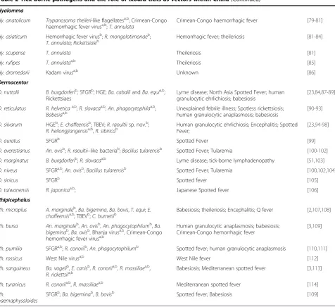
Table 2 Tick-borne pathogens and the role of ixodid ticks as vectors within China
Tick species Pathogens Diseases References
Ixodes
I. persulcatus B. burgdorferi; human granulocytcEhrlichia(HGE)b; Spotted Fever GroupRickettsia(SFGR)b;Ehrlichiab;
Anaplasma phagocytophilab; tick-borne Encephalitis virus (TBEV);Babesia bigemina,Ba. bovis
Lyme disease; Ehrlichiosis; spotted fever; human granulocytic anaplasmosis; babesiosis
[18-25]
I. kazakstani B. burgdorferia Lyme disease [26]
I. nipponensis B. afzeliia,b; TBEVa,b Lyme disease; tick-borne encephalitis; [27,28]
I. ovatus Ehrlichiaa,b; TBEV;R. japonicaa,b Ehrlichiosis; tick-borne encephalitis; Oriental spotted
fever
[29-31]
I. granulates B. burgdorferi Lyme disease [32]
I. acutitarsus B. burgdorferi Lyme disease [33]
I. sinensis R. monacensisb;Ehrlichia, Bartonella, and Borreliab;
B. gariniib Lyme disease; Mediterranean Spotted Fever [34]
Amblyomma
Am. geoemydae Reptile-associatedBorrelia spp.a,b;relapsing fever
Borrelia sp.a,b
Relapsing fever [35]
Am. helvolum SFGRa,b;Rickettsiasp.a,b Spotted fever [36,37]
Am. testudinarium R. tamuraesp. nov.a,b;Ehrlichia chaffeensisb; Human monocytic ehrlichiosis [38,39]
Haemaphysalis
H. longicornis New bunyavirusb;B. burodorferi;A. phagocytophilumb; SFGRa,b;Babesiaspa; Huaiyangshan virusb;Borrelia,
Bartonella,Anaplasma, andEhrlichiab;Theileria uilenbergib;
Severe fever with thrombocytopenia syndrome; Lyme disease; human granulocytic anaplasmosis; spotted fever; babesiosis; Huaiyangshan hemorrhagic fever
[40-47]
H. concinna B. gariniib; HGEb; SFGRb; TBEVb Human granulocytic Ehrlichiosis [23,48-50]
H. punctata B. burgdorferisensu strictob;Ba. majorandT. orientalis;
Crimean-Congo hemorrhagic fever virusa;Rickettsiaa,b; R. aeschlimanniia,b;An. phagocytophilumb;Flavivirusb
Lyme disease; Babesiosis; tick-borne encephalitis; Crimean-Congo hemorrhagic fever
[3,51-55]
H. cornigera R. heilongjiangensisa,b Spotted fever [31]
H. erinacei SFGRa,b Spotted fever [56]
H. flava Ehrlichiab;R. japonicaa,b EhIichiosis; Japanese Spotted fever [57,58]
H. formosensis R. asiaticasp. nov.a,b; Kyasanur Forest disease virusa,b;
R. japonicaa,b;An. phagocytophiluma,b
Spotted fever; Kyasanur Forest disease; [31,59-61]
H. hystricis An. phagocytophilumb;R. japonicaa,b Human granulocytic anaplasmosis; Japanese Spotted fever
[31,62]
H. japonica B. gariniib; TBEVa,b Lyme disease; tick-borne encephalitis [63,64]
H. kitaokai SFGRa,b Spotted Fever [65]
H. lagrangei Anaplasmaspp.a,b;Rickettsiaa,b; Human granulocytic ehrlichiosis; Rickettsioses [66,67]
H. bispinosa B. burodorferi;T. sergenti;Ba. bigemina Lyme disease; Piroplasmosis [32,68,69]
H. megaspinosa A. bovisandAn. phagocytophiluma,b; SFGRa,b Human granulocytic anaplasmosis; spotted fever; [65,70]
H. ornithophila SFGRa,b Spotted Fever [71]
H. phasiana TBEVa,b Tick-borne encephalitis [64]
H. qinghaiensis T. uilenbergib;An. phagocytophilumb;Theileriaspp.; Human granulocytic anaplasmosis; theileriosis [72-74]
H. spinigera Flavivirusa,b Kyasanur forest disease [3]
H. tibetensis GRD spirochetes Unknown [75]
H. wellingtoni Kyasanur forest disease virusa,b;Eubacteriumsp. strain
Hw124 andEubacteriumsp. strain Hw191a,b
Kyasanur forest disease; [66,76]
H. campanulata Coxiella burnetib Q fever [77]
Genus
Ixodes
Ixodes persulcatus
is undoubtedly the most notorious
tick within China, and is known to carry a wide range of
microorganisms, including
Borrelia,
Ehrlichia,
Rickettsia,
Anaplasma, and
Babesia
[18-25]. Lyme disease is mainly
transmitted by
Ixodes
ticks, and
Borrelia
spp. have been
isolated from or detected in
I. persulcatus,
I. kazakstani,
I. nipponensis,
I. granulates,
I. acutitarsus, and
I. sinesis
in China [18,26,27,32-34]. Tick-borne encephalitis virus
is carried by
I. persulcatus,
I. nipponensis, and
I. ovatus
[24,28,30], whereas spotted fever can only be transmitted
by
I. persulcatus
and
I. ovatus
[20,31].
Among these
Ixodes
species, only
I. kazakstani
and
I. nipponensis
have not yet been shown to carry Lyme
disease in China, because
B. burgdorferi
has not been
found in
I. kazakstani
collected in China [26], and
B.
afzelii
has not been detected in
I. nipponensis
distributed
in China [28]. Although their pathogens have not been
confirmed in China, the vector roles of these ticks are
widely recognized [26,28]. Tick-borne encephalitis virus
has not been found in
I. kazakstani
in China [27], whereas
Ehrlichia
and
R. japonica
have only been found in the
spe-cies
I. ovatus, distributed outside China [29,31].
Genus
Amblyomma
(
Am
.)
[image:4.595.60.537.95.531.2]Amblyomma geoemydae
[35],
Am. helvolum
[36,37], and
Am. testudinarium
[38,39], collected from Japan, Thailand,
and China, are known to carry pathogen DNA from
Table 2 Tick-borne pathogens and the role of ixodid ticks as vectors within China
(Continued)
Hyalomma
Hy. anatolicum Trypanosoma theileri-like flagellatesa,b; Crimean-Congo
haemorrhagic fever virusa,b;T. annulata
Crimean-Congo haemorrhagic fever [79-81]
Hy. asiaticum Hemorrhagic fever virusb;R. mongolotimonaeb;
T. annulata;Rickettsialeb Hemorrhagic fever; theileriosis [81-84]
Hy. scupense T. annulata Theileriosis [81]
Hy. rufipes T. annulataa,b Theileriosis [85]
Hy. dromedarii Kadam virusa,b Unknown [86]
Dermacentor
D. nuttalli B. burgdorferib; SFGRb; HGE;Ba. caballiandBa. equia,b;
Rickettsiaes
Lyme disease; North Asia Spotted Fever; human granulocytic ehrlichiosis; babesiosis
[23,84,87-89]
D. reticulatus R. helveticaa,b;R. slovacaa,b;An. phagocytophilaa,b;
Babesiaa,b Unexplained febrile illness; Spotless rickettsiosis;human granulocytic anaplasmosis; babesiosis [90-93]
D. silvarum HGEb;E. chaffeensisb; TBEV;R. raoultiisp. nov.b;
R. heilongjiangensisa,b,R. sibiricab
Human granulocytic ehrlichiosis; Encephalitis; Spotted Fever;
[23,94-98]
D. auratus SFGRb Spotted Fever [99]
D. everestianus An. ovisb;R. raoultii–like bacteriab;Bacillus tularensisb Spotted Fever; Tularemia [100-102]
D. marginatus B. burgdorferib;R. slovacaa,b Lyme disease; tick-borne lymphadenopathy [51,103]
D. niveus SFGRa,b;An. ovisb;Bacillus tularensisb Spotted Fever; Tularemia [100,102,104]
D. sinicus SFGRb Spotted fever [105]
D. taiwanensis R. japonicaa,b; Japanese Spotted fever [106]
Rhipicephalus
Rh. microplus A. marginaleb,Ba. bigemina,Ba. bovis,T. equi;E.
chaffeensisa,b; TBEVb;C. burnetiib Babesiosis; theileriosis; Encephalitis; Q fever [2,107,108]
Rh. bursa An. marginaleb,An. ovisb,An. phagocytophilumb,Ba.
bigeminab,Ba. ovisb, Bhanja virusa,b, Crimean-Congo hemorrhagic fever virusa,b
Human granulocytic anaplasmosis; babesiosis; Crimean-Congo hemorrhagic fever
[3,109]
Rh. pumilio SFGRa,b;R. conoriib;An. phagocytophilumb Spotted fever; human granulocytic anaplasmosis [110,111]
Rh. rossicus West Nile virusa,b West Nile fever [112]
Rh. sanguineus Ba. vogelib,E. canisb,R. conoriia,b,R. massiliaea,b,
R. rickettsiia,b
Babesiosis; Mediterranean spotted fever [3,113]
Rh. turanicus R. conoriia,b,R. massiliaea,b Mediterranean spotted fever [114]
Rh.
haemaphysaloides
SFGRb;Ba. bigeminab,B. bovisb Spotted fever; Babesiosis [109]
a
These pathogenic microorganisms have been recorded outside China. b
Borrelia,
Rickettsia, and
Ehrlichia, respectively.
How-ever, although all these species are found in China,
E.
chaffeensis, detected in
Am. testudinarium, is the only
bacterial species that has been found in specimens
col-lected within China [38].
Genus
Haemaphysalis
The majority of ixodid ticks found in China belong to
the genus
Haemaphysalis. Globally, 21 of the 44 species
found within China are known to be associated with
pathogens. Of these 21 species, 11 (H. longicornis,
H.
concinna,
H. punctata,
H. flava,
H. hystricis,
H. japonica,
H. bispinosa,
H. qinghaiensis,
H. tibetensis,
H.
campanu-lata, and
H. yeni) have been confirmed as pathogen
vec-tors in China (Table 2) [40-78]. The most commonly
detected diseases vectored by this genus of ticks are
spotted fever and human granulocytic anaplasmosis,
which are transmitted by 11 and six species, respectively.
Borrelia
is carried by at least five species of this genus,
and
Babesia
by at least four species (Table 2).
The ticks
H. longicornis,
H. punctata, and
H. concinna
support the greatest diversity of pathogenic
microorgan-isms, with
H. longicornis
the major vector of
B. burgdorferi,
Theileria
spp.,
Coxiella burnetti, Babesia
spp.,
Anaplasma
phagocytophilum,
Ehrlichia,
Bartonella,
spotted-fever-group rickettsiae, Huaiyangshan virus, and the recently
identified New bunyavirus (Table 2), which has caused
many deaths in China, Japan, and Korea [40-47].
Hae-maphysalis concinna
is mainly distributed in northern
China, where multiple outbreaks of
H. concinna-borne
disease have been reported since the early 20th century.
These outbreaks have been attributed to a diverse array
of pathogens, including
B. garinii, human granulocytic
Ehrlichia, spotted-fever-group Rickettsiae, and
encephal-itis viruses [23,48-50].
Haemaphysalis punctata
transmits
B. burgdorferi sensu stricto,
Ba. major
,
T. orientalis,
Cri-mean
–
Congo hemorrhagic fever virus,
Rickettsia,
R.
aeschlimannii,
An. phagocytophilum, and
Flavivirus,
resulting in diseases such as Lyme disease, babesiosis,
tick-borne encephalitis, and Crimean-Congo hemorrhagic
fever [3,51-55].
Haemaphysalis formosensis
has been
shown to carry pathogen DNA from a number of
bac-terial species, including
R. asiatica
sp. nov., Kyasanur
Forest disease virus,
R. japonica, and
An.
phagocytophi-lum, but these pathogens have not yet been detected in
this tick species within China. Among the pathogenic
mi-croorganisms transmitted by
Haemaphysalis
species, most
have been characterized with molecular techniques, and
some species have been shown to transmit particular
path-ogens under controlled experimental conditions (Table 2).
Genus
Hyalomma
(
Hy
.)
Five species of
Hyalomma
are known to harbor
patho-genic microorganisms (Table 2) [79-86], and three have
been confirmed as vectors within China:
Hy. anatolicum,
Hy. asiaticum, and
Hy. scupense. Hyalomma anatolicum
and
Hy. asiaticum
carry the greatest diversity of pathogens,
and each transmits at least three pathogens.
Theileria
annulata
is the most common pathogenic
microorgan-ism, and is transmitted by four of the five
Hyalomma
vec-tor ticks (Hy. anatolicum,
Hy. asiaticum,
Hy. scupense,
and
Hy. rufipes) [79,85].
Trypanosoma theileri-like
flagel-lates and Crimean-Congo hemorrhagic fever virus have
been detected in
Hy. anatolicum
outside China, whereas
T. annulata
was characterized from
Hy. anatolicum
within China [79-81]. Hemorrhagic fever virus and
R.
mongolotimonae
were detected in
Hy. asiaticum
in north
China [81-83], and
Hy. asiaticum
is the only tick species
that can transmit Rickettsiae [84].
Hyalomma dromedarii
has been shown to transmit Kadam virus outside China,
although the resulting symptoms are still unknown [86].
Genus
Dermacentor
Nine of the 12 species of
Dermacentor
found within
China can transmit pathogens, and seven of these
spe-cies (D. nuttali,
D. silvarum,
D. auratus,
D. everestianus,
D. marginatus,
D. niveus, and
D. sinicus) are of
epi-demiological importance in China (Table 2) [23,84-106].
The widely distributed
D. nuttalli,
D. reticulates, and
D.
silvarum
carry the largest numbers of different pathogenic
microorganisms, and Rickettsiae are the most commonly
found bacteria in this genus (Table 2). The causative agent
of human granulocytic ehrlichia has been detected in
D.
silvarum
and
D. nuttalli
within China [23], and
Babesia
is
commonly found in
D. nuttalli
and
D. reticulatus
outside
China [89].
Borrelia burgdorferi
has been found in
D.
marginatus
[51] and
D. nuttalli
within China [87];
An.
ovis
and
Bacillus tularensis
are most commonly found in
D. everestianus
[100] and
D. niveus
within China [104];
and
An. phagocytophila
is specifically detected in
D.
reticulatus
outside China [92].
Genus
Rhipicephalus
(
Rh
.)
In the genus
Rhipicephalus, seven tick species are known
to harbor pathogenic microorganisms, and five of these
species (Rh. microplus,
Rh. bursa,
Rh. pumilio,
Rh.
san-guineus, and
Rh. haemaphysaloides) are confirmed vectors
in China (Table 2) [107-114].
Rhipicephalus microplus
and
Rh. bursa
carry the largest numbers of different pathogens
in this genus.
Ehrlichia chaffeensis, Bhanja virus, and
Crimean
–
Congo hemorrhagic fever virus have not yet
been detected in
Rh. microplus
or
Rh. bursa
within China.
Babesia
is the most common microorganism transmitted
by
Rh. microplus
[2],
Rh. bursa,
Rh. sanguineus
[3], and
Rh. haemaphysaloides
[109], and various
Rickettsia
species
have been found in
Rh. pumilio
[110],
Rh. sanguineus,
Rh. haemaphysaloides, and
Rh. turanicus. West Nile virus
characterized solely in
Rh. rossicus
[112] and
Rh. bursa
[109], respectively, whereas
Ehrlichia
has been found in
both
Rh. microplus
[107] and
Rh. sanguineus
[113].
Ana-plasma marginale,
An. ovis, and
An. phagocytophilum
can
be acquired by
Rh. bursa
[3,113]. Among these bacterial
species, no spotted-fever-group
Rickettsia
has been
de-tected in
Rh. pumilio
collected in China; West Nile virus
has not been characterized in
Rh. rossicus
within China;
R.
conori,
R. massiliae, and
R. rickettsii
have not been detected
in
Rh. sanguineus
in China; and
R. massiliae
has not been
detected in
Rh. turanicus
within China.
Conclusion
Of the estimated 117 species of ticks in China, 36 have
been confirmed to carry or transmit one or more
gens, and 24 additional species are known to be
patho-genic vectors in other countries. Furthermore, 38 species
have been shown to carry multiple pathogens, indicating
the major roles they play in the spread and transmission
of these pathogens. Therefore, the number of pathogens
and the vector potential of ticks may still be
underesti-mated, because of the complex distributions and the
great diversity of tick species in diverse ecological
habi-tats in China. However, such knowledge will provide
clues to the further identification of tick-associated
path-ogens, especially in epidemic areas with multiple tick
species. Much more work is required to better
distin-guish between ticks that carry potential pathogens and
those that are competent to transmit pathogens to a
host. Targeted prevention methods will then be more
ef-fective in controlling tick-borne diseases.
Competing interests
The authors declare that they have no competing interests.
Authors’contributions
YZ-J and LJ-Z conceived and designed the study, drafted the manuscript, and critically revised the manuscript. W H, W T-H, S W-Y and YX-L participated in data collection and helped to revise the manuscript. All authors read and approved the final manuscript.
Acknowledgements
We would like to thank Dr. Alexander Smith from the Department of Biological Sciences at the University of Alberta, Canada for English editing. This work was supported by National Natural Science Foundation of China (31400342, 31272372), the Specialized Research Fund for the Doctoral Program of Higher Education of China (20131303130001), Natural Science Research Programs of Educational Department of Hebei Province (Q2012072), and Science Foundation of Hebei Normal University (L2012Z05; L2011B13).
Received: 13 October 2014 Accepted: 29 December 2014
References
1. de la Fuente J, Estrada-Pena A, Venzal JM, Kocan KM, Sonenshine DE. Overview: Ticks as vectors of pathogens that cause disease in humans and animals. Front Biosci. 2007;13:6938–46.
2. Jongejan F, Uilenberg G. The global importance of ticks. Parasitology. 2004;129:S3–14.
3. Dantas-Torres F, Chomel BB, Otranto D. Ticks and tick-borne diseases: a one health perspective. Trends Parasitol. 2012;28:437–46.
4. Chen Z, Yang XJ, Bu FJ, Yang XH, Yang XL, Liu JZ. Ticks (Acari: Ixodoidea: Argasidae, Ixodidae) of China. Exp Appl Acarol. 2010;51:393–404. 5. Wu XB, Na RH, Wei SS, Zhu JS, Peng HJ. Distribution of tick-borne diseases
in China. Parasit Vectors. 2013;6:119.
6. Li WX, Du XJ, Zhong DA. Investigation on human dermatitis caused by
Argas japonicasinbedroom. Chin J Parasitol Parasit Dis. 1986;4:77 (in Chinese).
7. Liu K, Ding XY, Chen LY, Mei JJ, Zhai LJ. A case report on the diagnosis of gosling spirochetosis. Jilin Anim Husb Vet Med. 2006;27:42–3 (in Chinese). 8. Singh KR, Goverdhan MK, Bhat UK. Transmission of Kyasanur Forest disease
virus by soft tick,Argas persicus(Ixodoidea: Argasidae). Indian J Med Res. 1971;59:213–8.
9. Rosenstein M. Paralysis in chickens caused by larvae of the poultry tick,
Argas persicus. Avian Dis. 1976;20:407–9.
10. Suitor EC, Weiss E. Isolation of aRickettsia-like microorganism (Wolbachia Persica, N. SP.) fromArgas persicus(Oken). J Infect Dis. 1961;108:95–106. 11. George TDS, Cybinski DH, Main AJ, McKilligan N, Kemp DH. Isolation of a
new arbovirus from the tickArgas robertsifrom a cattle egret (Bubulcus ibis coromandus) colony in Australia. Aust J Biol Sci. 1984;37:85–90.
12. Klein JM, Sureau P, Casals J, Piazak N, Korouri C, Calvo MA. Isolation of Quaranfil virus in Iran from ticksArgas vulgaris(Argasidae, arbovirus). Cahiers-ORSTOM Serie Entomologie Medicale et Parasitologie. 1980;17:201–6.
13. Gordeeva ZE, Kostyukov MA, Kuĭma AU, Daniyarov OA, Bulychev VP, Nemova NV, et al. Gissar virus-a new virus of the family Bunyaviridae isolated from the argasid tick Argas vulgaris Fil. in Tajikistan. Meditsinskaya Parazitologiya i Parazitarnye Bolezni. 1990;6:34–5.
14. Hutcheson HJ, Gorham CH, Machain-Williams C, Loroño-Pino MA, James AM, Marlenee NL, et al. Experimental transmission of West Nile virus (Flaviviridae: Flavivirus) by Carios capensis ticks from North America. Vector-Borne Zoonotic Dis. 2005;5:293–5.
15. Reeves WK, Loftis AD, Sanders F, Spinks MD, Wills W, Denison AM, et al.
Borrelia,Coxiella, andRickettsiainCarios capensis(Acari: Argasidae) from a brown pelican (Pelecanus occidentalis) rookery in South Carolina, USA. Exp Appl Acarol. 2006;39:321–9.
16. Lvov DK, Karas FR, Timofeev EM, Tsyrkin YM, Vargina SG, Veseloskaya OV, et al.“Issyk-Kul”virus, a new arbovirus isolated from bats andArgas (Carios) vespertilionis(Latr., 1802) in the Kirghiz SSR. Arch Virol. 1973;42:207–9. 17. Shao GN. Methods on investigation of the epidemic focus of tick-borne
relapsing fever. People’s Mil Sur. 1982;3:17–9 (in Chinese).
18. Cao YX, Zhang XT, Ma J, Zhang YG, Qian JQ, Wei GH.Borrelia burgdorferi
isolated fromIxodes persulcatusin Xinjiang. Endemic Dis Bull. 1988;4:13–5 (in Chinese).
19. Zhang ZQ, Wu YM, Feng L, Wang H, Yu M, Wang Z. Molecular epidemiology investigation on DNA ofEhrlichiain ticks in border region. Chin J Epidemiol. 2010;10:41–3 (in Chinese).
20. Tang K, Zuo SY, Zheng YC. Dynamic investigation on the co-infection status of two pathogens in ticks from tourist point in Heilongjiang province. Chin J Epidemiol. 2012;33:508–11 (in Chinese).
21. Fu WM, Ding DW, Li M, Cui ZQ, Zhao W, Cui YM. Preliminary investigation on theAnaplasma phagocytophiluminfection in the ticks from Port Areas of Heilongjiang Province, China. Acta Parasitol Med Entomol Sin. 2010;17:152–6 (in Chinese).
22. Zhao JX. Epidemiological investigation and prevention research of bovine babesiosis in Turpan. Urumqi: Xinjiang Agriculture University; 2012. in Chinese.
23. Gao DQ, Cao WC, Zhang XT, Zhao QM, Tong YG. The amplfying methods and application of the 16S rRNA genes of two kinds of human Ehrilichia species. Acta Parasitol Med Entomol Sin. 2000;8:175–80 (in Chinese). 24. Yan DC, Li YG, Wang SY, Chen LF, Ren GS, Liu YC. First detection of
tick-borne encephalitis virus in Daxing’anling forest regions, Inner Mongolia. Inner Mongolia Med J. 1996;7:65–7 (in Chinese).
25. Zhao QM, Cao WC, Zhang XT, Gao DQ, Zhang BH, Luo SF, et al. Detection of HE-like pathogen inIxodes persulcatuscollected from Heilongjiang. Chin J Zoonoses. 2001;17:28–30 (in Chinese).
26. Lane RS, Piesman J, Burgdorferi W.Lyme borreliosis: Relation of its causative agent to its vectors and hosts in North America and Europe. Ann Rev Entomol. 1991;36:587–609.
Korea, by PCR-RFLP analyses ofospCgene andrrf(5S)-rrl(23S) intergenic spacer. Microbiol Immunol. 2002;46:677–83.
28. Kim S, Yun S, Han MG, Lee IY, Lee NY, Jeong YE, et al. Isolation of tick-borne encephalitis viruses from wild rodents, South Korea. Vector-borne Zoonotic Dis. 2008;8:7–14.
29. Shibtata SH, Kawahara M, Rikihisa Y, Fujita H, Watanabe Y, Suto C, et al. New
Ehrlichiaspecies closely reIated toEhrlichia chaffeensisisoIated fromIxodes ovatusticks in Japan. J Clin Microbiol. 2000;38:1331–8.
30. Hou ZL, Huang WL, Bai D, Gong ZD, Lei YM. First characterization of tick-borne encephalitis virus from rodents and insectivores. Virol Sin. 1992;7:397–403 (in Chinese).
31. Ando S, Kurosawa M, Sakata A, Fujita H, Sakai K, Sekine M, et al. HumanRickettsia heilongjiangensisinfection, Japan. Emerg Infect Dis. 2010;16:1306–8.
32. Wan KL, Zhang ZF, Dou GL, Hou XX, Wang HY, Zhang JS, et al. Investigation on primary vectors ofBorrelia burgdorferiin China. Chin J Epidemiol. 1998;19:263–6 (in Chinese).
33. Sun Y, Xu RM. Experimental animal model of Lyme disease. Acta Parasitol Med Entomol Sin. 2001;8:20–6 (in Chinese).
34. Sun Y, Xu RM, Ge XF, Lu BL, Liu ZJ. Ability ofIxodes sinensisTeng,
Haemaphysalis yeniToumanoff andH.qinghaiensisTeng (Acari: Ixodidae) to transmitBorrelia gariniitransstadially. Acta Entomol Sin. 2003;46:667–73 (in Chinese).
35. Takano A, Sugimori C, Fujita H, Kadosaka T, Taylor KR, Tsubota T, et al. A novel relapsing feverBorreliasp. infects the salivary glands of the molted hard tick,Amblyomma geoemydae. Ticks Tick-borne Dis. 2012;3:259–61. 36. Sumrandee C, Hirunkanokpun S, Doornbos K, Kitthawee S, Baimai V,
Grubhoffer L, et al. Molecular detection ofRickettsiaspecies inAmblyomma
ticks collected from snakes in Thailand. Ticks Tick-borne Dis. 2014;5:632–40. 37. Doornbos K, Sumrandee C, Ruang-Areerate T, Baimai V, Trinachartvanit W,
Ahantarig A.Rickettsiasp. closely related toRickettsia raoultii(Rickettsiales: Rickettsiaceae) in anAmblyomma helvolum(Acarina: Ixodidae) tick from a
Varanus salvator(Squamata: Varanidae) in Thailand. J Med Entomol. 2013;50:217–20.
38. Cao WC, Gao YM, Zhang PH, Zhang XT, Dai QH, Dumler JS, et al. Identification ofEhrlichia chaffeensisbynested PCR in ticks from southern China. J Clin Microbiol. 2000;38:2778–80.
39. Fournier PE, Takada N, Fujita H, Raoult D.Rickettsia tamuraesp. nov., isolated fromAmblyomma testudinariumticks. Int J of Syst Evol Microbiol. 2006;56:1673–5.
40. Liu Y. Survey on ticks and detection of new bunyavirus in the endemic areas of fever, thrombocytopenia and leucopenia syndrome in Henan province. Zhengzhou: Zhengzhou University; 2012. in Chinese.
41. Liao DF, Chen JW, Xie P, Fu PP, Zhu ZQ. Progress in researches on the Lyme disease spirocheteBorrelia burgdoferi. Chin J Vet Parasitol. 2004;12:36–9 (in Chinese).
42. Zhang LJ. Progress in research ofAnaplasma phagocytophilumand anaplasmosis in China. Dis Survil. 2014;29:409–14 (in Chinese).
43. Lee JH, Park HS, Jung KD, Jang WJ, Koh SE, Kang SS, et al. Identification of the spotted fever grouprickettsiaedetected fromHaemaphysalis longicornis
in Korea. Microbiol Immunol. 2003;47:301–4.
44. Tsuji N, Battsetseg B, Boldbaatar D, Miyoshi T, Xuan XN, Oliver Jr JH, et al. Babesial vector tick defensin againstBabesiasp. parasites. Infect Immu. 2007;75:3633–40.
45. Zhang YZ, Zhou DJ, Qin XC, Tian JH, Xiong YW, Wang JB, et al. The ecology, genetic diversity, and phylogeny of Huaiyangshan virus in China. J Virol. 2012;86:2864–8.
46. Sun JM, Liu QY, Lu L, Ding GQ, Guo JQ, Fu GM, et al. Coinfection with four genera of bacteria (Borrelia,Bartonella,Anaplasma, andEhrlichia) in
Haemaphysalis longicornisandIxodes sinensisticks from China. Vector-Borne Zoonotic Dis. 2008;8:791–6.
47. Li YQ, Luo JX, Guan GQ, Ma ML, Liu AH, Liu JL, et al. Experimental transmission ofTheileria uilenbergiinfective for small ruminants by
Haemaphysalis longicornisandHaemaphysalis qinghaiensis. Parasitol Res. 2009;104:1227–31.
48. Sun Y, Xu RM. Ability ofIxodes persulcatus,Haemaphysalis concinnaand
Dermacentor silvarumticks to acquire and transstadially transmitBorrelia garinii. Exp Appl Acarol. 2003;31:151–60.
49. Zhang JZ, Fan MY, Bi DZ, Song XP. Isolation and identification of new species of Spotted Fever Group Rickettsiae. Chin J Zoonoses. 1996;12:2–8 (in Chinese).
50. Cai ZL, Lu ZX, Hu LM, Zhao ZL, Jin XT, He YX. Epidemiology survey of Russian-Spring summer encephalitis in the northeast areas, China. J Micobiol. 1996;16:19–22 (in Chinese).
51. Zhang L, Wang YZ, Chen CF, Li YX, Zhang K, Du JY, et al. Isolation ofBorrelia burgdorferiinixodesfrom four counties, in North Xinjiang. Chin J Epidem. 2014;35:262–5 (in Chinese).
52. L'Hostis M, Seegers H. Tick-borne parasitic diseases in cattle: current knowledge and prospective risk analysis related to the ongoing evolution in French cattle farming systems. Vet Res. 2002;33:599–611.
53. Hoogstraal H. The epidemiology of tick-borne Crimean-Congo hemorrhagic fever in Asia, Europe, and Africa. J Med Entomol. 1979;15:307–417. 54. Yin H, Lu W, Luo JX. Babesiosis in China. Trop Animal Health Pro.
1997;29:11S–5.
55. Fernández SP, Encinas GA, Pérez SR.Rickettsia aeschlimanniiin Spain: molecular evidence inHyalomma marginatumand five other tick species that feed on humans. Emerg Infect Dis. 2003;9:889–90.
56. Khaldi M, Socolovschi C, Benyettou M, Barech G, Biche M, Kernif T, et al.
Rickettsiaein arthropods collected from the North African Hedgehog (Atelerix algirus) and the desert hedgehog (Paraechinus aethiopicus) in Algeria. Comp Immunol Microbiol Infect Dis. 2012;35:117–22. 57. Wen B, Jian R, Zhang YZ, Chen R. Simultaneous detection ofAnaplasma
marginaleand a newEhrlichiaspecies closely related toEhrlichia chaffeensis
by sequence analyses of 16S ribosomal DNA inBoophilus microplusticks from Tibet. J Clin Microbiol. 2002;40:3286–90.
58. Fournier PE, Fujita H, Takada N, Raoult D. Genetic identification ofrickettsiae
isolated from ticks in Japan. J Clin Microbiol. 2002;40:2176–81.
59. Fujita H, Fournier PE, Takada N, Saito T, Raoult D.Rickettsia asiaticasp. nov., isolated in Japan. Int J Syst Evol Microbiol. 2006;56:2365–8.
60. Mehla R, Kumar SR, Yadav P, Barde PV, Yergolkar PN, Erickson BR, et al. Recent ancestry of Kyasanur Forest disease virus. Emerg Infect Dis. 2009;15:1431–7.
61. Gaowa W, Wu D, Yoshikawa Y, Ohashi N, Kawamori F, Sugiyama K, et al. Detection and characterization of p44/msp2 transcript variants of
Anaplasma phagocytophilumfrom naturally infected ticks and wild deer in Japan. Jpn J Infect Dis. 2012;65:79–83.
62. Fu XP, Wang JQ, He JR, Zhang JS. Establishment of real-time PCR assay to detectAnaplasma phagocytophilumMsp2 gene with TaqMan MGB probe. Dis Surveil. 2012;27:141–4 (in Chinese).
63. Li ZQ, Liu ZJ, Gong ZW, Zhang F, Fei JX, Liu T, et al. Molecular epidemiological studies onBorrelia burgdorferiiinfection in ticks from Shannxi Province. Acta Parasitol Med Entomol Sin. 2010;17:148–51 (in Chinese).
64. Ko S, Kang JG, Kim SY, Kim HC, Klein TA, Chong ST, et al. Prevalence of tick-borne encephalitis virus in ticks from southern Korea. J Vet Sci. 2010;11:197–203.
65. Ishikura M, Ando S, Shinagawa Y, Matsuura K, Hasegawa S, Nakayama T, et al. Phylogenetic analysis of spotted fever groupRickettsiaebased on gltA, 17-kDa, and rOmpA genes amplified by nested PCR from ticks in Japan. Microbiol Immunol. 2003;47:823–32.
66. Parola P, Cornet JP, Sanogo YO, Miller RS, Thien HV, Gonzalez JP, et al. Detection ofEhrlichiaspp.,Anaplasmaspp.,Rickettsiaspp., and other eubacteria in ticks from the Thai-Myanmar border and Vietnam. J Clin Microbiol. 2003;41:1600–8.
67. Ahantarig A, Malaisri P, Hirunkanokpun S, Sumrandee C, Trinachartvanit W, Baimai V. Detection ofRickettsiaand a novelHaemaphysalis shimoga
symbiont bacterium in ticks in Thailand. Curr Microbiol. 2011;62:1496–502. 68. Liu ZY, Ma YL, Ma TL, Guan MH, Gao GZ, Cao CY, et al. Research on the
transmission ability ofTheileria sergentibyHaemaphysalis bispinosain Jian of Jilin Province. Vet Sci China. 1982;11:28–31 (in Chinese).
69. Wu WH, Yang MS, Liao M, Yang L, Jia HJ. Diagnose and analysis on cattle babesiosis caused byBabesia bigemina. Shanghai J Animal Husb and Vet Med. 2008;1:112–4 (in Chinese).
70. Yoshimoto K, Matsuyama Y, Matsuda H, Sakamoto L, Matsumoto K, Yokoyama N, et al. Detection ofAnaplasma bovisandAnaplasma phagocytophilumDNA fromHaemaphysalis megaspinosain Hokkaido, Japan. Vet Parasitol. 2010;168:170–2.
71. Hirunkanokpun S, Kittayapong P, Cornet JP, Gonzalez JP. Molecular evidence for novel tick-associated spotted fever grouprickettsiaefrom Thailand. J Med Entomol. 2003;40:230–7.
73. Yang J, Liu ZJ, Guan GQ, Liu Q, Li YQ, Chen Z, et al. Prevalence of
Anaplasma phagocytophilumin ruminants, rodents and ticks in Gansu, north western China. J Med Microbiol. 2013;62:254–8.
74. Yin H, Guan GQ, Ma ML, Luo JX, Lua BY, Yuan GL, et al.Haemaphysalis qinghaiensisticks transmit at least two differentTheileriaspecies: one is infective to yaks, one is infective to sheep. Vet Parasitol. 2002;107:29–35. 75. Zhang PH, Cao JT, Li YC, Zhang QE. Ultra structure observation of newly GRD spirochetes isolate in Tibet. Corpus Epidemiol Invest. 1996;2:62–6 (in Chinese).
76. Bhat HR, Naik SV. Transmission of Kyasanur forest disease virus by
Haemaphysalis wellingtoniNuttall and Warburton, 1907 (Acarina: Ixodidae). Indian J Med Res. 1978;67:697–703.
77. Liu LZ, Li GP, Yu SR. Research on the experimental infection of Q-fever
rickettsiaeagainstHaemaphysalis canipanulata. Acta Acad Med Mil Tert. 1980;2:17–21 (in Chinese).
78. Chen M, Fan M. A review on the research of North-Asia tick-borne spotted fever. Chinese J Public Health. 1997;6:373–6 (in Chinese).
79. Morzaria SP, Latif AA, Jongejan F, Walker AR. Transmission of aTrypanosoma
sp. to cattle by the tickHyalomma anatolicum anatolicum. Vet Parasitol. 1986;19:13–21.
80. Williams RJ, Busaidy S, Mehta FR, Maupin GO, Wagoner KD, Awaidy SA, et al. Crimea-Congo haemorrhagic fever: a seroepidemiological and tick survey in the Sultanate of Oman. Trop Med Int Health. 2000;5:99–106.
81. Liu AH, Guan GQ, Liu JL, Li YQ, Ma ML, Niu QL, et al. Isolation and identification of the piroplasma species transmitted byHyalomma scupense
and preliminary study on its biological characteristics. Prog Vet Med. 2009;30:17–20 (in Chinese).
82. Xiao CE. Investigation on the vector host of hemorrhagic fever in Xinjiang. Endemic Dis Bull. 2004;19:50–4 (in Chinese).
83. Yu X, Fan M, Xu G, Liu Q, Raoult D. Genotypic and antigenic identification of two new strains of spotted fever group rickettsiae isolated from China. J Clin Microbiol. 1993;31:83–8.
84. Kang YJ, Diao XN, Zhao GY, Chen MH, Xiong YW, Shi M, et al. Extensive diversity ofRickettsialesbacteria in two species of ticks from China and the evolution of the Rickettsiales. BMC Evol Biol. 2014;14:167.
85. Dipeolu OO, Ogunji FO. The transmission ofTheileria annulatato a rabbit by the larvae of the tickHyalomma rufipes. Lab Anim. 1977;11:39–40. 86. Wood OL, Moussa MI, Hoogstraal H, Büttiker W. Kadam virus (Togaviridae,
flavivirus) infecting camel-parasitizingHyalomma dromedariiticks (Acari: Ixodidae) in Saudi Arabia. J Med Entomol. 1982;19:207–8. 87. Sun Y, Xu RM, Guo TY, Zhang BH, Cao WC. Failure to transmit Lyme
spirochetes transstadially inDermacentor silvarumandD. nuttalli
(Acari: Ixodidae). Acta Entomol Sin. 2002;45:578–82 (in Chinese). 88. Fan MY, Yu XJ, Bi DZ, Zhao LC, Zhang ML, Zong DG, et al. Study on
molecular epidemiology of North Asia Tick Borne Spotted Fever in China. Chin J Pub Health. 1992;11:67–72 (in Chinese).
89. Battsetseg B, Xuan X, Ikadai H, Bautista JL, Byambaa B, Boldbaatar D, et al. Detection ofBabesia caballiandBabesia equiinDermacentor nuttalliadult ticks. Int J Parasitol. 2001;31:384–6.
90. Dobec M, Golubic D, Punda-Polic V, Kaeppeli F, Sievers M.Rickettsia helvetica
inDermacentor reticulatusticks. Emerg Infect Dis. 2009;15:98–100. 91. Raoult D, Lakos A, Fenollar F, Beytout J, Brouqui P, Fournier PE. Spotless
rickettsiosis caused byRickettsia slovacaand associated withDermacentor
ticks. Clin Infect Dis. 2002;34:1331–6.
92. Sixl W, Petrovec M, Marth E, Wüst G, Stünzner D, Schweiger R, et al. Investigation ofAnaplasma phagocytophilainfections inIxodes ricinusand
Dermacentor reticulatusticks in Austria. Ann N Y Acad Sci. 2003;990:94–7. 93. Rar VA, Maksimova TG, Zakharenko LP, Bolykhina SA, Dobrotvorsky AK,
Morozova OV.BabesiaDNA detection in canine blood andDermacentor reticulatusticks in southwestern Siberia, Russia. Vector-Borne Zoonotic Dis. 2005;5:285–7.
94. Wei AM, Hu LM, Wu YM, Li HB, Zhang ZQ, Cao WC, et al.EhrlichiaDNA in ticks found in some parts of northeast China. J Prev Med Chin PLA. 2005;22:430–3 (in Chinese).
95. Liu GD, Quan LH. Immuno-fluorescence detection on North Asia Tick Borne Spotted Fever inDermacentor silvarum. Chin J Zoonoses. 1987;3:20–1 (in Chinese).
96. Mediannikov O, Matsumoto K, Samoylenko I, Drancourt M, Roux V, Rydkina E, et al.Rickettsia raoultiisp. nov., a spotted fever group rickettsia associated withDermacentorticks in Europe and Russia. Int J Syst Evol Microbiol. 2008;58:1635–9.
97. Tian ZC, Liu GY, Shen H, Xie JR, Luo J, Tian MY. First report on the occurrence ofRickettsia slovacaandRickettsia raoultiiinDermacentor silvarumin China. Parasit Vectors. 2012;5:19.
98. Wen J, Jiao D, Wang JH, Yao DH, Liu ZX, Zhao G, et al.Rickettsia raoultii, the predominant Rickettsia found inDermacentor silvarumticks in China–Russia border areas. Exp Appl Acarol. 2014;63:679–85.
99. Chen ZG, Chen M, Zhong JP, Bi DZ. Using PCR/ RFLP to detect spotted fever group rickettsia in ticks and rodents collected in Ninghua, Fujian province. Chin J Prev Med. 2002;36:106–8 (in Chinese).
100. Sun CQ, Liu JZ, Huang L, Luobu DZ, Wang YF, Liu ZJ, et al. Molecular prevalence ofAnaplasma ovisin Dangxiong county of China Tibet. Prog Vet Med. 2012;33:13–5 (in Chinese).
101. Wang YF, Liu ZJ, Yang JF, Chen Z, Liu JZ, Li YQ, et al.Rickettsia raoultii-like Bacteria inDermacentorspp. ticks, Tibet, China. Emerg Infect Dis. 2012;18:1532–4.
102. Zhou XR, Liu SK, Chang GS, Wang TX, Ma DX, Dou J, et al. Preliminary investigation on Tularemia in Ali prefecture, the Tibet Autonomous Region of China. Endem Dis Bull. 1993;8:73–7 (in Chinese).
103. Selmi M, Bertolotti L, Tomassone L, Manneli A.Rickettsia slovacain
Dermacentor marginatusand tick-borne lymphadenopathy, Tuscany, Italy. Emerg Infect Dis. 2008;14:817–20.
104. Shpynov S, Parola P, Rudakov N, Samoilenko I, Tankibaev M, Tarasevich I, et al. Detection and identification of spotted fever group rickettsiae in
Dermacentorticks from Russia and central Kazakhstan. Eur J Clin Microbiol Infect Dis. 2001;20:903–5.
105. He JF, Zheng K, Li W, Luo HM, Li LH, Bi DZ, et al. Study on spotted fever group Rickettsiae in Guangdong province. Chin J Epidemiol. 2003;24:700–3. (in Chinese).
106. Ishikura M, Fujita H, Ando S, Matsuura K, Watanabe M, Ishikura M, et al. Phylogenetic analysis of spotted fever group rickettsiae isolated from ticks in Japan. Microbiol Immunol. 2002;46:241–7.
107. Wen B, Cao W, Pan H.Ehrlichiaeand ehrlichial diseases in China. Ann N Y Acad Sci. 2003;990:45–53.
108. Yang LP, Zhang TS, Yuan XP, Zi DY. Two strain of Russian spring-summer Encephalitis virus isolated fromBoophilus microplusandHipposideros armiger
in Yunan province. Chin J Zoonoses. 1993;9:22–3 (in Chinese).
109. Liu ZL, Ma LH, Gao XS, Cheng XJ, Yang DJ, Wang SY. Study on babesiosis in buffaloes in Hubei province II Experimental infection demonstrated that
Rhicephilus haemaphysaloidesto be the vector of babesiosis in buffaloes. Acta Vet Zootech Sin. 1987;18:173–8 (in Chinese).
110. Chaudhry D, Garg A, Singh I, Tandon C, Saini R. Rickettsial diseases in Haryana: not an uncommon entity. J Assoc Physicians India. 2009;57:334–7. 111. Liu XM, Zhang GL, Zhao Y, Sun X, Zheng C. Detection of pathogens of
main tick-borne diseases in ticks from the desert area of Yuli, Xinjiang Uyghur Autonomous Region, China. Chin J Vector Biol Control. 2012;23:496–8 (in Chinese).
112. Hubálek Z, Halouzka J. West Nile fever-a reemerging mosquito-borne viral disease in Europe. Emerg Infect Dis. 1999;5:643–50.
113. Pan H, Chen XR, Ma YH, Sun Y, Yu Q, Tong SD, et al.Ehrlichia canis
DNA found in ticks in the south of China. Chin J Zoonoses. 1999;15:3–6 (in Chinese).
114. Estrada-Peňa A, Jongejan F. Ticks feeding on humans: a review of records on human-biting Ixodoidea with special reference to pathogen transmission. Exp Appl Acarol. 1999;23:685–715.
Submit your next manuscript to BioMed Central
and take full advantage of:
• Convenient online submission
• Thorough peer review
• No space constraints or color figure charges
• Immediate publication on acceptance
• Inclusion in PubMed, CAS, Scopus and Google Scholar
• Research which is freely available for redistribution