R E S E A R C H A R T I C L E
Open Access
Contribution of different pneumococcal virulence
factors to experimental meningitis in mice
Susanna Ricci
1*, Alice Gerlini
1, Andrea Pammolli
2, Damiana Chiavolini
1,5, Velia Braione
1,6, Sergio Antonio Tripodi
3,
Bruna Colombari
4, Elisabetta Blasi
4, Marco Rinaldo Oggioni
1, Samuele Peppoloni
4and Gianni Pozzi
1Abstract
Background:Pneumococcal meningitis (PM) is a life-threatening disease with a high case-fatality rate and elevated risk for serious neurological sequelae. In this study, we investigated the contribution of three major virulence factors ofStreptococcus pneumoniae, the capsule, pneumococcal surface protein A (PspA) and C (PspC), to the
pathogenesis of experimental PM.
Methods:Mice were challenged by the intracranial route with the serotype 4 TIGR4 strain (wt) and three isogenic mutants devoid of PspA, PspC, and the capsule. Survival, bacterial counts, and brain histology were carried out. To study the interaction betweenS. pneumoniaemutants and microglia, phagocytosis and survival experiments were performed using the BV2 mouse microglial cell line.
Results:Virulence of the PspC mutant was comparable to that of TIGR4. In contrast, survival of animals challenged with the PspA mutant was significantly increased compared with the wt, and the mutant was also impaired at replicating in the brain and blood of infected mice. Brain histology indicated that all strains, except for the
unencapsulated mutant, caused PM. Analysis of inflammation and damage in the brain of mice infected with TIGR4 or its unencapsulated mutant demonstrated that the rough strain was unable to induce inflammation and neuronal injury, even at high challenge doses. Results with BV2 cells showed no differences in phagocytic uptake between wt and mutants. In survival assays, however, the PspA mutant showed significantly reduced survival in microglia compared with the wt.
Conclusions:PspA contributed to PM pathogenesis possibly by interacting with microglia at early infection stages, while PspC had limited importance in the disease. The rough mutant did not cause brain inflammation, neuronal damage or mouse death, strengthening the key role of the capsule in PM.
Keywords:Experimental pneumococcal meningitis, Microglia, PspA, PspC, Capsule
Background
Streptococcus pneumoniae is a coloniser of the human nasopharynx and can also cause other diseases, includ-ing sinusitis, otitis media, pneumonia, sepsis and menin-gitis. The microorganism produces a plethora of virulence factors, including the polysaccharide capsule, several surface-located proteins, and the toxin pneumolysin [1,2]. The capsule is a major virulence determinant due to its anti-phagocytic activity [3-5]. Among the surface-associated proteins, the pneumococcal surface protein A (PspA) and
C (PspC) are the best characterised choline-binding pro-teins. PspA interferes with complement activation and de-position mediated by both the classical and alternative pathways [6-9] and also binds lactoferrin [10]. PspC inter-acts with human immunoglobin A and with the polymeric immunoglobulin receptor [11,12], thereby promoting adhe-sion and transcytosis of pneumococci across mucosal sur-faces [13,14]. PspC also shows anti-phagocytic properties due to its capability to bind to complement C3 [15] and factor H [11,16-18].
Pneumococcal meningitis (PM) is a life-threatening disease with high rates of mortality and neurological se-quelae [19,20]. The hallmark of meningitis is represented by cerebrospinal fluid (CSF) pleocytosis, which largely
* Correspondence:[email protected]
1Department of Medical Biotechnologies, Laboratory of Molecular
Microbiology and Biotechnology (LA.M.M.B.), University of Siena and Siena University Hospital, Siena 53100, Italy
Full list of author information is available at the end of the article
contributes to brain inflammation and damage [20,21]. It is well accepted that meningitis-induced brain injury depends on both the host inflammatory response and the direct bacterial toxicity [19-21]. The pneumococcal cell wall and pneumolysin initiate immune activation in the CSF by engaging the toll-like receptors 2 and 4, respectively [22]. Peptidoglycan and teichoic acid have been shown to trigger meningeal inflammation in ex-perimental PM [23,24]. Pneumolysin interferes with the beat frequency of brain ependymal cilia [25], damages the blood-brain-barrier (BBB) [26], mediates apoptosis of microglial and neuronal cellsin vitro[27] and partici-pates in hearing loss and cochlear damage associated with experimental PM [28]; moreover, a pneumolysin-deficient mutant showed reduced virulence in murine PM [29]. Other pneumococcal knock-out mutants have been analysed in PM models, including strains devoid of the neuroaminidases NanA and NanB [29], the hyal-uronidase [29] and the fibronectin-binding protein PavA [30]. To our knowledge, so far the role of PspA and PspC has not been assessed in experimental PM.
Microglial cells, comprising about 15% of brain cells, are located within the brain parenchyma and constitute the main phagocytic population of the central nervous system (CNS) [31,32]. Activated microglia can wield several effector functions, such as phagocytosis, inflam-matory responses and antigen presentation [32]. During infection of the CNS, microglial cells secrete pro-inflammatory mediators involved in the recruitment of peripheral immune cells to the site of infection and also exert antimicrobial activity towards invading pathogens [33]. Microglia is therefore considered a key player in the initial innate immune response against CNS infections.
In the present study, we tested the serotype 4 TIGR4 strain and three isogenic mutants deficient in PspA, PspC and capsule in an intracranic mouse model of PM. We also focused on anti-phagocytic pneumococcal viru-lence determinants and on microglial cells, key effectors of innate immunity and first line of defence against S. pneumoniae invading the brain, especially in the early phases of PM.
Methods
Bacterial strains and growth conditions
S. pneumoniaeTIGR4 (type 4) and the isogenic mutants FP23 (rough), FP28 (PspC-) and FP262 (PspA-) were used in this work. Bacteria were grown in Tryptic Soy Broth (TSB, Becton Dickinson, Milano, Italy) until mid-logarithmic phase and stored at −80°C with 10% gly-cerol. Solid media were prepared by addition of 1.5% agar and 3% defibrinated horse blood (Oxoid, Hamp-shire, UK) to TSB. Counts of colony forming units (cfu) were performed on blood-agar plates at 37°C with 5%
CO2. When necessary, chloramphenicol, erythromycin
and kanamycin were used at the concentrations of 2.5μg/ml, 1μg/ml and 500μg/ml, respectively.
Construction of knock-out mutants
All mutant strains were generated by gene SOEing [34]. Construction of the unencapsulated derivative of TIGR4 (FP23) and of the PspC-deficient mutant (FP28) has already been described [35,36]. To construct the PspA-deficient strain, the pspA gene was replaced with an erythromycin-resistance cassette (ermB) [37] using primers IF188 (5′ -AAGTGATTTGTGATTGTTGATG-3′) and IF189 (5′-ACCTCTTTAGCTCCTTGGAAG-3′) [38]. Primer pairs employed to amplify the regions up-stream (845 bp) and downup-stream (587 bp) of the pspA gene were IF215 (5′ -TTGGGCAGTAGTGAGAACTG-3′)/ IF216 (5′-CATCAACAATCACAAATCACTTCAG ACTATACTTATATTAAG-3′) and IF217 (5′-CTTCCA AGGAGCTAAAGAGGTGCCGATTAAATTAAAGCAT G-3′)/ IF218 (5′-ATCTTCGGTCGCCGTACAGA-3′), respectively. A 2571 bp-long PCR fragment was used to transform TIGR4, and an erythromycin-resistance mu-tant was selected and designated as strain FP262. Mu-tant construction was verified by PCR and sequencing.
compared to those of naïve uninfected control mice. Survival was recorded for 10 days. Assessment of PM by histology was carried out on the brain of animals (n = 3/ group) infected with 104 cfu/mouse of S. pneumoniae strains and sacrificed 48 h post-infection. For histo-logical evaluation of the role of capsule in PM, two groups of mice were infected with 105 cfu/mouse of TIGR4 (n = 6) or FP23 (n = 16) and sacrificed after 24 and 48 h (3–8 mice/time point). To strengthen the re-sults obtained with the dose of 105cfu, another group of mice (n = 4) was challenged with 107 cfu of FP23 and sacrificed 72 h post-infection. Control mice were inocu-lated with 50μl of phosphate buffered saline (PBS).
Sample collection
Blood and brain were collected from infected mice for histological analysis and cfu counts. Blood samples were obtained by the sub-mandibular vein. For cfu counts, 100 U/ml of heparin (MS Pharma, Milano, Italy) were added to blood samples to prevent coagulation, whereas brains were homogenised in 1 ml of TSB. Blood and brain samples were frozen at −80°C with 10% glycerol until use. Bacterial counts were performed by plating 10-fold dilutions onto blood-agar plates.
Brain histology
For histological analysis, brains were immediately fixed in formalin for 24 h and then embedded in paraffin according to standard procedures. The brains were en-tirely sectioned along a coronal plane. Sections were stained with haematoxylin-eosin according to standard techniques. The presence and degree of inflammation and neuronal damage were evaluated by using routine light microscopy (at least 100 power fields were exam-ined). Inflammation was estimated by counting the num-ber of polymorphonuclear cells (PMN) in four different brain regions: superficial meningeal regions over the convexities, frontal interhemispheric region, hippocam-pal fissure and third ventricle. Based on the number of PMN for each power field, a score was attributed as fol-lows: 0 (0 PMN), 1 (<10 PMN), 2 (10–50 PMN) and 3 (>50 PMN). For every animal, scores of each brain re-gion were summed up into a final inflammation score (IS). Neuronal damage was evaluated by estimating the percent of damaged neurons in one power field and cal-culating a score as follows: 0 (no damaged neurons), 1 (<10%), 2 (10-30%) and 3 (>30%). Both apoptotic and necrotic neurons were considered injured. Apoptosis was represented by cell shrinkage, homogenous chromatin condensation, nuclear shrinkage and nuclear transform-ation into apoptotic bodies. Cell swelling, eosinophilic degeneration of the cytoplasm, nuclear shrinkage with chromatin clumping were considered signs of necrosis. Four different brain areas were analysed: neocortex,
striatum, hippocampus/dentate gyrus and cerebellum. Scores of each brain region for each mouse were summed, and the resulting number represented the final damage score (DS).
Microglial cells
The murine microglial cell line BV2 [41] was main-tained in RPMI 1640 medium supplemented with 10% heat-inactivated fetal calf serum (hiFCS) (Defined Hyclone, Logan, UT, USA), gentamicin (50 μg/ml; Bio Whittaker, Verviers, Belgium) and L-glutamine (2 mM; EuroClone, Milan, Italy) (complete medium). Cells were detached biweekly by vigorous shaking, and fresh cul-tures were started at a concentration of 5 × 10⁵/ml.
Phagocytosis assay
For all fluorescence-based assays, pneumococci were thawed, washed and suspended at the desired concentra-tions. Staining of bacteria was performed by incubating 108 cfu/ml with 5 mM of Hoechst 33342 (Sigma-Aldrich, St. Louis, MO, USA) in the dark at 37°C for 1 h as described [42,43]. After labelling, pneumococci were washed four times with PBS and then suspended at the desired concentration in complete RPMI medium with-out antibiotics. To strengthen the attachment of BV2 cells to wells, Lab-Tek II chamber slides (Nalge Nunc International, Naperville, IL, USA) were pretreated with poly-L-lysine (Sigma-Aldrich; 10μg/well) for 30 min and then washed with PBS. BV2 cells (106/ml, 100 μl/well) were seeded, incubated for 15 min and infected (moi = 10) with 100μl of 107/ml cfu of Hoechst 33342-labelled S. pneumoniae in RPMI containing L-glutamine and normal FCS (nFCS). After incubation for 3 h, cells were treated with trypan blue for 5 min to quench the fluores-cence of bound bacteria, washed with PBS to remove extracellular bacteria and fixed for 30 min with 4% para-formaldehyde (PFA) (Sigma-Aldrich) in PBS. Finally, BV2 cells were washed with PBS and treated with Pro-Long Gold Antifade Reagent (Molecular Probes, Invitrogen, St. Louis, Mo, USA) to suppress the photo-bleaching effect and preserve the signals of fluorescent molecules. Remaining fluorescence of phagocytosed bac-teria was visualised by epifluorescence microscopy. At least 200 microglial cells from each sample were exam-ined, and the percentage of cells with intracellular bac-teria was defined as the ratio of the number of BV2 cells containing one or more bacteria to the total number of cells examined.
Phagolysosome acidification assay
of the acidotropic dye LysoTracker Red DND-99 (Molecu-lar Probes, Invitrogen) at a final concentration of 5 μM. Thirty minutes before the end of incubation, an additional volume (4 μl) of the same dye was added. Finally, 5 min before the end of incubation, 100 μl of trypan blue were dispensed into each well. After PFA fixing, BV2 cells were washed and treated with ProLong Gold Antifade Reagent (Molecular Probes, Invitrogen). Acidification of phagosomes containing Hoechst 33342-labelled bacteria was visualised by epifluorescence microscopy by the simul-taneous appearance of LysoTracker Red DND-99 (red) and Hoechst 33342 (blue) fluorescence within the phagosomes, resulting in purple fluorescence when merging images. For quantitative analysis, the number of bacteria-containing acidic phagosomes per image was determined by counting the number of purple phagosomes within phagocytic cells. The percentage of colocalisation was then calculated as the number of cells with bacteria-containing acidic phagosomes over the total number of phagocytic cells.
Epifluorescence microscopy
Prior to visualisation, Lab-Tek II chamber slides were washed with PBS and treated with Prolong Gold antifade Reagent (Molecular Probes, Invitrogen). Epifluorescence and differential interference contrast (DIC) microscopy were performed using a Nikon Eclipse 90i imaging system equipped with Nomarski DIC optics (Nikon Instruments Inc., Melville, NY, USA). Samples were photographed with a DS-2Mv Nikon digital camera, and the resulting photo-graphs were analysed by using the Nikon NIS-ELEMENTS version D3.1 software.
Intracellular survival assay
Bacterial survival inside microglial cells was assessed by performing an antibiotic-protection assay as previously described [42,43]. Briefly, BV2 cells (106/ml) were incu-bated for 3 h with bacteria (moi = 10) in RPMI with nFCS. Cells were washed with PBS to remove extracellu-lar bacteria and exposed for 1.5 h to gentamicin (150 μg/ml) and vancomycin (10 μg/ml) in RPMI with L-glutamine. BV2 cells were washed twice with PBS and suspended in complete RPMI without antibiotics (time 0). Following 4 h of incubation (time 4), cells were lysed with 0.2% (v/v) Triton X-100 for 15 sec to release intracellular bacteria, and serial dilutions of the lysates were plated onto blood-agar plates. After 36-48 h cfu were counted, and the survival index (SI) of each strain was calculated as the number of cfu at time 4 h divided by the number of cfu at time 0 h. In all assays where microglial BV2 cells were exposed to S. pneumoniae, the viability of infected cells was tested and found comparable to that of uninfected control cells.
Statistical analyses
[image:4.595.308.537.457.537.2]Detailed data on mouse survival are described in Additional file 1: Table S1 which reports the median survival time (h) with 95% confidence interval (CI) for each mouse group to-gether with the related statistical analysis (Log Rank test). Additional file 1: Tables S2 and S3 describe the mean and standard deviation (SD) of log cfu counts in the brain and blood of infected mice, respectively. Complete data on phagocytosis and intracellular survival ofS. pneumoniaein BV2 microglial cells are reported (mean ± SD) in Additional file 1: Table S4. Differences between TIGR4 and the mu-tants in Additional file 1: Tables S2, S3 and S4 were analysed by the Bootstrap (BCa method) performed on 1000 stratified resampling [44]. Data on IS and DS from mice challenged with TIGR4 and FP23 are shown as me-dian with interquartile range (IQR). Analysis of differences in DS and IS was carried out by the Mann–Whitney U test (Table 1). P values < 0.05 were considered as statistically significant.
Results
Analysis of virulence of TIGR4 and the isogenic mutants FP28 (PspC-), FP262 (PspA-) and FP23 (rough) was performed both in vivo in a PM mouse model and in vitro using a murine microglial cell line. Although a few results on FP23 have already been published [42], we still decided to include this strain in all experiments to provide the reader with a complete comparative view
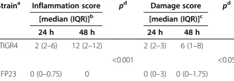
Table 1 Brain inflammation and damage over time in mice infected with TIGR4 or FP23
Straina Inflammation score pd Damage score pd
[median (IQR)]b [median (IQR)]c
24 h 48 h 24 h 48 h
TIGR4 2 (2–6) 12 (2–12) 2 (2–3) 6 (1–8)
<0.001 <0.05
FP23 0 (0–0.75) 0 0 (0–3) 0 (0–1.75)
a
Two groups of MF1 mice were infected via the i.c. route with 105
cfu/mouse of TIGR4 (n = 6) or FP23 (n = 16). Animals were sacrificed at 24 or 48 h, and brains were removed and treated for haematoxylin-eosin staining. Three (TIGR4) or 8 (FP23) mice per time-point were used.
b
Inflammation in the brain was evaluated by counting the number of PMN in one power field (× 40) and calculating a score as follows: 0 (0 PMN), 1 (<10 PMN), 2 (10–50 PMN) 3 (>50 PMN). Four different brain regions were analysed: superficial meningeal regions over the convexities, frontal interhemispheric region, hippocampal fissure and third ventricle. For every animal, scores of each brain region were summed up into a total inflammation score (IS). Results are expressed as the median IS with the interquartile range (IQR) for each mouse group at 24 and 48 h.
c
Neuronal damage was evaluated by estimating the percent of damaged neurons in one power field (× 40) and calculating a score as follows: 0 (no damaged neurons), 1 (<10%), 2 (10-30%), 3 (>30%). Four different brain areas were analysed: neocortex, striatum, hippocampus/dentate gyrus and cerebellum. For each mouse, scores of each brain region were summed, and the resulting number represented the total damage score (DS). Results are expressed as the median DS with the interquartile range (IQR) for each mouse group at 24 and 48 h.
d
of the different behaviours of pneumococcal mutants de-void of three major virulence determinants.
Analysis of mouse survival after infection with pneumococcal strains
Based on previous data on lethal doses ofS. pneumoniae killing 50% of animals (LD50) in experimental PM [39],
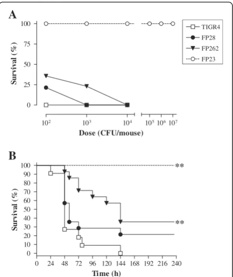
mice were infected by the i.c. route with different doses ofS. pneumoniaestrains TIGR4, FP28, FP262 and FP23. Analysis of mouse clinical parameters (body temperature and weight) showed no significant differences among the groups infected with the encapsulated strains (data not shown). All animals died following challenge with 102 cfu of TIGR4, while survival percentages of mice infected with FP28 and FP262 were 21 and 36, respect-ively (Figure 1A). At 103cfu/mouse, survival dropped to 0 and 23% in the groups infected with FP28 and FP262, respectively (Figure 1A). No animals survived challenge with 104 cfu of TIGR4, FP28 and FP262 (Figure 1A). The LD50 of all strains, except for FP23, were below
102 cfu/mouse. Upon time-to-death analysis, mice infected with the PspA-deficient strain FP262 showed significantly prolonged survival at all doses (Figure 1B and Additional file 1: Table S1,p< 0.01). In order to un-ravel differences among the groups, a Kaplan-Meyer analysis was carried out only on data from mice infected with the smallest dose of 102cfu. Median survival times of rodents challenged with TIGR4, FP28, FP262 and FP23 were 48, 56, 144 and >240 h with significant differ-ences for the groups TIGR4-FP262 and TIGR4-FP23 (Figure 1B and Additional file 1: Table S1, p< 0.01 for both groups). In accordance with previously published data [42], mice injected with the rough mutant FP23 presented clinical signs comparable to uninfected naïve animals and survived all challenge doses.
Replication of pneumococcal strains in the brain and blood of mice following i.c. infection
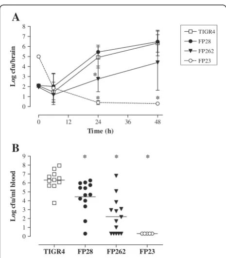
Analysis of the growth features of the mutants in stand-ard media showed no differences compared with TIGR4 (data not shown).To evaluate their capability to replicate in the CNS, viable counts were determined over time (at 6, 24 and 48 h) in the brain of infected mice. No differ-ences in bacterial titers were observed between FP28 and TIGR4 at any time-point. In contrast, the FP262 bacterial load in the brain was significantly lower than that of TIGR4 at 24 h post-infection (Figure 2A and Additional file 1: Table S2; p< 0.05). This finding sug-gests that the PspA mutant is impaired at replicating in the CNS in the early phase of infection. As previously reported [42], the rough strain FP23 was cleared from the brain by 24 h despite injection of animals with a high bacterial inoculum. As our PM model is characterised by the occurrence of both meningitis and sepsis [39],
pneumococci were enumerated in the blood 24 h after i.c. infection with different bacterial doses (102-104cfu/mouse). At the lowest inoculum of 102cfu/mouse, the number of cfu counts was significantly different between mice infected with TIGR4 and those challenged with the mutants FP28, FP262 and FP23 with a progressively decreasing trend in mean log cfu/ml of blood (Figure 2B and Additional file 1: Table S3;p< 0.05). In addition, significant differences could also be observed for the groups TIGR4-FP262 (at 103cfu/ mouse; p< 0.05) and TIGR4-FP23 (at 103 and 104 cfu/ mouse,p< 0.05), (Additional file 1: Table S3).
Evaluation of PM development in mice infected i.c. withS.
pneumoniaestrains
To assess whether the mutant strains were able to in-duce PM, mice were infected i.c. with 104cfu of TIGR4 and the three mutants. Brains were collected 24 h after infection and subjected to histological analysis. Results
102 103 104
0 25 50 75
100 TIGR4
FP28 FP262 FP23
105106107 Dose (CFU/mouse)
Survi
val
(
%
)
A
0 24 48 72 96 120 144 168 192 216 240 0
10 20 30 40 50 60 70 80 90 100
Time (h)
Survi
val
(
%
)
B
[image:5.595.306.539.88.365.2]** **
showed the presence of granulocytic infiltrations involv-ing both the subarachnoid and ventricular spaces of the brain from mice inoculated with all the encapsulated strains, and no major differences could be observed be-tween animals infected with TIGR4 and those challenged with the mutants FP28 and FP262 (data not shown). In contrast, no inflammation was detected in the brain of animals challenged with the rough FP23 mutant.
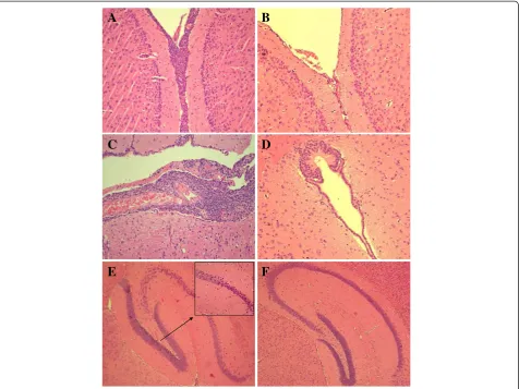
To further investigate this evidence, two groups of mice were inoculated with 105 cfu of TIGR4 and FP23 and euthanised 24 and 48 h later. Histological analysis of the brain from mice injected with TIGR4 showed severe inflammation characterised by massive infiltrations of PMN both on the meninges (Figure 3A) and in the ven-tricles, where accumulation of fibrin was also observed (Figure 3C). In contrast, the unencapsulated FP23 strain failed to cause inflammation on the meninges (Figure 3B) and in the ventricular spaces (Figure 3D). The TIGR4 strain also induced brain injury in the dentate gyrus of
hippocampus, where shrunk neurons with picnotic nu-clei were found (Figure 3E). No damage was found in samples from mice infected with FP23 (Figure 3F). A semi-quantitative analysis of brain inflammation and damage over time was carried out by determining the number of infiltrating PMN and the percentage of in-jured neurons in different brain regions, respectively, and by assigning each animal with an inflammation score (IS) and a damage score (DS). All mice infected with TIGR4 presented mild to severe inflammation that increased over time reaching a median IS (with IQR) of 12 (2–12) at 48 h post-infection (Table 1). Likewise, all animals challenged with TIGR4 showed neuronal injury of different degrees peaking at 48 h with median DS (IQR) of 6 (1–8) (Table 1). In contrast, median IS and DS from mice infected with the FP23 strain were equal to 0 at both time-points (Table 1), although some mice still showed mild brain damage (data not shown). The above data were also confirmed by infecting animals with a larger dose of FP23 (107cfu/mouse) and analys-ing their brains at a later time-point (72 h) (data not shown). The brain tissue of control mice injected with PBS appeared normal. Differences in brain inflamma-tion (p< 0.001) and neuronal damage (p< 0.05) between the groups infected with TIGR4 and FP23 were statisti-cally significant.
In vitrointeraction ofS. pneumoniaewith microglia The susceptibility of TIGR4 and FP23 to BV2 microglial cells was recently investigated to elucidate the import-ance of the capsule in phagocytosis and killing by brain macrophages [42]. To understand the role of PspC and PspA in the interaction ofS. pneumoniaewith microglia, phagocytosis and intracellular survival of FP28 and FP262 were assessed in comparison with the parental strain TIGR4. The rough strain FP23 was employed as a control. By using a previously established fluorescent assay that allows to distinguish attached from internalised bacteria [42,43], the number of phagocytic cells was mea-sured at 3 h post-infection. Levels of phagocytosis were similar for all strains (Figure 4A and Additional file 1: Table S4), suggesting that in a serotype 4 background PspC and PspA do not significantly affect uptake by BV2 microglial cells. As the TIGR4 strain was shown to resist intracellular killing by BV2 cells despite being phagocy-tosed to the same extent of its isogenic unencapsulated strain FP23 [42], we investigated the behaviours of FP28 and FP262 in microglial cells. Bacteria associated with acidic phagosomes (phagolysosomes) were visualised by incubating Hoechst-labelled pneumococci with BV2 cells in the presence of LysoTracker, a marker of phagosome acidification. The number of phagolysosomes within each microglial cell was determined, and the percentage of colocalisation was calculated. At 3 h post-infection, the
A
0 12 24 36 48
0 1 2 3 4 5 6 7 8
TIGR4 FP28 FP262 FP23
Time (h)
L
og cf
u/
brai
n
* *
*
B
*
* *
TIGR4 FP28 FP262 FP23 0
1 2 3 4 5 6 7 8 9
L
o
g c
fu/
m
l bl
[image:6.595.57.290.85.350.2]ood
Figure 2Bacterial loads in the brain and blood of mice i.c. challenged withS. pneumoniae. A.Mice were infected with 102cfu/mouse of strains TIGR4 (wt, open squares), FP28 (PspC-,
closed circles) and FP262 (PspA-, closed triangles). Control mice were inoculated with 105cfu/mouse of the mutant FP23 (rough, open
circles). Animals were sacrificed at different time-points (6 , 24 and 48 h) after infection, and brains were collected to determine the viable counts. Results are represented as mean log (± SD) cfu/brain over time.B.Animals were infected with 102cfu/mouse of strains
mean percentage of TIGR4-containing phagolysosomes was significantly lower (p< 0.05) than those of all mutant strains, suggesting that PspC and PspA may influence phagosome maturation in microglia (Figure 4B and Additional file 1: Table S4). Finally, to examine the capability of FP28 and FP262 to survive within microglia, an intracellular survival assay was carried out by infecting BV2 cells with bacteria for 3 h, followed by treatment with antibiotics, and then counting the number of intracellular surviving pneumococci at 4 h post-phagocytosis. Consistently with previous data [42], the survival index of FP23 was significantly lower (13-folds; p< 0.05) than that of TIGR4. Survival of the PspA mutant FP262 was also significantly lower (4-folds;p< 0.05) compared with the wt strain. No differences were observed between TIGR4 and FP28 (Figure 4C and Additional file 1: Table S4).
Discussion
Animal models of disease have significantly improved our knowledge on the interaction betweenS. pneumoniae and the host, and on the pathophysiological mechanisms involved in inflammation and brain damage during PM. Nevertheless, several issues remain to be clarified, includ-ing the role of different pneumococcal virulence factors in the disease. So far, only pneumolysin [29] and PavA [30] have been shown to contribute to PM development when tested via the i.c. route.
In this study, we used a mouse model of PM based on the inoculation of bacteria into the subarachnoid space [39] to test the pathogenicity of three different pneumo-coccal mutants in a type 4 background. PspA and PspC are well-known pneumococcal virulence factors [1,2], but their impact on PM has not been investigated before.
B
A
D
C
[image:7.595.62.539.89.446.2]F
E
Figure 3Brain histology of mice infected with TIGR4 or FP23.Mice were infected i.c. with 105cfu/mouse of TIGR4 or FP23 and sacrificed
Mice infected with the PspA-deficient strain FP262 showed increased survival and prolonged time-to-death for all bacterial inocula as well as lower viable counts in both blood (at the doses of 102and 103cfu/mouse) and brain (24 h post-infection) compared with TIGR4. Lower loads of FP262 in the blood may be due to decreased or delayed systemic spread of bacteria from the CNS and/ or higher clearance from the bloodstream. PspA plays a key role in immune escape ofS. pneumoniae by inter-fering with complement deposition/activation [6-9]. At-tenuation of PspA mutants in sepsis models is well documented [6,7,45-48], and the pspA gene was also shown to be upregulated in experimental sepsis [49]. Therefore, reduced virulence of FP262 in our PM model may be partially caused by its impaired virulence in the bloodstream. In addition, the ability of the FP262 strain to multiply in the CNS was initially hampered, as evidenced by the kinetics of viable bacterial counts in the brain. Nonetheless, the FP262 was still able to in-duce inflammation in the brain. In contrast, virulence of the PspC-deficient strain FP28 was moderately reduced compared with TIGR4, and the only condition able to unveil such difference was bacterial enumeration in the blood following infection with 102cfu/mouse. Although PspC seems to have a limited impact in the PM model, our data do not exclude a possible role in crossing the BBB, an early pathogenic step bypassed by i.c. infection. It was in fact suggested that S. pneumoniaetropism for the CNS may be partially due to PspC (CbpA), which participates in bacterial translocation from the blood to the CSF [50], most likely by binding to the laminin re-ceptor present on the endothelial cells of BBB [51].
A
B
*
*
*
TIGR4 FP28 FP262 FP23
0 5 10 15 20
P
h
a
g
o
cy
to
si
s (%
)
20 30 40 50 60
C
o
lo
ca
lis
a
tio
n
(%
)
C
*
*
TIGR4 FP28 FP262 FP23
0 10
TIGR4 FP28 FP262 FP23
0 10 20 30 40
Sur
vi
val
i
nde
[image:8.595.57.290.87.612.2]x
Figure 4Phagocytosis and intracellular survival ofS.
pneumoniaestrains in microglial cells. A.BV2 cells were infected
for 3 h with bacteria (moi = 10). A minimum of 200 BV2 cells were examined, and any cell containing one or more bacteria was considered as phagocytic.B.Quantification of acidic phagosomes in microglial cells infected withS. pneumoniae. Hoechst-labelled bacteria were exposed to BV2 cells (moi = 10) for 3 h, and the acidotropic dye LysoTracker Red DND-99 was added. Accumulation of the dye in phagosomes containing Hoechst-labelled bacteria was observed by epifluorescence microscopy. At least 200 BV2 cells were counted, and the number of cells with bacteria-containing acidic phagosomes was scored. The percentage of colocalisation was determined as the number of BV2 cells with pneumococci-containing phagolysosomes over the total number of phagocytes.C.
OnceS. pneumoniaeenters the CNS after crossing the BBB [52], the brain resident macrophages act as key ef-fectors of initial innate immunity, by clearing bacteria and recruiting peripheral blood cells to the site of infec-tion [33]. To investigate the role of PspA and PspC in the interaction with microglia, in vitro phagocytosis as-says were performed using the well-established mouse microglial cell line BV2 [41]. Bacterial uptake by BV2 cells was comparable among the strains, in accordance with previous observations on TIGR4 and FP23 [42]. Nonetheless, when the fate of the different mutants in-side microglia was analysed by evaluating colocalisation of intracellular pneumococci with acidic phagosomes and bacterial survival, both the unencapsulated strain FP23 and the PspA mutant FP262 showed significantly increased association with phagolysosomes and killing compared with the wt. As previously shown for the type 4 polysaccharide capsule [42], these data suggest that PspA may also participate in pneumococcal resistance to microglial killing, possibly by interfering with phago-some maturation. In contrast, survival of the PspC mu-tant FP28 in BV2 cells was similar to that of TIGR4, despite an increased association with phagolysosomes. This observation seems to disagree with a previous work reporting that the lack of PspC increased the susceptibil-ity of pneumococcal killing by microglia [53]. Such discrepancy may be explained by the different pneumo-coccal strains employed in the assays, a serotype 3 PspC mutant [53] and a serotype 4 PspC-deficient strain (this work), which are resistant and susceptible to phagocyt-osis by microglia, respectively. In summary, the data on PspA and PspC in our i.c. model (characterised by concurrent sepsis and meningitis) confirm previous reports on their key roles in experimental sepsis [6,7,36,45-48,54]. In addition, the decreased ability of FP262 of replicating in the brain at early time-points (24 h post-infection) together with its increased suscepti-bility to microglial killing also suggest that PspA may play a role in early stages of CNS infection byS. pneumoniae.
Rough strains are virtually unable to cause pneumo-coccal invasive disease [55], and to support this observa-tion, fresh isolates from patients with pneumococcal infection are encapsulated [56]. However, early studies in a PM rabbit model showed that large inocula (107 cfu/ ml) of rough pneumococci could also be lethal, and that CSF inflammation could be induced by heat-killed unencapsulated S. pneumoniae (R6 strain) or their iso-lated cell walls, but not by heat-killed encapsulat-ed pneumococci or their capsular polysaccharides [23,24,57]. Bacterial cell walls are potent inflammatory components, and the threshold of bioactivity of pneumococcal cell wall (PCW) corresponds to ~105cfu/ ml of intact bacteria [58,59]. In our study, infection with up to 107cfu (in 50 μl) of unencapsulated FP23 bacteria
(corresponding to 2×10⁸cfu/ml of CSF) caused no men-ingeal inflammation or animal death ([42] and this work). Other than the PCW concentration, different fac-tors may be responsible of the discrepancies observed, including the choice of different readouts to assess men-ingeal inflammation (i.e. cytochemical features of CSF versus histological analysis), the use of heat-inactivated versus live bacteria, and/or the type of PCW fragments released in the CSF of infected animals. As only specific motifs (i.e. trimeric stem peptides) of PCW are highly inflammatory [59], these structures may have not been accessible in FP23-infected mice, while being available in rabbits inoculated with heat-inactivated R6 bacteria [23]. Despite the lack of brain inflammation in mice injected with the FP23 mutant, a degree of neuronal damage was still found in the dentate gyrus of the hippocampus of some mice. As the effector mechanisms of neuronal damage in PM are both the host inflammatory response and the direct citotoxicity of bacterial components [19-21], the brain damage observed in mice infected with FP23 may be due to toxic molecules of S. pneumoniae, including pneumolysin and/or H2O2in
ac-cordance to previous animal studies [21,27,28].
Conclusions
The pathogenesis of PM is highly complex and multifac-torial, and it is difficult to ascribe a precise role to a bac-terial virulence factor in the disease. The results obtained with our i.c. model indicate a limited role for PspC, whereas PspA participates in PM pathogenesis possibly by interacting with microglial cells at an early phase of infec-tion. The unencapsulated pneumococcal mutant failed to induce meningeal inflammation, brain injury and animal death even at high challenge doses, emphasising the piv-otal role played by the capsule in invasive pneumococcal disease. The virulence factors evaluated in this study are either antigens of existing vaccines or strong candidates for vaccine development. The fact that they have been shown to contribute, to various degrees, to experimental PM support their use in current/future vaccine formula-tions againstS. pneumoniaebecause vaccine efficacy may be enhanced by impairment of pneumococcal virulence. In conclusion, the data presented here may be relevant for translational research studies aimed at improving or devel-oping effective and sustainable preventive measures against infectious diseases such as PM.
Additional files
Abbreviations
PspA:Pneumococcal surface protein A; PspC: Pneumococcal surface protein C; wt: Wild type; cfu: Colony forming units; PMN: Polymorphonuclear cells; CNS: Central nervous system; BBB: Blood-brain-barrier; CSF: Cerebrospinal fluid; PCW: Pneumococcal cell wall; IC: Intracranial; SI: Survival index; IS: Inflammation score; DS: Damage score; FCS: Fetal calf serum; PBS: Phosphate buffered saline; PFA: Paraformaldehyde; IQR: Interquartile range; CI: Confidence interval; SD: Standard deviation.
Competing interests
The authors declare that they have no competing interests.
Authors’contributions
SR, co-ordination and design of the study, data analysis and interpretation, supervision of experimental work, writing of manuscript. AG, animal experiments and microbiological analysis. AP, statistical analyses. DC, animal experiments and manuscript revision. VB, animal experiments. SAT, histological analysis. BC, phagocytosis/survival experiments with microglia. EB, co-ordination of phagocytosis experiments. MRO, experimental design and critical reading of manuscript. SP, co-ordination of phagocytosis experiments, data evaluation, and manuscript revision. GP, co-ordination and design of the study, data evaluation. All authors read and approved the manuscript.
Acknowledgements
The work was supported by Italian MIUR 2008 (to G. Pozzi).
Author details
1Department of Medical Biotechnologies, Laboratory of Molecular
Microbiology and Biotechnology (LA.M.M.B.), University of Siena and Siena University Hospital, Siena 53100, Italy.2Department of Physiopathology, Experimental Medicine and Public Health, University of Siena, Siena, Italy. 3Department of Pathology, Siena University Hospital, Siena, Italy.
4Department of Diagnostics, Clinical and Public Health Medicine, University
of Modena and Reggio Emilia (Unimore), Emilia-Romagna, Italy.5Present address: Evans Medical Research Center, Boston University School of Medicine, 650 Albany Street, Boston, MA 02118, USA.6Present address: Sanofi-Aventis S.p.a., Brindisi 72100, Italy.
Received: 27 May 2013 Accepted: 13 September 2013 Published: 24 September 2013
References
1. Kadioglu A, Weiser JN, Paton JC, Andrew PW:The role ofStreptococcus
pneumoniaevirulence factors in host respiratory colonization and disease.Nat Rev Microbiol2008,6:288–301.
2. Mitchell AM, Mitchell TJ:Streptococcus pneumoniae: virulence factors and variation.Clin Microbiol Infect2010,16:411–418.
3. Brown EJ, Joiner KA, Cole RM, Berger M:Localization of complement component 3 onStreptococcus pneumoniae:anti-capsular antibody causes complement deposition on the pneumococcal capsule.Infect Immun1983,39:403–409.
4. Abeyta M, Hardy GG, Yother J:Genetic alteration of capsule type but not PspA type affects accessibility of surface-bound complement and surface antigens ofStreptococcus pneumoniae.Infect Immun2003, 71:218–225.
5. Hyams C, Camberlein E, Cohen JM, Bax K, Brown JS:TheStreptococcus
pneumoniaecapsule inhibits complement activity and neutrophil phagocytosis by multiple mechanisms.Infect Immun2010,78:704–715. 6. Tu AH, Fulgham RL, McCrory MA, Briles DE, Szalai AJ:Pneumococcal
surface protein A inhibits complement activation byStreptococcus
pneumoniae.Infect Immun1999,67:4720–4724.
7. Ren B, McCrory MA, Pass C, Bullard DC, Ballantyne CM, Xu Y, Briles DE, Szalai AJ: The virulence function ofStreptococcus pneumoniaesurface protein A involves inhibition of complement activation and impairment of complement receptor-mediated protection.J Immunol2004,173:7506–7512. 8. Ren B, Szalai A, Hollingshead SK, Briles DE:Effects of PspA and antibodies
to PspA on activation and deposition of complement on the pneumococcal surface.Infect Immun2004,72:114–122.
9. Mukerji R, Mirza S, Roche AM, Widener RW, Croney CM, Rhee DK, Weiser JN, Szalai AJ, Briles DE:Pneumococcal surface protein A inhibits complement
deposition on the pneumococcal surface by competing with the binding of C-reactive protein to cell-surface phosphocholine.J Immunol 2012,189:5327–5335.
10. Hammerschmidt S, Bethe G, Remane PH, Chhatwal GS:Identification of pneumococcal surface protein A as a lactoferrin-binding protein of
Streptococcus pneumoniae.Infect Immun1999,67:1683–1687.
11. Dave S, Carmicle S, Hammerschmidt S, Pangburn MK, Mc Daniel LS:Dual roles of PspC, a surface protein ofStreptococcus pneumoniae, in binding human secretory IgA and factor H.J Immunol2004, 173:471–477.
12. Hammerschmidt S, Tilling MP, Wolff S, Vaerman JP, Chhatwal GS: Species-specific binding of human secretory component to SpsA protein of
Streptococcus pneumonievia a hexapeptide motif.Mol Microbiol2000, 36:726–736.
13. Zhang JR, Mostov KE, Nanno M, Shimida S, Ohwaki M, Tuomanen E:The polymeric immunoglobulin receptor translocates pneumococci across human nasopharyngeal epithelial cells.Cell2000,102:827–837. 14. Elm C, Braathen R, Bergmann S, Frank R, Vaerman JP, Kaetzel CS, Chhatwal
GS, Johansen FE, Hammerschmidt S:Ectodomains 3 and 4 of human polymeric Immunoglobulin receptor (hpIgR) mediate invasion of
Streptococcus pneumoniaeinto the epithelium.J Biol Chem2004, 279:6296–6304.
15. Smith BL, Hostetter MK:C3 as substrate for adhesion ofStreptococcus
pneumoniae.J Infect Dis2000,182:497–508.
16. Janulczyk R, Iannelli F, Sjöholm AG, Pozzi G, Björck L:Hic, a novel surface protein ofStreptococcus pneumoniaethat interferes with complement function.J Biol Chem2000,275:37257–37263.
17. Jarva H, Janulczyk R, Hellwage J, Zipfel PF, Björck L, Meri S:Streptococcus
pneumoniaeevades complement attack and opsonophagocytosis by expressing thepspClocus-encoded Hic protein that binds to short consensus repeats 8–11 of factor H.J Immunol2002,168:1886–1894. 18. Duthy TG, Ormsby RJ, Giannakis E, Ogunniyi DA, Stroeher UH, Paton JC,
Gordon DL:The human complement regulator factor H binds
pneumococcal surface protein PspC via short consensus repeats 13 to 15. Infect Immun2002,70:5604–5611.
19. Mook-Kanamori BB, Geldhoff M, van der Poll T, van de Beek D:Pathogenesis and pathophysiology of pneumococcal meningitis.Clin Microbiol Rev2011, 24:557–591.
20. Koedel U, Klein M, Pfister HW:New understandings on the pathophysiology of bacterial meningitis.Curr Opin Infect Dis2010,23:217–223.
21. Gerber J, Nau R:Mechanisms of injury in bacterial meningitis.Curr Opin Neurol2010,23:312–318.
22. Klein M, Obermaier B, Angele B, Pfister HW, Wagner H, Koedel U, Kirschning CJ:Innate immunity to pneumococcal infection of the central nervous system depends on toll-like receptor (TLR) 2 and TLR4.J Infect Dis2008, 198:1028–1036.
23. Tuomanen E, Tomasz A, Hengstler B, Zak O:The relative role of bacterial cell wall and capsule in the induction of inflammation in pneumococcal meningitis.J Infect Dis1985,151:535–540.
24. Tuomanen E, Liu H, Hengstler B, Zak O, Tomasz A:The induction of meningeal inflammation by components of the pneumococcal cell wall. J Infect Dis1985,151:859–868.
25. Hirst RA, Sikand KS, Rutman A, Mitchell TJ, Andrew PW, O’Callaghan C: Relative roles of pneumolysin and hydrogen peroxide from
Streptococcus pneumoniaein inhibition of ependymal ciliary beat frequency.Infect Immun2000,68:1557–1562.
26. Zysk G, Schneider-Wald BK, Hwang JH, Bejo L, Kim KS, Mitchell TJ, Hakenbeck R, Heinz HP:Pneumolysin is the main inducer of cytotoxicity to brain microvascular endothelial cells caused byStreptococcus
pneumoniae.Infect Immun2001,69:845–852.
27. Braun JS, Sublett JE, Freyer D, Mitchell TJ, Cleveland JL, Tuomanen EI, Weber JR:Pneumococcal pneumolysin and H2O2mediate brain cell apoptosis during meningitis.J Clin Invest2002,109:19–27.
28. Winter AJ, Comis SD, Osborne MP, Tarlow MJ, Stephen J, Andrew PW, Hill J, Mitchell TJ:A role for pneumolysin but not neuraminidase in the hearing loss and cochlear damage induced by experimental pneumococcal meningitis in guinea pigs.Infect Immun1997,65:4411–4418.
29. Wellmer A, Zysk G, Gerber J, Kunst T, von Mering M, Bunkowski S, Eiffert H, Nau R:Decreased virulence of a pneumolysin-deficient strain of
30. Pracht D, Elm C, Gerber G, Bergmann S, Rohde M, Seiler M, Kim KS, Jenkinson HF, Nau R, Hammerschmidt S:PavA ofStreptococcus
pneumoniaemodulates adherence, invasion, and meningeal inflammation.Infect Immun2005,73:2680–2689.
31. Vilhardt F:Microglia: phagocyte and glia cell.Int J Biochem Cell Biol2005, 37:17–21.
32. Hauwel M, Furon E, Canova C, Griffiths M, Neal J, Gasque P:Innate (inherent) control of brain infection, brain inflammation and brain repair: the role of microglia, astrocytes,“protective”glial stem cells and stromal ependymal cells.Brain Res Rev2005,48:220–233.
33. Rock RB, Gekker G, Hu S, Sheng WS, Cheeran M, Lokensgard JR, Peterson PK: Role of microglia in central nervous system infections.Clin Microbiol Rev 2004,17:942–964.
34. Horton RM, Cai Z, Ho SN, Pease LR:Gene splicing by overlap extension: tailor-made genes using the polymerase chain reaction.Biotechniques 1990,8:528–535.
35. Ali F, Lee ME, Iannelli F, Pozzi G, Mitchell TJ, Read RC, Dockrell DH:
Streptococcus pneumoniae-associated human macrophage apoptosis after bacterial internalization via complement and Fcgamma receptors correlates with intracellular bacterial load.J Infect Dis2003,188:1119–1131. 36. Iannelli F, Chiavolini D, Ricci S, Oggioni MR, Pozzi G:Pneumococcal surface protein C contributes to sepsis caused byStreptococcus pneumoniaein mice.Infect Immun2004,72:3077–3080.
37. Pearce BJ, Iannelli F, Pozzi G:Construction of new unencapsulated (rough) strains ofStreptococcus pneumoniae.Res Microbiol2002,153:243–247. 38. Oggioni MR, Memmi G, Maggi T, Chiavolini D, Iannelli F, Pozzi G:
Pneumococcal zinc metalloproteinase ZmpC cleaves human matrix metalloproteinase 9 and is a virulence factor in experimental pneumonia.Mol Microbiol2003,49:795–805.
39. Chiavolini D, Tripodi S, Parigi R, Oggioni MR, Blasi E, Cintorino M, Pozzi G, Ricci S:Method for inducing experimental pneumococcal meningitis in outbred mice.BMC Microbiol2004,4:36.
40. Sandgren A, Albiger B, Orihuela CJ, Tuomanen E, Normark S, Henriques-Normark B:Virulence in mice of pneumococcal clonal types with known invasive disease potential in humans.J Infect Dis2005,192:791–800. 41. Blasi E, Barluzzi R, Bocchini V, Mazzolla R, Bistoni F:Immortalization of
murine microglial cells by a v-raf/v-myc carrying retrovirus.J Neuroimmunol1990,27:229–237.
42. Peppoloni S, Ricci S, Orsi CF, Colombari B, De Santi MM, Messinò M, Fabio G, Zanardi A, Righi E, Braione V,et al:The encapsulated strain TIGR4 of
Streptococcus pneumoniaeis phagocytosed but is resistant to
intracellular killing by mouse microglia.Microbes Infect2010,12:990–1001. 43. Peppoloni S, Colombari B, Beninati C, Felici F, Teti G, Speziale P, Ricci S,
Ardizzoni A, Manca L, Blasi E:The Spr1875 protein confers resistance to the microglia-mediated killing ofStreptococcus pneumoniae.Microb Pathog2013,59–60:42–47.
44. Efron B, Tibshirani RJ:An introduction to the Bootstrap.New York: Chapman and Hall; 1994.
45. McDaniel LS, Yother J, Vijayakumar MN, McGarry L, Guild WR, Briles DE:Use of insertional inactivation to facilitate studies of biological properties of pneumococcal surface protein A (PspA).J Exp Med1987,165:381–394. 46. Berry AM, Paton JC:Additive attenuation of virulence ofStreptococcus
pneumoniaeby mutation of the genes encoding pneumolysin and other putative pneumococcal virulence proteins.Infect Immun2000,
68:133–140.
47. Yuste J, Botto M, Paton JC, Holden DW, Brown JS:Additive inhibition of complement deposition by pneumolysin and PspA facilitates
Streptococcus pneumoniaesepticemia.J Immunol2005,175:1813–1819. 48. Quin LR, Moore QC III, McDaniel LS:Pneumolysin, PspA, and PspC
contribute to pneumococcal evasion of early innate immune responses during bacteremia in mice.Infect Immun2007,75:2067–2070.
49. Ogunniyi DA, Giammarinaro P, Paton JC:The genes encoding virulence-associated proteins and the capsule ofStreptococcus pneumoniaeare upregulated and differentially expressed in vivo.Microbiology2002, 148:2045–2053.
50. Orihuela CJ, Gao G, Francis KP, Yu J, Tuomanen EI:Tissue-specific contributions of pneumococcal virulence factors to pathogenesis.J Infect Dis2004,190:1661–1669.
51. Orihuela CJ, Mahdavi J, Thornton J, Mann B, Wooldridge KG, Abouseada N, Oldfield NJ, Self T, Ala’Aldeen DA, Tuomanen EI:Laminin receptor initiates bacterial contact with the blood brain barrier in experimental meningitis models.J Clin Invest2009,119:1638–1646.
52. Ring A, Weiser JN, Tuomanen EI:Pneumococcal trafficking across the blood–brain barrier. Molecular analysis of a novel bidirectional pathway. J Clin Invest1998,102:347–360.
53. Peppoloni S, Colombari B, Neglia R, Quaglino D, Iannelli F, Oggioni MR, Pozzi G, Blasi E:The lack of pneumococcal surface protein C (PspC) increases the susceptibility ofStreptococcus pneumoniaeto the killing by microglia.Med Microbiol Immunol2006,195:21–28.
54. Kerr AR, Paterson GK, McCluskey J, Iannelli F, Oggioni MR, Pozzi G, Mitchell TJ:The contribution of PspC to pneumococcal virulence varies between strains and is accomplished by both complement evasion and complement-independent mechanisms.Infect Immun2006,74:5319–5324. 55. Griffith F:The significance of pneumococcal types.J Hyg1928,
27:113–159.
56. Austrian R:Some observations on the pneumococcus and on the current status of pneumococcal disease and its prevention.Rev Infect Dis1981, 3:S1–S17.
57. Tomasz A, Saukkonen K:The nature of cell wall-derived inflammatory components of pneumococci.Pediatr Infect Dis1989,8:902–903. 58. Weber JR, Moreillon P, Tuomanen EI:Innate sensors for Gram-positive
bacteria.Curr Opin Immunol2003,15:408–415.
59. Moreillon P, Majcherczyk PA:Proinflammatory activity of cell-wall constituents from Gram-positive bacteria.Scand J Infect Dis2003, 35:632–641.
doi:10.1186/1471-2334-13-444
Cite this article as:Ricciet al.:Contribution of different pneumococcal virulence factors to experimental meningitis in mice.BMC Infectious Diseases201313:444.
Submit your next manuscript to BioMed Central and take full advantage of:
• Convenient online submission
• Thorough peer review
• No space constraints or color figure charges
• Immediate publication on acceptance
• Inclusion in PubMed, CAS, Scopus and Google Scholar
• Research which is freely available for redistribution