JOURNAL OF VIROLOGY,Dec. 1993,p.7373-7382 Vol. 67,No. 12 0022-538X/93/127373-10$02.00/0
Copyright © 1993,American
Society
forMicrobiologyA
Novel Class of
Transcripts Expressed with
Late
Kinetics in the
Absence of
ICP4 Spans
the
Junction between the Long and Short
Segments
of
the
Herpes
Simplex
Virus Type 1
Genome
LILY YEH ANDPRISCILLAA. SCHAFFER*
Division of Molecular Genetics, Dana-Farber Cancer Institute, and DepartmentofMicrobiology and Molecular Genetics, HarvardMedicalSchool,Boston, Massachusetts 02115
Received 15 July1993/Accepted 12 September 1993
A novel family oftranscripts that span thejunction between the long and short segments of the herpes simplex virus type 1 genome has been identified. These transcripts, designated L/S junction-spanning
transcripts (L/STs),are synthesizedinabundance in a
variety
of cells infected with mutant virusesdefective in the gene forICP4,themajor transcriptional regulatoryprotein ofthevirus.Transcriptionof abundant 2.3-and 8.5-kb series ofL/STs was shown to initiatewithin the same sequences as less abundant 4.2-, 7.3-, and >9.5-kbtranscripts byNorthern (RNA)blotanalysis.Sinucleaseanalysis revealedasingle5' terminus 28bp downstream of a TATA box and 6 bp downstream of a consensus ICP4 binding site. The location of the transcriptional start site indicates that the promoter of the LISTs likely corresponds to the bidirectional promoterdescribedbyBohenzkyetal. (R. A.Bohenzky,A.G.Papavassiliou,I. H.Gelman, and S.Silverstein, J. Virol. 67:632442, 1993). The ISTs accumulate with late kinetics in ICP4 mutant-infected cells andare polyadenylated.Mutantvirusesencodingformsof ICP4unable tobind the consensussite,ATCGTC, exhibited abundant expressionof the L/STs,whereas mutantsencoding forms ofICP4 able tobind this siteexpressed nodetectableL/STs,suggestingthat ICP4plays acriticalroleinrepressingLIST expression. Their synthesis inICP4 mutant-infectedcells isinhibitedbytheproteinsynthesisinhibitorcycloheximide, indicatingthatthey areinducedeither byanimmediate-earlyviralproteinother thanICP4orbyavirus-induced cellularprotein. Preliminary evidence indicates that the L/STs are not present in latently infected ganglia. The abundant expression of the L/STswith late kinetics onlyin the absence offunctional ICP4and the sensitivity oftheir synthesistocycloheximideindicate that they are not members of any of therecognizedkinetic classes ofherpes simplexvirus type 1transcripts butconstitutea newclass of viraltranscript.Theexpression of herpes simplex virus(HSV)genesduring
productive infection has long beenrecognizedtoproceed ina coordinate and sequential manner (27, 28; reviewed in refer-ences 3 and 32). The classification of HSVgenes into broad
groups-immediate-early (IE),early(E), delayed early (DE), andlate(L)-isbasedonthe kinetics ofsynthesis of individual viral transcripts andproteins, the effects of various metabolic inhibitorsonDNA, RNA, andproteinsynthesis, and studies of viral mutants.
IEproteins are the firstto be synthesized in infected cells and are the major regulatory proteins of the virus. They are required for the synthesis ofE,DE,andLproteins and for the repression of theirown synthesis (16a, 17, 19, 23, 27, 28, 35, 39a,41).Transcription ofIE genesis activatedbyalate protein in infecting virions, VP16 (reviewed in reference 24). IE transcripts and proteins are detectable at 1 h postinfection
(hpi). Peaksynthesis of IEproteins occurs at3 to 4 hpi and declinesrapidly thereafter, althoughlowlevelsofsynthesisare detectable at later timespostinfection (27). Productive infec-tion is blocked in thepresenceofinhibitors ofprotein
synthe-sis, such that onlyIE mRNAs aremade (27). IEproteins are requiredtoinduce thesynthesisof Etranscripts. Transcription of E genes andsynthesis ofEproteins begins by 3 hpi, peaksat about6hpi, and declines thereafter.Eproteinsareinvolvedin nucleotide precursor metabolism and viral DNA synthesis. Transcription of DE (but not L) genes occurs at low levels from input genomes prior to the initiation of viral DNA
*Correspondingauthor. Electronic mail address:
Priscilla_Schaffer
@DFCI.harvard.edu.
synthesisandthus does not depend stringently onviralDNA
synthesis (26, 57). As viral DNA synthesis begins, high-level
expression ofDE genes occursand L geneexpression begins. DEandLproteinsynthesiscontinues throughout the remain-der of the replication cycle. DE and L proteins include envelopeglycoproteins, capsidproteins,and othercomponents ofmaturevirusparticles.
In contrast to the complex sequence of events that occurs during productive infection, viral gene expression during la-tencyisconsiderablylesscomplicated.Inlatently infected cells, viral geneexpression is limitedtothe latency-associated
tran-scripts (LATs),afamily oftranscripts rangingin size from2.0 to >8kb (21).Whether the LATs encode functional proteins
and what role they play in the viral life cycle remain to be determined.Although much is known about viralgene expres-sionduringproductive infection,verylittleisknownabout the factors that mediate the switch from productive infection to latency and vice versa. Whatever the mechanism underlying this switch, theregulatory activities of the IEproteins, which orchestrate eventsduringproductive infection, must be over-ridden to establish latency and reinitiated during reactivation fromlatency.
Physical mapping studieshave shown thatfour ofthe five IE regulatory genes are located wholly or in part within b a c repeat sequences flanking the unique long
(Ut_)
and uniqueshort
(Us)
regionsof the genome(12, 36, 38) (Fig. 1),whereas nearly allE,DE,and L genesarelocated inunique-sequenceDNA. One consequence of this arrangement is that IE regu-latorygenes andother elements located totally within repeat sequencesarediploid. Inadditionto IEregulatorygenes,the 7373
on November 9, 2019 by guest
http://jvi.asm.org/
A. L
B. 1117 118 120 122 124 126 128 130 132 134
I b ~~~~~~~~~~~~~~acI Us
12 -
~~~I
~ ~ H4iH:l4iR H 00 4 0 ,- Hi i)) )
CA)Qm -a to U iJOvUm o X4Cn'U u R.
Ow E U U MI z v zc,mzz XU En Uv i)E z u7
8.3 khk
kb__| ' F
~~~~~A+
r6.0k- W_ A
LATS 2.0 kb -
A-1l. 5 kb _
A-A
ICP0
A4-9 ICP34.5
A
ICP4
4- BbSLAT EBNc3-LAT (S) 4-- 4- LAT/4Sma
EBNc3-IAT 4 b4Sma
EBN9-IAT 94 d EBNH2-IAT
IdlLAT 8
StuI-BssHII* *
n212
I
- ~OrisRNA2
.4. OrisRNA1
- l-A+
Oris ICP22
22SS 22KS
tsB2 n215 n214 n12 n6
lI I I I
dl20
xdl56 Cd2
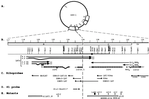
FIG. 1. Physicalmapof the internalrepeatregionof HSV-l DNA.(A) Diagramof theHSV-lgenome. UL, unique longsegment;US,unique shortsegment; b,inverted repeatsequencebracketing UL;c,invertedrepeatsequencebracketingUs;a, 317-bpsequence between the b andc
repeatsequences.Twocopiesof the ba crepeatsexist in circulargenomes. (B) Expandedmapof internalrepeatsequences lyingbetweenmap
units 117 and 134.5onthephysicalmapofthe HSV-1 genome(12, 36, 38).Beneath the scale of kilobasepairsareshown the locations oftheb, a,andcsequences and relevant restriction sites in KOS DNA. Beneath themapofrestriction sitesare shown the locations of the genesand
cis-actingelementscontained within thesequencefrom 118to 134kb.Specifically, themapshows the locations ofsequencesspecifyingthesmall
(1.5- and 2.0-kb) and large (8.3- and putative 6-kb) LATs, the transcripts encoding ICPO, ICP34.5, ICP4, and ICP22, sequences inoriS, and
sequencesspecifyingthetranscriptsdesignatedoriS RNA1 and oriS RNA2.ORFsareshownashatched bars.(C)DNAsequencesspecifyingthe
riboprobes (arrows)used in thisstudy.The arrowsrepresenttheorientation of thesesequences inpGEMvectorsasdrivenbythe SP6promoter.
(D) Sequencespecifyingthe DNAprobeused forS1nucleasemapping.Theprobewaslabelledatthe BssHII site(asterisk). (E)Locations of the mutations in themutantviruses used in these studies. n212 containsa nonsensemutation in the ICPOgene(6). n6, nl2, n214,and n215 contain
nonsensemutations in the ICP4gene(20).Theopenboxes indicate thesequencesdeleted ind1LATI.8,whichspecifiesnodetectable LATs(34),
andd2, d120,andd156 ICP4,whichare nullmutants(17). Thesmall black box indicates the approximate locationof the mutationin tsB2(23).
ba crepeatscontain thejunction betweenthe longand short regions of thegenome(the L/Sjunction)aswellasothergenes
and cis-acting elements of fundamental significance to both productive infection and latency. These include (i) the se-quencesspecifyingthe LATs(2, 21, 33, 37, 51, 56), (ii) thegene
encoding a neurovirulence factor, ICP34.5 (8-10), (iii) the a
sequence, which contains cis-acting elements involved in cir-cularization, recombination, andpackaging of the viralgenome
(15, 16, 39, 47, 48, 54, 55), and (iv) oriS,anorigin of viral DNA
replication (49, 52, 53, 55, 58).
During thecourseof effortstofinemapthe LATs within the
ba crepeats,we chosetoisolate and characterize RNA from cells infected with an ICP4 null mutant in order to enhance LAT expression, as ICP4 has been reported to repress
tran-scription from the LAT promoter (2). Using a series of strand-specific riboprobes in Northern (RNA) blot analysis,we
identified a family of previously undescribed 5' coterminal
transcripts ranginginsize from 2.3to>9.5kb thatspantheL/S
junction. These transcripts, designated L/S junction-spanning transcripts (L/STs), appeartoinitiate inb sequences immedi-atelytotheright of the bidirectional ICPOpromoterdescribed by Bohenzky et al. (4). They cross the L/S junction in the a
sequence and terminate in c sequences near the poly(A) addition site atposition 127040 (2.3-kb transcript) or beyond (8.5-kb and larger species). Thus, these transcripts are anti-sense to the genesencoding ICP34.5 and ICP4. Because the DNAtemplate specifyingthe L/STs includes the L/S junction, it is diploid in circulargenomes. Timecourse studies indicate that the L/STs accumulate in ICP4mutant-infected cells with late kinetics. The studies described herein demonstrate that expression of the L/STs is repressed by ICP4 and stimulated eitherbyother IEproteinsorbyvirus-induced cellular factors.
Because they are not expressed in the presence of cyclohexi-mide,the L/STsare not IE transcripts. The fact thatthey are
expressedathighlevels and with latekinetics in the absence of C. Riboprobas
D. Si probe E. Mutants
1%
on November 9, 2019 by guest
http://jvi.asm.org/
[image:2.612.65.555.74.402.2]HSV-1 L/STs 7375
ICP4,
yet are inhibitedby
cycloheximide,
indicates that theL/STs
representa newclassof viraltranscript.
MATERIALSAND METHODS
Cells and viruses.Africangreen
monkey kidney
cells(Vero;ATCC CCL
81),
EScells(Vero
cells stablytransformed with thewild-type
genefor ICP4[17]),
0-28cells(Vero
cellsstablytransformed with the
wild-type
gene forICPO[42]),
and 3-3 cells(Vero
cellsstably
transformed with thewild-type
genefor ICP27[35])
were grown and maintained in Dulbecco's modi-fiedEagle
medium(GIBCO Laboratories,
Inc.,Gaithersburg,Md.)
aspreviously
described(41).
Mouse neuroblastoma cells(NB41A3;
ATCCCCL147)
were propagated in F10 medium(GIBCO) supplemented
with2.5%fetal calf serum, 15% horse serum, 100 Uofpenicillinperml,and 100 ,ug ofstreptomycinper ml. Rat
pheochromocytoma (PC12)
cells were kindlyprovided by
JohnWagner
(Cornell University
MedicalCol-lege,NewYork, N.Y.)andwerepropagatedandmaintainedas described
previously (25).
The KOS wild-type strain of HSV-1 (46) and a series of
temperature-sensitive (ts),
nonsense, and deletion mutants derived from KOS cellswereused in these studies. The ICP4 nonsenseanddeletion mutants,n6,n12,
n214, n215,d2, d120,
andd156(17, 20),
weregrownandassayed
inE5 cells(17).
TheICPO nonsense mutant, n212, was grown in Vero cells and
assayed
on0-28 cells(6).
The ICP27deletion mutant,5d11.2,
was grown and
assayed
in 3-3 cells(35).
KOS, the ICP4 ts mutant, tsB2(23),
the ICP22 nonsense mutant,22/n199
(1),
andtheLATdeletion mutant,dlLAT1.8(34),
weregrownandassayed
in Vero cells(41).
The locations of the mutations in thesevirusesare shown inFig.
1E.Riboprobes. The
riboprobes
usedin thisstudyareshown inFig.
1C. The BamHI Kfragment
containing theL/Sjunctionand partof the ba crepeatsfrompSG28 (44)wascloned into
pGEM3Zf(+) (Promega, Madison, Wis.).
The 1,750-bpNcoIfragment
frompBamK(map
units 124to125.8)wassubcloned into theNcoI site inpGEM5Zf(+)
toyield pNco. To generateriboprobe
EBNc3-LAT, pNcowas linearized at theStul site,and the StuI-NcoI sequencewas transcribed as instructedby
the manufacturer
(Promega).
To generate EBNc3-LAT(S),pNco
was cleaved with Stul and HinclI(HinclI
is in thepolylinker beyond
theright-hand
NcoIsite)
andreligated
such thatonly
the NcoI-StuIfragment
remained. EBNc3-LAT(S)wastranscribed from this sequence.A
plasmid
containingthe Notlsubfragment (map
units 124.9to125.7)
frompNcocloned into the NotI site ofpGEM5Zf(+)
was transcribed to yieldEBN9-LAT.
Riboprobe
EBNH2-LAT was derived from theNotI-HincII fragment
frompBamK,andriboprobe
LAT/4Smawas derived from the SmaI
fragment
from pnll (19),which contains thewild-type
ICP4 gene.Riboprobe
4Sma, used to detect the ICP4 transcript,was derived from the same SmaIfragment
from pnll but cloned in the opposite direction.Riboprobe
BbSLATwas derived from the 786-bpSphI
frag-ment from the BamHI B
fragment
of KOS DNA, which contains the sequencesspecifying
the 2-kb LATs.Riboprobes
22SSand 22KSwere derived from theEcoNI-SphI
andSphlI-KpnI fragments,
respectively, containing most of the ICP22 gene and somedownstream sequences. The sizes and orienta-tion of clonedfragments
were verified by restriction enzymeanalysis. Riboprobes
weretranscribed from the SP6 promoter in thevector asinstructed bythemanufacturer(Promega).Northern blotanalysis.Approximately4 x
106
NB41A3or 2 x10⁶
Vero or E5 cells were seeded in 100-mm-diameterpetri
dishes 24 hprior
to infection. Cells were infected at amultiplicity
of 10 PFU per cell in 0.5 ml of inoculum. Afterabsorption for 1 h at 37°C, medium was added to infected cells, and incubation was continued at 37°C for the indicated times. Total cell RNAwas harvested as described previously (29). Briefly, monolayers were washed twice with cold phosphate-buffered saline and scraped into 0.5 ml of GIT buffer (4 M guanidine isothiocyanate, 25 mM sodium acetate, 100 mM
13-mercaptoethanol).
The volumewasadjusted
to3.0 mlwithGIT buffer, and the cell suspension was subjected to Vortex mixing for 15 s to shear DNA. The GIT-RNA solution was loaded onto a 2-ml cesium chloride cushion (5.7 M cesium chloride, 25 mM sodium acetate), and the sample was
centri-fugedat35,000rpminanSWi5O.1orSWi55.1rotor at20°C for 18h. The RNApelletwasresuspended in diethyl pyrocarbon-ate-treated water, ethanolprecipitated, and resuspended again in 100 ,ul of diethyl pyrocarbonate-treated water. RNA con-centrationswere determinedbyoptical density at 260 nm.
ForNorthern blotanalysis, 15 ,ugofRNAsamplewasheat denatured in 1x MOPS (20 mM 3-N-[morpholino]propane-sulfonic acid, 1 mM sodium acetate, 1 mM EDTA)-50% formamide-17.5%formaldehyde (15 min, 68°C), loaded onto anagarosegel (1% agarose,16.6%formaldehyde,1x MOPS), andelectrophoresedovernightat35 Vin 1 x MOPS bufferas described previously (29). The gel was washed (15 min per
wash) onceinwater andfour times in 10x SSC(10x SSC is 1.5 M sodium chloride plus 0.15 M sodium citrate, pH 7.0) before transfer to a Magnagraph nylon membrane (Micron
Separations,Inc.,Westboro,Mass.) in 10x SSC. The blotwas either fixedby bakingat85°Cundervacuumfor 2 horexposed to UV light at 1,200 ,uJ (x 100) (UV Stratalinker 2400;
Stratagene, La Jolla, Calif.) and prehybridized overnight at
68°C in 50% formamide-5x Denhardt's solution (5 mg of Ficoll [type 400; Pharmacia, Piscataway, N.J.]perml, 5 mg of
polyvinylpyrrolidone per ml, 5 mg of bovine serum albumin
[fraction 5; Sigma, St. Louis, Mo.] per ml)-6x SSPE (0.9 M sodium chloride, 60 mM sodium phosphate [monobasic], 6 mMEDTA, pH7.5)-0.2% sodium dodecyl sulfate (SDS)-100 ,ugof salmontestisDNAperml. 32P-labeledriboprobeswere added to the blot in prehybridization buffer for incubation
overnight at 68°C. The blot was rinsed once in 2x SSC-1% SDSandwashed twice for15minin 2x SSC-1% SDSatroom temperature,twice for 15 min in 0.1x SSC-0.1% SDSat68°C, and oncefor 15 min in 0.1x SSC-0.1% SDS at 85°C. Blots were exposed to XAR-5 film (Kodak, Rochester, N.Y.), and bandswerevisualizedby autoradiography.
S1 nuclease analysis. The S1 nuclease mapping procedure used in these studies has been described elsewhere (30). To map the 5' end of the L/STs, 50 jig of plasmid pNco was
digestedwithBssHII, end labeled with
32p,
anddigested with StuItoyielda443-bp double-strandedDNAprobe labeled at theBssHII site (Fig. 1D). Theprobe and 5jig
of total RNA weredenaturedat85°C,hybridizedat65°C overnight, digested with 1,000 U ofS1 nuclease(GIBCO)at40°C for 40 min, andelectrophoresedon a5%polyacrylamide-8Mureasequencing
gel together with a sequence ladder of the same DNA.
Sequencingwasperformed by theSangermethod (45), using Sequenase version 2.0 reagents (United States Biochemical,
Cleveland, Ohio). The primer sequence was 5'-CGCGC
CGCGGCTCGTGGG-3',of which the5'-terminal nucleotide
corresponds tothe labeled nucleotideofthe S1 probe. Isolation of mRNA. Polyadenylated mRNA and
nonpoly-adenylatedRNAintotal cell RNA wereseparated by usingthe
PolyATract mRNA isolation system (Promega). Total cell RNA wasisolatedasdescribed above from NB41A3 cells mock infectedorinfected with 10 PFU of eithern12orKOS per cell andharvestedat 24hpi.
VOL.67, 1993
on November 9, 2019 by guest
http://jvi.asm.org/
A.
oU) C) U) U)~~~~~~~~~~1- -Il -,
U CN .I)
9.73
-'.1-._
-9. 49
-I 46
2 . 3
-..3- O
EBN C--A f'-BN7A:i C-!-B'NH2C.-A.
U)i U) U U
-1 10 -U _0 0
C-C (Z C 'I C M C
>9.5
-73 - - -'.46
4 3 .4s
4 2
--u_ _ _ -2 q
- - -1.35
N i4 i.A37
.1 Q' U1
I9.
-8 0 @| U 9'4
5- -9.49
11I
_>_ ~~~-7.46 4 3 =
4 2
:2.3
-FIG. 3. Physical mapping of viral transcripts present in total cell RNA from KOS- and n12-infected NB41A3 cells by Northern blot analysis. NB41A3 cellswere mockinfected or infected with KOS or
n12at10 PFU per cell. At 18 hpi, total RNA was harvested, separated, and transferred to Magnagraph paper as described in the text. The
RNAblot was cut into fourstrips, and each strip was probed with the riboprobes indicated. No signal was detected by any probe in mock-infected cell RNA (data not shown). RNA size markers are shown on the right in kilobases, and the sizesof the transcripts detected are
indicatedonthe left in kilobases.
-4.4C -2.37
- 35
FIG. 2. Northern blot analysis of total RNA from KOS- and
mutantvirus-infected cells. (A)NB4lA3 cellsweremock infectedor
infected with KOS or mutant viruses nl2 and d120 (ICP4), n212
(ICPO), 22/n199 (ICP22), 5d11.2 (ICP27), andd1LAT1.2 (LATs)at a
multiplicity of 10 PFU per cell. Total RNAwas isolated at 18hpi, separated electrophoretically, and transferred to Magnagraph paper.
The viral transcripts were detected by Northern blot analysis using riboprobe EBN9-LAT(Fig. IC).The locations of RNA size markers
are indicated on the right in kilobases; the approximate sizes of the
transcriptsdetectedareindicatedonthe left in kilobases.(B)NB41A3 andE5 cells (ICP4-expressingVero cells [17])weremock infected or
infected with KOSorn12. Total RNAwasanalyzed byNorthern blot
hybridization asdescribed above.
RESULTS
A novel family of viral transcripts specified in part by b repeatsequencesissynthesizedincells infected with ICP4 null
mutant viruses. As part of efforts to fine map the LATs expressed from the b a c repeat sequences in cells of neural origin,weperformed Northern blot analysisof RNAobtained from NB41A3 cells infected withwild-type HSV-1 strain KOS
orKOSmutantviruses. ICP4nullmutantsn12 and d120(Fig. IE) were used in these experiments in order to increase the levels of detectable LATs, since ICP4 has been shown in transient assays tosuppress LATexpression (2). Viruses with mutations in the genes for ICPO (n212), ICP27 (5dll.2), and
ICP22 (22/n199)were also used todeterminewhether, in the absence of these IE proteins, the LATs might be expressed
moreefficiently. The LAT deletionmutantd1LAT1.8(Fig.IE)
was used as a LAT-minus control virus.
Asshown in Fig. 2A, riboprobe EBN9-LAT detected abun-dant transcripts of approximately 2.3 kb, and less abundant transcripts of 4.2, 7.3, 8.5, and >9.5 kb, inn12-infected cells but
not in cells infected with KOS, n212, 22/nl99, 5d1l.2 or
d1LAT1.8. In cells infected with the ICP4 deletion mutant
d120,asingle abundant transcript of approximately 4.3 kbwas
detected. Given the size ofthe deletion in d120 (4.1 kb), the 4.3-kb transcript in d120-infected cells may be a stable but
deleted form ofthe larger 8.5-kb speciesseen innl2-infected cells. Closeinspection of underexposed gels indicates that the 2.3-kb species synthesized in n12-infected cells consists of a series oftranscripts differing in size byauniform unit length. It was notpossibletodetect the4.2-, 7.3-,and >9.5-kb transcripts reproducibly orin sufficient quantity to mapbecause oftheir low abundance in these tests (see also Fig. 3, 5, and 6). Consequently, the relationship of these transcriptstothemore abundant2.3- and8.5-kb species remains unclear.Transcripts corresponding to the low-abundance 6.3- and 8.0-kb LATs (Fig. 1B) were notdetected in theseexperiments.
The resultsof initialNorthern blot analysis using riboprobe EBN9-LAT as a probe demonstrated that a series of tran-scripts encoded in part by sequences in the b repeats is expressed at high levels in the absence of ICP4, but not in KOS-infectedcells orin cellsinfected with mutantsdefective in ICPO, ICP22, ICP27, or the LATs. These results also demonstrated that L/ST expression is not dependent on the synthesis of viral DNA, sincenoviral DNA issynthesized in nl2-infected cells (20, 23). Identical resultswere obtained in Vero,HEL, and PC12 cells (data notshown).
Evidence that expression ofthe L/STs is repressed in the presence of ICP4 was obtained by infecting ICP4-expressing E5cells withn12and KOS (Fig. 2B).In these tests, low levels ofthe2.3-kbtranscriptweredetectedinn12-infectedbutnotin KOS-infected E5 cells. In contrast, in n12-infected NB41A3 (Fig. 2B) and Verocells (not shown), substantialand approx-imately equal amounts of the 2.3-kb transcripts were synthe-sized, whereas no such transcripts were evident in KOS-infectedNB41A3 or Vero cells. Because E5 cells express ICP4 only at low levels (i.e., levels that are insufficient to fully complement ICP4 null mutants
[18]),
synthesis of the 2.3-kb transcriptswas notfully suppressed in these cells. RepressionofL/STexpression by ICP4 will beconsidered furtherbelow. Physicalmapping of the transcripts. We next used aseries ofcontiguous strand-specific riboprobes to better define the limits of the transcripts in nl2-infected NB41A3 cells. RNA from KOS-infected cells was used as the negative control. When infected NB41A3 cell RNAwasharvestedat18hpiand examined byNorthern blot analysis, the L/STswere detected in n12- but not in KOS-infected cells (Fig. 3). In three independenttests,the abundant 2.3-kbtranscriptwasdetected
on November 9, 2019 by guest
http://jvi.asm.org/
[image:4.612.110.263.76.340.2] [image:4.612.365.525.76.189.2]HSV-1 L/STs 7377 by using probes EBNc3-LAT and EBNH2-LAT; however,
probes able to detect upstream and downstream sequences [EBNc3-LAT(S) and LAT/4Sma, respectively] did not detect this transcript. A riboprobe derived from the Xcml-HincII fragment (map units 126.6 to 128.2) (Fig. IC) also failed to detect the 2.3-kb transcript (data not shown), indicating that the3' terminus of the 2.3-kbspecies isnearthe XcmI site. The larger, less abundant 8.5-kb transcript was detected with probes EBNc3-LAT, EBNH2-LAT, and LAT/4Sma but not withprobe EBNc3-LAT(S) (Fig. 3). The absence of detectable hybridization with EBNc3-LAT(S) suggests that the 2.3- and 8.5-kbtranscriptsare5' coterminalnearthe StuI site(Fig. IB).
The 8.5-kb transcript was also detected with riboprobes 22SS and 22KS (Fig. IC), which hybridize to the ICP22 transcript
(data not shown).
Collectively, physical mapping of the 2.3- and 8.5-kb tran-scriptssuggeststhat a5' start site issharedbybothspeciesand that this site lies in theb repeats near the StuIsite (Fig. IB).
Both transcripts span the L/S junction, and the 2.3-kb
tran-script likely terminates in the c repeats near the XcmI site. Given its estimated size in gels and assuming a start site near
the StuI site in the b repeats, the 8.5-kb transcript probably
terminates neartheSphIsite in ICP22codingsequencesin
Us
(Fig. iB). Because these novel transcripts span the L/Sjunc-tion, they have been designated L/STs.
Mapping ofthe5' end of theL/STs. Toidentify the 5' start site of the L/STs, SI nuclease mapping was performed. The probeusedin thesetestswasthe443-bpStuI-BssHIIfragment,
labeled atthe BssHII terminus (Fig. ID). Asshown inFig. 4,
a single 5' terminus correspondingto a C residuethat lies 28 bp downstream of a TATA box and 6 bp downstream ofan ICP4 consensus binding site (ATCGTC) was identified. The identification ofasingle5' startsitenearthecenter(bp 221)of the 443-bp StlI-BssHIIprobe and thefailure to detect
hybrid-izable RNA by using the EBNc3-LAT(S) riboprobe (Fig. 3)
suggest that at least the abundant 2.3- and 8.5-kb species of L/ST begin here.
The L/STs are expressed with late kinetics in ICP4 null mutant virus-infected cells. To examine the kinetics of L/ST expression, a time course experiment was
performed
with RNAfromn12-infected NB41A3 cells(Fig.5).Total RNAwasharvested at 6-h intervals through 24 hpi, and Northern blots were probed with riboprobe EBN9-LAT. The 2.3-, 4.2-, and 8.5-kb L/STswere firstevident at6 hpi and accumulated with time through24hpi.In thesetests,the 7.3-kbspecieswas also visible at 24 hpi. No transcripts were detected in RNA preparations from KOS-infected cellsat6, 12,or 18hpi, buta
veryfaint bandwhich maycorrespondtothe 2.3-kbspecieswas
detectedat24hpi.Thekinetics ofaccumulation of the4.2-and 8.5-kbtranscripts (and to alesser extent the 7.3-kb
transcript)
in parallel with the 2.3-kb transcript likelyreflects a commonpromoter for these transcripts.
TheL/STsarepolyadenylated. Thepolyadenylationstatusof theL/STswas nextdetermined. Total cell RNAwas
separated
into polyadenylated and nonpolyadenylated RNA by
using
apoly(dT) affinity separation system. RNA
species
were sepa-rated and examinedbyNorthern blot analysis using riboprobeEBN9-LAT (Fig. IC). As shown in Fig. 6, the L/STs were
detected among polyadenylated RNAs (lane 5). A duplicate
blotwasprobedwithacombination ofriboprobesBbSLATto
detect the LATs (lane 9) and 4Sma to detect the ICP4
transcript (lanes 11 and 12) as controls for
poly(A)-
andpoly(A)+ RNAs, respectively.
L/ST expression isrepressed byICP4peptidesable tobind theconsensussequence, ATCGTC. Asnoted
above,
the 5'startsite of the L/STs is located 6 bp downstream ofa consensus
(N10 0~
C AG aC o.
1
-A T
C ___
A
L*_
T C
T
C*_
-E
A _
FIG. 4. SI nuclcase analysisof the 5'endof theL/STs. RNAfrom NB4IA3 cells mock infected or infected with KOS or n12 was
[image:5.612.382.474.79.355.2]harvested at 18hpi. Five micrograms of RNAwas hybridized to the Stul-BssHIIprobe (Fig. ID)anddigestedwith 1,000UofSI nuclease. DNA sequencing was performed by the Sanger method (45). The nucleotidetowhich then12bandcorrespondsistheC.indicatedbythe asterisk. The sequence upstream of the transcriptional start site, includingaTATAbox and aconsensusICP4bindingsite(ATCGTC), is shown onthe left.
ICP4
binding
site,suggesting
that L/STsynthesis might
berepressed
in thepresence but not in the absence of1CP4. To testthishypothesis,
wemadeuseof theseriesofKOS-derivedICP4 ts, nonsense, and deletion mutant viruses
generated
inthis
laboratory (16a,
19, 20,23)
(Fig. IE).
These mutants encode forms of ICP4 that differ in theability
tolocalizetothe>00~~~~~~~~~~~~~
T~~~-_~ 40
4.3
-4.s
'.. .1
I
_- -9 4 9
- 46
-4.40
-2 .37,
-2.375
FIG. 5. Kinetics of
expression
of theL/STsinNB41A3
cells. RNAfrom
NB4lA3
cells infected with 10 PFUofKOSorn12percellwasharvested at 6-h intervals
through
24hpi.
Mock-infected cells wereharvestedat24
hpi.
RNAwasanalyzed by
Northernblothybridization
with
riboprobe
EBN9-LAT(Fig.
IC).
The sizes ofthe fourL/STsareindicatedontheleftinkilobases;RNAsize markersareshownonthe
right
inkilobases. VOL. 67, 1993on November 9, 2019 by guest
http://jvi.asm.org/
[image:5.612.350.513.557.659.2]EBN9-LAT polyA(-) polyA(+)
U (N u) U N U)
.M
. 0 0.-: 0i
s
.-4
w..
BbSI_AT& 'Sina
poiyA(-) puc1yA(+
C) N U) V (N On E C CO
..1. sl. ._ ....
A
-9 . 4 9
-7. 4 6
L/STS -4 .40
-2 37
-1 3 5
u
_h o
o
U N a 1 -14 ,.4 0)
0 - u)X ' N N N .4
4fi4i 9:
--9.49
*caN
~~~~~~~7.
46-4.40
-2.37
e ig 09 :!~~4 1.3 5
B
ICP4
[image:6.612.110.267.73.207.2]1 2 3 4 5 6 7 8 9 10 11 12
FIG. 6. Polyadenylation of the L/STs. RNA from NB41A3 cells mock infected or infected with 10 PFU of KOS orn12percell was harvested at 24 hpi. Then 120 ,ug of RNA was separatedintopoly(A)+ andpoly(A)- species by using the PolyATract mRNAisolationsystem (Promega); 15 ,ug of poly(A)- RNAand one-fourth the totalyield of poly(A)+RNAwereloaded in theappropriatelanes. Lanes 1 to 6 were probedwithEBN9-LAT(Fig. 1C) forthe presenceof theL/STs;lanes 7to12 wereprobed with BbSLATand 4Sma(Fig.IC)for the presence of theLATsand ICP4transcripts, respectively. The locations of the L/STs are indicated byfilled arrowheads; the locationoftheLATsis indicatedby the bracket. Theposition ofICP4 mRNAis indicatedby the open arrowhead. Sizes areindicatedinkilobase pairs.
nucleus, bind to the consensus sequence ATCGTC, and acti-vate expression of viral genes. As shown in Fig. 7, the L/STs were abundant in cells infected with nonsense mutants n12
(positivecontrol), n214, d2, and d156 but were not detectable in cells infected with n6 or n215. Inaddition, the L/STs were notdetected in cells infected with tsB2incubated at 34°C, the permissive temperature (negative control). In tsB2-infected cellsincubatedat39.6°C, thenonpermissivetemperature,only low levels of the 2.3-kb L/ST were observed. With the excep-tion of themutant form of ICP4 synthesized in tsB2-infected cells at 39.6°C, which has notbeen well characterized, abun-dant L/STs were present only in cells infected with mutant viruses thatspecify ICP4 peptides unable to bind the consensus site, ATCGTC(20). Mutant virusestsB2 (34°C), n6, and n215, encoding ICP4peptides able to bind tothis site, induced no detectableL/STs. Evidence that the ICP4 genes of allmutants were expressed is shown by the presence of abundant ICP4 transcripts in all lanes but tsB2 (34°C) (Fig. 7B); ICP4 tran-scriptsare notabundant intsB2-infected cells late ininfection at34°C(23). These findings strongly suggest that ICP4serves to repress expression of the L/STs through interactions with theATCGTC sequence in the L/ST promoter.
L/ST synthesis requires new protein synthesis. To deter-mine whether the L/STsaremade in the presence ofinhibitors ofprotein synthesis,Northern blotanalysiswasperformedwith total cell RNA from KOS- and n12-infected NB41A3 cells incubated in the presence of 50
pLg
of cycloheximide per ml (Fig. 8). KOS-infected cell extracts were tested by Western blot (immunoblot) analysis for the presence of ICP4 to confirm the effectiveness of the cycloheximide treatment. No ICP4 was detected in treated cells, whereas a single major band of approximately 175 kDa was detected in untreated cells (data notshown). Asinthe Northern blot analyses described above, all fivespecies of L/STs were present in untreated n12-infected cells, and no L/STs were detected in RNA from untreated KOS-infectedcells. The L/STs were not detected, however, in extractsderived from cycloheximide-treated cells infected with either n12orKOS. Inthe same experiment, RNA from both KOS- andn12-infectedcells, treated and untreated, containedclass: 2 (1) nd 1 2 3 2 2 localization: C/N(N>C)(C>N) C/N C>N (N/C)NXC C>N
DNAbindinq: - (+) nd + - + -
-FIG. 7. Northern blot analysis of total RNA from ICP4 mutant virus-infected cells. NB41A3 cells were mock infected orinfected with ICP4 mutant virus n12, n6, n214, n215, d2, d156, or tsB2 at a
multiplicity of 10 PFU per cell. Cells infected with nonsense and deletion mutants were incubated at 37°C, and cells incubated with tsB2 wereincubatedat34and39.6°C.Total cellRNA wasisolatedat 18hpi, separated electrophoretically,and transferredtoMagnagraph paper. Inpanel A, the L/STs(indicatedbyarrowheads)weredetected by Northernblotanalysis using riboprobeEBN9-LAT(Fig.1C).Ona
duplicateblot(B), the ICP4transcripts (indicated by thearrowhead) weredetected byusing riboprobe4Sma(Fig. 1C).Foreachmutant,the class of the ICP4 mutation (1, 2, or 3), cellular localization (C, cytoplasmic;N,nuclear),and DNAbinding ability of themutantICP4 protein as defined by DeLuca and Schaffer are listed (20). These properties of tsB2 at 34°C areinferred from thoseof wild-typeICP4. The localization of tsB2 ICP4 at 39.6°C was determined in this laboratory byZhu(59).The localization of n215 hasbeen showntobe both nuclear andcytoplasmic, dependingonthemultiplicity of infec-tion(20, 59). nd,notdetermined.
ICPO-specificRNA(data not shown). Together, these findings indicate that expression of the L/STs is dependent upon the de novo synthesis of other viral and/or cellular proteins in n12-infected cells.
no treatment cycloheximide
0 (I U) U N E)
o - 0 0 4 0
i
~ E c 4
>9.5
-8 . 5
-7.3 -am
4.
3-4.2
2.3
-9. 49
-7.46
-4.40
-2.37
-1 . 3 5
FIG. 8. Effect of cycloheximide on expression of the L/STs. NB41A3cells were treated with 50 ,ugof cycloheximide per ml for1h priortomockinfectionorinfection with 10 PFU of KOSorn12per cell.Untreated cells wereincluded as controls. RNA was harvested at
12hpi and analyzed by Northern blot hybridization using riboprobe EBN9-LAT(Fig.IC).The sizes oftheL/STsareindicatedontheleft inkilobases;RNAsize markers are shown ontheright in kilobases.
on November 9, 2019 by guest
http://jvi.asm.org/
[image:6.612.354.533.74.228.2] [image:6.612.368.522.499.657.2]HSV-1 L/STs 7379
124 126 128 130 132 134
b a c Us
U2Z4-) 0-PZ ~ ~ r
° 0 0u p080 0-1 4J14 R
z Z zZ 0. mfz z ) tq::un x d
I.~~~~~~~~~
I
4L $ * ~~~A+
Al
-ICP4
O Ori RNAi 0- 0-' =s-A+
Orls ICP22
Dd StuI E4TF1
AGGCCTCTTG CAAGTTTTTA ATTACCATAC CGAAGTGD GCGCCCGCCC AGTGGGCGGT AGTTACCGCC CAGTGGGCCG GCCCGAAGAC
NotI MscI
TCGGCGGACG CTGGTTGGCC GGCCCGCC GCGCTGGCGG CCGCCGATTG GCCAGTCCCG CCCCCGAGGC GGCCCGCCCT GTGAGGGCGG
100
200
ICP4 binding o SacI SacI
GCTGGCTCCA AGC*CATATAtCGCGGCTC CTGCaTCGT aCTCCGGAG AGCGGCTTGG TGCGGAGCTC CCGGGAGCTC CGCGGAAGAC 300
CCAGGCCGCC TCGGGTGTAA CGTTAGACCG AGTTCGCCGG GCCGGCTCCG CGGGCCAGGG CCCGGGCACG GGCCTCGGGC CCCAGGCACG 400
[image:7.612.80.516.71.410.2]BssHII GCCCGATGAC CGCCTCGGCC TCCCCCCCC GGCGCCCGAA CCGAGCCCCGG TCGGCCCGCT CGCGGGCCCA CGAGCCGCGG CGCGC
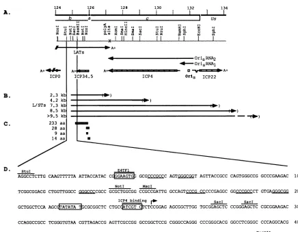
FIG. 9. Expanded physical mapof the region of HSV-1 DNA encoding the L/STs. (A) Beneath the scale of kilobase pairs are shown the
locations of theb,a,andcsequencesand of relevantrestriction sites in KOS DNA. Beneath themapofrestriction sitesareshown the 3' end of theLATs, the 5' end of the ICPO transcript, and the intact transcripts encodingICP34.5, ICP4, and ICP22. OriSRNAI and RNA2arealso shown. OriS is located between the5' startsites of the ICP4 and ICP22 transcripts. (B) Locations ofsequences specifying the L/STs. The directionof
transcription is indicated by thearrows.The 5' ends of thetranscripts lie between the NotI andSaclsites. The 3' endsof thetranscripts havenot
beenmapped andareshown inparentheses. (C) The locations of the four potential ORFs within the abundant 2.3-kbL/STareshownasboxes.
aa,amino acids.(D) Nucleotidesequenceof HSV DNA between the StuI andBssHIIsites. TheE4TF1 recognitionsite(31)and the ICP4binding site(ATCGTC)areshownasclosed boxes beneath lines.The TATA box is shownas aclosed box. Thesequencespecifyingthe N terminus of the
233-amino-acidORF is shaded. The transcriptional startsite is indicatedbyanarrow.
DISCUSSION
The L/STs: a new class of HSV-1 transcript. A family of transcripts that span thejunction between the L and S
com-ponentsof the HSV-1 genome,the L/STs, hasbeenidentified by Northern blot analysis in RNA from cells of neural and nonneuralorigin infected with ICP4 nullmutants(Fig. 9). The abundant expression of the L/STs only in the absence of functional ICP4, their expression with late kinetics, and the sensitivityoftheirsynthesis tocycloheximide inICP4
mutant-infectedcells indicate that theyarenotmembersofanyof the
recognized kinetic classes of HSV-1 transcripts but constitutea
new class of viraltranscript.
Location and properties of the L/STs. The location of a
single 5' start site at nucleotide 125042, together with the failure to detect hybridizable transcripts with riboprobe EBNc3-LAT(S), 710 bptothe left of the StuI siteatnucleotide 124818, indicate that the L/STsare5' coterminal and that their
expressionisregulated by the bidirectionalpromoterdescribed by Bohenzkyetal. (4). The fact that theL/STsareexpressedin
abundance onlyin the absence ofICP4,or in thepresence of
ICP4peptides thatare unabletobindATCGTC, is consistent with the presence of a consensus ICP4 binding site 6 bp upstream of the start site and may explain the difficulties experienced by Bohenzky et al. in identifying and mapping these transcripts in RNA from wild-type virus-infected cells. Although we have not yet mapped the 3' end of the 2.3-kb L/ST species,weestimatethat theterminationsite liesnearthe
polyadenylation sites utilized by ICP4 mRNA and the large LATs (Fig. 9A and B). Notably, the abundant 2.3-kb species
appearstoconsist ofaseries oftranscriptsthat differ in sizeby
auniformlength. Shouldthese abundanttranscriptsbe both 5' and 3'coterminal, it ispossiblethat these small size differences arise from the presence ofserially repeated DNA sequences
located to the right of the a sequence which vary in copy
number among genomes within a given virus preparation as
describedbyDavison and Wilkie(12) andMcGeoch et al.(36, 38).
Thesequence of the HSV-1 strain 17genome encodingthe
2.3-kbL/STshas been determinedbyDavison and Wilkie(12) andMurchie and McGeochet al. (38), and the sequences of
A.
LATs
A+41
ICP0
Al * ICP34.5
B.
C.
2.3 kb 4.2 kb
L/STs 7.3 kb 8.5 kb >9.5 kb
233 aa 28 aa
9 aa
14 aa
U
U
d
VOL.67, 1993
4
on November 9, 2019 by guest
http://jvi.asm.org/
thebidirectional promoterand thea repeathave been deter-mined for strain KOS (4, 48). The DNA template specifying the 2.3-kb L/STs contain one long continuous open reading
frame (ORF) and three short ORFs (Fig. 9C). The first and longest ORFstarts at nucleotide 125180 and extends for 702 bp to nucleotide 125881. It is 233 amino acids in length and extremely arginine rich. A search of the protein data base revealed limited homology between this L/ST ORF, the early proteinEPOof suid herpesvirus1 (7), andEBNA-1of Epstein-Barr virus (43). The second ORF consists of only 87 bp (nucleotides 125833 to 125919). The third and fourth ORFs
are evensmaller, consisting of only 30 and 45 bp,respectively.
None of the fourORFs extends into theasequence(125955to
126373). Notably, ORF1,and indeed the L/STs themselves, do not appear to be essential for virus replication because the mutations in severalviablemutantsin theICP34.5gene,which
also disrupt theL/STs andORF1, have beenisolated by Chou et al. (8). On the other hand, because the mutations in the ICP34.5 gene disrupt the L/STs, it is unclear whether the
altered virulence of the ICP34.5 mutants in mice is a
conse-quence of mutations in sequences specifying ICP34.5 or the
L/STs.
Factors affecting L/ST expression. A distinguishing feature of any gene or cis-acting element contained totally within
repeat sequences is that it is diploid. For genes encoding
trans-acting factors, the amplification of the protein product that results from the expression of both genes has significant
implications for gene regulation. Forexample, the expression oftwocopies of ICPO rather thanone doubles the viral yield
andtheefficiencyofvirusreactivation from latency ina mouse
model (5). Anadditional instance of diploidy involves the L/S junction. Any gene containing the L/S junction would be diploid only when the genome is circular, i.e., immediately
after the genome enters the nucleus during productive infec-tion, and during latency. In addition, junction-spanninggenes
wouldoccurinhighercopynumber in concatemericmolecules generated during viral DNA replication relative to single, linear packagedgenomes.
Because the expression of ICP4 is essential forDNA repli-cationbutrepressive for expression of the L/STs, and because L/ST expressionwasnot detected in KOS-infected cellsatthe timeofpeakDNAreplication (6to12hpi), the L/STsarenot
likely expressed from concatemers. Our studies show that the L/STsaresynthesizedin abundance in cells infectedwith ICP4 null mutants andat low levels in cells expressinglow levelsof
ICP4(E5 cells) butareundetectable in cells infectedwithKOS or ICPO, ICP22, ICP27, and LAT null mutants (all of which
express high levels of ICP4). Moreover, L/ST synthesis is completely inhibited in ICP4 null mutant-infected cells in the
presence of cycloheximide. Collectively, these observations suggest that ICP4 represses expression of the L/STs and that
L/ST expression requires either one or more of the viral
proteins synthesized in ICP4 null mutant-infected cells (i.e., ICPO, ICP6, ICP22, ICP27, or ICP47) and/or a cellular
pro-tein(s) induced byoneof these viral proteins.Theproximityto
theL/STstart site ofan ICP4 binding site long recognizedto
mediate the repressive effects of ICP4 and the failure of mutant forms of ICP4 unabletobind this siteto inhibit L/ST
expression support the hypothesis that ICP4 represses L/ST
expression.
Amongthe viral regulatoryproteins expressed in ICP4null
mutant-infected cells that may, in theory, be involved in
activating L/ST expression, Bohenzky et al. have shown that
the L/ST promoter is only modestly activated by ICPO and
weakly activatedby VP16. Hence, the probability that either
ICPO or VP16 alone induces L/ST expression at such high
levels is not great. ICP22 has recently been shown to repress the transactivating activities of ICPO and ICP4(1), and
ICP27
is also able to repress ICPO- and ICP4-induced activation of viral genes. By virtue of their ability to repress the transacti-vating activity (albeit not thesynthesis) of ICP4, eitherof these proteins might play a role in activating L/ST expression. Finally, ICP6 has recently been reported to possess phospho-rylating activity in addition to its role as the large subunit of ribonucleotide reductase (11). ICP6 is present in abundance at IE times and may function as a protein kinase to regulate the activities of other IE and cellular proteins.When do the L/STs function? Generally speaking, the kinetics of expression of individual viral genes parallels the time period during viral replication when the products of these genes function. If one assumes that this is the case for the L/STs, when might they function during replication? Given the observations that the L/STs accumulate in abundance with late kinetics in the absence of ICP4, that they would be expressed at maximum levels from a diploid template in circular ge-nomes, and that they likely require other HSVIE proteins or cellular proteins for their synthesis, when during the life cycle of HSV-1 are the L/STs likely to be synthesized in greatest abundance? Available evidence indicates that ICP4 is not expressed (or is expressed only at low levels) (i) in productive infection prior to IE gene expression, (ii) at late times in the viral replication cycle, and (iii) during latency. Because the L/STs are expressed with late kinetics in the absence of ICP4, it is unlikely that they are synthesized prior to IE gene expression in productive infection. High levels of ICP4 are present at IE and E times, and low levels of ICP4 persist even late in infection. Moreover, the synthesis of low levels of L/STs in n12-infected E5 cells which express low levels of ICP4 indicates that they might be synthesized late in infection when levels of ICP4 have dropped. The expression of very low levels of the 2.3-kb L/STs late in KOS infection (Fig. 5) would support this hypothesis. That the L/STs function late in productive infection is plausible but unlikely because (i) virus replication was complete when the L/STs were detected in KOS-infected cells (24 hpi; Fig. 5) and (ii) the extremely low levels of L/STs detected at late times argue against their functioning maximally at this time. On the other hand, it may be that the L/ST gene product functions during the succeeding replication cycle as does VP16 (24). One must also consider the stages of the viral life cycle that include the establishment, maintenance, and reactivation of latency as times when the ICP4 gene is repressed. We have examined mouse ganglia latently infected with wild-type strain KOS for L/ST expression and have detected none. Furthermore, previous studies using L/ST template sequences as probes in in situ hybridization tests didnot detect these transcripts in latently infected ganglia (14, 33, 37,40, 50). Thus, it appears as if the L/STs are not present (at least not at high levels) in latently infected ganglionic neurons during the maintenance of latency.
The absence of L/ST expression in latently infected gangli-onic neurons is in sharp contrast to the abundant expression of the LATs in these cells. Indeed, expression of these two families of transcripts appears to be mutually exclusive both in vivo, as just noted, and in vitro, where abundant LATs are detected only in the presence of ICP4, whereas L/STs are detected only in the absence of ICP4. Thus, we attribute the absence of L/ST expression in dlLAT1.8-infected cells to the expression of ICP4. The definitive test of the relationship of theLATs and the L/STs will require construction of an L/ST-mutant virus (which should express the LATs), and a LAT-ICP4- mutant virus (which should express the L/STs).
One must also consider the possibility that the L/STs are
on November 9, 2019 by guest
http://jvi.asm.org/
HSV-1 L/STs 7381 expressed only during a brief period of time during HSV-1
infection, such as during the switch from lytic infection to latency, or during the sequence of events that lead to reacti-vation. ICP4 is known to be actively repressed during the establishment of latency and immediately prior to reactivation (50), yet at both stages of latency, the viral genome is circular and otherIEproteins and cellular proteins may be synthesized, facilitating expression of the L/STs. To determine whether this is indeed the case, Northern blot analysis of infected ganglia during the establishment of and reactivation from latency must be conducted.
In this report, we have described a new class of HSV-1 transcript represented by the L/STs. Identification of the specific function(s) of the L/STs will necessitate the isolation and characterization of mutant viruses which fail to synthesize one or more of the L/STs. Whether the L/STs are the only representatives of this new class of transcript or whether additional transcriptsof this class are encoded by otherregions of the viral genome remain to be determined.
ACKNOWLEDGMENTS
We thank Christine Dabrowski, Robert Jordan, and DavidFrazier for helpful discussions, Lauren Liptak for assistance in plasmid construction, and Monica Shea for preparation of the manuscript.
This study was supported by research grants R37CA20260fromthe National Cancer Institute and P01AI24010 from the National
Insti-tute of Allergy and Infectious Diseases. L.Y. was supported in part by National Science Foundation predoctoral fellowship 75982.
REFERENCES
1. Astor, T. A., S. Rundle, C. L. Bogard, W. Cai, and P. A.Schaffer.
Unpublished data.
2. Batchelor, A. H., and P. O'Hare. 1990. Regulation and
cell-type-specific activity of a promoter located upstream of the latency-associated transcript of herpes simplex virus type 1. J. Virol. 64:3269-3279.
3. Batterson, W., and B. Roizman. 1983. Characterization of the herpes simplex virion-associatedfactor responsible for the
induc-tion of the alpha genes. J. Virol. 46:371-377.
4. Bohenzky, R. A., A. G. Papavassiliou, I. H. Gelman, and S.
Silverstein. 1993. Identification of a promoter mappingwithin the
reiterated sequences that flank theherpes simplex virus type 1UL region. J. Virol. 67:632-643.
5. Cai, W., T. L. Astor, L. M. Liptak, C. Cho, D. M. Coen,and P. A.
Schaffer. 1993. The herpes simplex virus type 1 regulatory protein
ICP0 enhances virus replication during acute infection and reac-tivation from latency. J. Virol. 67:7501-7512.
6. Cai, W., and P. A.Schaffer. 1989.Herpes simplex virus type 1 ICPO plays a critical role in the de novo synthesis of infectious virus
following transfection of viral DNA. J. Virol.63:4579-4589. 7. Cheung, A. K. 1991. Cloning of the latency gene and the early
protein 0gene of pseudorabies virus. J. Virol. 65:5260-5271.
8. Chou, J., E. R. Kern, R. J. Whitley, and B. Roizman. 1990.
Mapping of herpes simplex virus-I neurovirulence to y, 34.5, a
gene nonessential for growthin culture. Science 250:1262-1266. 9. Chou, J., andB. Roizman. 1986. The terminal a sequenceofthe
herpes simplex virus genome contains the promoter of a gene
located in the repeat sequences of the L component. J. Virol.
57:629-637.
10. Chou, J., and B. Roizman. 1992. The yl 34.5 gene of herpes simplex virus1 precludesneuroblastoma cellsfromtriggeringtotal
shutoff of protein synthesis characteristic of programmed cell
death in neuronal cells.Proc.Natl.Acad. Sci. USA 89:3266-3270.
11. Conner, J., J. Cooper, J. Furlong, and J. B. Clements. 1992. An
autophosphorylatingbut not transphosphorylating activity is
asso-ciatedwith the unique N terminusof the herpessimplexvirus type
I ribonucleotide reductase large subunit. J.Virol. 66:7511-7516. 12. Davison, A. J., and N. M. Wilkie. 1981. Nucleotide sequences of
the joint between the L and S segments ofherpes simplex virus
types1 and2. J. Gen. Virol. 55:315-331.
13. Deatly, A. M., J.G. Spivack, E. Lavi, and N. W. Fraser. 1987. RNA
from an immediate early region of the type 1herpessimplexvirus genome is present in the trigeminal ganglia of latently infected mice. Proc. Natl. Acad. Sci. USA 84:3204-3208.
14. Deatly, A. M., J.G. Spivack, E. Lavi, D. R. O'BoyleII,and N.W.
Fraser. 1988. Latent herpes simplex virus type 1 transcripts in peripheral and central nervous system tissues of mice map to similarregionsof the viralgenome. J. Virol. 62:749-756. 15. Deiss, L. P., J. Chou, and N. Frenkel. 1986. Functional domains
within the a sequence involved in the cleavage-packagingofherpes simplex virus DNA. J. Virol. 59:605-618.
16. Deiss, L. P., and N. Frenkel. 1986.Herpes simplex virus amplicon: cleavageofconcatemericDNAis linkedtopackagingandinvolves amplification of the terminally reiterated a sequence. J. Virol. 57:933-941.
16a.DeLuca, N. A.,M. A. Courtney, and P. A.Schaffer. 1984. Temper-ature-sensitive mutants in herpes simplex virus type 1 ICP4 permissive forearly gene expression. J. Virol. 52:767-776.
17. DeLuca, N. A., M. McCarthy, and P. A. Schaffer. 1985. Isolation and characterization of deletion mutants ofherpes simplex virus type 1 in the gene encoding immediate-early regulatory protein ICP4. J. Virol. 56:558-570.
18. DeLuca, N. A., and P.A.Schaffer. 1985.Activation of immediate-early, immediate-early, and late promoters by temperature-sensitive and wild-type forms of herpes simplexvirus type 1 protein ICP4. Mol. Cell. Biol. 5:1997-2008.
19. DeLuca, N. A., and P. A. Schaffer. 1987. Activities of herpes simplex virus type 1 (HSV-1) ICP4 genes specifying nonsense peptides. Nucleic Acids Res. 15:4491-4511.
20. DeLuca, N. A., and P. A. Schaffer. 1988. Physical and functional domains of the herpes simplex virus transcriptional regulatory protein ICP4. J. Virol. 62:732-743.
21. Devi-Rao, G., S. A.Goodart, L. M. Hecht, R. Rochford, M. A.Rice, and E. K. Wagner. 1991. Relationship between polyadenylated and nonpolyadenylated herpes simplex virus type 1 latency-asso-ciated transcripts. J. Virol. 65:2179-2190.
22. DiDonato, J. A., J. R. Spitzner, and M. T. Muller. 1991. A predictive modelfor DNA recognition by the herpes simplex virus protein ICP4. J. Mol. Biol. 219:451-470.
23. Dixon, R. A. F., and P. A. Schaffer. 1980. Fine-structure mapping and functional analysis of temperature-sensitive mutants in the gene encoding the herpes simplex virus type 1 immediate early protein VP175. J. Virol.36:189-203.
24. Goding, C. R., and P. O'Hare. 1989. Herpes simplex virus Vmw65-octamer binding protein interaction: a paradigm for combinatorial control oftranscription. Virology 173:363-367.
25. Greene, L. A., and A. S.Tischler. 1982. PC12 pheochromocytoma cutures in neurobiological research. Adv. Cell. Neurobiol. 3:373-414.
26. Holland, L. E., K. P. Anderson, C. Shipman, Jr., and E. K.
Wagner. 1980. Viral DNA synthesis is required for the efficient expression of specificherpes simplex virus type 1 mRNA species. Virology 101:10-24.
27. Honess, R. W., and B. Roizman. 1974. Regulation of herpesvirus macromolecular synthesis. I. Cascade regulation of the synthesis of three groups of viral proteins. J. Virol. 14:8-19.
28. Honess, R. W., and B. Roizman. 1975. Regulation of herpesvirus macromolecular synthesis: sequential transition of polypeptide synthesis requires functional viral polypeptides. Proc. Natl. Acad. Sci. USA72:1276-1280.
29. Imbalzano, A. N., D. M. Coen, and N. A. DeLuca. 1991. Herpes simplexvirus transactivator ICP4 operationally substitutes forthe cellular transcription factor Spl for efficient expression of the viral thymidine kinase gene. J. Virol. 65:565-574.
30. Imbalzano, A. N., A. A. Shepard, and N. A. DeLuca. 1990. Functional relevance of specific interactions between herpes sim-plex virus type I ICP4 and sequences from the promoter-regula-tory region of the viral thymidine kinase gene. J. Virol. 64:2620-263 1.
31. Jones, N. C., P. W. J. Rigby, and E. B. Zif. 1988. Trans-acting protein factors and the regulation of eukaryotic transcription: lessons from studies on DNA tumor viruses. Genes Dev. 2:267-281.
VOL.67, 1993
on November 9, 2019 by guest
http://jvi.asm.org/
32. Jones, P. C., and B. Roizman. 1979. Regulation of herpesvirus macromolecular synthesis. VIII.The transcription program
con-sists of three phases duringwhich bothextentoftranscription and accumulation of RNA in the cytoplasm are regulated. J. Virol.
31:299-314.
33. Krause,P. R., K. D. Croen, S. E.Straus,andJ. M.Ostrove. 1988. Detection and preliminary characterization of herpes simplex virus type transcripts in latently infected human trigeminal ganglia. J. Virol. 62:4819-4823.
34. Lieb, D. A.,C. L. Bogard, M. Kosz-Vnenchak, K. A. Hicks, D. M. Coen, D. M. Knipe, and P. A.Schaffer. 1989.Adeletionmutantof the latency-associated transcript of herpes simplex virus type 1
reactivates from the latentstatewith reduced frequency. J. Virol. 63:2893-2900).
35. McCarthy, A. M., L. McMahan, and P. A. Schaffer. 1989.Herpes
simplex virus type I ICP27 deletion mutants exhibit altered
patternsoftranscription and are DNAdeficient. J. Virol.
63:18-27.
36. McGeoch, D. J., A. Dolan, S. Donald, and F. J. Rixon. 1985. Sequencedetermination and geneticcontentofthe short unique regioninthegenomeof herpes simplex virustype1.J. Mol. Biol. 181:1-13.
37. Mitchell, W. J., R. P. Lirette, and N. W. Fraser. 1990.Mappingof lowabundance latency-associated RNA in thetrigeminalganglia of micelatently infectedwith herpes simplex virustype 1.J. Gen.
Virol. 71:125-132.
38. Murchie,M.J., andD.J.McGeoch. 1982. DNAsequenceanalysis
ofanimmediate-earlygeneregion of the herpes simplex virustype
1 genome.J. Gen. Virol.62:1-15.
39. Nasseri, M., and E. S.Mocarski. 1988.Thecleavage recognition signaliscontained withinsequencessurroundingthea-ajunction
in herpes simplexvirus DNA.J. Virol. 167:25-30.
39a.O'Hare,P., and G. S.Hayward. 1985. Threetrans-acting
regula-toryproteinsofherpessimplexvirusimmediate-earlygene
expres-sion in apathway involving positive andnegative feedback
regu-lation. J. Virol. 56:723-733.
40. Rock, D. L.,A. B.Nesburn,H. Ghiasi, J. Ong,T. L.Lewis,J. R. Lokensgard, and S. L. Wechsler. 1987. Detection of
latency-related viral RNAs in trigeminal ganglia of rabbits latently
in-fected with herpes simplexvirustype 1.J. Virol.61:3820-3826.
41. Sacks, W. R., C. C.Greene, D. P.Aschman, and P. A. Schaffer.
1985.Herpessimplexvirustype ICP27 isanessentialregulatory
protein.J. Virol. 55:796-805.
42. Sacks, W. R.,and P.A. Schaffer. 1987. Deletion mutants in the
gene encoding the herpes simplex virus type I immediate-early
protein ICPO exhibit impaired growth in cell culture. J. Virol. 61:829-839.
43. Sample, J.,M. Hummel, D. Braun, M.Birkenbach,and E.Kieff. 1986. Nucleotide sequences of mRNAs encoding Epstein-Barr
virus nuclear proteins: a probable transcriptional initiation site.
Proc. Natl. Acad.Sci. USA83:5096-5100.
44. Sandri-Goldin, R. M., M. Levine, and J. C. Glorioso. 1981.
Method for introduction of mutations inphysically definedregions ofthe herpes simplexvirus genome. J. Virol. 38:41-49.
45. Sanger, F., S.Nicklen, and A.R.Coulson.1977. DNAsequencing with chain-terminating inhibitors. Proc. Natl. Acad. Sci. USA 74:5463-5467.
46. Schaffer, P.A., V. C. Carter,and M.C.Timbury. 1878. Collabo-rativecomplementationstudy oftemperature-sensitivemutantsof herpessimplexvirus types 1 and 2. Virology 27:490-504. 47. Smiley, J. R., J. Duncan, andM.Howes.1990. Sequence
require-mentsfor DNA rearrangements induced by the terminal repeatof herpes simplex virus type 1 KOS DNA. J. Virol.64:5036-5050. 48. Smiley, J. R., C. Lavery,and M.Howes.1992. Theherpessimplex
virus type 1 (HSV-1) a sequence serves as a cleavage/packaging signal but does not drive recombinational genome isomerization when it is insertedinto the HSV-2 genome. J. Virol.66:7505-7510. 49. Spaete, R. R., and N. Frenkel. 1982. The herpes simples virus amplicon: a new eucaryotic defective-virus cloning-amplifying vector.Cell 30:295-304.
50. Spivack, J. G., and N. W. Fraser. 1988. Expression of herpes simplex virus type 1 latency-associated transcripts in thetrigeminal gangliaof mice during acute infection and reactivation of latent infection. J. Virol. 62:1479-1485.
51. Stevens, J. G., E. K. Wagner, G.B.Devi-Rao,M. L.Cook,and L. T.
Feldman. 1987.RNAcomplementary to aherpesvirus alpha gene mRNA is prominent in latently infected neurons. Science 235: 1056-1059.
52. Stow, N. D. 1982. Localization of an origin of DNA replication within the TRs/IRs repeated region of the herpes simplex virus typeI genome. EMBO J. 1:863-867.
53. Stow,N.D., andE.C.McMonagle. 1983.Characterization of the TRs/IRsorigin of DNA replication of herpes simplex virus type 1. Virology 130:427-438.
54. Varmuza, S. L.,and J. R. Smiley. 1985. Signals for site-specific cleavage of HSV DNA: maturation involvestwoseparatecleavage
events atsites distal to therecognition sequences. Cell 41:793-802. 55. Vlazny, D. A., A. Kwong, and N. Frenkel. 1982. Site-specific cleavage/packagingofherpessimplex virus DNA and the selective maturation of nucleocapsids containing full-length viral DNA.
Proc. Natl.Acad. Sci. USA 79:1423-1427.
56. Wagner, E. K.,W. M.Flanagan,G. Devi-Rao, Y.-F. Zhang, J.M.
Hill,K. P.Anderson, and J. G. Stevens.1988.The herpessimplex virus latency-associated transcript is spliced during the latent phase ofinfection. J. Virol.62:4577-4585.
57. Weinheimer,S.P.,andS. L.McKnight. 1987.Transcriptional and post-transcriptional controls establish the cascade of herpes sim-plexvirusprotein synthesis.J. Mol.Biol. 195:819-833.
58. Weller, S.K.,K.J. Lee,D.J.Sabourin,andP.A.Schaffer. 1983. Genetic analysis of temperature-sensitive mutants which define the gene for themajorherpes simplex virus type I DNA-binding protein. J. Virol. 45:354-366.
59. Zhu,Z. M.Personal communication.