
0022-538X/95/$04.0010
Copyrightq1995, American Society for Microbiology
The NS1 Polypeptide of the Murine Parvovirus Minute Virus of Mice
Binds to DNA Sequences Containing the Motif [ACCA]
2–3SUSAN F. COTMORE,1JESPER CHRISTENSEN,1JURG P. F. NU¨ ESCH,1†
ANDPETER TATTERSALL1,2*
Departments of Laboratory Medicine1and Genetics,2Yale University School
of Medicine, New Haven, Connecticut 06510
Received 22 September 1994/Accepted 1 December 1994
A DNA fragment containing the minute virus of mice 3*replication origin was specifically coprecipitated in immune complexes containing the virally coded NS1, but not the NS2, polypeptide. Antibodies directed against the amino- or carboxy-terminal regions of NS1 precipitated the NS1-origin complexes, but antibodies directed against NS1 amino acids 284 to 459 blocked complex formation. Using affinity-purified histidine-tagged NS1 preparations, we have shown that the specific protein-DNA interaction is of moderate affinity, being stable in 0.1 M salt but rapidly lost at higher salt concentrations. In contrast, generalized (or nonspecific) DNA binding by NS1 could be demonstrated only in low salt. Addition of ATP orgS-ATP enhanced specific DNA binding by wild-type NS1 severalfold, but binding was lost under conditions which favored ATP hydrolysis. NS1 molecules with mutations in a critical lysine residue (amino acid 405) in the consensus ATP-binding site bound to the origin, but this binding could not be enhanced by ATP addition. DNase I protection assays carried out with wild-type NS1 in the presence ofgS-ATP gave footprints which extended over 43 nucleotides on both DNA strands, from the middle of the origin bubble sequence to a position some 14 bp beyond the nick site. The DNA-binding site for NS1 was mapped to a 22-bp fragment from the middle of the 3*replication origin which contains the sequence ACCAACCA. This conforms to a reiterated motif (ACCA)2–3, which occurs, in more or
less degenerate form, at many sites throughout the minute virus of mice genome (J. W. Bodner, Virus Genes 2:167–182, 1989). Insertion of a single copy of the sequence (ACCA)3was shown to be sufficient to confer NS1
binding on an otherwise unrecognized plasmid fragment. The functions of NS1 in the viral life cycle are reevaluated in the light of this result.
The small (5-kb), single-stranded DNA genome of the mu-rine parvovirus minute virus of mice (MVM) encodes two major blocks of open reading frame, with overlapping tran-scripts from one block programming the synthesis of the var-ious capsid polypeptides, while the other gives rise to a series of nonstructural (NS) proteins (14). MVM encodes at least four closely related NS proteins (17), but of these only one, the 83-kDa NS1 polypeptide, is essential for productive infection in all cell types (8, 34, 40). NS1, an abundant and long-lived nuclear phosphoprotein (13), performs many disparate func-tions in the viral life cycle. It serves as an initiator of viral DNA replication, introducing a single-strand nick into a specific site in concatemeric replication intermediates generated during MVM’s unique rolling-hairpin mode of DNA synthesis (11, 12, 18). This initiation step allows resolution and replication of the viral telomeres and leaves NS1 covalently attached to the new
59 end generated at each nick site (15), where its helicase
activity (30, 40a, 52) is thought to contribute to the movement
of the replication fork. Its eventual retention on the exposed 59
termini of encapsidated progeny DNA, even after their release from the host cell, suggests that NS1 may also be involved in the packaging and/or export of progeny virions (16). In addi-tion, NS1 acts as a potent transactivator of transcription from both the MVM P4 and P38 promoters (10, 20, 21, 27, 46), modulates the activity of various heterologous promoters (24, 31, 51), and serves multiple, as yet poorly defined, cytostatic or
cytotoxic functions (6, 7, 31, 33) which are presumed to facil-itate takeover of the host cell’s synthetic machinery by the invading virus.
NS1 must interact directly with MVM DNA at the replica-tion initiareplica-tion site, since an active-site tyrosine, most likely amino acid 210 in the NS1 sequence (40a), becomes covalently attached to a specific consensus DNA sequence during this reaction (18). However, in most prokaryotic rolling-circle rep-lication systems, such active-site recognition is weak, and a second domain in the initiator protein specifically recognizes and binds to an adjacent DNA sequence in the origin (4, 35, 43, 45). To examine whether NS1 functioned in a similar way, we set about reexamining the question of whether NS1 can bind to the origin in a site-specific manner. Since previous attempts to demonstrate such an interaction between NS1 and the viral DNA had met with little success (1, 26), we chose to reexamine this question by using an alternative technique which involves incubation of the candidate DNA-binding protein with a mix-ture of labeled DNA fragments of which only one contains the candidate binding site. The protein is then reisolated by spe-cific immunoprecipitation, and the complex is analyzed for coprecipitated DNA fragments (39). In this way, we were able to show that NS1 does bind to a specific DNA sequence in the
MVM 39replication origin and that this binding is potentiated
by the presence of ATP but eliminated by conditions which favor ATP hydrolysis. The core element in the binding site is
the motif [ACCA]2, and more or less degenerate forms of this
sequence are scattered throughout the viral genome (5). This observation suggests that, in addition to initiating viral DNA replication and trans regulating transcription, NS1 may per-form a number of previously unsuspected roles in the viral life cycle.
* Corresponding author. Mailing address: Department of Labora-tory Medicine, Yale University School of Medicine, 333 Cedar St., New Haven, CT 06510. Phone: (203) 785-4586. Fax: (203) 785-7340.
† Present address: Angewandte Tumorvirologie, Deutches Krebs-forschungzentrum, Im Neuenheimer Feld 280, D-6900 Heidelberg 1, Germany.
1652
on November 9, 2019 by guest
http://jvi.asm.org/
MATERIALS AND METHODS
Plasmids.Plasmid pL1-2TC, which contains the minimal 39replication origin of MVM on a 75-bp viral fragment cloned into the polylinker of the vector plasmid pCR II (Invitrogen, San Diego, Calif.), pL2.1-1 and pL4-2, which con-tain deleted forms of this origin, and pL1-2GAA, which concon-tains an inactive origin, have all been described previously (18). Plasmids pOri-Core and pATF-box were obtained by annealing overlapping oligonucleotides derived from the origin region (as shown in Fig. 5), extending them with Amplitaq (Perkin-Elmer Cetus, Norwalk, Conn.), and cloning the product into pCR II. Plasmid p[ACCA]3was similarly derived by using synthetic oligonucleotides. All insert
DNA in these plasmids was sequenced by using Sequenase (U.S. Biochemical, Cleveland, Ohio) as instructed by the supplier.
Plasmid digests used in binding assays. Plasmids pL1-2TC, pL1-2GAA, pL2.1-1, pL4-2, p[ACCA]3, and their parent vector, pCR II, were cut with
restriction enzymes Sau3A and NarI so that each gave 30 fragments ranging in size from 9 to 985 bp. The 39 ends of all DNA strands were filled in with Sequenase in the presence of three unlabeled nucleotides and [32
P]dGTP or [32
P]dATP, as indicated in the text, to yield mixtures of labeled, blunt-ended fragments. Plasmids pOri-Core and pATF-box were cut with HinfI and AflIII to yield 11 fragments, which were then similarly end labeled with Sequenase in the presence of [32P]dATP.
Preparation of NS1.Wild-type NS1 and NS2P(the major form of NS2) were
translated in a coupled reticulocyte T7 transcription-translation extract (Pro-mega, Madison, Wis.), using high-level TM1 expression constructs (42) in which the viral proteins are encoded downstream of an encephalomyocarditis virus leader sequence and a T7 promoter.
NS1 genes were modified to include an amino-terminal histidine tag sequence comprising six histidine residues separated from the authentic amino terminus of NS1 by an enterokinase cleavage site. These proteins were expressed in HeLa cells from recombinant vaccinia virus vectors and purified by nickel chelate chromatography as will be described elsewhere (40a). Wild-type NS1 bearing an amino-terminal histidine tag was also expressed in insect cells from a recombi-nant baculovirus vector, kindly provided by David Pintel, University of Missouri, and affinity purified in a similar manner (9a). NS1 preparations were routinely eluted in 17% glycerol–10 mM N-2-hydroxyethylpiperazine-N9-2-ethanesulfonic acid (HEPES)-KOH (pH 7.8)–50 mM NaCl–0.1 mM EDTA–1 mM dithiothrei-tol–80 mM imidazole (buffer A) and stored as aliquots at2808C, usually follow-ing the further addition of 5 mM MgCl2.
NS1 DNA binding assays.Binding assays were carried out at 48C in 100ml of 20 mM Tris-HCl (pH 8.0)–10% glycerol–1% Nonidet P-40, 5 mM dithiothreitol– 100 mM NaCl (buffer B) unless otherwise specified in the text. Site-specific binding assays contained 100 to 200 ng of poly(dI-dC), and nucleotides were added to 0.5 mM as indicated. NS1 was supplied in the form of a crude trans-lation extract (4ml) or as 1ml of the affinity-purified product containing approx-imately 50 ng of protein (in buffer A supplemented with 5 mM MgCl2). The
double-stranded competitor oligonucleotides Ori-core, ATF-box, and [ACCA]3
were prepared by annealing synthetic complementary single strands. These were then kinase treated and self-ligated so that they were present as mixtures of fragments ranging in size from 12 to approximately 500 bp. Assays described here contained 40 ng of competitor DNA. Other reaction components were added as indicated. In standard assays, NS1 was incubated for 10 min on ice with the appropriate mixture of poly(dI-dC), competitor oligonucleotide, EDTA, MgCl2,
and nucleoside triphosphate as indicated before the32P-labeled DNA fragments
(10 ng) were added. Samples were incubated for 30 min on ice before 2ml of the appropriate antiserum was added, and the incubation continued for a further hour. Thirty microliters of buffer B containing 1.5 mg of protein A-Sepharose and 20 ng of poly(dI-dC) was added, and the mixtures were tumbled for 30 min at 8 to 108C. After removal of the supernatant, the immunoprecipitates were washed twice with 1 ml of cold buffer B and the DNA was deproteinized by incubation with proteinase K in the presence of 0.5% sodium dodecyl sulfate (SDS) for 1 h at 558C. Samples were analyzed by electrophoresis on 2.5% agarose gels containing a 3:1 mixture of standard and NuSieve agarose (FMC, Rockland, Maine). Gels were fixed with 7% trichloroacetic acid, blotted, dried, and exposed to film and/or quantitated with a Molecular Dynamics Phosphor-Imager.
DNase I protection assays.Plasmid pL1-2TC was cut with either HindIII or XbaI and 39end labeled by incubation with [32P]dATP or [32P]dCTP,
respec-tively, in the presence of Sequenase and three unlabeled nucleotides. Labeled DNA was ethanol precipitated, digested with XbaI or HindIII, and gel purified from agarose. Extensively purified NS1 (500 ng) obtained by using the baculo-virus expression system was incubated with 53104
cpm of end-labeled probe in buffer B supplemented with 2mg of poly(dI-dC) per ml and 0.25 mMgS-ATP in a total volume of 50ml on ice for 1 h. An equal volume of prewarmed buffer B containing 4 mM MgCl2and variable amounts of DNase I (0.2 to 1.6 U per
reaction; Promega, Madison, Wis.) was added, and the reactions were allowed to proceed for 2.5 min at 258C before being terminated by the addition of 0.3 ml of 10 mM Tris-HCl–10 mM EDTA–0.5% SDS (pH 8.0) containing 300mg of proteinase K. After incubation for 45 min at 508C, the samples were extracted with phenol-chloroform, ethanol precipitated, and redissolved in gel loading buffer. Samples were analyzed by electrophoresis through denaturing acrylamide
gels (6%) against similarly labeled probe DNA which had been chemically cleaved at G residues by the procedure of Maxam and Gilbert (36).
RESULTS
A DNA fragment containing the MVM 3* origin is specifi-cally coimmunoprecipitated with NS1 but not NS2. Plasmid
pL1-2TC, which contains the minimal 39 replication origin of
MVM on a 75-bp viral fragment, and its parent vector, pCR II, were cut with restriction enzymes Sau3A and NarI, so that each
gave 30 fragments ranging in size from 9 to 985 bp. The 39ends
of all DNA fragments were then labeled with [32P]dGTP, and
the resulting mixtures were incubated with wild-type NS1 or
NS2Psynthesized in vitro in a combined
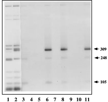
[image:2.612.328.540.74.274.2]transcription-transla-tion system. NS1 was then reisolated by immunoprecipitatranscription-transla-tion with antibodies directed against specific domains in the polypeptide, and any coprecipitated DNA fragments were an-alyzed by agarose gel electrophoresis. When pL1-2TC frag-ments were incubated with NS1 and the complexes were im-munoprecipitated with an antibody directed against the carboxy-terminal 16 amino acids of NS1, a 309-bp fragment containing the MVM origin was specifically coprecipitated (Fig. 1, lane 6). This fragment was not present if NS1 was omitted from the mixture (lane 4) or if nonimmune serum was used (lane 5). When pCR II sequences were substituted for the pL1-2TC fragments, the equivalent 234-bp polylinker fragment was not precipitated (lane 3). Similarly, the 309-bp origin frag-ment could be precipitated if a mixture of pL1-2TC fragfrag-ments and NS1 was precipitated with an antibody directed against a
FIG. 1. The MVM 39origin sequence specifically coprecipitates with NS1. The autoradiograph of an agarose gel shows [32
P]dGTP-labeled Sau3A-NarI double-digest fragments derived from pCR II (lane 1) and pL1-2TC (lane 2), prior to immunoprecipitation. Lanes 1 and 2 contain the equivalent of 1% of the input radioactivity present in the binding assays analyzed in lanes 3 through 11. Binding reactions contained pCR II DNA fragments (lane 3) or pL1-2TC DNA fragments (lanes 4 to 11). These reactions also contained in vitro translation mixes containing no NS proteins (lane 4), NS1 (lanes 3, 5 to 8, 10, and 11), or NS2P
(lane 9). After incubation, these mixes were immunoprecipitated with prebleed serum (lane 5), an antiserum directed against the carboxy-terminal 16 amino acids of NS1 (lanes 3, 4, and 6), an antiserum, designated B, directed against NS1 amino acids 284 to 459 (lane 7), or an antiserum directed against the amino-terminal peptide of NS1 (lanes 8 and 9). Samples in lanes 10 and 11 were both precipitated with the antiserum directed against the NS1 carboxy terminus, but the sample in lane 10 also received antiserum B early in the reaction, while that in lane 11 received antiserum B just prior to immunoprecipitation. Vector DNA fragments of 105 and 248 bp are indicated, together with the 309-bp fragment from pL1-2TC which contains the MVM 39origin.
VOL. 69, 1995 SITE-SPECIFIC DNA BINDING BY MVM NS1 1653
on November 9, 2019 by guest
http://jvi.asm.org/
91-amino-acid domain from the amino-terminal region of NS1
(lane 8), but this was not precipitated if NS2P, the major form
of NS2, another nonstructural polypeptide which shares this amino-terminal domain, was substituted for NS1 in the mixture (lane 9). Thus, this interaction appears to be specific for the NS1 polypeptide. While all lanes contain trace amounts of higher-molecular-weight fragments precipitated nonspecifi-cally, there are also two plasmid fragments, of 248 and 105 bp, which specifically coprecipitate with NS1, albeit less efficiently than the origin fragment. These interactions will be discussed in a subsequent section dealing with the NS1 recognition se-quence.
A third anti-NS1 antibody, directed against amino acids 284 through 459 in the 672-amino-acid NS1 sequence (nucleotides 1110 to 1638 in the MVM sequence [3]), previously called antiserum B (15), was also examined for its ability to precipi-tate complexes of NS1 with the origin fragment. The peptide recognized by this antibody comprises just over a quarter of the NS1 sequence, starting downstream of the putative active-site tyrosine residue at amino acid 210 (40a), extending through the middle of the protein and into the P loop of the consensus ATP-binding site centered around lysine 405. Unlike the anti-NS1 carboxy- or amino-terminal peptide sera, antibodies against the middle of NS1 were unable to precipitate the origin complexes (Fig. 1, lane 7) and could block complex formation if added to the reaction mixture early. This was demonstrated by adding antiserum B to the reaction mixture at the same time as the target DNA and then precipitating any resulting com-plexes with one of the competent antibodies (lane 10). A con-trol precipitate (lane 11) shows that antiserum B added to the reaction immediately prior to the final adsorption step did not interfere with origin coprecipitation, indicating that the immu-noadsorbant used was able to scavenge the additional antibody present in these reactions. Thus, complexes formed between NS1 and the origin sequence can be sequestered by using
antibodies directed against either end of the NS1 polypeptide chain, but antibodies recognizing sequences in the middle of the protein specifically block complex formation.
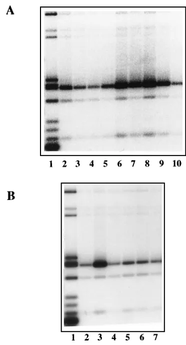
Affinity-purified, histidine-tagged NS1 specifically copre-cipitates the origin fragment.Wild-type and mutant NS1 genes were modified to include an amino-terminal histidine tag se-quence, expressed in HeLa cells from a recombinant vaccinia virus vector, and purified by nickel ion chromatography as described elsewhere (40a). Figure 2A shows the wild-type NS1 preparation used in subsequent assays. Although NS1 is by far the major component, this preparation is not entirely homo-geneous, containing a minor contaminant with a molecular weight of around 56,000 which does not react with anti-NS1 antibodies. Several other NS1 preparations were also tested and gave results comparable to those presented here. When this material was used in a standard coprecipitation experiment with pL1-2TC fragments, it specifically associated with the origin fragment (Fig. 2B, lanes 4 and 8). Thus, affinity-purified preparations of NS1 carrying an amino-terminal histidine tag are also able to mediate the specific origin-binding reaction.
Nonspecific DNA binding by NS1 can be demonstrated only in low salt, but the specific NS1-origin interaction is of sub-stantially higher affinity. The effects of varying the salt con-centration were examined by using histidine-tagged, affinity-purified NS1. Reaction mixtures containing 50 or 100 mM NaCl allowed efficient coprecipitation of the origin fragment (Fig. 2B, lanes 3, 4, 7, and 8), although precipitates obtained in 50 mM NaCl (lanes 3 and 7) were more heavily contaminated with other DNA fragments than those precipitated in 100 mM NaCl (lanes 4 and 8). However, complex formation was se-verely impaired in 150 mM NaCl (lanes 5 and 9), whether or not 0.5 mM ATP was included in the reaction mixture (dis-cussed in detail below). Thus, the specific interaction between NS1 and the origin sequence is of moderate affinity. In con-trast, if nonspecific DNA [poly(dI-dC)] was omitted from the
FIG. 2. Effects of NaCl concentration on specific and nonspecific DNA binding by NS1. (A) Protein molecular weight markers and an affinity-purified, histidine-tagged NS1 preparation (NS1 estimated at 1.75mg by comparison with standard proteins) electrophoresed on a Laemmli SDS-acrylamide gel and stained with Coomassie brilliant blue. (B) Autoradiograph of an agarose gel in which lane 1 contains the equivalent of 2.5% of the input [32P]dGTP-labeled pL1-2TC DNA
fragments used in each of the assays analyzed in lanes 2 through 9. Binding reaction mixtures containing this DNA and 50 ng of affinity-purified NS1 were incubated with (lanes 2 to 5) or without (lanes 6 to 9) 0.5 mM ATP under standard conditions except that the NaCl concentration was varied from 50 mM (lanes 2, 3, 6, and 7), through 100 mM (lanes 4 and 8), up to 150 mM (lanes 5 and 9). Samples were precipitated with prebleed serum (lanes 2 and 6) or with an antiserum against the carboxy-terminal peptide of NS1. (C) Autoradiograph of an agarose gel in which lane 1 again contains the equivalent of 2.5% of the input pL1-2TC DNA fragments used in each binding assay. Binding reactions containing this DNA and 50 ng of affinity-purified NS1 were carried out in the absence of poly(dI-dC) competitor DNA and in NaCl concentrations which varied from 25 mM (lanes 2 and 3), through 50 mM (lanes 4 and 5), to 100 mM (lanes 6 and 7). Samples were precipitated with prebleed serum (lanes 2, 4, and 6) or with an antiserum against the carboxy terminus of NS1 (lanes 3, 5, and 7).
on November 9, 2019 by guest
http://jvi.asm.org/
reaction, similar assays could be used to show that NS1 also exhibits nonspecific DNA-binding activity, but this is of sub-stantially lower affinity since it could be demonstrated only in low-salt conditions (up to a maximum of 25 mM NaCl, as seen in Fig. 2C, lane 3). At higher salt concentrations, nonspecific binding is progressively lost, so that at 100 mM NaCl (Fig. 3C, lane 7), site-specific binding predominates despite the lack of unlabeled nonspecific competitor DNA.
Binding of NS1 to the origin is enhanced by the addition of
ATP.Affinity-purified NS1 preparations were assayed for their
ability to interact with the viral 39 origin under a variety of
assay conditions. In the experiment shown in Fig. 3, incubation of NS1 with pL1-2TC fragments under standard assay condi-tions resulted in the precipitation of around 7% of the input origin fragment (Fig. 3A, lane 2). If scavenger amounts of EDTA (0.1 mM) were added to this mixture, the proportion of
origin fragments precipitated dropped marginally (to 3.9%;
Fig. 3A, lane 3), but further addition of 0.5 mM MgCl2had
little effect on this percentage (2.9% precipitated; lane 4), and the subsequent addition of 0.5 mM rGTP enhanced precipita-tion only twofold, to 6.1% of the input fragment. In contrast,
addition of 0.5 mM ATP (lane 6) orgS-ATP (lane 7) raised the
percentage precipitated to 21.1 or 18.8%, respectively, of the input fragment, representing a six- to sevenfold enhancement over unstimulated levels under the same assay conditions. This stimulation upon addition of 0.5 mM ATP has been seen consistently over many assays and varies between 4- and 10-fold. In addition, dATP has been found to be as effective as rATP in stimulating specific binding. In Fig. 2B, comparison of lanes 3 and 4 with lanes 7 and 8 suggests that this ATP-mediated enhancement in origin binding is, at least in part, mediated by an increase in the affinity of the interaction, since in the absence of ATP, standard assay mixtures containing 100 mM NaCl are too stringent for maximal binding. These obser-vations suggest that ATP brings about a change in the confor-mation or oligomerization state of the NS1 polypeptide which favors site-specific DNA binding.
In the presence of 0.5 mM ATP, further addition of MgCl2
to a final concentration of 1 mM had little effect (Fig. 3A, lane 8; 26.9% of the input fragment precipitated), but concentra-tions of 2 mM (lane 9) and 4 mM (lane 10) progressively impaired the interaction so that only 10.4 and 3.0%, respec-tively, of the input origin fragment was captured in the immu-noprecipitate. We believe that this effect might be due to enhanced ATP hydrolysis. Early steps in these assays are rou-tinely carried out on ice, but the final adsorption step is
per-formed in a cold box at around 108C, and thus limited ATP
hydrolysis is possible at this time.
NS1 molecules with mutations at lysine 405, in the consen-sus ATP-binding site, coprecipitate with the viral origin but do not respond to the addition of ATP.NS1 genes containing a lysine-to-arginine (K405R) or lysine-to-methionine (K405M) mutation at a critical residue in the consensus ATP-binding site were tagged with the same amino-terminal histidine se-quence used for the wild-type molecule. We have shown pre-viously that these mutant forms of NS1 are completely unable to initiate viral DNA replication or transactivate transcription from the viral P38 promoter (42). Mutant genes were recom-bined into vaccinia virus vectors, and the proteins were affinity purified as before. Protein concentrations were matched to wild-type NS1 by SDS-polyacrylamide gel electrophoresis fol-lowed by staining with Coomassie brilliant blue, and wild-type and mutant proteins were assayed for site-specific DNA bind-ing against pL1-2TC fragments under standard conditions. In the absence of added ATP, all NS1 preparations coprecipi-tated the origin fragment to similar extents (Fig. 3B, lanes 2, 4, and 6), but whereas addition of 0.5 mM ATP enhanced the proportion of the origin fragment precipitated by wild-type NS1 fivefold (from 5.1 to 26.2%; Fig. 3B, lanes 2 and 3), similar additions had little effect on the percentage precipitated by the ATP-binding-site mutants (Fig. 3B, lanes 5 and 7). This result suggests that the mutants do not undergo the same steric changes as the wild-type molecule upon addition of ATP. Since ATP binding, but not hydrolysis, is required to enhance DNA binding by wild-type NS1, this observation may indicate that neither the K405R nor the K405M mutant binds ATP effi-ciently.
NS1 protects much of the minimal origin sequence from digestion with DNase I.DNase I protection assays were carried out with affinity-purified wild-type NS1 in the presence of 0.5
mMgS-ATP and under the same stringent reaction conditions
[image:4.612.80.266.68.412.2]as used for the previous DNA binding assays. These assays
FIG. 3. Effects of ATP addition on origin-specific DNA binding by wild-type and mutant NS1. (A) PhosphorImager reconstruction of an agarose gel showing, in lane 1, the equivalent of 5% of the input [32P]dGTP-labeled pL1-2TC DNA
fragments used in each of the assays analyzed in lanes 2 through 10. Binding reaction mixtures containing this DNA and 50 ng of NS1 were incubated under standard assay conditions (lane 2) or following the addition of EDTA to 0.1 mM (lanes 3 to 10), MgCl2to 0.5 mM (lanes 4 to 7), to 1 mM (lane 8), to 2 mM (lane
9), or to 4 mM (lane 10), rGTP to 0.5 mM (lane 5), rATP to 0.5 mM (lanes 6 and 8 to 10), orgS-ATP to 0.5 mM (lane 7). Samples were immunoprecipitated with antiserum directed against the carboxy terminus of NS1. (B) Autoradiograph of an agarose gel in which lane 1 again contains the equivalent of 5% of the input [32P]dGTP-labeled pL1-2TC DNA fragments used in each binding assay.
Bind-ing reaction mixtures containBind-ing this DNA and 50 ng of wild-type NS1 (lanes 2 and 3) or the ATP-binding site mutant K405M (lanes 4 and 5) or K405R (lanes 6 and 7) in the absence of ATP (lanes 2, 4, and 6) or in the presence of 0.5 mM ATP (lanes 3, 5, and 7) were immunoprecipitated with antiserum against the carboxy terminus of NS1.
VOL. 69, 1995 SITE-SPECIFIC DNA BINDING BY MVM NS1 1655
on November 9, 2019 by guest
http://jvi.asm.org/
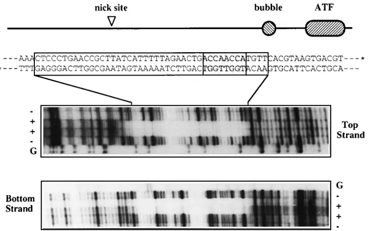
revealed a footprint of some 43 bp extending over both DNA strands of the minimal origin, the landmarks of which are
depicted in Fig. 4. The 39hairpin of MVM contains a stem in
which there is a mismatched bubble sequence where a GA doublet opposes a GAA triplet. During replication, this hairpin is copied to form an imperfect palindrome which bridges ad-jacent genomes in a dimer duplex intermediate, leaving the two bubble sequences embedded in potential replication origins on either side of the axis of symmetry. The sequence surrounding the doublet is a potent origin, but the analogous region con-taining the triplet is completely inactive (18). The active origin is about 50 bp long, extending from an Activated Transcription Factor (ATF)-binding site at one end to a position some 7 bp beyond the major initiation site, to which NS1 ultimately be-comes covalently attached. The actual sequence of the GA doublet is unimportant, but insertion of any third nucleotide here inactivates the origin, indicating that it represents a crit-ical spacer element. The NS1 footprint shown in Fig. 4 extends from a position 14 nucleotides beyond the nick site, across much of the essential origin sequence to the first nucleotide of the bubble region.
NS1 interacts with sequences in the middle of the 3* repli-cation origin which contain the motif ACCAACCA.As shown
in Fig. 5A, the active 39replication origin of MVM is present
in pL1-2TC, whereas the inactive form of the origin, in which the triplet GAA is substituted for the doublet TC in the bubble sequence, is present in the equivalent plasmid pL1-2GAA. Digestion of pL1-2GAA with Sau3A and NarI gives a 310-bp fragment which corresponds to the pL1-2TC 309-bp origin fragment. When assayed for its ability to interact with NS1, the GAA fragment bound to NS1 as efficiently as the competent origin (Fig. 5B; compare lane 6, in which 28.8% of the input TC fragment is precipitated, with lane 7, in which 28.5% of the input GAA fragment is precipitated).
Two deleted forms of the active origin, pL2.1-1 and pL4-2,
do not support DNA replication in vitro when NS1 is supplied in trans in a nuclear extract (18), but they do bind to NS1 in the present assay, albeit with slightly reduced efficiency (Fig. 5B; compare lane 8, in which 19.3% of the input 2.1-1 fragment is precipitated, and lane 9, where 22.9% of the input 4-2 frag-ment is precipitated). This result indicates that the major
NS1-binding element in the 39origin is contained within a 30-base
sequence extending from the nick site to the bubble.
We next cloned a pair of synthetic oligonucleotides encoding 22 nucleotides from the middle of the origin sequence into the vector pCR II. This sequence is designated Ori-core in Fig. 5A. For comparison, we also cloned an overlapping 21-nucleotide MVM sequence centered on the adjacent ATF-binding site, referred to as the ATF box. These two clones were digested with the enzymes HinfI and AflIII and end labeled with
[32P]dATP, and the fragments compared for their abilities to
bind NS1. The Ori-core and ATF box sequences are contained on DNA fragments of 388 and 387 nucleotides, respectively, in such digests, but whereas the Ori-core fragment precipitates efficiently (Fig. 5C, lane 4), the corresponding ATF box frag-ment does not precipitate at all (lane 5).
[image:5.612.128.488.69.295.2]Two additional plasmid fragments of 853 and 517 bp specif-ically precipitate with NS1 in both of these digests, although less efficiently than the Ori-core sequence. These two frag-ments contain pCR II sequences 2290 to 2548 and 2953 to 3058, respectively, previously encountered as NS1-associated 248- and 105-bp fragments in Sau3A digests of pL1-2TC (e.g., Fig. 1). The 248-bp plasmid fragment contains the seven-nu-cleotide sequence CAACCAA, while the 105-bp plasmid frag-ment contains the more complex configuration [TTGGT]CT GACAGTT[ACCAA], in which two pentanucleotide ACCAA sequences (one is represented here as its complement, TTG GT) are spaced nine nucleotides apart. Since no other simi-larities to the Ori-core sequence were observed in these pCR II fragments, it seemed probable that the ACCA motifs were
FIG. 4. NS1 protects 43 bp of the origin fragment from digestion with DNase I. A diagram of the major elements in the minimal 39origin of MVM is aligned above the DNA sequence from this region. The bubble sequence is an important spacer element in the origin, and ATF indicates an ATF-binding site. Asterisks on the 39
ends of the DNA indicate the positions of the 3932P label used to label separately the top and bottom strands. Autoradiographs show lanes of sequencing gels in which
DNase I digest products obtained from each end-labeled strand in the presence (1) and absence (2) of NS1. The lanes marked G indicate the products of G-specific cleavage reactions (34) run on each substrate. Top and bottom strands are as designated in the diagram above. Sequences protected by the NS1 footprint are boxed in the DNA sequence above, and the position within this footprint of the ACCAACCA motif discussed in the text is indicated by shading.
on November 9, 2019 by guest
http://jvi.asm.org/
responsible for the interaction between NS1 and the vector fragments. These observations clearly focus attention on the
eight-nucleotide [ACCA]2sequence within the 39 replication
origin, suggesting that this may form the core of the NS1-binding site.
NS1 binds efficiently to a cloned copy of the oligonucleotide [ACCA]3. More or less degenerate reiterated forms of the
ACCA motif, up to 12 nucleotides in length, are found at many positions throughout the MVM genome (5). To determine whether this sequence could be recognized by NS1, we cloned
oligonucleotides containing the motif [ACCA]3 into the
polylinker of pCR II and set up competition assays between
fragments derived from this construct and the full 39 origin
sequence, using limiting NS1 concentrations. Both the full
origin sequence and the [ACCA]3fragment bound efficiently
to NS1 (Fig. 6, lanes 3 and 4, respectively). When fragments from these two plasmids were premixed and 10 ng of the mixture was incubated with an equal aliquot of NS1 (50 ng), the two candidate sequences competed approximately equally for the available NS1 (lane 5). Moreover, when the NS1 was reduced to approximately 10 ng (one-fifth of the standard
concentration) and the origin and [ACCA]3plasmid fragments
were assayed separately (lanes 9 and 10, respectively) or
to-gether (lane 11), binding of all fragments was substantially reduced, but both sequences competed equally well for the available NS1. If plasmid mixtures were incubated with 50 ng of NS1 (as in lane 5) but ligated forms of the ATF box oligo-nucleotide (40 ng) were added to act as competitor, there was essentially no reduction in the precipitation of either the origin
or [ACCA]3fragment (lane 6). In contrast, addition of ligated
forms of the Ori-core or [ACCA]3 oligonucleotides totally
inhibited precipitation of both fragments (lanes 7 and 8, re-spectively). Thus, NS1 is able to recognize and bind efficiently to a DNA sequence which contains only three tandem copies of the ACCA motif, and tandem multimers of this motif are able to compete effectively with the entire origin sequence for available NS1.
DISCUSSION
We have shown that the MVM NS1 polypeptide binds to the
viral 39 replication origin through a DNA sequence which
contains the motif [ACCA]2and that this binding is enhanced
by the addition of rATP, dATP, orgS-ATP. Previous attempts
to demonstrate a specific interaction between NS1 and viral DNA sequences had met with little success, leading to the suggestion that NS1 must establish its many interactions with the viral genome through a series of intermediary cellular molecules (1, 26). While it is likely that NS1 does indeed bind to cellular proteins which are themselves able to bind directly to the viral DNA, it is now clear that these are secondary, or potentiating, interactions rather than the primary means by which NS1 is directed to the viral genome.
FIG. 5. NS1 interacts with sequences in the middle of the replication origin. (A) Diagram of the MVM 39replication origin showing the position of the GAA substitution in the bubble sequence of pL1-2GAA and the viral insert sequences present in plasmids pL2.1-2, pL4-2, pOri-Core, and pATF box. (B) Autoradio-graph of an agarose gel in which lanes 1 through 4 contain the equivalent of 5% of the [32
P]dGTP-labeled input DNA fragments used in the corresponding bind-ing reactions (lanes 5 to 9). Samples: pL1-2TC input (lane 1) and specific precipitate (lane 6); pL1-2GAA input (lane 2) and specific precipitate (lane 7), pL2.1-1 input (lane 3) and specific precipitate (lane 8), pL4-2 input (lane 4) and specific precipitate (lane 9). Lane 5 shows the result of precipitating a binding reaction containing NS1 and the pL1-2TC fragments with nonimmune prebleed serum. (C) Equivalent of 5% of the [32P]dATP-labeled, HinfI-AflIII-digested,
input fragments from plasmids pOri-Core (lane 1) and pATF box (lane 2) and their specific precipitates (lanes 4 and 5, respectively). Lane 3 shows a control precipitate obtained with prebleed serum from a reaction in which NS1 was incubated with pOri-Core fragments. Reactions equivalent to the specific pre-cipitate shown in lane 4 were also set up in the presence of ligated oligonucle-otide competitors (80 ng) as follows: Ori-core (lane 6) and ATF box (lane 7). The repeating sequence of ligated ATF box is [GATC]TTCACGTAAGTGACGT GATGA, and that of ligated Ori-core is [GATC]AGAACTGACCAACCATGT TCAC. In both cases, the [GATC] sequence is derived from a four-base, 59
[image:6.612.328.540.75.297.2]overhang synthesized on each strand of their component complementary oligo-nucleotide to promote ligation. A viral insert fragment of 388 bp and two vector fragments of 853 and 517 bp are indicated.
FIG. 6. NS1 binds efficiently to sequences containing the motif [ACCA]3.
Autoradiograph of an agarose gel in which lane 1 contains the equivalent of 5% of the 10 ng of [32P]dATP-labeled, Sau3A-NarI-digested, input DNA fragments
of plasmids pL1-2TC (lane 1) and p[ACCA]3and their specific precipitates with
50 ng of NS1 (lanes 3 and 4, respectively) and with 10 ng of NS1 (lanes 9 and 10, respectively). Reactions containing 5 ng of DNA from each plasmid were also precipitated after incubation with 50 ng of NS1 (lane 5). Reactions equivalent to the one shown in lane 5 were also set up in the presence of ligated oligonucle-otide competitors (40 ng) as follows: ATF box (lane 6), Ori-core (lane 7), and [ACCA]3(lane 8). Lane 11 shows the precipitate obtained with 5 ng of each
plasmid DNA in the presence of reduced amounts (10 ng) of NS1. Fragments of 309 and 248 bp, carrying the origin insert and the [ACCA]3insert, respectively,
are indicated.
VOL. 69, 1995 SITE-SPECIFIC DNA BINDING BY MVM NS1 1657
on November 9, 2019 by guest
http://jvi.asm.org/
The immunoprecipitation technique described here had a number of advantages over methods used previously, such as gel retardation assays. For example, we were able to control and manipulate individual assay conditions and temperatures relatively easily, and this allowed us to optimize specific inter-actions. Such assays rapidly demonstrated that while addition
of ATP orgS-ATP enhanced specific binding, conditions which
allowed ATP hydrolysis disrupted the interaction, and with this knowledge we were able to set up DNase I protection assays in such a way that the assay conditions themselves did not destroy site-specific binding. We have also shown that NS1 recognizes a DNA sequence which occurs so frequently in the viral ge-nome that any MVM-infected cell extracts used in previous, unsuccessful, analyses may simply have carried their own po-tent competitor DNA.
The coprecipitation approach has other potential advan-tages which derive from the very presence of the antibodies. For example, it has been shown that the monoclonal antibody PAb 421, used to sequester complexes between the cellular anti-oncogene p53 and its cognate site (23), is able to do this because it induces a conformational change in p53 which is essential for site-specific DNA binding (28). Such extreme dependence on the precipitating antibody is unlikely in the case of NS1, since antibodies to both its carboxy- and amino-terminal peptides can be used to scavenge DNA-protein com-plexes and because we can footprint NS1 on its binding site in the absence of antibody. However, an alternative possibility, that antibodies promote the specific DNA-protein interaction simply by aggregating NS1, seems plausible, especially in the light of subsequent DNase I protection experiments (9a), which have shown that under stringent conditions, either an antibody or ATP is absolutely required in order to obtain an NS1 footprint on the origin sequence. This suggests that under the assay conditions used here, an antibody is likely to be required either to create or to stabilize and enhance the DNA-NS1 interaction, at least in those reactions from which ATP is omitted.
At present we do not know exactly why ATP enhances site-specific DNA binding. A somewhat analogous binding pattern is seen for RecA, which binds to single-stranded DNA in dif-ferent ways in the presence and absence of ATP, giving rise to two quite different physical structures (49). Of these, only the complex formed in the presence of ATP is biologically active, but subsequent ATP hydrolysis induces a conformational change which causes RecA to release the DNA (44, 50). An alternative, and perhaps more likely, explanation is that ATP induces NS1 to oligomerize in a specific way, and that these oligomers bind DNA more efficiently than monomeric or low-er-order aggregates of NS1. This type of interaction is seen between simian virus 40 T antigen and the simian virus 40 origin, since in the absence of ATP, tetrameric or smaller forms of the protein bind to and protect a 35-bp DNA se-quence in site II of the viral origin, but when ATP is added, DNA binding is enhanced 10-fold and a 70-bp sequence con-taining the entire origin is protected by two hexamers of T antigen (25). As with the MVM complexes, these T antigen-origin interactions formed in the presence of ATP have a short half-life and cannot be demonstrated by standard gel retarda-tion assays (19).
The analogy with T antigen is further strengthened by results reported here for assays using the NS1 ATP-binding-site mu-tants K405M and K405R. We have previously shown that these same mutants are unable to cotranslocate other NS1 molecules into the cell nucleus, a defect which suggests that they do not oligomerize correctly in vivo (41). The current observation, that these mutants can recognize the cognate DNA sequence
(at least in the presence of a divalent antibody) but that their binding is not enhanced by ATP addition, could thus be ex-plained by their failure to oligomerize correctly. ATP-binding-site mutants of NS1 are generally impaired for functions such as transcriptional transactivation and cytotoxicity, which do not obviously depend on the presence of an intact ATP-binding site (33, 31, 39a). Such defects may thus ultimately reflect the fact that these mutants are unable to bind DNA effectively.
Since the NS1 preparations used in this study were not purified to homogeneity, it is formally possible that the inter-actions documented here are mediated by a minor cellular
contaminant which itself binds to both NS1 and the [ACCA]2–3
DNA motif. We consider this to be highly unlikely for several reasons. First, NS1 similarly purified from both vaccinia virus-infected HeLa cells and baculovirus-virus-infected insect cells bind viral DNA equally well (9a). Second, an analogous protein, Rep68, encoded by the helper virus-dependent parvovirus ad-eno-associated virus type 2, has been purified to homogeneity (29) and does function as a site-specific DNA-binding protein, albeit with rather different binding characteristics (9, 29, 38). Finally, prokaryotic and geminivirus systems which employ rolling-circle replication invariably encode their own origin recognition proteins (2, 4).
We have previously shown that the replication origin at the
39end of the viral genome is a sequence of some 50 bp derived
from one arm of the palindromic dimer bridge structure present in replicative intermediate forms of viral DNA (18). At one end of this sequence is a consensus ATF-binding element which is essential for origin activity and which must itself be positioned at a critical distance from an adjacent and previ-ously uncharacterized recognition element. This positioning is achieved by the so-called bubble sequence (Fig. 5), such that insertion of a single additional base into the sequence destroys origin function. This inactivation mechanism appears to be employed by the virus to segregate active origin sequences from nonactive forms which are themselves presumably des-tined to serve as upstream control elements for the nearby transcriptional promoter (18). In the present report, we show that this second recognition element in fact binds NS1, sug-gesting that the critical steric interaction which creates the origin may be an interaction between an ATF molecule and NS1 itself. How such an interaction would operate is not clear at present, although the simplest mechanism would be to es-tablish a ternary complex in which NS1 is positioned optimally for nicking the nearby consensus initiation site.
Details of the NS1 footprint tend to support this idea.
Al-though the core NS1 binding element, [ACCA]2, is located
immediately next to the bubble region, the NS1 footprint stretches some 43 nucleotides and is highly asymmetric, so that it covers the nick site and, in fact, projects some 14 nucleotides beyond this point. In contrast, the border of the footprint near the NS1-binding site is abrupt and falls in the middle of the bubble sequence, as would be expected if NS1 made a critically spaced contact with another protein at this position. Similar large asymmetric footprints have also been observed, in the presence of ATP, for all other NS1 binding sites in the MVM genome analyzed to date (9a).
The [ACCA]2–3DNA sequence which forms the core of the
NS1-binding site has long been recognized as one of the major repetitive elements in the MVM genome. Bodnar (5) mapped the distribution of a nine-nucleotide sequence, AACCAA CCA, through the viral genome and showed that this particular size element makes up approximately 10% of viral DNA. A perfect or eight-of-nine match to this motif occurs 22 times in the genome, or once every 234 bases on average, while a seven-of-nine match occurs 47 times, or once every 74 bases
on November 9, 2019 by guest
http://jvi.asm.org/
(5). Since in this study we show that NS1 coprecipitates a 248-bp pCR II sequence which only contains a seven-of-nine match to this sequence, it is quite likely that all 47 MVM sites are able to interact with NS1 to some degree.
This observation suggests that NS1-binding sites are distrib-uted throughout the viral genome, a possibility which has ma-jor consequences for the way we view a number of previous experimental observations. For example, NS1 is known to transactivate transcription from the viral P38 promoter (21, 46), but a controversy exists as to the mechanism involved. While some investigators claim that a specific upstream DNA sequence, the transactivation-responsive element, is critical for this activation (47), others claim that this sequence is irrelevant (1). However, the transactivation-response region of MVM
contains a cluster of [ACCA]nsequences and binds NS1
effi-ciently (9a), suggesting that its only function may be to seques-ter NS1 near the upstream end of the P38 promoseques-ter. This view is supported by the recent observation that the carboxy-termi-nal domain of NS1 fused in frame to sequences encoding the DNA-binding domain of the lexA repressor can function as a classic acidic transcriptional activator for promoters containing
lexA operator sequences (32).
NS1 is expressed to a very high level in infected cells and is very stable (17), but relatively little is needed to accomplish its known functions (15, 20). However, if, as suggested by the current in vitro studies, NS1 binds all over replicating viral DNA, it may also play a more structural role in the intracel-lular part of the viral life cycle, perhaps serving in place of histones or mediating the concomitant synthesis and packaging of progeny single strands. At present, we know relatively little about the proteins associated with replicating viral DNA in vivo. When digested with micrococcal nuclease, MVM DNA from infected cells does not give the characteristic nucleosome repeat pattern associated with cellular DNA, but it does exist in some sort of nucleoprotein complex which can be separated into 110S and a 40S forms by velocity sedimentation (22). The 40S complex is of particular interest because it contains a form of viral DNA which sediments faster than monomeric duplex DNA but more slowly than comparable cellular chromatin fragments. When cross-linked with psoralen and viewed in the electron microscope, these 40S complexes gave looped struc-tures, which are thought to indicate the presence of proteins.
However, these structures contained DNA fragments 90629
nucleotides in length, in contrast to the 160-bp nucleosomes derived from cellular chromatin (22). It is tantalizing to spec-ulate that these loops might reflect the spacing of NS1 mole-cules along the genome. Moreover, if replicating viral DNA is complexed with multiple copies of NS1, it is hard to see how this could fail to influence packaging. Progeny DNA is
pre-sumed to be encapsidated in a 59-to-39 direction, but the
ex-treme 59end of the progeny strand is itself covalently attached
to NS1 and remains outside the virion structure throughout the entire packaging process (16). This would appear to put NS1 in an ideal position to mediate packaging by self-association and might suggest why genomes containing cellular genes, rather than viral sequences, are packaged relatively poorly by MVM vectors (37, 48). If NS1 interactions do mediate packaging in this way, transgene constructs engineered to contain NS1-bind-ing sites might offer a substantially improved packagNS1-bind-ing sub-strate.
ACKNOWLEDGMENTS
We are indebted to Natalie Salome and David Pintel for the kind gift of the baculovirus vector expressing His-tagged NS1.
This work was supported by Public Health Service grants AI26109 and CA29303 from the National Institutes of Health. J.C. received
salary support from the Danish Agricultural and Veterinary Research Council.
REFERENCES
1. Ahn, J. K., Z. W. Pitluk, and D. C. Ward. 1992. The GC box and TATA transcription control elements in the P38 promoter of the minute virus of mice are necessary and sufficient for transactivation by the nonstructural protein NS1. J. Virol. 66:3776–3783.
2. Arguello-Astorga, G. R., R. G. Guevara-Gonzalez, L. R. Herrera-Estrella,
and R. F. Rivera-Bustamante.1994. Geminivirus replication origins have a group-specific organization of iterative elements: a model for replication. Virology 203:90–100.
3. Astell, C. R., M. Thomson, M. Merchlinsky, and D. C. Ward. 1983. The complete DNA sequence of minute virus of mice, an autonomous parvovi-rus. Nucleic Acids Res. 11:999–1018.
4. Baas, P. D., and H. S. Jansz. 1988. Single-stranded DNA phage origins. Curr. Top. Microbiol. Immunol. 136:31–70.
5. Bodnar, J. W. 1989. Sequence organization in regulatory regions of DNA of minute virus of mice. Virus Genes 2:167–182.
6. Brandenburger, A., D. Legendre, B. Avalosse, and J. Rommelaere. 1990. NS-1 and NS-2 proteins may act synergistically in the cytopathogenicity of parvovirus MVMp. Virology 174:576–584.
7. Caillet-Fauquet, P., M. Perros, A. Brandenburger, P. Spegelaere, and J.
Rommelaere.1990. Programmed killing of human cells by means of an inducible clone of parvoviral genes encoding non-structural proteins. EMBO J. 9:2989–2995.
8. Cater, J. E., and D. J. Pintel. 1992. The small non-structural protein NS2 of the autonomous parvovirus minute virus of mice is required for virus growth in murine cells. J. Gen. Virol. 73:1839–1843.
9. Chiorini, J. A., S. M. Wiener, R. A. Owens, S. R. M. Kyostio, R. M. Kotin,
and B. Safer.1994. Sequence requirements for stable binding and function of Rep68 on the adeno-associated virus type 2 inverted terminal repeat. J. Virol. 68:7448–7457.
9a.Christensen, J., et al. Unpublished data.
10. Christensen, J., T. Storgaard, B. Viuff, B. Aasted, and S. Alexandersen. 1993. Comparison of promoter activity in Aleutian mink disease parvovirus, minute virus of mice, and canine parvovirus: possible role of weak promoters in the pathogenesis of Aleutian mink disease parvovirus infection. J. Virol.
67:1877–1886.
11. Cotmore, S. F., J. P. F. Nu¨esch, and P. Tattersall.1992. In vitro excision and replication of 59telomeres of minute virus of mice DNA from cloned pal-indromic concatemer junctions. Virology 190:365–377.
12. Cotmore, S. F., J. P. F. Nu¨esch, and P. Tattersall.1993. Asymmetric reso-lution of a parvovirus palindrome in vitro. J. Virol. 67:1579–1589. 13. Cotmore, S. F., and P. Tattersall. 1986. The NS-1 polypeptide of the
auton-omous parvovirus MVM is a nuclear phosphoprotein. Virus Res. 4:243–250. 14. Cotmore, S. F., and P. Tattersall. 1987. The autonomously replicating
par-voviruses of vertebrates. Adv. Virus Res. 33:91–174.
15. Cotmore, S. F., and P. Tattersall. 1988. The NS-1 polypeptide of minute virus of mice is covalently attached to the 59termini of duplex replicative-form DNA and progeny single strands. J. Virol. 62:851–860.
16. Cotmore, S. F., and P. Tattersall. 1989. A genome-linked copy of the NS-1 polypeptide is located on the outside of infectious parvovirus particles. J. Virol. 63:3902–3911.
17. Cotmore, S. F., and P. Tattersall. 1990. Alternate splicing in a parvoviral nonstructural gene links a common amino-terminal sequence to downstream domains which confer radically different localization and turnover charac-teristics. Virology 177:477–487.
18. Cotmore, S. F., and P. Tattersall. 1994. An asymmetric nucleotide in the parvoviral 39hairpin directs segregation of a single active origin of DNA replication. EMBO J. 13:4145–4152.
19. Dean, F. B., M. Dodson, H. Echols, and J. Hurwitz. 1987. ATP-dependent formation of a specialized nucleoprotein structure by simian virus 40 (SV40) large tumor antigen at the SV40 replication origin. Proc. Natl. Acad. Sci. USA 84:8981–8985.
20. Doerig, C., B. Hirt, J. P. Antonietti, and P. Beard. 1990. Nonstructural protein of parvoviruses B19 and minute virus of mice controls transcription. J. Virol. 64:387–396.
21. Doerig, C., B. Hirt, P. Beard, and J. P. Antonietti. 1988. Minute virus of mice non-structural protein NS-1 is necessary and sufficient for trans-activation of the viral P39 promoter. J. Gen. Virol. 69:2563–2573.
22. Doerig, C., G. McMaster, J. Sogo, H. Bruggmann, and P. Beard. 1986. Nucleoprotein complexes of minute virus of mice have a distinct structure different from that of chromatin. J. Virol. 58:817–824.
23. el Deiry, W. S., S. E. Kern, J. A. Pietenpol, K. W. Kinzler, and B. Vogelstein. 1992. Definition of a consensus binding site for p53. Nat. Genet. 1:45–49. 24. Faisst, S. R., S. Faisst, C. Grangette, J. R. Schlehofer, and J. Rommelaere.
1993. NF kappa B upstream regulatory sequences of the HIV-1 LTR are involved in the inhibition of HIV-1 promoter activity by the NS proteins of autonomous parvoviruses H-1 and MVMp. Virology 197:770–773. 25. Fanning, E., and R. Knippers. 1992. Structure and function of simian virus
40 large tumor antigen. Annu. Rev. Biochem. 61:55–85.
VOL. 69, 1995 SITE-SPECIFIC DNA BINDING BY MVM NS1 1659
on November 9, 2019 by guest
http://jvi.asm.org/
26. Gu, M. L., and S. L. Rhode. 1992. Trans-activation of H-1 parvovirus P38 promoter is correlated with increased binding of cellular protein(s) to the trans-activation responsive element (tar). Virology 190:116–123.
27. Hanson, N. D., and S. L. Rhode. 1991. Parvovirus NS1 stimulates P4 expres-sion by interaction with the terminal repeats and through DNA amplifica-tion. J. Virol. 65:4325–4333.
28. Hupp, T. R., D. W. Meek, C. A. Midgley, and D. P. Lane. 1992. Regulation of the specific DNA binding function of p53. Cell 71:875–886.
29. Im, D.-S., and N. Muzyczka. 1990. The AAV origin binding protein Rep68 is an ATP-dependent, site-specific endonuclease with DNA helicase activity. Cell 61:447–457.
30. Jindal, H. K., C. B. Yong, G. M. Wilson, P. Tam, and C. R. Astell. 1994. Mutations in the NTP-binding motif of minute virus of mice (MVM) NS-1 protein uncouple ATPase and DNA helicase functions. J. Biol. Chem. 269: 3283–3289.
31. Legendre, D., and J. Rommelaere. 1992. Terminal regions of the NS-1 protein of the parvovirus minute virus of mice are involved in cytotoxicity and promoter trans inhibition. J. Virol. 66:5705–5713.
32. Legendre, D., and J. Rommelaere. 1994. Targeting of promoters for trans activation by a carboxy-terminal domain of the NS-1 protein of the parvo-virus minute parvo-virus of mice. J. Virol. 68:7974–7985.
33. Li, X., and S. L. Rhode III. 1990. Mutation of lysine 405 to serine in the parvovirus H-1 NS1 abolishes its functions for viral DNA replication, late promoter trans activation, and cytotoxicity. J. Virol. 64:4654–4660. 34. Li, X., and S. L. Rhode III. 1991. Nonstructural protein NS2 of parvovirus
H-1 is required for efficient viral protein synthesis and virus production in rat cells in vivo and in vitro. Virology 184:117–130.
35. Matson, S. W., W. C. Nelson, and B. S. Morton. 1993. Characterization of the reaction product of the oriT nicking reaction catalyzed by Escherichia coli DNA helicase I. J. Bacteriol. 175:2599–2606.
36. Maxam, A. M., and W. Gilbert. 1980. Sequencing end-labeled DNA with base-specific chemical cleavages. Methods Enzymol. 65:499–560. 37. Maxwell, I. H., C. J. Long, J. O. Carlson, S. L. Rhode III, and F. Maxwell.
1993. Encapsidation of a recombinant LuIII parvovirus genome by H1 virus and the fibrotropic or lymphotropic strains of minute virus of mice. J. Gen. Virol. 74:1175–1179.
38. McCarty, D. M., J. H. Ryan, S. Zolotukhin, X. Zhou, and N. Muzyczka. 1994. Interaction of the adeno-associated virus Rep protein with a sequence within the A palindrome of the viral terminal repeat. J. Virol. 68:4998–5006. 39. McKay, R. D. 1981. Binding of a simian virus 40 T antigen-related protein to
DNA. J. Mol. Biol. 145:471–488.
39a.Moir, R., et al. Unpublished data.
40. Naeger, L. K., J. Cater, and D. J. Pintel. 1990. The small nonstructural protein (NS2) of the parvovirus minute virus of mice is required for efficient DNA replication and infectious virus production in a cell-type-specific man-ner. J. Virol. 64:6166–6175.
40a.Nu¨esch, J. P. F., et al.Unpublished data.
41. Nu¨esch, J. P. F., and P. Tattersall.1993. Nuclear targeting of the parvoviral replicator molecule NS1: evidence for self-association prior to nuclear trans-port. Virology 196:637–651.
42. Nu¨esch, J. P. F., S. F. Cotmore, and P. Tattersall.1992. Expression of functional parvoviral NS1 from recombinant vaccinia virus: effects of muta-tions in the nucleotide-binding motif. Virology 191:406–416.
43. Pansegrau, W., G. Ziegelin, and E. Lanka. 1990. Covalent association of the traI gene product of plasmid RP4 with the 59-terminal nucleotide at the relaxation nick site. J. Biol. Chem. 265:10637–10644.
44. Radding, C. M. 1991. Helical interactions in homologous pairing and strand exchange driven by RecA protein. J. Biol. Chem. 266:5355–5358. 45. Reygers, U., R. Wessel, H. Muller, and H. Hoffmann-Berling. 1991.
Endo-nuclease activity of Escherichia coli DNA helicase I directed against the transfer origin of the F factor. EMBO J. 10:2689–2694.
46. Rhode, S. L., III. 1985. trans-Activation of parvovirus P38promoter by the
76K noncapsid protein. J. Virol. 55:886–889.
47. Rhode, S. L., III, and S. M. Richard. 1987. Characterization of the trans-activation-responsive element of the parvovirus H-1 P38 promoter. J. Virol.
61:2807–2815.
48. Russell, S. J., A. Brandenburger, C. L. Flemming, M. K. Collins, and J.
Rommelaere.1992. Transformation-dependent expression of interleukin genes delivered by a recombinant parvovirus. J. Virol. 66:2821–2828. 49. Stasiak, A., E. H. Egelman, and F. P. Howard. 1988. Structure of helical
RecA-DNA complexes. III. The structural polarity of RecA filaments and functional polarity in the RecA-mediated strand exchange reaction. J. Mol. Biol. 202:659–662.
50. Story, R. M., and T. A. Steitz. 1992. Structure of the recA protein-ADP complex. Nature (London) 355:374–376.
51. Vanacker, J. M., V. Laudet, G. Adelmant, D. Stehelin, and J. Rommelaere. 1993. Interconnection between thyroid hormone signalling pathways and parvovirus cytotoxic functions. J. Virol. 67:7668–7672.
52. Wilson, G. M., H. K. Jindal, D. E. Yeung, W. Chen, and C. R. Astell. 1991. Expression of minute virus of mice major nonstructural protein insect cells: purification and identification of ATPase and helicase activities. Virology
185:90–98.
![FIG. 6. NS1 binds efficiently to sequences containing the motif [ACCA]Autoradiograph of an agarose gel in which lane 1 contains the equivalent of 5%of the 10 ng of [of plasmids pL1-2TC (lane 1) and p[ACCA]50 ng of NS1 (lanes 3 and 4, respectively) and with](https://thumb-us.123doks.com/thumbv2/123dok_us/1287498.81562/6.612.328.540.75.297/efciently-sequences-containing-autoradiograph-contains-equivalent-plasmids-respectively.webp)
