Whitefly Interactions
Monika Götz,aSmadar Popovski,bMario Kollenberg,aRena Gorovits,cJudith K. Brown,dJoseph M. Cicero,dHenryk Czosnek,c Stephan Winter,aand Murad Ghanimb
Leibniz Institute DSMZ-German Collection of Microorganisms and Cell Cultures, Plant Virus Department, Braunschweig, Germanya
; Department of Entomology, Volcani Center, Bet Dagan, Israelb
; Institute of Plant Sciences and Genetics in Agriculture, Robert H. Smith Faculty of Agriculture, Food and Environment, Hebrew University of Jerusalem, Rehovot, Israelc
; and School of Plant Sciences, University of Arizona, Tucson, Arizona, USAd
The whitefly
Bemisia tabaci
(Gennadius) is a major cosmopolitan pest capable of feeding on hundreds of plant species and
trans-mits several major plant viruses. The most important and widespread viruses vectored by
B. tabaci
are in the genus
Begomovi-rus, an unusual group of plant viruses owing to their small, single-stranded DNA genome and geminate particle morphology.
B.
tabaci
transmits begomoviruses in a persistent circulative nonpropagative manner. Evidence suggests that the whitefly vector
encounters deleterious effects following
Tomato yellow leaf curl virus
(TYLCV) ingestion and retention. However, little is
known about the molecular and cellular basis underlying these coevolved begomovirus-whitefly interactions. To elucidate these
interactions, we undertook a study using
B. tabaci
microarrays to specifically describe the responses of the transcriptomes of
whole insects and dissected midguts following TYLCV acquisition and retention. Microarray, real-time PCR, and Western blot
analyses indicated that
B. tabaci
heat shock protein 70 (HSP70) specifically responded to the presence of the monopartite
TYLCV and the bipartite
Squash leaf curl virus. Immunocapture PCR, protein coimmunoprecipitation, and virus overlay
pro-tein binding assays showed
in vitro
interaction between TYLCV and HSP70. Fluorescence
in situ
hybridization and
immunolo-calization showed coloimmunolo-calization of TYLCV and the bipartite
Watermelon chlorotic stunt virus
virions and HSP70 within midgut
epithelial cells. Finally, membrane feeding of whiteflies with anti-HSP70 antibodies and TYLCV virions showed an increase in
TYLCV transmission, suggesting an inhibitory role for HSP70 in virus transmission, a role that might be related to protection
against begomoviruses while translocating in the whitefly.
T
omato yellow leaf curl virus
(TYLCV) is a complex of
single-stranded-DNA plant viruses of the genus
Begomovirus
in the
family
Geminiviridae
that causes severe damage in tomatoes by
stopping or interfering with normal plant growth, thus
signifi-cantly affecting crop yields. Begomoviruses exhibit tissue tropism
in the plant phloem, and some are effectively transmitted by
phloem feeders, such as the whitefly
Bemisia tabaci
(
17
,
18
).
TYLCV is transmitted exclusively by
B. tabaci
, and many of the
parameters for acquisition, transmission, and retention of the
vi-rus by
B. tabaci
have been studied in depth (
10
,
11
,
55
,
68
);
how-ever, the molecular interactions that underlie the persistence of
the virus in its vector are unknown for the most part. TYLCV is
transmitted by
B. tabaci
in a persistent circulative nonpropagative
manner (
31
); however, it has been shown that virus genes may be
transcribed in the whitefly vector (
70
). TYLCV is ingested by
B.
tabaci
with the phloem sap, passes through the food canal in the
stylet, reaches the chitin-lined esophagus in the thorax, and enters
the filter chamber which connects the midgut with the hindgut
(
28
,
29
). The majority of TYLCV particles are absorbed into the
hemolymph in the filter chamber (
16
,
25
,
71
), while some move
along the descending and ascending midgut and are also absorbed
by midgut epithelial cells. Virus particles circulate in the
hemo-lymph and reach the salivary glands, where they are internalized
into the primary salivary glands, move along the salivary duct, and
are injected with the saliva back into the plant phloem (
13
,
16
,
28
,
29
,
39
). Interaction of TYLCV with the insect is mediated by the
coat protein (CP) of the virus. CP is thought to be the only viral
protein required for insect-mediated transmission (
3
,
8
,
36
,
62
).
The intimate relationships between the virus and
B. tabaci
sug-gest an active molecular response of whitefly genes and proteins to
the presence of the virus. These relationships include association
of TYLCV with the entire life of the B biotype of
B. tabaci
(recently
termed Middle East-Asia Minor 1 (MEAM1) (
19
,
21
,
68
). This
long-term association was correlated with a decrease in longevity
and fertility of the insect (
68
). Similar results were obtained with a
TYLCV isolate from China (
Tomato yellow leaf curl China virus
[TYLCCV]) in two
B. tabaci
cryptic species, MEAM1 and Asia II 2
(
40
). TYLCV was shown to be transovarially transmitted to the
progeny of viruliferous whiteflies, and the viruliferous progenies
were able to transmit the virus to tomato test plants (
31
). Similar
results were obtained with
Tomato yellow leaf curl Sardinia virus
(TYLCSV); however, the transmission occurred to one
genera-tion, and the progenies were unable to transmit the virus to test
plants (
7
). TYLCV was also shown to be transmitted from
virulif-erous males to nonvirulifvirulif-erous females and vice versa through
mating, and again the individuals that received the virus via
mat-ing were able to transmit the virus to tomato test plants (
30
).
During the translocation of begomoviruses in the whitefly, it is
likely that they interact with proteins that facilitate transport from
the digestive tract to the hemolymph and from the hemolymph to
Received7 April 2012Accepted11 September 2012
Published ahead of print26 September 2012
Address correspondence to Murad Ghanim, [email protected]. This is contribution 508/12 from the ARO, The Volcani Center, Bet Dagan, Israel.
Supplemental material for this article may be found athttp://jvi.asm.org/.
Copyright © 2012, American Society for Microbiology. All Rights Reserved.
doi:10.1128/JVI.00880-12
on November 7, 2019 by guest
http://jvi.asm.org/
the salivary glands. These interactions may induce innate
immu-nity and stress-responsive whitefly genes. To date, only a small
number of proteins were shown to be indirectly involved in the
transmission of begomoviruses. Similar to the peach potato aphid
Myzus persicae-Potato leafroll virus
system, a 63-kDa GroEL
pro-tein produced by endosymbionts of
B. tabaci
was shown to be
involved in TYLCV transmission by
B. tabaci
(
37
,
56
,
57
,
77
,
78
).
This protein is abundant in the hemolymph of the insect and
exhibits binding affinity for TYLCV (
56
). It has been shown that
feeding whiteflies with anti-GroEL antiserum prior to acquisition
of virions reduced TYLCV transmission by more than 80%. It has
also been shown that TYLCV particles that reach the hemolymph
interact with GroEL on their way to the salivary glands, forming a
complex that protects virions from rapid proteolysis (
56
).
Immu-nogold labeling with anti-GroEL antibodies suggested that this
GroEL is produced by a secondary symbiont in bacteriosomes of
B. tabaci
MEAM1 (
57
). This GroEL protein was shown to be
pro-duced by the secondary symbiont
Hamiltonella
in
B. tabaci
MEAM1, thus making this species a much more efficient vector
for TYLCV than
B. tabaci
Mediterranean (MED), which does not
harbor
Hamiltonella
(
32
). Only a small number of nonbacterial
proteins were shown to be indirectly involved in the transmission
of begomoviruses. In order to search for such proteins, the CP was
used as bait in a yeast two-hybrid screen against a cDNA library
constructed from
B. tabaci
MED, formerly known as the Q biotype
(
19
,
21
), to search for proteins interacting with the virus (
64
).
While very few responsive proteins were identified, only a 16-kDa
small heat shock protein (named BtHSP16) belonging to the
HSP20/alpha crystallin family was shown to bind to the TYLCSV
CP and identified as good candidate for involvement in virus
translocation.
A recent study profiled the response of
B. tabaci
MEAM1 to the
presence of TYLCV from China (TYLCCV), using a
whole-tran-scriptome approach. One thousand six hundred six genes were
significantly regulated in viruliferous whiteflies. These genes were
involved in 157 biochemical pathways, including cell cycle,
pri-mary metabolism, activation of the whitefly immune responses,
and induction of the autophagy mechanism in ovaries and fat
bodies (
47
). TYLCCV also suppressed whitefly Toll-like and
mi-togen-activated protein kinase (MAPK) pathways, which are
in-volved in immune responses.
In this study,
B. tabaci
DNA microarrays were used to search
for genes that respond to the acquisition and retention of TYLCV
in
B. tabaci
MEAM1. Among the several responsive genes
identi-fied, we found that a heat shock 70 gene (
hsp70
) specifically
re-sponds to the acquisition and retention of two begomoviruses that
were tested. Several
in vitro
and
in vivo
experiments confirmed the
specific interaction of the virus with HSP70. These results suggest
that the expression of
hsp70
is induced in
B. tabaci
as a protective
response to TYLCV. This overexpression seems to have a role in
reducing the deleterious effect of the virus, since anti-HSP70
an-tibody feeding leads to an increase in TYLCV transmission by
B
.
tabaci
.
MATERIALS AND METHODS
Maintenance of viruses and synchronization of whitefly population. Experiments were conducted in two locations: Volcani Center in Israel and Leibniz Institute-DSMZ German Collection of Microorganisms and Cell Cultures in Germany. In Israel, allB. tabacipopulations used were reared on cotton plants (Gossypium hirusutumcv. Akala) grown in
insect-proof cages at 26°C⫾2°C. The populations of the MEAM1 (Middle East-Asia Minor 1, formerly biotype B [21,24]) used in this study har-bored the primary symbiontPortieraand the secondary symbionts Ham-iltonellaandRickettsia.In Germany,B. tabaciMEAM1 was used and kept under the same rearing conditions and harbored the symbiotic bacteria PortieraandHamiltonellaand the secondary endosymbiontWolbachia. All experiments were conducted in insect-proof cages kept at 26°C⫾2°C in an insect-proof growth chamber. The whiteflies used in all experiments were synchronized 4 to 7 days after emergence and were used for main-taining the virus by whitefly-mediated transmission from infected to healthy plants. After 3 to 4 weeks, the plants were visually monitored for the appearance of disease symptoms, and DNA was extracted (20) and subjected to PCR and dot blot analyses to detect the presence of the virus (31). For virus overlay protein binding assays, whiteflies were collected once a week by aspiration and stored at⫺80°C until protein extraction.
RNA isolation and cDNA synthesis.Whiteflies were collected and homogenized in TRI reagent (MRC Inc., Cincinnati, OH). All samples were treated with RQ1 DNase (RNase free) (Promega Corporation, Mad-ison, WI) to avoid DNA contamination. The RNA served as a template for reverse transcription reactions in a reverse transcription-PCR (RT-PCR) approach and for linear amplification (4) for labeling in microarray hy-bridizations. Oligo(dT) primer served for first-strand synthesis. For mi-croarrays, whiteflies were given an acquisition access period for 48 h on TYLCV-infected tomato plants and then were transferred to cotton non-TYLCV-host plants for 3 days for clearing the effect of the infected plant on gene expression. As a control, whiteflies were reared on healthy tomato plants and then cotton plants for the same periods of time. Total RNA was isolated from 100 adult female whiteflies that had acquired the virus and from 100 control whiteflies. The same was also done from 100 heads with the first segment of the thorax (to include the salivary glands) and 125 dissected midguts and their control counterparts. Each of these RNA preparations was replicated three times. Microarray methods and data analyses are given in File S1 in the supplemental material.
qRT-PCR analysis.To verify the enhanced expression of thehsp70 gene, a quantitative RT- PCR (qRT-PCR) approach was used. The primer pair used for the amplification is 5=-ACTCCGACAATCAACCTGGT-3=
for the forward primer and 5=-TCCCGAGGAGATTGTTGTCC-3=for the reverse primer, with a predicted product size of 81 bp. Actin was used as a normalization gene with the forward primer 5=-TCTTCCAGCCATCCT TCTTG-3=and the reverse primer 5=-CGGTGATTTCCTTCTGCATT-3=
with an expected product size of 81 bp. Amplifications were performed using 1⫻Quantitect SYBR green PCR mix (Qiagen, Hilden, Germany) and 5 pmol of each primer. To ensure the validity of the data, the expres-sion of each gene was tested in triplicate in each of three biologically independent experiments. Each RNA sample in each replicate was pre-pared from 50 individuals. The cycling conditions were as follows: 15 min of activation at 95°C and 45 cycles of 10 s at 95°C, 20 s at 60°C, and 25 s at 72°C. A melting ramp from 60°C to 99°C was used, with a 1°C rise at each step, waiting 5 s between steps. The channel source was 470 nm, with a detector at 510 nm. A Rotor-Gene 6000 machine (Corbett Robotics Pty. Ltd., Brisbane, Australia) and the accompanying software were used for qPCR data normalization and quantification. Average expression of actin cDNA, which was not regulated after the virus acquisition experiments, was used as a reference as stated above.
HSP70 and TYLCV CP coimmunoprecipitation.For immunopre-cipitation, protein A-Sepharose beads (Pharmacia) were incubated with an excess of either anti-CP or anti-HSP70, or their corresponding preim-mune serums as controls, in binding buffer (50 mM Tris-HCl [pH 7.5], 150 mM NaCl, 2 mM EDTA, and 0.5% NP-40) for 2 h at 4°C. After unbound antibodies were removed, whitefly soluble protein extracts were added and incubated for 10 h at 4°C. Immunoprecipitated proteins were eluted by boiling in PAGE buffer. Each immunodetection was repeated at least three times. Incubation with antibodies was followed by ECL detec-tion (Amersham, United Kingdom). Protein A-Sepharose beads bound to
on November 7, 2019 by guest
http://jvi.asm.org/
anti-GroEL antibody were used to show a lack of interaction between HSP70 and GroEL as a negative control.
Immunodetection of HSP70 and virus overlay protein binding as-says.To extract soluble and insoluble proteins from whiteflies, frozen insects were homogenized in ice-cold protein extraction buffer (100 mM Tris-HCl, 1 mM EDTA, 1.5% glycerol, 1% Triton X-100, and 1% protei-nase inhibitor mix M [Serva Electrophoresis GmbH, Heidelberg, Ger-many]) with a cordless motor (VWR, Darmstadt, Germany) and spun for 30 min at 14,000⫻gat 4°C. The supernatant was collected, and extraction was repeated. Supernatants were combined and referred to as soluble protein fraction. The pellet was washed twice with extraction buffer. The proteins of the soluble protein fraction and the pellet were precipitated with ice-cold acetone overnight. After 20 min of centrifugation at 14,000⫻gand 4°C, the acetone was discharged and the pellets were dried in a speed vacuum concentrator (Speed Vac SC 110; Thermo Fisher Sci-entific GmbH, Schwerte, Germany) for 2 min. The proteins were solved in extraction buffer, and protein concentration was determined using a 2-D Quant kit (GE Healthcare, Freiburg, Germany).
SDS electrophoresis and electrotransfer of proteins.A total of 80g insect protein per lane was loaded on an SDS-polyacrylamide gel (15%) and separated at a constant 140 V (Hoefer mini VE vertical electrophoresis system; Hoefer Scientific Instruments, San Francisco, CA). Identical gels were run for Coomassie staining, Western analysis, and virus overlay pro-tein binding assays. After electrophoresis, propro-teins were either fixed (10% acetic acid and 40% ethanol) and stained with colloidal Coomassie bril-liant blue (Sigma-Aldrich, Munich, Germany) or were transferred onto nitrocellulose membranes (0.2m, Whatman Protran BC 83; Whatman GmbH, Dassel, Germany) at 350 mA for 2 h in ice-cold Towbin buffer (25 mM Tris [pH 8.3], 192 mM glycine, 15% methanol, and 0.1% SDS) for Western analysis and virus overlay protein binding assays (Hoefer mini VE electrotransfer unit). Nitrocellulose blots were stained with Ponceau S solution (Serva Electrophoresis GmbH, Heidelberg, Germany), and the positions of visible protein bands were recorded by scanning. After the stain was stripped with TBS buffer (10 mM Tris-HCl [pH 7.5], 150 mM sodium chloride), the membranes were blocked with skimmed milk (5%) in TBS buffer at 4°C overnight.
Western blot immunodetection of HSP70 and tubulin.Blocked membranes were incubated with mouse monoclonal antibody against HSP70 (diluted 1:1,000 in TBS buffer, ab74072; Abcam, Cambridge, United Kingdom) for 1 h and after intensive washing with rabbit anti-mouse IgG-alkaline phosphatase (AP) (1:5,000 in TBS; Dako, Cytoman-tion GmbH, Hamburg, Germany) for 45 min. The detecCytoman-tion of HSP70 was done after application of 5-bromo-4-chloro-3-indolylphosphate–ni-troblue tetrazolium (BCIP-NBT) (Promega Corporation, Madison, WI). Detection of tubulin followed the same protocols using a rat monoclonal antibody (diluted 1:1,000 in TBS buffer, ab6160; Abcam, Cambridge, United Kingdom) and a respective secondary antibody conjugated to AP. Virus overlay protein binding analysis (VOPBA).Blocked mem-branes were incubated with purified virus particles of TYLCV andYam sphaerical aureus virus(YSV), as described in File S1 in the supplemental material (40g per ml in TBS buffer), overnight at 4°C. Control blots were incubated with TBS buffer without virions. After washing, mem-branes were incubated with the primary antibodies DSMZ AS-546/1 (1g ml⫺1in TBS; 4 h) for the detection of TYLCV and DSMZ AS-517 (1g ml⫺1in TBS, 2 h) for YSV. Virus-antibody complexes were visualized after incubation with alkaline phosphatase-conjugated rabbit anti-mouse IgG (1:5,000 in TBS; 10 mM Tris-HCl [pH 7.5] and 150 mM NaCl; Dako, Cytomantion GmbH, Hamburg, Germany) and goat anti-rabbit IgG-AP (1:30,000 in TBS; Sigma-Aldrich, Munich, Germany), respectively, and BCIP-NBT color development substrate (Promega Corporation). All de-tection steps were performed at room temperature with rocking. The reaction was stopped when bands had developed. Membranes were scanned and compared to the Ponceau S images, and protein bands cor-responding to the virus binding bands on the membranes were selected in the Coomassie stained gels, picked, and analyzed by electrospray
ioniza-tion mass spectrometry (ESI-MS) (Henning Urlaub, Bioanalytical Mass Spectrometry, Max Planck Institute for Biophysical Chemistry, Göttin-gen, Germany). Peptide mass spectra were analyzed using the software program Scaffold3 (Matrix Science).
IC-PCR.Interaction between HSP70 and TYLCV was detected by im-munocapture-PCR assay (IC-PCR) (32). The buffers used for IC-PCR are described by the manufacturer (Bioreba, Ebringen, Germany) and were used as instructed by the manufacturer. PCR tubes were filled with 200l of anti-HSP70 antiserum (1:1,000-diluted in coating buffer), incubated for 3 h at 37°C, and washed five times for 5 min (each time) with 200l washing buffer. Homogenates from 30 to 40 whiteflies caged with TYLCV-infected tomato plants for 48 h were incubated for 18 h at 4°C in the coated PCR tubes in 200l of extraction buffer. The tubes were washed five times, 5 min each time, with 200l washing buffer and dried. PCR amplification of the viral DNA from the virions bound to the HSP70 protein, which was itself bound to the antibody-coated tubes, was per-formed with the TYLCV-specific primers V61 and C473 (31). All experi-ments were replicated 3 to 5 times.
Transmission of TYLCV after feeding on HSP70 antibodies.To as-sess the implication of HSP70 in the transmission of TYLCV, synchro-nized 5- to 8-day-old whiteflies were fed on antibodies against HSP70 (1:200, ab310010; Abcam, Cambridge, United Kingdom) in 15% sucrose solution through Parafilm M (Brand GmbH⫹Co, Wertheim, Germany). After 1 day, the medium was replaced by a sucrose solution containing purified TYLCV particles (10g ml⫺1) and HSP70 antibodies (1:200), and whiteflies were fed for another day. Whiteflies fed on preimmune serum (1:200) in 15% sucrose solution following preimmune serum (1: 200) in 15% sucrose solution containing TYLCV virions (10g ml⫺1) served as a control. Subsequently, single whiteflies were transferred to tomato plants (2- to 3-leaf stage) and incubated in Plexiglas tubes under greenhouse conditions for 7 days. The whiteflies that were incubated with the plants were tested for the presence of TYLCV by PCR, and only plants that were caged with whiteflies that tested positive for TYLCV were used for disease symptom monitoring after 28 days postinoculation. Symp-tomless tomato plants were tested for TYLCV infection by PCR. The ex-periments were performed twice with 26 plants for each treatment.
Detection of HSP70 antibodies after feeding on antibody-amended sucrose solution.The ingestion of HSP70 antibodies was proven as de-scribed previously by Morin et al. (57) with modifications. Whiteflies were collected 5 to 8 days after emergence and were fed on a sucrose solution amended with HSP70 antibodies or preimmune serum as control for 2 days (as described for the transmission of TYLCV after antibody feeding). After 4 h of starvation, whiteflies were frozen and homogenized in ice-cold TBS buffer with a cordless motor. Homogenates were used as primary antibodies. Pro-tein blots of soluble and insoluble whitefly proPro-teins (80g protein per lane) were incubated with the whitefly homogenates or with HSP70 antibodies (1:200 in TBS, ab310010; Abcam) for 20 h at 4°C and after washing with the secondary antibodies goat anti-rabbit IgG-AP (1:30,000 in TBS; Sigma-Al-drich, Munich, Germany) for 1 h. The detection was done after application of BCIP-NBT. Forin situdetection of the ingested antibody in the midgut, whiteflies were fed with the first anti-HSP70 antibody for 24 h from artificial diet or with preimmune serum in the control group and then were fed with a goat-anti rabbit Alexa Fluor 568-nm secondary antibody diluted 1:100 for additional 24 h. Midguts were dissected from both groups, whole mounted in 1⫻phosphate-buffered saline (PBS) solution containing 1l of 10 mg ml⫺1 4=,6-diamidino-2-phenylindole (DAPI) solution, and viewed under an IX81Olympus FluoView500 confocal microscope.
Additional methods are given in File S1 in the supplemental material.
RESULTS
Microarray results.
The transcriptional response of
B. tabaci
MEAM1 female adults that had acquired and retained the two
begomoviruses TYLCV and SLCV was investigated using DNA
microarrays. The overall microarray results clearly indicated a
transcriptional response after virus acquisition and retention. The
on November 7, 2019 by guest
http://jvi.asm.org/
transcriptional response of
B. tabaci
to TYLCV was not
pro-nounced, and only a low number of genes responded to the
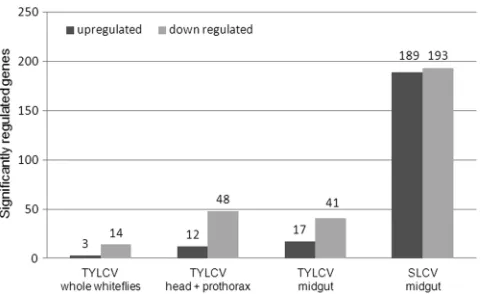
pres-ence of the virus. Overall, 14 genes were downregulated and 3
genes were upregulated when whole whiteflies were analyzed.
However, proportionally more genes were significantly regulated
from head-prothorax (12 up- and 48 downregulated) and from
dissected midguts (17 up- and 41 downregulated) than was the
case for whole whiteflies, suggesting a masking effect when whole
whiteflies were analyzed (
Fig. 1
). Taking the fact that the number
of genes regulated from the midgut was greater than the number
of regulated genes from whole whiteflies, we analyzed the
re-sponse of
B. tabaci
females to another begomovirus, SLCV.
Sur-prisingly, a much higher number of midgut genes were
signifi-cantly regulated in response to this virus, with 189 upregulated
and 193 downregulated genes (
Fig. 1
). The regulated genes were
examined for their fold change in expression and possible functional
relevance to virus translocation, macromolecule movement, defense
or stress response, and other functions that might be relevant to the
acquisition and retention of the viruses (
Table 1
). The response of the
[image:4.585.44.285.65.212.2]FIG 1Significantly induced (black) and repressed (gray) genes based on their fold change in expression in the microarray experiments following the acqui-sition of either TYLCV or SLCV. Analyzed samples were whole whiteflies, head-prothorax, and midguts for TYLCV and midguts for SLCV.
TABLE 1Differentially transcribed ESTs in the midgut ofB. tabacifollowing TYLCV and SLCV acquisition and retentiona
EST clone Best hit in GenBank
GenBank
accession no. GO term Biological function
Microarray expression in midgut
TYLCV SLCV
HBT008_F09_T3_078 70-kDa heat shock EE597405 000940 Response to heat, protein folding 1.65 1.51 TOMOV-BT003_A05 OS-D-like EE599211 0009700 Sensory perception 2.81 BT-H-034-1-H5-T3_H05 Pantothenate kinase 4 EE596485 0015937 Coenzyme A biosynthesis 5.74 BT-H-032-1-C9-T3_C09 Transmembrane assembly EE596401 0006928 Cellular component movement 3.96 BT-TYLCV-053-1-H9-T3_H09 Checkpoint-like EE601807 0000075 Cell cycle checkpoint 3.22 BT-H-020-1-E2-T3_E02 Cathepsin B protease EE595866 000691 Autophagy, proteolysis response to stress 2.09 BT-TYLCV-018-1-H4-T3_H04 Sugar transporter EE600420 000535 Glucose transmembrane transporter 2.01 BT-TOMOV-040-1-G8-T3_G08 Btk-1 EE598908 Immunity 1.59 HBT006_G06_T3_051 Cathepsin F protease EE597298 000658 Proteolysis, receptor-mediated
endocytosis
1.59
BT-TYLCV-056-1-F6-T3_F06 ATP synthase EE601889 0042776 ATP synthesis-coupled proton transport -1.7 -2.6 BT-TOMOV-042-1-F9-T3_F09 NADH dehydrogenase subunit 1 EE599001 0005747 Mitochondrial respiratory chain
complex I
2.7
BT-TYLCV-057-1-E4-T3_E04 Nuclear envelope membrane protein
EE601922 001503 Transmembrane transport 2.58
BT-TYLCV-051-1-A8-T3_A08 Fatty acid binding protein EE601683 000521 Transporter activity 1.97 TOMOV-BT012_G05 Sugar transporter EE599789 000535 Glucose transmembrane transporter
activity
2.01
BT-HINST-004-1-C2-T3_C02 ATP synthase subunit C EE602876 0040007 Cytoplasmic membrane-bounded vesicle 1.88 TOMOV-BT012_C02 Phosphoglycerate mutase 1 EE599756 0006096 Glycolysis 1.55 BT-TYLCV-045-1-A6-T3_A06 Cytochromecoxidase subunit III EE601430 0006123 Mitochondrial electron transport 1.55 BT-TOMOV-028-1-A9-T3_A09 Adenine nucleotide translocator EE598307 0055085 Transmembrane transport 1.54 BT-TYLCV-042-1-B2-T3_B02 ATP-binding FecE EE601334 0006811 Ion transport 1.53 BT-TYLCV-018-1-H4-T3_H04 Acyl-coenzyme A esterase EE600420 0004091 Protein binding 2.01 BT-TYLCV-051-1-A8-T3_A08 Putative fatty acid binding protein EE601683 000521 Transporter activity 1.97 TOMOV-BT012_C02 Phosphoglycerate mutase 1 EE599756 0006096 Glycolysis 1.55 BT-TOMOV-020-1-D2-T3_D02 Vitellogenin EE597946 0006869 Lipid transport 1.67 TYLCV006_F04 Cytochrome P450 CYP6CX2 EE602200 0055114 Oxidation reduction 1.78 BT-TYLCV-058-1-F5-T3_F05 Cytochrome oxidase I EE601978 000612 Mitochondrial electron transport ⫺1.78 BT-TOMOV-028-1-E7-T3_E07 Sodium-dependent glucose
transporter
EE598330 000864 Carbohydrate transport ⫺1.63 BT-HINST-020-1-B11-T3_B11 Apolipophorin III EE603648 0008289 Lipid binding ⫺1.69 BT-TYLCV-025-1-B7-T3_B07 Cytochromecoxidase subunit II EE600708 000609 Precursor metabolites and energy ⫺1.57
aEST (expressed sequence tag) clone name, best hit in GenBank, GenBank accession no. of the EST, GO (gene ontology) category, biological function of the expected protein, and
the average ratio of three highly correlated biological replicates following the microarray experiments comparing viruliferous and nonviruliferous midguts with TYLCV or SLCV are shown.
on November 7, 2019 by guest
http://jvi.asm.org/
[image:4.585.50.541.318.698.2]genes was not high as measured by their fold change in expression.
The majority of the genes showed downregulation with TYLCV,
while in the case of SLCV, the numbers of up- and downregulated
genes were similar. Examining the functional classification of the
genes (
Table 1
) suggests that the presence of begomoviruses,
espe-cially SLCV, in
B. tabaci-
induces genes related to mitochondrial
res-piration, metabolism, stress, and transport of macromolecules (
Table
1
). Interestingly, little overlap was observed between classes of genes
that were significantly regulated in response to the two investigated
begomoviruses.
The
hsp70
gene is overexpressed and results in higher HSP70
protein concentrations in viruliferous whiteflies.
As observed in
our microarray experiments,
hsp70
was overexpressed by more
than 1.5-fold after acquisition and retention of TYLCV and SLCV
(
Fig. 2A
). Interestingly, it is the only gene that was upregulated
after uptake and retention of both viruses. This result was
con-firmed for TYLCV using a quantitative real-time RT-PCR
ap-proach, in which an overexpression of more than 1.7-fold was
observed (
Fig. 2A
). The overexpression of
hsp70
was accompanied
by an accumulation of HSP70 amounts with time as demonstrated
by Western blot analysis, best seen 7 days after TYLCV acquisition
(
Fig. 2C
). HSP70 accumulation was quantified 7 days after
TYLCV acquisition relative to a tubulin control using the Scion
Image densitometry software program, and the results showed an
increase by 1.6-fold.
HSP70 Co-IP and interacts with TYLCV in virus overlay
as-says and immunocapture PCR.
Following the overexpression of
hsp70
in response to TYLCV at the gene and protein levels, we
tested whether this response involves direct interaction
be-tween TYLCV and the HSP70 protein. Three techniques were
used to test this hypothesis. (i) Coimmuniprecipitation
(Co-IP) of TYLCV coat protein (CP) with anti-HSP70 specific
an-tibody and vice versa, to detect both proteins in crude extracts
and to immunoprecipitate the other interacting protein. In
both cases where anti-CP or anti-HSP70 antibodies were used
for Co-IP, both antibodies coimmunoprecipitated the other
interacting protein as detected by Western blotting, suggesting
a specific interaction between CP and HSP70 (
Fig. 3A
and
B
).
Control preimmune serums did not immunoprecipitate either
protein, indicating the specific interaction between the two
proteins (
Fig. 3A
and
B
). An additional control used was the
GroEL protein produced by the endosymbiotic bacteria of
B.
tabaci
, which was shown to interact with TYLCV CP (
32
).
GroEL did not Co-IP with HSP70 (
Fig. 3C
), suggesting that the
interaction between TYLCV CP and HSP70 is specific. (ii)
Vi-rus overlay protein binding assays (VOPBA) were performed to
further validate the interaction of purified TYLCV virions and
HSP70. HSP70 was detectable in the soluble and insoluble
pro-tein fractions of
B. tabaci
after Western blot analysis but in a
FIG 2(A) Fold change in overexpression ofhsp70in viruliferous whiteflies, calculated after microarray and qRT-PCR analyses. Both analyses were further performed with nonviruliferous whiteflies, and the qRT-PCR results were nor-malized to the expression of an actin gene. (B and C) Increase in the HSP70 concentration after acquisition of TYLCV. Total proteins were extracted from whole whiteflies after virus acquisition and retention for 0, 1, and 7 days, subjected to SDS-PAGE (B), and blotted and incubated with HSP70-specific antibodies to follow the HSP70 levels over time (C). The expression ofB. tabaci tubulin did not change following virus acquisition and retention and is given as a loading control.
FIG 3Coimmunoprecipitation of HSP70 and TYLCV CP with anti-TYLCV CP and anti-HSP70, respectively, in TYLCV viruliferous whitefly extracts. (A) Membranes reacted with anti-HSP70 antibody. HSP70 is detected in crude extracts and in extract incubated with protein A-Sepharose beads bound to anti-CP antibody but not in extract incubated with beads bound to preimmune serum (Pre-Im). (B) Membranes reacted with anti-TYLCV CP antibody. TYLCV CP is detected in crude extracts and in extract incubated with protein A-Sepharose beads bound to anti-HSP70 antibody but not in extract incubated with beads bound to preimmune serum. Both HSP70 and TYLCV CP proteins specifically coimmunoprecipitated with the other interacting protein. (C) Membrane is reacted with anti-GroEL antibody. GroEL is detected in whitefly crude extracts and in extracts incubated with protein A-Sepharose beads bound to anti-GroEL antibody, but HSP70 is not detected when protein A-Sepharose beads are bound to anti-HSP70. IgG molecules for confirming the presence of the antibodies in the membranes were also detected with the secondary antibody used.
on November 7, 2019 by guest
http://jvi.asm.org/
[image:5.585.86.236.65.339.2] [image:5.585.135.450.558.648.2]much lesser amount in the insoluble fraction (
Fig. 4A
). Virus
binding to soluble and insoluble whitefly proteins was
evalu-ated after protein separation, blotting, and incubation of the
membranes with purified TYLCV virions with TYLCV-specific
antibodies. Purified
Yam sphaerical aureus virus
(YSV) particles
(a spherical virus detected with YSV-specific antibody from
purified virions, as shown in
Fig. 4A
, lanes 5 and 6, that is not
transmitted by or detected in
B. tabaci
, as shown in
Fig. 4A
, lane
7) and YSV-specific antibodies were applied to demonstrate the
specificity of virus binding. TYLCV virions bound, among
oth-ers, to a 70-kDa band of the soluble protein fraction shown by
a prominent signal (
Fig. 4B
, lane 2, band 1). The main protein
component of this band was identified as HSP70 (
Table 2
). No
signal was detected for the 68-kDa band (
Fig. 4C
, lanes 1 and 2,
band 2) of the insoluble protein fraction which also contained
HSP70. In the insoluble protein fraction, a 65-kDa band
con-taining a putative HSP70 band showed a strong signal (
Fig. 4C
,
lanes 1 and 2, band 3). Unspecific binding of anti-TYLCV
an-tibodies and secondary anan-tibodies was never detected. In
addi-tion, YSV particles did not bind to any whitefly proteins, a
result that points to a specific binding of the begomovirus
par-ticles and not to nonspecific stickiness of HSP70. (iii) The third
assay used to validate the interaction between TYLCV and
HSP70 was an immunocapture-PCR assay (IC-PCR). A
TYLCV-specific PCR product similar to a positive control (
Fig.
5
, lanes 1 and 2) was obtained when extracts of viruliferous
whiteflies were applied to PCR tubes coated with an
HSP70-specific antibody and a sequential PCR was performed on the
complexes bound to the antibody (
Fig. 5
, lanes 3 and 4).
Neg-ative TYLCV PCR results were obtained when extracts from
nonviruliferous whiteflies or uncoated tubes were used as
con-trols (
Fig. 5
, lanes 5 to 8), suggesting a specific interaction
between HSP70 and TYLCV, very likely via the formation of
HSP70 and TYLCV complexes.
HSP70 colocalizes with TYLCV and WmCSV in
B. tabaci
midgut.
To follow the spatial distribution of HSP70 and TYLCV
[image:6.585.316.526.64.144.2]FIG 4(A) Immunodetection of HSP70 in the soluble (lanes 1 and 2) and insoluble (lanes 3 and 4) protein fractions ofB. tabaci. The proteins were subjected to SDS-PAGE (lanes 1 and 3), followed by Western analysis with anti-HSP70 antibodies (lanes 2 and 4). M, molecular weight marker. Immu-nodetection of purified YSV (15g protein per lane, lanes 5 and 6) and the soluble protein fraction ofB. tabaci(120g protein per lane, lane 7) was done as a control. The proteins were subjected to SDS-PAGE (lane 5), followed by Western analysis with anti-YSV antibodies (DSMZ AS-517, 1g ml⫺1; lanes 6 and 7). M, molecular weight marker. (B and C) Virus overlay protein binding assay with the soluble protein (B) or the insoluble protein fractions (C) ofB. tabaci. After SDS-PAGE (lane 1), proteins were blotted and incubated with purified TYLCV virions (lane 2), purified YSV virions (lane 4), and buffer without virus (lanes 3 and 5). Virus binding was shown after application of primary antibodies against TYLCV (lanes 2 and 3) and YSV (lanes 4 and 5), secondary antibodies, and NBT-BCIP. M, molecular weight marker; band 1, heat shock protein 70; band 2, heat shock protein 70; for band 3, seeTable 2.
TABLE 2Analysis of protein bands that bound purified TYLCV virions in virus overlay protein binding assaysa
Band Identified protein
Accession no.
Theoretical mass (kDa)
Exptl mass (kDa)
Protein identification probability (%)
Coverage (%)
1 70-kDa heat shock protein,Bemisia tabaci gi|156254075 71 70 100 60 2 70-kDa heat shock protein,Bemisia tabaci gi|156254075 71 68 100 56 3 GI22533,Drosophila mojavensis gi|193917024 68 65 100 7
Hsp70, putative,Ixodes scapularis gi|215501872 67 65 100 8 GTP-binding protein LepA,Rickettsiaendosymbiont of
Ixodes scapularis
gi|239947763 67 65 100 5
Similar to Igf2 mRNA binding protein, putative, Nasonia vitripennis
gi|156541968 64 65 100 7
Similar to CG2812-PA,Apis mellifera gi|110748705 64 65 100 4
a
Protein bands were excised from corresponding Coomassie-stained gels and analyzed by electrospray ionization mass spectrometry. Peptide mass spectra were analyzed using the software program Scaffold3. Only proteins with an identification probability of 100%. are displayed. Numbering of the bands corresponds to the bands in Fig. 4.
FIG 5Immunocapture PCR assay to detect HSP70-TYLCV CP interaction in viruliferous whiteflies. A TYLCV-specific PCR product. as seen in lanes 1 and 2, was obtained when extracts of dissected midguts of viruliferous whiteflies were applied to PCR tubes coated with an anti-HSP70 specific antibody, and a PCR was performed on the caught complexes (lanes 3 and 4). Control treat-ments with midgut extracts of nonviruliferous whiteflies (lanes 5 and 6) and with uncoated tubes (lanes 7 and 8) were negative.
on November 7, 2019 by guest
http://jvi.asm.org/
[image:6.585.65.261.65.305.2] [image:6.585.38.552.588.707.2]and their possible colocalization in whitefly midguts, we
per-formed colocalization studies using an immunological detection
of HSP70 and fluorescence
in situ
hybridization (FISH) or
immu-nological detection of TYLCV and
Watermelon chlorotic stunt
vi-rus
(WmCSV) viral DNA in dissected midguts from viruliferous
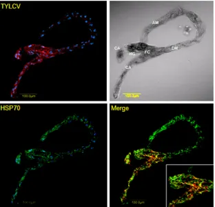
whiteflies. The highest TYLCV concentration shown by red
fluo-rescence was found in the filter chamber and the cecae (
Fig. 6
,
upper row), where HSP70 localization was also observed among
the other parts of the midgut (lower row, green fluorescence).
Colocalization of both HSP70 and TYLCV was clearly seen (
Fig. 6
,
lower row merge) as yellow patches in the filter chamber and
cecae, while no such signal was observed in other parts of the
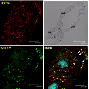
midgut. Colocalization of HSP70 and WmCSV was further
ob-served in the ascending midgut (
Fig. 7
) when specific antibodies
were used against WmCSV CP and HSP70, confirming the
possi-ble direct
in vivo
interaction between begomoviruses and HSP70.
Feeding whiteflies with anti-HSP70 antibody increases
TYLCV transmission.
Transmission experiments were
per-formed with whiteflies fed with anti-HSP70 antibodies to further
investigate the involvement of HSP70 in TYLCV transmission by
B. tabaci
.
B. tabaci
MEAM1 adults were first fed on artificial
me-dium containing HSP70 antibodies and subsequently fed purified
TYLCV virions. Whiteflies fed with preimmune serum instead of
anti-HSP70 antibodies served as controls. Interestingly the
trans-mission rates were enhanced, with 14 out of 24 plants in the first
experiment and 23 out of 26 plants in the second experiment
testing as being infected after feeding on anti-HSP70 antibodies,
while only 10 out of 23 and 13 out of 25 plants tested as infected in
the control experiments, in which whiteflies were fed with
preim-mune serum only. Altogether, 73% transmission rates, on average,
were obtained after feeding with anti-HSP70 antibody, compared
to 47% when the whiteflies were fed with preimmune serum (
Fig.
8A
), clearly demonstrating the involvement of HSP70 in virus
transmission.
To demonstrate that HSP70 antibodies were internalized by
the whiteflies, homogenates of insects previously fed on
anti-bodies and insects fed on preimmune serum were used as the
first antibody for the detection of HSP70 from whole whitefly
protein extracts in Western analysis. While no signals were
detectable after incubation of whitefly proteins with
homoge-nates of insects previously fed on preimmune serum (
Fig. 8B
,
lanes 3 and 4), faint signals were visible after application of
homogenates of insects fed on anti-HSP70 antibodies,
demon-strating the presence of the anti-HSP70 antibodies (
Fig. 8B
,
lanes 1 and 2). Additionally, midguts were dissected from
whiteflies fed on the first antibody against HSP70 or
preim-mune serum and subsequently fed on a goat-anti rabbit Alexa
Fluor 568-nm secondary antibody. A clear signal was seen in
the midguts from whiteflies fed with the HSP70 antibody, while
no signal was observed in the control midgut dissected from
whiteflies fed with preimmune serum (
Fig. 8C
).
FIG 6Colocalization of HSP70 and TYLCV in midguts of viruliferous whiteflies. HSP70 immunolocalization using specific first antibody and secondary antibody conjugated to Alexa Fluor (488 nm) (green) and TYLCV detection using a specific DNA probe conjugated to Cy3 (red). Note the yellow patches indicating colocalization of TYLCV and HSP70 in the filter chamber (FC) and cecae (CA) and the absence of TYLCV and colocalization signal in other parts of the midgut. Blue indicates DAPI staining of the nuclei. DM, descending midgut; AM, ascending midgut; HG, hindgut.
on November 7, 2019 by guest
http://jvi.asm.org/
[image:7.585.138.450.67.367.2]DISCUSSION
We have performed microarray experiments to investigate the
response of
B. tabaci
to the presence of the Old World
monopar-tite TYLCV and the New World biparmonopar-tite SLCV. A low number of
genes responded to the uptake and retention of TYLCV in the
whitefly. In comparison, a higher number of genes were
signifi-cantly regulated after the uptake of SLCV. This difference in the
response is likely related to the coevolution between the Old
World
B. tabaci
MEAM1 with the Old World TYLCV
begomovi-rus, which is well adapted to the whitefly and to a lower
compat-ibility with the New World SLCV (
9
,
16
). The transcriptional
changes are likely to be related to virus interaction with insect
organs and not the feeding on the infected plants. This is because
the setup of the experiment included rearing the viruliferous and
nonviruliferous whiteflies used for RNA extraction on
TYLCV-nonhost cotton plants for sufficient time to prevent residual
ef-fects from the infected plant. Genes that were differently expressed
due to virus uptake were involved in several biological processes,
including stress response, macromolecule transport, metabolism,
and mitochondrial functions, as already shown for TYLCCV by
Luan et al. (
47
). Remarkably, the genes significantly regulated in
response to TYLCV and SLCV were significantly different,
sug-gesting a differential response to the acquisition and retention of
these two viruses that belong to the same genus. Only one gene was
significantly upregulated in response to both TYLCV and SLCV.
This gene encodes the stress-related heat shock protein 70 (
hsp70
).
The elevated expression of
hsp70
was confirmed using a qRT-PCR
approach (
Fig. 2
). Interestingly,
hsp70
was upregulated at the same
order of magnitude after the uptake of
Watermelon chlorotic stunt
virus
(WmCSV), another whitefly-transmitted begomovirus (data
not shown). The upregulation of
hsp70
resulted in elevated
con-centrations of the heat shock protein HSP70 as demonstrated by
Western blot analysis (
Fig. 2
). The specificity of the upregulation
of
hsp70
in response to begomoviruses was further confirmed by
performing a control experiment in which the expression of the
hsp70
gene was analyzed 2 and 9 days after the uptake of
Faba bean
necrotic stunt virus
, an aphid-transmitted virus which is not
trans-mitted by
B. tabaci
. The results showed that the expression of
hsp70
was not enhanced (data not shown). This result
demon-strates that the uptake of different begomoviruses that are
trans-mitted by
B. tabaci
results in
hsp70
upregulation, while no
upregu-lation is observed when a virus that is not transmitted by
B. tabaci
is acquired.
HSP70 is ubiquitously expressed, with unusually high
conser-vation in protein sequence and functional features (
6
,
42
), and the
functions are well established (
23
,
34
,
35
,
53
). HSPs and their
constitutively synthesized relatives known as heat shock cognates
or HSC proteins belong to a multicomponent and
multifunc-tional chaperone family that plays a role in various cellular
pro-cesses, such as disaggregation of proteins from large aggregates or
FIG 7Colocalization of HSP70 and WmCSV in midguts of viruliferous whiteflies. HSP70 immunolocalization using specific first antibody and secondary antibody conjugated to Alexa Fluor (635 nm) (red) and WmCSV immunolocalization using specific first antibody and secondary antibody conjugated to Alexa Fluor (488 nm) (green). Note the yellow areas indicating colocalization of WmCSV and HSP70 in the ascending midgut (AM). Blue indicates DAPI staining of the nuclei. DM, descending midgut; CA, cecae; HG, hindgut; FC, filter chamber.
on November 7, 2019 by guest
http://jvi.asm.org/
[image:8.585.137.451.67.380.2]assemblies, prevention of aggregation of denatured proteins,
aid-ing of the renaturation or foldaid-ing of proteins to reach their proper
conformation, direction of proteins to degradative pathways, and
binding of proteins to restrain their function, making them
avail-able for ligand binding or allowing them to translocate across
membranes. Although some of the classes of HSPs clearly have
distinct activities, they also exhibit overlapping functions and may
share proteins that act as cofactors, known as cochaperones (
41
,
46
,
65
,
72
). Extracellular and membrane-bound HSPs are
in-volved in immunological functions (
58
,
61
). Previous findings
have implicated members of the HSP70 family in the targeted
transport of proteins to specific cellular domains (
66
,
67
,
75
).
Fur-thermore, HSP70s were shown to play various roles in the life
cycles of plant and animal viruses (
33
,
74
). Some plant viruses,
such as closteroviruses, encode their own HSP70 homolog, which,
in combination with the virus coat protein, facilitates transport
through plasmodesmata and thus enables cell-to-cell movement
(
1
,
2
,
54
). In tombusviruses, cytosolic HSP70s play multiple roles
in
Tomato bushy stunt virus
(TBSV) replication, such as affecting
the subcellular localization and membrane insertion of the viral
replication proteins, as well as the assembly of the viral replicase
(
79
). Krenz et al. (
43
) showed a targeted transport of
Abutilon
mosaic virus
movement protein toward the chloroplasts shuttled
by a heat shock cognate 70-kDa protein. For several animal
vi-ruses, the interaction with HSP70s appears to be involved in virion
assembly, which may take place in the cytoplasm, nucleus, or
en-doplasmatic reticulum depending on the requirements of
differ-ent viruses (
15
,
45
). Oh & Song (
63
) demonstrated an interaction
of the coat protein of
West Nile virus
(
Flavivirus
) with HSP70
using the yeast two-hybrid system and purified proteins.
Coutu-rier et al. (
14
) found high-affinity binding between an inducible
HSP70 and the measles virus nucleoprotein (which requires an
HSP40 cochaperone). In most cases, HSP70 displays stability and
activity control reactions, and binding to HSP70 results in
en-hanced transcription and replication of these viruses. For animal
viruses, HSP70 promotes gene expression for several viral families
(
12
), e.g., for baculoviruses (
49
). An involvement of HSP70 in
viral cell-to-cell transport has not yet been reported for animal
cells. The role of HSP70 in interaction of plant viruses with their
vector insects is unknown as well.
Enhanced
hsp70
expression and elevated HSP70
concentra-tions were found especially within the filter chamber, where the
largest amounts of the virus were detected by
in situ
hybridization
and immunolocalization (
25
,
26
; also unpublished data). These
results are supported by previous observations that the majority of
TYLCV particles are translocated from the filter chamber into the
hemolymph (
25
,
26
,
71
). Colocalization of TYLCV and HSP70
was mainly observed in the filter chamber; however,
colocaliza-tion was also observed in other parts of the midgut. These results
were confirmed by immunolocalization studies with the bipartite
WmCSV and HSP70 that resulted in high colocalization rates.
These results point to a direct interaction
in vivo
. To validate this
interaction, further experiments are required to visualize this
in-teraction at a higher resolution level: for instance, using
transmis-sion electron microscopy and immunolabeling.
All results indicate a direct or indirect response of whiteflies to
begomovirus uptake. Several experiments we performed,
includ-ing Co-IP (
Fig. 3
), VOPBA (
Fig. 4
), and IC-PCR (
Fig. 5
), proved
the possibility of binding of HSP70 and TYLCV
in vitro
. In
addi-tion, HSP70 and virions were to some extend colocalized in the
filter chamber and the ascending midgut, pointing to a direct
in-teraction
in vivo
(
Fig. 6
and
7
). One possible role of HSP70 could
be the aided/directed shuttling of the TYLCV virions through the
insect midgut cells to the hemolymph, thus minimizing virus
con-tact with insect tissue. In this case, the insect promotes virus
trans-mission to minimize harmful effects caused by the virus, which
was previously shown to cause negative effects to the insect,
in-cluding a decrease in the longevity and fecundity of viruliferous
whiteflies (
17
,
68
). A previous study showed the binding of
TYLCV particles to the chaperone GroEL, which is produced by
Hamiltonella
, a secondary symbiont of
B. tabaci
, thus supporting
the hypothesis that proteins produced within the vector aid in
virus transmission (
32
). Following this hypothesis, one would
ex-pect reduced virus transmission after blocking HSP70 by feeding
HSP70 antibodies to the whiteflies. In contrast to these
expecta-tions, TYLCV transmission was enhanced after HSP70
anti-body uptake (
Fig. 8
). The enhanced transmission is not a result of
uptake of proteins that stabilize virus particles, as hypothesized by
Bencharki et al. (
5
), who found stimulated transmission of virus
FIG 8(A) Enhancement of TYLCV transmission rates after feeding of white-flies on artificial medium containing anti-HSP70 antibodies and preimmune serum, respectively, for 1 day followed by feeding on the same medium con-taining purified TYLCV virions for another day. (B) Detection of ingested HSP70 antibodies in whiteflies fed on artificial medium containing anti-HSP70 antibodies for 2 days. The soluble (lanes 1, 3, and 5) and insoluble (lanes 2, 4, and 6) protein fractions ofB. tabaciwere subjected to SDS-PAGE and blotting followed by immunodetection of HSP70. Homogenates of white-flies fed on artificial medium amended with anti-HSP70 antibodies (lanes 1 and 2) and preimmune serum (lanes 3 and 4) were used as a source for HSP70 antibodies. HSP70 antibodies were used as a positive control (lanes 5 and 6). (C) Detection of ingested HSP70 antibodies in the midgut after feeding with anti-HSP70 first antibody for 24 h (left) or with preimmune serum (right) and subsequently feeding with a goat antirabbit Alexa Fluor 568-nm secondary antibody for another 24 h. M, molecular weight marker.
on November 7, 2019 by guest
http://jvi.asm.org/
[image:9.585.44.283.65.356.2]by aphids after uptake of several soluble proteins, because feeding
B. tabaci
with bovine serum albumin and anti-WmCSV
antibod-ies (DSMZ AS830) did not result in higher transmission rates for
TYLCV (data not shown). Since TYLCV was shown to cause
neg-ative effects on
B. tabaci
, the overexpression of HSP70 at the gene
and protein levels could result from a stress or immune response
of the vector in response to the virus. In other species, such as
leafminers (
38
),
Drosophila
(
73
,
80
), and even desert lizards (
76
,
81
), the inductive and conserved role of HSP70 in the stress
re-sponse is well documented. The overexpression of
hsp70
in
re-sponse to stress was reported for mammalian systems with
auto-immune disorders, supporting the possibility that overexpression
of
hsp
contributes to immunopathological changes (
82
). The
overexpression of
hsp70
might be direct, by recognizing the
pres-ence of the virus, or indirect, through a conserved stress response
pathway. The heat shock protein response, although related to
heat, can also be related to other stresses, such as infection,
inflam-mation, radiation, hypoxia, and chemical stressors (
50
). The
abil-ity of
B. tabaci
to respond to the presence of begomoviruses and
other invaders was previously documented for a number of
invad-ers, including begomoviruses (
47
) and parasitoids (
52
). These
re-sponses were related to several stress response pathways, such as
the Toll-like, Imd, and mitogen-activated protein kinase
(MAPK)-related pathways in the case begomoviruses and
parasi-toids and heat shock, mitochondrial, and cytoskeleton-related
genes in the case of heat stress (
51
).
The transcriptional response of
hsp70
activation starts in the
cytoplasm with trimerization of heat shock factors (HSF) that
enter the nucleus and bind to the heat shock elements (HSE) on
the DNA (
69
). HSFs are the first molecules to sense the stress and
respond accordingly, and thus it is likely that HSFs sense the
pres-ence of the high concentration of TYLCV virions in the cytoplasm
of the cell. The function of HSP70 as a refolder and stabilizer of
proteins through direct interaction with them suggests possible
binding to TYLCV particles in the cell cytoplasm, as observed in
our results (
Fig. 6
and
7
). Since a main function for HSP70 is
selectively targeting cytosolic proteins to lysosomes for
degrada-tion, it is likely that interaction with TYLCV particles might be a
protective response for degrading TYLCV virions. The results of
the feeding experiments support this hypothesis. Uptake of
anti-HSP70 antibodies into the midgut was proven by using extracts of
fed and discharged whiteflies as antibodies for HSP70 in Western
blot analysis and by localization after confocal laser scanning
mi-croscopy (CLSM) (
Fig. 8
). The enhanced TYLCV transmission
rates after feeding on HSP70 antibodies suggest an inhibition of
HSP70-virus interaction/binding and presenting them for
degra-dation. Thus, more TYLCV virions could pass the midgut barrier
and enter the hemolymph; still, further experiments have to be
conducted to confirm this hypothesis.
ACKNOWLEDGMENTS
This research was supported by research grant no. 908-42.12/2006 from the German-Israeli Foundation (GIF) to S.W., H.C., and M.G. and grant no. IS-4062-07 from the United States-Israel Binational Agricultural Re-search and Development Fund (BARD) to M.G., H.C., and J.K.B. and in part by an Israel Science Foundation research grant, no. 884/07, to M.G. and H.C.
We thank Henning Urlaub (Bioanalytical Mass Spectrometry, Max Planck Institute for Biophysical Chemistry, Göttingen, Germany) for an-alyzing proteins using LC-ESI-MS.
REFERENCES
1.Alzhanova DV, Napuli AJ, Creamer R, Dolja VV. 2001. Cell-to-cell movement and assembly of a plant closterovirus: roles for the capsid pro-teins and Hsp70 homolog. EMBO J.20:6997–7007.
2.Aoki K, Kragler F, Xoconostle-Cazares B, Lucas WJ.2002. A subclass of plant heat shock cognate 70 chaperones carries a motif that facilitates trafficking through plasmodesmata. Proc. Natl. Acad. Sci. U. S. A.99: 16342–16347.
3.Azzam O, et al.1994. Whitefly transmission and efficient ssDNA accu-mulation of bean golden mosaic geminivirus require functional coat pro-tein. Virology204:289 –296.
4.Baugh LR, Hill AA, Brown EL, Hunter CP.2001. Quantitative analysis of mRNA amplification by in vitro transcription. Nucleic Acids Res.29: E29. doi:10.1093/nar/29.5.e29.
5.Bencharki B, et al.2010. Phloem protein Partners of Cucurbit aphid borne yellows virus: possible involvement of phloem proteins in virus transmission by aphids. Mol. Plant Microbe Interact.23:799 – 810. 6.Boorstein WR, Ziegelhoffer T, Craig EA.1994. Molecular evolution of
the HSP70 multigene family. J. Mol. Evol.38:1–17.
7.Bosco D, Mason G, Accotto GP.2004. TYLCSV DNA, but not infectivity, can be transovarially inherited by the progeny of the whitefly vector Bemisia tabaci(Gennadius). Virology323:276 –283.
8.Briddon RW, Pinner MS, Stanley J, Markham PG.1990. Geminivirus coat protein gene replacement alters insect specificity. Virology177: 85–94.
9.Brown JK.2010. Phylogenetic biology of the Bemisia tabaci sibling species group, p 31– 67.InStansley PA, Naranjo SE (ed), Bemisia: bionomics and management of a global pest. Springer, New York, NY.
10. Brown JK, Czosnek H. 2002. Whitefly transmission of plant viruses, p 65–100.InPlumb RT (ed), Advances in botanical research, vol 36: plant virus vector interactions. Academic Press, New York, NY.
11. Caciagli P, Bosco D.1997. Quantitation over time of tomato yellow leaf curl geminivirus DNA in its whitefly vector. Phytopathology87: 610 – 613.
12. Carsillo T, et al.2009. Major histocompatibility complex haplotype de-termines hsp70-dependent protection against measles virus neuroviru-lence. J. Virol.83:5544 –5555.
13. Cicero JM, Hiebert E, Webb SE.1995. The alimentary canal ofBemisia tabaciandTrialeurodes abutilonea(Homoptera, Sternorrhynchi): histol-ogy, ultrastructure and correlation to function. Zoomorphology
115:31–39.
14. Couturier M, et al.2010. High affinity binding between Hsp70 and the C-terminal domain of the measles virus nucleoprotein requires an Hsp40 co-chaperone. J. Mol. Recognit.23:301–315.
15. Cripe TP, Delos SE, Estes PA, Garcea RL.1995. In vivo and in vitro association of hsc70 with polyomavirus capsid proteins. J. Virol.69:7807– 7813.
16. Czosnek H, Ghanim M, Ghanim M.2002. Circulative pathway of bego-moviruses in the whitefly vectorBemisia tabaci—insights from studies withTomato yellow leaf curl virus.Ann. Appl. Biol.140:215–231. 17. Czosnek H, et al.2001. Whiteflies: vectors— or victims?— of
geminivi-ruses, p 291–322.InMaramorosch K (ed), Advances in virus research, vol 57. Academic Press, New York, NY.
18. Czosnek H, Laterrot H.1997. A worldwide survey of tomato yellow leaf curl viruses. Arch. Virol.142:1391–1406.
19. De Barro PJ, Liu SS, Boykin LM, Dinsdale AB.2011.Bemisia tabaci: a statement of species status. Annu. Rev. Entomol.56:1–19.
20. Dellaporta S, Wood J, Hicks JB.1983. A plant DNA minipreparation: version II. Plant Mol. Biol. Rep.1:19 –21.
21. Dinsdale A, Cook L, Riginos C, Buckley YM, De Barro P.2010. Refined global analysis ofBemisia tabaci(Hemiptera: Sternorrhyncha: Aleyroidae: Aleyrodidae) mitochondrial cytochrome oxidase 1 to identify species level genetic boundaries. Ann. Entomol. Soc. Am.103:196 –208.
22. Reference deleted.
23. Flynn GC, Pohl J, Flocco MT, Rothman JE. 1991. Peptide-binding specificity of the molecular chaperone BiP. Nature353:726 –730. 24. Frohlich D, Torres-Jerez I, Bedford ID, Markham PG, Brown JK.1999.
A phylogeographic analysis of theBemisia tabacispecies complex based on mitochondrial DNA markers. Mol. Ecol.8:1683–1691.
25. Ghanim M, Brumin M, Popovski S.2009. A simple, rapid and inex-pensive method for localization ofTomato yellow leaf curl virusand
on November 7, 2019 by guest
http://jvi.asm.org/
Potato leafroll virusin plant and insect vectors. J. Virol. Methods159: 311–314.
26. Ghanim M, Medina V.2007. Localization of Tomato yellow leaf curl virus in its whitefly vector Bemisia tabaci, p 171–183.InCzosnek H (ed), Tomato yellow leaf curl virus disease. Springer, Dordrecht, The Netherlands. 27. Reference deleted.
28. Ghanim M, Morin S, Czosnek H.2001. Rate ofTomato Yellow Leaf Curl Virus(TYLCV) translocation in the circulative transmission pathway of its vector, the whiteflyBemisia tabaci.Phytopathology91:188 –196. 29. Ghanim M, et al.2001. Microscopic analysis of the digestive, salivary and
reproductive organs ofBemisia tabaci(Gennadius) (Hemiptera: Aleyro-didae) biotype B. J. Morphol.248:22– 40.
30. Ghanim M, Czosnek H. 2000.Tomato yellow leaf curl geminivirus (TYLCV-Is) is transmitted among whiteflies (Bemisia tabaci) in a sex-related manner. J. Virol.74:4738 – 4745.
31. Ghanim M, Morin S, Zeidan M, Czosnek H.1998. Evidence for transo-varial transmission of tomato yellow leaf curl virus by its vector the white-flyBemisia tabaci.Virology240:295–303.
32. Gottlieb Y, et al.2010. The transmission efficiency of Tomato yellow leaf curl virus is correlated with the presence of a specific symbiotic bacterium species. J. Virol.84:9310 –9317.
33. Hafren A, Hofius D, Rönnholm G, Sonnewald U, Makinen K.2010. HSP70 and its cochaperone CPIP promote potyvirus infection in Nicoti-ana benthamiNicoti-anaby regulating viral coat protein functions. Plant Cell
22:523–535.
34. Hartl FU, Hayer-Hartl M.2002. Molecular chaperones in the cytosol: from nascent chain to folded protein. Science295:1852–1858.
35. Hartl FU.1996. Molecular chaperones in cellular protein folding. Nature
381:571–579.
36. Hofer P, Bedford ID, Markham PG, Jeske H, Frischmuth T.1997. Coat protein gene replacement results in whitefly transmission of an insect nontransmissible geminivirus isolate. Virology236:288 –295.
37. Hogenhout SA, van der Wilk F, Verbeek M, Goldbach RW, van den Heuvel JFJM.1998. Potato leafroll virus binds to the equatorial domain of the aphid endosymbiotic GroEL homologue. J. Virol.72:358 –365. 38. Huang LH, Kang L.2007. Cloning and interspecific altered expression of
heat shock protein genes in two leafminer species in response to thermal stress. Insect Mol. Biol.16:491–500.
39. Hunter WB, Hiebert E, Webb SE, Tsai JH, Polston JE.1998. Location of geminiviruses in the whiteflyBemisia tabaci(Homoptera: Aleyrodidae). Plant Dis.82:1147–1151.
40. Jiu M, et al.2007. Vector-virus mutualism accelerates population in-crease of an invasive whitefly. PLoS One 2:e182. doi:10.1371/ journal.pone.0000182.
41. Johnston JA, Ward CL, Kopito RR. 1998. Aggresomes: a cellular re-sponse to misfolded proteins. J. Cell Biol.143:1883–1898.
42. Karlin S, Brocchieri L.1998. Heat shock protein 70 family: multiple sequence comparisons function and evolution. J. Mol. Evol.47:565– 577.
43. Krenz B, Windeisen V, Wege C, Jeske H, Kleinow T.2010. A plastid-targeted heat shock cognate 70 kDa protein interacts with theAbutilon mosaic virusmovement protein. Virology401:6 –17.
44. Reference deleted.
45. Liberman E, et al.1999. Activation of the grp78 and grp94 promoters by hepatitis C virus E2 envelope protein. J. Virol.73:3718 –3722.
46. Lindquist SL.1986. The heat-shock response. Annu. Rev. Biochem.55: 1151–1191.
47. Luan JB, et al.2011. Global analysis of the transcriptional response of whitefly toTomato yellow leaf curl china virusreveals their relationship of coevolved adaptations. J. Virol.85:3330 –3340.
48. Reference deleted.
49. Lyupina YV, et al.2010. An important role of the heat shock response in infected cells for replication of baculoviruses. Virology406:336 –341. 50. Macario AJ, de Macario EC.2000. Stress and molecular chaperones in
disease. Int. J. Clin. Lab Res.30:49 – 66.
51. Mahadav A, Kontsedalov S, Czosnek H, Ghanim M.2009. Thermotol-erance and gene expression following heat stress in the whiteflyBemisia tabaciB and Q. biotypes. Insect Biochem. Mol. Biol.39:668 – 676. 52. Mahadav A, Gerling D, Gottlieb Y, Czosnek H, Ghanim M. 2008.
Parasitization by the waspEretmocerus mundusinduces transcription of genes related to immune response and symbiotic bacteria proliferation in the whiteflyBemisia tabaci.BMC Genomics9:342. doi:10.1186/1471-2164-9-342.
53. Mayer MP.2005. Recruitment of Hsp70 chaperones: a crucial part of viral survival strategies. Rev. Physiol. Biochem. Pharmacol.153:1– 46. 54. MedinaV Peremyslov VV, Hagiwara Y, Dolja VV. 1999. Subcellular
localization of the HSP70-homolog encoded by beet yellows closterovirus. Virology260:173–181.
55. Mehta P, Wyman JA, Nakhla MK, Maxwell DP.1994. Transmission of tomato yellow leaf curl geminivirus byBemisia tabaci(Homoptera: Aley-rodidae). J. Ecol. Entomol.87:1291–1297.
56. Morin S, Ghanim M, Sobol I, Czosnek H.2000. The GroEL protein of the whiteflyBemisia tabaciinteracts with the coat protein of transmissible and non-transmissible begomoviruses in the yeast two-hybrid system. Vi-rology276:404 – 416.
57. Morin S, et al.1999. A GroEL homologue from endosymbiotic bacteria of the whiteflyBemisia tabaciis implicated in the circulative transmission of Tomato yellow leaf curl virus.Virology30:75– 84.
58. Multhoff G.2007. Heat shock protein 70 (Hsp70): membrane location, export and immunological relevance. Methods43:229 –237.
59. Reference deleted. 60. Reference deleted.
61. Nishikawa M, Takemoto S, Dakakura Y.2008. Heat shock protein de-rivatives for delivery of antigens to antigen presenting cells. Int. J. Pharm.
354:23–27.
62. Noris E, et al.1998. Amino acids in the capsid protein of tomato yellow leaf curl virus that are crucial for systemic infection, particle formation, and insect transmission. J. Virol.72:10050 –10057.
63. Oh W, Song J.2006. Hsp70 functions as a negative regulator of West Nile virus capsid protein through direct interaction. Biochem. Biophys. Res. Commun.347:994 –1000.
64. Ohnesorge S, Bejarano ER.2009. Begomovirus coat protein interacts with a small heatshock protein of its transmission vector (Bemisia tabaci). Insect Mol. Biol.18:693–703.
65. Pelham HR.1986. Speculations on the functions of the major heat shock and glucose-regulated proteins. Cell46:959 –961.
66. Pishvaee B, et al.2000. A yeast DNA J protein required for uncoating of clathrin-coated vesicles in vivo. Nat. Cell Biol.2:958 –963.
67. Pratt WB, Silverstein AM, Galigniana MD.1999. A model for the cyto-plasmic trafficking of signalling proteins involving the hsp90-binding im-munophilins and p50. Cell Signal.11:839 – 851.
68. Rubinstein G, Czosnek H.1997. Long-term association of tomato yellow leaf curl virus (TYLCV) with its whitefly vectorBemisia tabaci: effect on the insect transmission capacity, longevity and fecundity. J. Gen. Virol.
78:2683–2689.
69. Sarge KD, Murphy SP, Morimoto RI.1993. Activation of heat shock gene transcription by heat shock factor 1 involves oligomerization, acquisition of DNA-binding activity, and nuclear localization and can occur in the absence of stress. Mol. Cell. Biol.13:1392–1407.
70. Sinisterra XH, McKenzie CL, Hunter WB, Powell CA, Shatters RG, Jr.
2005. Differential transcriptional activity of plant-pathogenic begomovi-ruses in their whitefly vector (Bemisia tabaci, Gennadius: Hemiptera Aley-rodidae). J. Gen. Virol.86:1525–1532.
71. Skaljac M, Ghanim M.2010.Tomato yellow leaf curl virusand the plant-virus-vector interactions. Isr. J. Plant Sci.58:103–111.
72. Sorensen JG, Kristensen GTN, Loeschcke V.2003. The evolutionary and ecological role of heat shock proteins. Ecol. Lett.6:1025–1037.
73. Sorensen JG, Dahlgaard J, Loeschcke V. 2001. Genetic variation in thermal tolerance among natural populations of Drosophila buzzatii: down regulation of HSP70 expression and variation in heat-stress resis-tance traits. Funct. Ecol.15:289 –296.
74. Sullivan CS, Pipas JM.2001. The virus-chaperone connection. Virology.
287:1– 8.
75. Tsai MY, Morfini G, Szebenyi G, Brady ST.2000. Release of kinesin from vesicles by hsc70 and regulation of fast axonal transport. Mol. Biol. Cell
11:2161–2173.
76. Ulmasov KA, Shammakov S, Karaev K, Evgenev MB.1992. Heat-shock proteins and thermoresistance in lizards. Proc. Natl. Acad. Sci. U. S. A.
89:1666 –1670.
77. van den Heuvel JFJM, Verbeek M, van der Wilk F.1994. Endosymbiotic bacteria associated with circulative transmission ofpotato leafroll virusby Myzus persicae.J. Gen. Virol.75:2559 –2565.
78. van den Heuvel JFJM, et al. 1997. The N-terminal region of the luteovirus readthrough domain determines virus binding toBuchnera GroEL and is essential for virus persistence in the aphid. J. Virol.71: 7258 –7265.
on November 7, 2019 by guest
http://jvi.asm.org/
79. Wang RY, Stork J, Pogany J, Nagy PD.2009. A temperature sensitive mutant of heat shock protein 70 reveals an essential role during the early steps of tombusvirus replication. Virology394:28 –38.
80. Zatsepina OG, et al.2001. A Drosophila melanogaster strain from sub-equatorial Africa has exceptional thermotolerance but decreased HSP70 expression. J. Exp. Biol.204:1869 –1881.
81. Zatsepina OG, et al.2000. Thermotolerant desert lizards characteristi-cally differ in terms of heat-shock system regulation. J. Exp. Biol.203