0022-538X/07/$08.00⫹0 doi:10.1128/JVI.00895-07
Copyright © 2007, American Society for Microbiology. All Rights Reserved.
Mamu-B*08
-Positive Macaques Control Simian
Immunodeficiency Virus Replication
䌤
John T. Loffredo,
1Jess Maxwell,
1Ying Qi,
2Chrystal E. Glidden,
1Gretta J. Borchardt,
1Taeko Soma,
1Alex T. Bean,
1Dominic R. Beal,
1Nancy A. Wilson,
1William M. Rehrauer,
3Jeffrey D. Lifson,
4Mary Carrington,
2and David I. Watkins
1,3*
Wisconsin National Primate Research Center, University of Wisconsin-Madison, Madison, Wisconsin 537151; Laboratory of Genomic Diversity, SAIC-Frederick, Inc., National Cancer Institute, Frederick, Maryland 217022; Department of
Pathology and Laboratory Medicine, University of Wisconsin-Madison, Madison, Wisconsin 537153; and AIDS Vaccine Program/Basic Research Program, SAIC-Frederick, Inc.,
National Cancer Institute, Frederick, Maryland 021724
Received 26 April 2007/Accepted 20 May 2007
Certain major histocompatibility complex (MHC) class I alleles are associated with the control of human immunodeficiency virus and simian immunodeficiency virus (SIV) replication. We have designed sequence-specific primers for detection of the rhesus macaque MHC class I alleleMamu-B*08by PCR and screened a cohort of SIV-infected macaques for this allele. Analysis of 196 SIVmac239-infected Indian rhesus macaques revealed that
Mamu-B*08was significantly overrepresented in elite controllers; 38% of elite controllers wereMamu-B*08positive compared to 3% of progressors (Pⴝ0.00001).Mamu-B*08was also associated with a 7.34-fold decrease in chronic phase viremia (Pⴝ0.002).Mamu-B*08-positive macaques may, therefore, provide a good model to understand the correlates of MHC class I allele-associated immune protection and viral containment in human elite controllers.
Certain HLA class I alleles strongly influence whether individ-uals become slow or rapid progressors after human immunode-ficiency virus (HIV) infection (8, 18–20, 28, 30, 32, 39). Long-term nonprogressor/elite controller (EC) cohorts are enriched for
HLA-B27 and HLA-B57. Numerous studies have implicated these molecules in the presentation of epitopes that elicit effective HIV-specific CD8⫹T-lymphocyte responses (2, 3, 13, 17, 21, 22, 24, 31, 32). Unfortunately, there are many difficulties associated with understanding the basis for these HLA associations in HIV-infected people, including inoculum variability, allelic diversity, and access to acute-phase samples. Studying simian immunode-ficiency virus (SIV)-infected rhesus macaques that control viral replication may shed some light on why certainHLA-B27- and
HLA-B57-positive humans control HIV infection.
Although certain major histocompatibility complex (MHC) class I alleles, includingMamu-A*01andMamu-B*17, are as-sociated with slow disease progression in SIV-infected ma-caques (33–35, 38, 41, 42), independently, the presence of these alleles is not predictive for disease outcome. In the case ofMamu-B*17, only one-fourth ofMamu-B*17-positive ma-caques become ECs, controlling SIV replication below 1,000 viral RNA (vRNA) copies/ml of plasma during the chronic phase of SIV infection (41). Even Mamu-B*17-positive ma-caques containing identical MHC class I haplotypes have widely divergent disease courses (40).
Recently, we defined novel Vif- and Nef-specific CD8⫹ T-cell responses in three EC macaques. We discovered that these epitopes were recognized by T cells restricted by the MHC
class I molecule, Mamu-B*08 (J. T. Loffredo et al., unpub-lished data). Therefore, we investigated the impact of Mamu-B*08on SIV disease progression in a cohort of 196 SIVmac
239-infected Indian rhesus macaques.
Development of PCR-SSP for Mamu-B*08. To identify
Mamu-B*08-positive animals, rhesus macaques (Macaca mulatta) of Indian descent were genotyped for the MHC class I allele,Mamu-B*08, using PCR amplification with sequence-specific DNA priming (PCR-SSP) methodology as previously described (27, 36). The nucleotide sequences of the primers used for typingMamu-B*08were as follows: forward, 5⬘-CGT GAG GCG GAG CAG GTC-3⬘; and reverse, 5⬘-CCA CAG CTC CGA TGA ACA CAG-3⬘. Because Mamu-B*08 and
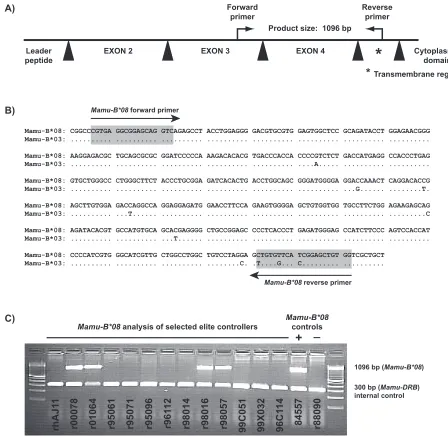
Mamu-B*03are strikingly similar in the regions encoding the alpha-1 to alpha-3 domains (6), we designed the reverse primer for the Mamu-B*08 PCR-SSP reaction to target multiple unique polymorphisms present in the exon encoding the trans-membrane region of Mamu-B*08 (Fig. 1A and B). Primers were diluted to a working concentration of 1M.
Thermal cycling conditions were identical to methods pre-viously published (27), with the exception of the second an-nealing temperature being 67.9°C for 21 cycles. At the com-pletion of 30 thermal cycles, the PCR underwent a final extension at 72°C for 8 min, followed by a terminal hold at 25°C. Subsequently, PCR products were electrophoresed on 2% agarose gels at a constant voltage in 0.5⫻sodium borate buffer (7). The correspondingMamu-B*08allele-specific am-plicon was⬃1,096 bp (Fig. 1C). EachMamu-B*08typing re-action also included primers to target an⬃300-bp fragment of the class IIMamu-DRBas an internal control (23). MHC class I typing reactions were not considered valid without the pres-ence of this internal control amplification product. Amplicon length was measured relative to a 100-bp DNA ladder (Invitro-gen, Carlsbad, CA). Amplification specificity of the
Mamu-* Corresponding author. Mailing address: Department of Pathology and Laboratory Medicine, University of Wisconsin-Madison, 555 Sci-ence Dr., Madison, WI 53711. Phone: (608) 265-3380. Fax: (608) 265-8084. E-mail: [email protected].
䌤Published ahead of print on 30 May 2007.
8827
on November 8, 2019 by guest
http://jvi.asm.org/
B*08typing primers was confirmed by using previously estab-lished sequencing methods (18a) in 44 Mamu-B*08-positive Indian rhesus macaques.
The remaining MHC class I alleles (Mamu-A*01, -A*02,
-A*08, -A*11, -B*01, -B*03, -B*04, -B*17, and -B*29) were typed as previously described (18a, 27).
Colony frequencies of Mamu-B*08. After completion of PCR-SSP development, we screened eight different Indian rhesus macaque colonies to determine the frequency of Mamu-B*08(Table 1). After testing⬎2,900 macaques, we found that
the overall frequency ofMamu-B*08was⬃5.8%. At the Wis-consin National Primate Research Center (WNPRC), the fre-quency of Mamu-B*08 was 6.8% (1,271 macaques tested). Overall, the frequency ranged from 0.9 to 14.1% across the various macaque colonies.
Mamu-B*08 is significantly enriched in a cohort of elite controller macaques and is associated with lower viremia in the chronic phase of SIV infection.We next investigated the frequency ofMamu-B*08in a cohort of previously identified EC macaques (41). Four of the thirteen ECs expressed
Mamu-FIG. 1. Design of PCR-sequence specific primers (PCR-SSP) to amplify the MHC class I alleleMamu-B*08from genomic DNA. (A) Primers were designed to amplify a 1,096-bp product that contains part of exon 3 (77 bp), intron 3 (573 bp), exon 4 (276 bp), intron 4 (106), and 64 bp of the transmembrane region (exon 5) ofMamu-B*08. Black triangles representMamu-B*08introns. (B) Sequence similarity betweenMamu-B*08 and Mamu-B*03. To avoid amplification of Mamu-B*03, the Mamu-B*08 primers were designed such that the forward primer amplifies Mamu-B*08orMamu-B*03and the reverse primer takes advantage of three unique polymorphisms in the transmembrane region (exon 5), allowing for specific detection ofMamu-B*08only. (C) PCR-SSP genotyping ofMamu-B*08from genomic DNA of selected elite controller macaques. Macaques 84557 and r88090 were used asMamu-B*08positive and negative controls, respectively. Previously, a cDNA library isolated from 84557 indicated that this macaque expressesMamu-B*08.Mamu-B*08was not found in a cDNA library from macaque r88090 (6). PCR-SSP primers that amplify a portion ofMamu-DRB(⬃300 bp) were included as an internal control to minimize false negatives. A 100-bp DNA ladder was used to interpret amplicon sizes.
on November 8, 2019 by guest
http://jvi.asm.org/
[image:2.585.59.507.66.504.2]B*08(Fig. 1C). Interestingly, three of the four Mamu-B*17 -negative ECs, r00078, r01064, and r98057, wereMamu-B*08
positive. After testing a cohort of 192 SIV-infected macaques forMamu-B*08, we identified sevenMamu-B*08-positive ma-caques, four of which were from this initial EC cohort.
To investigate the role ofMamu-B*08in control of
patho-genic SIV replication, we recently infected four additional
Mamu-B*08-positive Indian rhesus macaques with SIVmac239
(100 50% tissue culture infective doses, intravenously). Two of theseMamu-B*08-positive macaques, r00032 and r02019, had viral loads below 1,000 vRNA copies/ml at 22 weeks postinfec-tion. We have included them in Table 2 as ECs due to their low plasma viremia early into the chronic phase of SIV infection. All four recently infectedMamu-B*08-positive macaques were included in the statistical analysis.
PROC LOGISTIC regression analysis was performed on 196 SIV-infected Indian rhesus macaques from two indepen-dent cohorts (143 animals from D. Watkins’ studies and 53 animals from J. Lifson’s studies) to determine whether the frequency ofMamu-B*08was enriched in EC macaques. The analysis methods were performed as previously described (41), except the program used was SAS 9.1 (SAS Institute, Cary, NC). All macaques were infected with the molecular clone SIVmac239, and plasma virus concentrations were monitored
as previously described (10, 26) at multiple time points for a minimum of 10 weeks postinfection. Although vaccinated an-imals are included in the 196 animal cohort, none of these macaques received a vaccine regimen that demonstrated effec-tive and durable control of SIV replication.
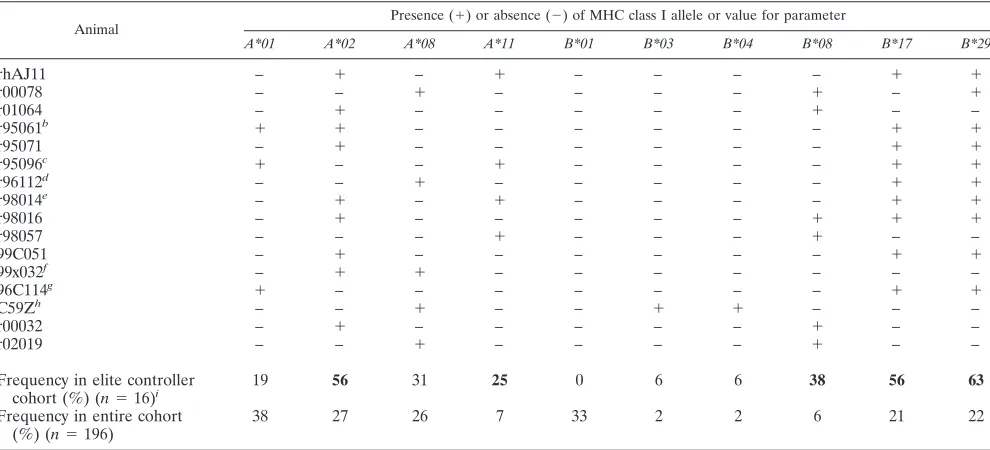
[image:3.585.43.283.89.236.2]Including the two recently infectedMamu-B*08-positive EC animals, 16 SIVmac239-infected macaques were identified as
TABLE 1. Frequency ofMamu-B*08in different Indian rhesus macaque colonies
Colonya
No. of
Mamu-B*08⫹
animals
Total no. of animals
Frequency (%)
Southern Research Institute (Birmingham, AL)
1 113 0.9
Yerkes NPRC 3 141 2.1
Various NIH/NCI-funded projects
8 380 2.1
Tulane NPRC 3 122 2.5
Caribbean Primate Research Center
18 451 4.0
California NPRC 11 163 6.7
Wisconsin NPRC 86 1271 6.8
Oregon NPRC 41 290 14.1
Total 171 2,931 5.8
a
NPRC, National Primate Research Center; NIH/NCI, National Institutes of Health and National Cancer Institute.
TABLE 2. Mamu-B*08is significantly overrepresented among elite controller macaquesa
Animal
Presence (⫹) or absence (⫺) of MHC class I allele or value for parameter
A*01 A*02 A*08 A*11 B*01 B*03 B*04 B*08 B*17 B*29
rhAJ11 – ⫹ – ⫹ – – – – ⫹ ⫹
r00078 – – ⫹ – – – – ⫹ – ⫹
r01064 – ⫹ – – – – – ⫹ – –
r95061b ⫹ ⫹ – – – – – – ⫹ ⫹
r95071 – ⫹ – – – – – – ⫹ ⫹
r95096c ⫹ – – ⫹ – – – – ⫹ ⫹
r96112d – – ⫹ – – – – – ⫹ ⫹
r98014e – ⫹ – ⫹ – – – – ⫹ ⫹
r98016 – ⫹ – – – – – ⫹ ⫹ ⫹
r98057 – – – ⫹ – – – ⫹ – –
99C051 – ⫹ – – – – – – ⫹ ⫹
99x032f – ⫹ ⫹ – – – – – – –
96C114g ⫹ – – – – – – – ⫹ ⫹
C59Zh – – ⫹ – – ⫹ ⫹ – – –
r00032 – ⫹ – – – – – ⫹ – –
r02019 – – ⫹ – – – – ⫹ – –
Frequency in elite controller cohort (%) (n⫽16)i
19 56 31 25 0 6 6 38 56 63
Frequency in entire cohort (%) (n⫽196)
38 27 26 7 33 2 2 6 21 22
aSixteen SIV
mac239-infected Indian rhesus macaques that maintained chronic phase (ⱖ10 weeks) plasma virus concentrations of⬍1,000 vRNA copies/ml were identified as ECs. This 1,000 vRNA copies/ml EC threshold represents a⬎2-log reduction in chronic-phase plasma virus concentrations compared to the chronic-phase geometric mean of the entire cohort of 196 SIV-infected macaques (223,800 vRNA copies/ml). Viral control was maintained for⬎1 year during the chronic phase of SIVmac239 infection or from week 10 until the animal was assigned to another study. All ECs were infected for⬎39 weeks (9 months) with the exception of r96112 (38 weeks), along with r00032 and r02019 (22 weeks each in an ongoing study). Animals were vaccine naive except where indicated.
bDNA/MVA Gag
181-189CM9 epitope only. The vaccine results were previously published (1).
cLipopeptide Gag
181-189CM9 epitope only (35).
dCpG in incomplete Freund adjuvant (41).
eIntramuscular injections of 107irradiated PBMC from SIV-naive animal rh2055 (41).
fDNA SIVgagprime/Ad5 SIVgagboost. The vaccine results were previously published (9, 29). gWhole inactivated SIV virions. The vaccine results were previously published (25).
hCpG in PBS (J. Lifson, unpublished results).
iSignificant enrichments of the elite controller cohort for expression of a particular MHC class I allele are indicated in bold. Enrichment of this elite controller group
for MHC class I alleles was determined with PROC LOGISTIC in SAS 9.1 (SAS Institute, Cary, NC).Mamu-A*02, P⫽0.01;Mamu-A*11,P⫽0.01;Mamu-B*08,P⫽
0.00001;Mamu-B*17,P⫽0.001;Mamu-B*29,P⫽0.0003.
on November 8, 2019 by guest
http://jvi.asm.org/
[image:3.585.45.540.378.603.2]ECs, defined as maintaining a chronic phase (ⱖ10 weeks) plasma viremia geometric mean below 1,000 vRNA copies/ml (Table 2).Mamu-B*08was dramatically enriched in this EC group. Thirty-eight percent of this EC cohort expressed
Mamu-B*08compared to just three percent of the 180 SIV-infected macaques that did not control viral replication to the EC threshold (P ⫽ 0.00001). Overall, 6 of 11 (⬃55%) ma-caques identified asMamu-B*08-positive became ECs. Two of the elevenMamu-B*08-positive macaques also displayed what we refer to as controller status, limiting viral replication to
⬍22,000 viral RNA copies/ml (one log reduction of the co-hort’s chronic phase geometric mean, 223,800 vRNA copies/ ml) for at least 10 weeks in the chronic stage of SIV infection. Hence, a majority ofMamu-B*08-positive macaques (8 of 11 [73%]) displayed better-than-typical viral control of patho-genic SIVmac239 replication.
Animal C59Z, a Mamu-B*03-positive macaque, was also recently identified as 1 of the 16 EC macaques (Table 2). AlthoughMamu-B*03was previously associated with slow SIV disease progression (12), it has been difficult to conduct addi-tional studies withMamu-B*03due to the low frequency of this allele (overall,⬍1%; 18a). Interestingly, a previous study dem-onstrated that Mamu-B*03 and HLA-B27 bind peptides with similar motifs (11). Mamu-B*03 and Mamu-B*08 are almost identical in amino acid sequence (6). There are only two amino acid differences between Mamu-B*03 and Mamu-B*08 in re-gions that influence peptide binding and antigen recognition (5, 16). Both differences reside in the alpha-1 domain (exon two); the alpha-2 domains (exon 3) of B*03 and Mamu-B*08 are identical. Therefore, it is likely that Mamu-Mamu-B*08 also shares this HLA-B27 binding profile.
We analyzed the association between MHC class I alleles in both acute- and chronic-phase viremia for each MHC class I allele in the 196 SIVmac239-infected macaque cohort. With the
exception of the program used (SAS 9.1; SAS Institute, Cary, NC), the analysis methods were performed as previously de-scribed (41). PROC REG was used to estimate the relative log geometric mean acute phase peak (single highest value from weeks 1 to 3 for each macaque) plasma virus concentration. PROC MIXED was used to estimate the relative log geometric mean for chronic-phase (ⱖ10 weeks) plasma viremia. All typed MHC class I alleles, EC status, and vaccine status were in-cluded as covariates in the models.
Although none of the MHC class I alleles correlated with changes in peak viremia during the acute phase of SIV infec-tion (Fig. 2A), several MHC class I alleles affected chronic phase viremia (Fig. 2B). The 11 Mamu-B*08-positive ma-caques had a 7.34-fold decrease in geometric mean chronic phase plasma virus concentrations compared to the rest of the cohort (P⫽0.002). The reduction in the relative log geometric mean ofMamu-B*08-positive macaques was similar to that of macaques expressingMamu-B*17(7.93-fold), an allele previ-ously associated with slow disease progression (35, 41).
[image:4.585.318.516.64.569.2]We identified another Mamu-B*08-positive EC macaque (r99006) not included in the 196 SIV-infected animal cohort described in the present study. Animal r99006 was part of a separate study in whichMamu-A*01-positive andMamu-B*17 -positive macaques were infected with an engineered CD8⫹ T-cell escape variant virus (14, 15) to prevent the development of immunodominant CD8⫹ T-cell responses thought to be
FIG. 2.Mamu-B*08influences chronic-phase plasma virus concentra-tions in SIVmac239-infected Indian rhesus macaques. (A) Acute-phase
peak viremia (highest plasma virus concentration between weeks 1 and 3 postinfection) is not affected by the presence of the typed MHC class I alleles in 196 SIVmac239-infected Indian rhesus macaques. (B)
Contribu-tion of typed MHC class I alleles to variaContribu-tion in chronic-phase plasma viremia in the same cohort of 196 macaques. The chronic-phase geomet-ric mean was calculated, and the plasma virus concentration time points ranged fromⱖ10 weeks to⬍7.16 years postchallenge. MHC class I effects on SIV plasma viremia are shown with the changes in geometric mean displayed as dots for each MHC class I allele (Mamu-A*01,-A*02,-A*08, -A*11,-B*01,-B*03/-B*04,-B*08,-B*17, and -B*29) relative to the whole cohort and 95% confidence intervals (bars). Probabilities (Pvalues) for MHC class I alleles with statistically significant reductions from the rela-tive log geometric mean are displayed.Mamu-B*03and -B*04were an-alyzed together because the threeMamu-B*03-positive macaques were also positive forMamu-B*04.
on November 8, 2019 by guest
http://jvi.asm.org/
associated with viral control (35). Macaque r99006 controlled viremia to⬍1,000 vRNA copies/ml for almost 4 years after SIV infection before being assigned to another study. We have also identified three additional Mamu-B*08-positive macaques from other investigators (D. Evans and G. Franchini, personal communications). At 25 weeks postinfection, two of the threeMamu-B*08-positive macaques limited replication of SIVmac239 or SIVmac251 isolates at or below ⬃1,000 vRNA
copies/ml. In total, 11 of 15 (73%)Mamu-B*08-positive ma-caques controlled replication of pathogenic strains of SIV.
It has been previously reported thatMamu-B*08segregates on the same chromosome asMamu-B*06in rhesus macaques (37). Interestingly, when investigating MHC class I cDNA li-braries from several Mamu-B*08-positive Indian rhesus caques at the WNPRC, we also noticed that all of these ma-caques possessed Mamu-B*06 transcripts (data not shown). None of the cDNA libraries from Mamu-B*08-negative ma-caques possessedMamu-B*06transcripts. However, control of SIV replication is unlikely due to Mamu-B*06. From our stud-ies, Mamu-B*06 does not appear to be involved in the presen-tation of SIV epitopes (unpublished data). Mamu-B*08, but not Mamu-B*06, was detected by one-dimensional isoelectric focusing (6). This suggests thatMamu-B*06may not encode an expressed MHC class I protein.
Mamu-B*08-positive Indian rhesus macaques might provide an insightful model for understanding the influence of partic-ular MHC class I alleles on viral control. We have recently identified several Mamu-B*08-restricted CD8⫹T-cell epitopes in the chronic phase of SIV infection (Loffredo et al., unpub-lished). We are currently determining whether CD8⫹immune responses restricted by Mamu-B*08 exert strong patterns of immunodominance similar to those associated withHLA-B27
andHLA-B57(2, 4, 22). Mamu-B*08-restricted SIV epitopes should be useful for future pathogenesis studies, including the generation of MHC class I tetramers, to study differences in the CD8⫹T-cell responses between progressor and EC Mamu-B*08-positive macaques. Finally, given the independent influ-ence of this allele on the outcome of SIV infection, it might be prudent to removeMamu-B*08-positive macaques from vac-cine studies.
We thank Debra L. Fisk for MHC class I sequencing assistance and Richard Rudersdorf and Lyle Wallace for the construction of MHC class I cDNA libraries. We are also grateful to Danilo R. Casimiro, David T. Evans, Genoveffa Franchini, Adrian McDermott, Eva Rakasz, Thomas Friedrich, Levi Yant, and Matthew Reynolds for providing plasma virus concentrations and SIV disease status of ma-caques used in their studies. Sean Spencer, Laura Valentine, and David O’Connor provided helpful discussions. We also thank the vi-rology, genetics, and immunology core laboratories at the National Primate Research Center, University of Wisconsin-Madison (WNPRC) for technical assistance.
This research was supported by a National Institutes of Health (NIH) contract HHSN266200400088C and NIH grants R24 RR015371, R24 RR016038, R01 AI049120, and R01 AI052056 to D.I.W. in addition to P51 RR000167 to the WNPRC. This project has been funded in whole or in part with federal funds from the National Cancer Institute, National Institutes of Health, under contract N01-CO-12400. This research was supported in part by the Intramural Research Program of the NIH, National Cancer Institute, Center for Cancer Research. This study was conducted in part at a facility con-structed with support from Research Facilities Improvement grant numbers RR15459-01 and RR020141-01 (WNPRC).
The content of this publication does not necessarily reflect the views or policies of the U.S. Department of Health and Human Services, nor does mention of trade names, commercial products, or organizations imply endorsement by the U.S. Government.
REFERENCES
1.Allen, T. M., P. Jing, B. Calore, H. Horton, D. H. O’Connor, T. Hanke, M. Piekarczyk, R. Ruddersdorf, B. R. Mothe, C. Emerson, N. Wilson, J. D. Lifson, I. M. Belyakov, J. A. Berzofsky, C. Wang, D. B. Allison, D. C. Montefiori, R. C. Desrosiers, S. Wolinsky, K. J. Kunstman, J. D. Altman, A. Sette, A. J. McMichael, and D. I. Watkins. 2002. Effects of cytotoxic T lymphocytes (CTL) directed against a single simian immunodeficiency virus (SIV) Gag CTL epitope on the course of SIVmac239 infection. J. Virol. 76:10507–10511.
2.Altfeld, M., E. T. Kalife, Y. Qi, H. Streeck, M. Lichterfeld, M. N. Johnston, N. Burgett, M. E. Swartz, A. Yang, G. Alter, X. G. Yu, A. Meier, J. K. Rockstroh, T. M. Allen, H. Jessen, E. S. Rosenberg, M. Carrington, and B. D. Walker. 2006. HLA alleles associated with delayed progression to AIDS contribute strongly to the initial CD8⫹T cell response against HIV-1. PLoS Med.3:e403.
3.Bailey, J. R., T. M. Williams, R. F. Siliciano, and J. N. Blankson.2006. Maintenance of viral suppression in HIV-1-infected HLA-B*57⫹elite sup-pressors despite CTL escape mutations. J. Exp. Med.203:1357–1369. 4.Bihl, F., N. Frahm, L. Di Giammarino, J. Sidney, M. John, K. Yusim, T.
Woodberry, K. Sango, H. S. Hewitt, L. Henry, C. H. Linde, J. V. R. Chisholm, T. M. Zaman, E. Pae, S. Mallal, B. D. Walker, A. Sette, B. T. Korber, D. Heckerman, and C. Brander.2006. Impact of HLA-B alleles, epitope bind-ing affinity, functional avidity, and viral coinfection on the immunodomi-nance of virus-specific CTL responses. J. Immunol.176:4094–4101. 5.Bjorkman, P. J., M. A. Saper, B. Samraoui, W. S. Bennett, J. L. Strominger,
and D. C. Wiley.1987. The foreign antigen binding site and T-cell recogni-tion regions of class I histocompatibility antigens. Nature329:512–518. 6.Boyson, J. E., C. Shufflebotham, L. F. Cadavid, J. A. Urvater, L. A. Knapp,
A. L. Hughes, and D. I. Watkins.1996. The MHC class I genes of the rhesus monkey: different evolutionary histories of MHC class I and II genes in primates. J. Immunol.156:4656–4665.
7.Brody, J. R., and S. E. Kern.2004. Sodium boric acid: a Tris-free, cooler conductive medium for DNA electrophoresis. BioTechniques36:214–216. 8.Carrington, M., G. W. Nelson, M. P. Martin, T. Kissner, D. Vlahov, J. J.
Goedert, R. Kaslow, S. Buchbinder, K. Hoots, and S. J. O’Brien.1999. HLA and HIV-1: heterozygote advantage and B*35-Cw*04 disadvantage. Science 283:1748–1752.
9.Casimiro, D. R., F. Wang, W. A. Schleif, X. Liang, Z. Q. Zhang, T. W. Tobery, M. E. Davies, A. B. McDermott, D. H. O’Connor, A. Fridman, A. Bagchi, L. G. Tussey, A. J. Bett, A. C. Finnefrock, T. M. Fu, A. Tang, K. A. Wilson, M. Chen, H. C. Perry, G. J. Heidecker, D. C. Freed, A. Carella, K. S. Punt, K. J. Sykes, L. Huang, V. I. Ausensi, M. Bachinsky, U. Sadasivan-Nair, D. I. Watkins, E. A. Emini, and J. W. Shiver.2005. Attenuation of simian immu-nodeficiency virus SIVmac239 infection by prophylactic immunization with DNA and recombinant adenoviral vaccine vectors expressing Gag. J. Virol. 79:15547–15555.
10.Cline, A. N., J. W. Bess, M. Piatak, Jr., and J. D. Lifson.2005. Highly sensitive SIV plasma viral load assay: practical considerations, realistic per-formance expectations, and application to reverse engineering of vaccines for AIDS. J. Med. Primatol.34:303–312.
11.Dzuris, J. L., J. Sidney, E. Appella, R. W. Chesnut, D. I. Watkins, and A. Sette.2000. Conserved MHC class I peptide binding motif between humans and rhesus macaques. J. Immunol.164:283–291.
12.Evans, D. T., D. H. O’Connor, P. Jing, J. L. Dzuris, J. Sidney, J. da Silva, T. M. Allen, H. Horton, J. E. Venham, R. A. Rudersdorf, T. Vogel, C. D. Pauza, R. E. Bontrop, R. DeMars, A. Sette, A. L. Hughes, and D. I. Watkins. 1999. Virus-specific cytotoxic T-lymphocyte responses select for amino-acid variation in simian immunodeficiency virus Env and Nef. Nat. Med.5:1270– 1276.
13.Feeney, M. E., Y. Tang, K. A. Roosevelt, A. J. Leslie, K. McIntosh, N. Karthas, B. D. Walker, and P. J. Goulder.2004. Immune escape precedes breakthrough human immunodeficiency virus type 1 viremia and broadening of the cytotoxic T-lymphocyte response in an HLA-B27-positive long-term-nonprogressing child. J. Virol.78:8927–8930.
14.Friedrich, T. C., E. J. Dodds, L. J. Yant, L. Vojnov, R. Rudersdorf, C. Cullen, D. T. Evans, R. C. Desrosiers, B. R. Mothe, J. Sidney, A. Sette, K. Kunstman, S. Wolinsky, M. Piatak, J. Lifson, A. L. Hughes, N. Wilson, D. H. O’Connor, and D. I. Watkins.2004. Reversion of CTL escape-variant immunodeficiency viruses in vivo. Nat. Med.10:275–281.
15.Friedrich, T. C., A. B. McDermott, M. R. Reynolds, S. Piaskowski, S. Fuenger, I. P. De Souza, R. Rudersdorf, C. Cullen, L. J. Yant, L. Vojnov, J. Stephany, S. Martin, D. H. O’Connor, N. Wilson, and D. I. Watkins.2004. Consequences of cytotoxic T-lymphocyte escape: common escape mutations in simian immunodeficiency virus are poorly recognized in naive hosts. J. Vi-rol.78:10064–10073.
16.Garrett, T. P., M. A. Saper, P. J. Bjorkman, J. L. Strominger, and D. C.
on November 8, 2019 by guest
http://jvi.asm.org/
Wiley.1989. Specificity pockets for the side chains of peptide antigens in HLA-Aw68. Nature342:692–696.
17.Goulder, P. J., R. E. Phillips, R. A. Colbert, S. McAdam, G. Ogg, M. A. Nowak, P. Giangrande, G. Luzzi, B. Morgan, A. Edwards, A. J. McMichael, and S. Rowland-Jones.1997. Late escape from an immunodominant cyto-toxic T-lymphocyte response associated with progression to AIDS. Nat. Med. 3:212–217.
18.Hendel, H., S. Caillat-Zucman, H. Lebuanec, M. Carrington, S. O’Brien, J. M. Andrieu, F. Schachter, D. Zagury, J. Rappaport, C. Winkler, G. W. Nelson, and J. F. Zagury.1999. New class I and II HLA alleles strongly associated with opposite patterns of progression to AIDS. J. Immunol.162: 6942–6946.
18a.Kaizu, M., G. J. Borchardt, C. E. Glidden, D. L. Fisk, J. T. Loffredo, D. I. Watkins, and W. M. Rehrauer.Molecular typing of major histocompatibility complex class I alleles in the Indian rhesus macaque which restricts SIV CD8⫹T cell epitopes. Immunogenetics, in press.
19.Kaslow, R. A., M. Carrington, R. Apple, L. Park, A. Munoz, A. J. Saah, J. J. Goedert, C. Winkler, S. J. O’Brien, C. Rinaldo, R. Detels, W. Blattner, J. Phair, H. Erlich, and D. L. Mann.1996. Influence of combinations of human major histocompatibility complex genes on the course of HIV-1 infection. Nat. Med.2:405–411.
20.Kaslow, R. A., C. Rivers, J. Tang, T. J. Bender, P. A. Goepfert, R. El Habib, K. Weinhold, and M. J. Mulligan.2001. Polymorphisms in HLA class I genes associated with both favorable prognosis of human immunodeficiency virus (HIV) type 1 infection and positive cytotoxic T-lymphocyte responses to ALVAC-HIV recombinant canarypox vaccines. J. Virol.75:8681–8689. 21.Kelleher, A. D., C. Long, E. C. Holmes, R. L. Allen, J. Wilson, C. Conlon, C.
Workman, S. Shaunak, K. Olson, P. Goulder, C. Brander, G. Ogg, J. S. Sullivan, W. Dyer, I. Jones, A. J. McMichael, S. Rowland-Jones, and R. E. Phillips.2001. Clustered mutations in HIV-1 gag are consistently required for escape from HLA-B27-restricted cytotoxic T lymphocyte responses. J. Exp. Med.193:375–386.
22.Kiepiela, P., A. J. Leslie, I. Honeyborne, D. Ramduth, C. Thobakgale, S. Chetty, P. Rathnavalu, C. Moore, K. J. Pfafferott, L. Hilton, P. Zimbwa, S. Moore, T. Allen, C. Brander, M. M. Addo, M. Altfeld, I. James, S. Mallal, M. Bunce, L. D. Barber, J. Szinger, C. Day, P. Klenerman, J. Mullins, B. Korber, H. M. Coovadia, B. D. Walker, and P. J. Goulder.2004. Dominant influence of HLA-B in mediating the potential co-evolution of HIV and HLA. Nature432:769–775.
23.Knapp, L. A., E. Lehmann, M. S. Piekarczyk, J. A. Urvater, and D. I. Watkins.1997. A high frequency of Mamu-A*01 in the rhesus macaque detected by polymerase chain reaction with sequence-specific primers and direct sequencing. Tissue Antigens50:657–661.
24.Leslie, A. J., K. J. Pfafferott, P. Chetty, R. Draenert, M. M. Addo, M. Feeney, Y. Tang, E. C. Holmes, T. Allen, J. G. Prado, M. Altfeld, C. Brander, C. Dixon, D. Ramduth, P. Jeena, S. A. Thomas, A. S. John, T. A. Roach, B. Kupfer, G. Luzzi, A. Edwards, G. Taylor, H. Lyall, G. Tudor-Williams, V. Novelli, J. Martinez-Picado, P. Kiepiela, B. D. Walker, and P. J. Goulder. 2004. HIV evolution: CTL escape mutation and reversion after transmission. Nat. Med.10:282–289.
25.Lifson, J. D., M. J. Piatak, J. L. Rossio, J. J. Bess, E. Chertova, D. Schneider, R. Kiser, V. Coalter, B. Poore, R. Imming, R. C. Desrosiers, L. E. Henderson, and L. O. Arthur.2002. Whole inactivated SIV virion vaccines with func-tional envelope glycoproteins: safety, immunogenicity, and activity against intrarectal challenge. J. Med. Primatol.31:205–216.
26.Loffredo, J. T., B. J. Burwitz, E. G. Rakasz, S. P. Spencer, J. J. Stephany, J. P. Vela, S. R. Martin, J. Reed, S. M. Piaskowski, J. Furlott, K. L. Weisgrau, D. S. Rodrigues, T. Soma, G. Napoe, T. C. Friedrich, N. A. Wilson, E. G. Kallas, and D. I. Watkins.2007. The antiviral efficacy of simian immunodeficiency virus-specific CD8⫹ T cells is unrelated to epitope specificity and is abrogated by viral escape. J. Virol.81:2624– 2634.
27.Loffredo, J. T., J. Sidney, S. Piaskowski, A. Szymanski, J. Furlott, R. Rudersdorf, J. Reed, B. Peters, H. D. Hickman-Miller, W. Bardet, W. M. Rehrauer, D. H. O’Connor, N. A. Wilson, W. H. Hildebrand, A. Sette, and D. I. Watkins.2005. The high frequency Indian rhesus macaque MHC class I molecule, Mamu-B*01, does not appear to be involved in CD8⫹T lymphocyte responses to SIVmac239. J. Immunol.175:5986–5997. 28.Mann, D. L., R. P. Garner, D. E. Dayhoff, K. Cao, M. A. Fernandez-Vina, C.
Davis, N. Aronson, N. Ruiz, D. L. Birx, and N. L. Michael.1998. Major histocompatibility complex genotype is associated with disease progression
and virus load levels in a cohort of human immunodeficiency virus type 1-infected Caucasians and African Americans. J. Infect. Dis.178:1799–1802. 29.McDermott, A. B., D. H. O’Connor, S. Fuenger, S. Piaskowski, S. Martin, J. Loffredo, M. Reynolds, J. Reed, J. Furlott, T. Jacoby, C. Riek, E. Dodds, K. Krebs, M. E. Davies, W. A. Schleif, D. R. Casimiro, J. W. Shiver, and D. I. Watkins.2005. Cytotoxic T-lymphocyte escape does not always explain the transient control of simian immunodeficiency virus SIVmac239 viremia in adenovirus-boosted and DNA-primed Mamu-A*01-positive rhesus macaques. J. Virol.79:15556–15566.
30.McNeil, A. J., P. L. Yap, S. M. Gore, R. P. Brettle, M. McColl, R. Wyld, S. Davidson, R. Weightman, A. M. Richardson, and J. R. Robertson.1996. Association of HLA types A1–B8-DR3 and B27 with rapid and slow pro-gression of HIV disease. QJM89:177–185.
31.Migueles, S. A., A. C. Laborico, W. L. Shupert, M. S. Sabbaghian, R. Rabin, C. W. Hallahan, D. Van Baarle, S. Kostense, F. Miedema, M. McLaughlin, L. Ehler, J. Metcalf, S. Liu, and M. Connors.2002. HIV-specific CD8⫹ T-cell proliferation is coupled to perforin expression and is maintained in nonprogressors. Nat. Immunol.3:1061–1068.
32.Migueles, S. A., M. S. Sabbaghian, W. L. Shupert, M. P. Bettinotti, F. M. Marincola, L. Martino, C. W. Hallahan, S. M. Selig, D. Schwartz, J. Sullivan, and M. Connors.2000. HLA B*5701 is highly associated with restriction of virus replication in a subgroup of HIV-infected long-term nonprogressors. Proc. Natl. Acad. Sci. USA97:2709–2714.
33.Mothe, B. R., J. Weinfurter, C. Wang, W. Rehrauer, N. Wilson, T. M. Allen, D. B. Allison, and D. I. Watkins.2003. Expression of the major histocom-patibility complex class I molecule Mamu-A*01 is associated with control of simian immunodeficiency virus SIVmac239 replication. J. Virol.77:2736– 2740.
34.Muhl, T., M. Krawczak, P. Ten Haaft, G. Hunsmann, and U. Sauermann. 2002. MHC class I alleles influence set-point viral load and survival time in simian immunodeficiency virus-infected rhesus monkeys. J. Immunol.169: 3438–3446.
35.O’Connor, D. H., B. R. Mothe, J. T. Weinfurter, S. Fuenger, W. M. Rehrauer, P. Jing, R. R. Rudersdorf, M. E. Liebl, K. Krebs, J. Vasquez, E. Dodds, J. Loffredo, S. Martin, A. B. McDermott, T. M. Allen, C. Wang, G. G. Doxiadis, D. C. Montefiori, A. Hughes, D. R. Burton, D. B. Allison, S. M. Wolinsky, R. Bontrop, L. J. Picker, and D. I. Watkins.2003. Major histocompatibility complex class I alleles associated with slow simian immunodeficiency virus disease progression bind epitopes recognized by dominant acute-phase cytotoxic-T-lymphocyte responses. J. Virol.77:9029–9040.
36.Olerup, O., and H. Zetterquist.1991. HLA-DRB1*01 subtyping by allele-specific PCR amplification: a sensitive, allele-specific and rapid technique. Tissue Antigens37:197–204.
37.Otting, N., C. M. Heijmans, R. C. Noort, N. G. de Groot, G. G. Doxiadis, J. J. van Rood, D. I. Watkins, and R. E. Bontrop.2005. Unparalleled complexity of the MHC class I region in rhesus macaques. Proc. Natl. Acad. Sci. USA 102:1626–1631.
38.Pal, R., D. Venzon, N. L. Letvin, S. Santra, D. C. Montefiori, N. R. Miller, E. Tryniszewska, M. G. Lewis, T. C. VanCott, V. Hirsch, R. Woodward, A. Gibson, M. Grace, E. Dobratz, P. D. Markham, Z. Hel, J. Nacsa, M. Klein, J. Tartaglia, and G. Franchini.2002. ALVAC-SIV-gag-pol-env-based vac-cination and macaque major histocompatibility complex class I (A*01) delay simian immunodeficiency virus SIVmac-induced immunodeficiency. J. Virol. 76:292–302.
39.Tang, J., S. Tang, E. Lobashevsky, A. D. Myracle, U. Fideli, G. Aldrovandi, S. Allen, R. Musonda, and R. A. Kaslow.2002. Favorable and unfavorable HLA class I alleles and haplotypes in Zambians predominantly infected with clade C human immunodeficiency virus type 1. J. Virol.76:8276–8284. 40.Wojcechowskyj, J. A., L. J. Yant, R. W. Wiseman, S. L. O’Connor, and D. H.
O’Connor.2007. Control of simian immunodeficiency virus SIVmac239 is not predicted by inheritance of Mamu-B*17-containing haplotypes. J. Virol. 81:406–410.
41.Yant, L. J., T. C. Friedrich, R. C. Johnson, G. E. May, N. J. Maness, A. M. Enz, J. D. Lifson, D. H. O’Connor, M. Carrington, and D. I. Watkins.2006. The high-frequency major histocompatibility complex class I allele Mamu-B*17 is associated with control of simian immunodeficiency virus SIVmac239 replication. J. Virol.80:5074–5077.
42.Zhang, Z. Q., T. M. Fu, D. R. Casimiro, M. E. Davies, X. Liang, W. A. Schleif, L. Handt, L. Tussey, M. Chen, A. Tang, K. A. Wilson, W. L. Trigona, D. C. Freed, C. Y. Tan, M. Horton, E. A. Emini, and J. W. Shiver.2002. Mamu-A*01 allele-mediated attenuation of disease progression in simian-human immunodeficiency virus infection. J. Virol.76:12845–12854.