Differentially Regulate Its Activities and Enhance Viral Replication
Heba H. Mostafa, Thornton W. Thompson, David J. Davido
Department of Molecular Biosciences, University of Kansas, Lawrence, Kansas, USA
The herpes simplex virus 1 (HSV-1) infected cell protein 0 (ICP0) is an immediate-early phosphoprotein that transactivates viral
gene expression. Evidence suggests that phosphorylation regulates the functions of ICP0, and three regions (termed regions I, II,
and III) in the protein are known to be phosphorylated. Mutation of the putative phosphorylation sites within region I, termed
Phos 1, which lies in the N-terminal portion of ICP0, impairs the E3 ubiquitin (Ub) ligase and ND10-disrupting activities of
ICP0 in cell culture and diminishes viral replication. To identify the specific phosphorylation site(s) or residues responsible for
the phenotypes observed with Phos 1, individual residues within region I were mutated to alanine (S224A, T226A, T231A, and
T232A) and one double mutant S224A/T226A was constructed. Tissue culture studies demonstrated that the S224A, S224A/
T226A, T231A, and T232A mutants were unable to dissociate the cellular protein PML from ND10 and that the S224/T226A
mu-tant was defective in its ability to dissociate the cellular protein Sp100 from ND10. Additionally, the transactivation activity of
ICP0 was impaired in the S224A and S224A/T226A mutants. The S224A and S224A/T226A mutant forms were more stable than
wild-type ICP0, suggesting that their ability to autoubiquitinate was limited. Moreover, one ICP0 ubiquitination target, USP-7,
was also more stable after infection with these two mutants. Lastly, the replication of the S224A and S224A/T226A mutant
vi-ruses was reduced in cell culture and
in vivo
. Overall, our data suggest that specific phosphorylation sites within region I
differ-entially regulate the activities of ICP0, which are required for efficient viral replication.
H
erpes simplex virus 1 (HSV-1) is a major human pathogen
that infects approximately 80% of the world’s population (
1
).
HSV-1 infection can range from being asymptomatic to causing
severe diseases such as blindness and encephalitis. As part of its life
cycle, the virus can latently infect the sensory neurons and
reacti-vate from latency by stress. can be reactireacti-vated by stress. The lytic
phase of infection is characterized by the expression of the viral
genes in a temporal cascade of immediate-early (IE), early (E), and
late (L) genes (
2
). The latent phase of infection, on the other hand,
is characterized by the overall lack of productive viral gene
expres-sion, with the exception of the latency-associated transcripts
(LATs) (
3
). Of the five IE proteins, the phosphoprotein ICP0
(in-fected cell protein 0) is required for efficient lytic and latent
infec-tions and for reactivation from latency (
4
–
7
).
ICP0 is a RING finger motif (
8
,
9
)-containing protein that acts
as a potent transactivator of all three categories of HSV-1 genes
(
10
), a function that is required for efficient lytic replication (
11
,
12
). The transactivation activity of ICP0 is regulated in part by its
E3 ubiquitin (Ub) ligase activity. The E3 ubiquitin ligase activity of
ICP0 directs the proteosomal degradation of several cellular
pro-teins, two of which, promyelocytic leukemia protein (PML) and
Sp100, are contained in the subnuclear organelle ND10 (nuclear
domain 10) (
13
–
17
). ND10 is associated with cellular
prolifera-tion, differentiaprolifera-tion, senescence, and apoptosis but is also noted
for possessing antiviral activity (
18
–
21
). ICP0-mediated
degrada-tion of ND10-associated proteins disrupts the structure of ND10,
facilitating viral replication (
22
). Ubiquitin-specific protease 7
(USP-7), which is also called herpesvirus-associated
ubiquitin-specific protease (HAUSP), is another cellular target of ICP0
ubiq-uitination (
23
). The relation between USP-7 and ICP0 is unique:
USP-7 initially stabilizes ICP0 levels, and later during infection,
ICP0 directs the degradation of USP-7 (
23
,
24
). In addition to
disrupting ND10 and altering the stability of cellular proteins,
ICP0 impairs the host cell innate and intrinsic antiviral defenses,
which include interferon (
25
–
30
) and DNA damage responses
(
31
,
32
), both of which are triggered by viral infection.
ICP0 has been shown to be phosphorylated (
33
); however, the
exact function phosphorylation plays in regulating its activities is
largely unknown. The observation that specific cellular kinase
in-hibitors impair the transactivating activity of ICP0 suggested a
potential link between phosphorylation and this key activity of
ICP0 (
34
). Tandem mass spectrometric analysis identified three
regions on ICP0 that are phosphorylated; these are named regions
I, II, and III (
35
). Mutational analyses of the 11 putative
phos-phorylation residues clustered in the three regions identified
re-gion I as having the greatest impact on ICP0 functions and HSV-1
replication (
36
,
37
). Specifically, clustered mutation of four amino
acid residues in region I (termed Phos 1) impaired the ability of
ICP0 to dissociate two ND10-associated proteins, PML and
Sp100, from ND10, reduced its E3 Ub ligase activity, impaired its
transactivation activity, and impaired acute replication of HSV-1
in cell culture (
36
). Not surprisingly, Phos 1 mutant virus showed
diminished acute replication and reactivation when tested in a
mouse model of HSV-1 latency and reactivation (
37
).
In the current study, we wanted to determine which sites in
region I are responsible for the phenotypes observed with Phos 1.
Specifically, we sought to examine the contributions of serine 224
and threonine 226, which have been shown by tandem mass
spec-trometric analyses to be phosphorylated in cell culture (M. C.
Smith, W. S. Lane, and D. J. Davido, unpublished data; M. S.
Received20 September 2012Accepted27 November 2012
Published ahead of print5 December 2012
Address correspondence to David J. Davido, [email protected]. Copyright © 2013, American Society for Microbiology. All Rights Reserved.
doi:10.1128/JVI.02588-12
on November 7, 2019 by guest
http://jvi.asm.org/
Chaurushiya, A. Aslanian, and M. D. Weitzman, unpublished
data), as well as those of the potentially phosphorylated
thre-onines 231 and 232. In addition, we wanted to determine whether
there is differential regulation of the functions of ICP0 through
the phosphorylation or potential phosphorylation of different
res-idues within region I. To address these questions, several
phos-phorylation site mutations (S224A, T226A, S224A/T226A,
T231A, and T232A) were constructed, and the corresponding
mu-tant viruses were generated. Our data indicate that of the four
serines and threonines identified in region I, serine 224 had the
greatest impact on ICP0 functions and viral replication.
Interest-ingly, combined mutation of this site and the threonine 226 site
further reduced the transactivation activity of ICP0 and impaired
its ability to dissociate Sp100 from ND10. Our results strongly
suggest a model in which specific amino acids in region I can
differentially regulate ICP0 activities that are subsequently
re-quired for efficient viral replication.
MATERIALS AND METHODS
Cells and viruses.Vero, Hep-2, and HeLa cells were obtained from the American Type Culture Collection (ATCC) and were grown in Dulbec-co’s modified Eagle’s medium (DMEM) supplemented with 5% fetal bo-vine serum (FBS), 2 mML-glutamine, 100g/ml penicillin, and 100 U/ml streptomycin as described previously (38). L7 cells, a Vero cell line stably transfected with the HSV-1 ICP0 gene, were grown in the same medium as Vero cells and were used to determine the titers of all the ICP0 mutant viruses, including the ICP0-null mutant, 7134 (12). The titers of wild-type strain KOS (passage 11) and marker rescue (MR) viruses were determined on Vero cells.
Construction of region I phosphorylation mutations. Plasmid pAlter-1⫹ICP0 was used to make all region I mutations by using the pAlter site-directed mutagenesis kit according to the manufacturer’s pro-tocol (Promega). The primers (IDT) used for the mutagenesis are S224A (5=-GGGCCCTGTCGCCCAC-3=), T226A (5= -GTCGCCCACCCACCC-3=), S224A/T226A (5=-GGGCCCTGTCGCCCACCCACCC-3=), T231A (5=-CTGAGCCGGCCACGGA-3=), and T232A (5=-CTGAGCCCACCGC GGA-3=) (underlined letters represent the mutated codon or codons). Each primer was constructed such that a restriction enzyme site was cre-ated or elimincre-ated. Mutations were identified by restriction enzyme digest analyses and were confirmed by DNA sequencing. The double site muta-tion was made because these sites are phosphorylated in HSV-1 ICP0 and are conserved between the ICP0 homologues of HSV-1 and HSV-2 (M. C. Smith and D. J. Davido, unpublished data).
Generation of ICP0 phosphorylation mutant viruses.Region I mu-tant viruses were created by marker transfer. Briefly, Vero cells were plated on 60-mm dishes at 4⫻105cells per plate, and 24 h later, cells were cotransfected with 1g of 7134 viral DNA and 2.5g of pAlter-1 express-ing mutant forms of ICP0 digested with EcoRI and HindIII. Transfections were carried out by using Fugene 6 or Fugene HD (Roche) at a ratio of 3:1 (l of transfection reagent perg of DNA) according to the manufactur-er’s recommendations. Mutants were identified by blue/white selection. White plaques were picked and purified in at least 3 rounds in the presence of 5-bromo-4-chloro-3-indolyl--D-galactopyranoside (X-gal). Insertion of region I mutations was initially identified by PCR and restriction en-zyme digestions (seeFig. 4) and was confirmed by restriction enzyme digestion and Southern blot analyses. Marker rescue viruses were gener-ated by cotransfecting the mutant viral genomic DNA with plasmid pAlter-1⫹ICP0 digested with EcoRI and HindIII. Rescuants were iden-tified by PCR amplification of region I and restriction enzyme and/or Southern blot analyses. All the marker rescue viruses were plaque purified at least 3 times.
Immunofluorescence studies. (i) ND10 and ICP0 staining.Hep-2 cells were plated on glass coverslips in 12-well plates for 24 h (5⫻104cells per well). Cells were then transfected with 1g of plasmid DNA and
Fugene 6 or Fugene HD using a 3:1 dilution ratio as described above. Sixteen to 18 h posttransfection, cells were fixed with 5% formaldehyde and 2% sucrose in phosphate-buffered saline (PBS) for 5 min, washed with PBS, and permeabilized in 0.5% NP-40 and 10% sucrose in PBS for 15 min at 4°C. Cells were washed and blocked with PBS-FB (PBS, 1% fetal bovine serum, 1% bovine serum albumin [BSA], and 0.05% sodium azide) for 1 h at 37°C. The primary antibodies used were a mouse mono-clonal antibody against ICP0 (11060; catalog no. sc-53070; Santa Cruz Biotechnology) diluted 1:500 and a rabbit polyclonal antibody against PML (A301-167A; Bethyl Laboratories) diluted 1:100 or a rabbit poly-clonal antibody against Sp100 (AB1380; Chemicon International) diluted 1:100. Cells were incubated in primary antibodies for 30 min at 37°C and were then washed at least 3 times. Each secondary antibody was added for 30 min at 37°C. The secondary antibodies used were a DyLight 594-con-jugated donkey anti-rabbit antibody (catalog no. 711-515-152; Jackson ImmunoResearch) diluted 1:1,000 and a DyLight 488-conjugated donkey anti-mouse antibody (catalog no. 715-295-151; Jackson Immuno-Research) diluted 1:1,000. Cells were then washed 3 times with PBS, mounted with the ProLong Antifade kit (Invitrogen), and examined by immunofluorescence microscopy (with a Nikon Eclipse TE-2000-U4 mi-croscope).
(ii) Subcellular localization of ICP0.HeLa and Hep-2 cells were in-fected at a multiplicity of infection (MOI) of 4 with KOS, 7134, Phos1, or each region I phosphorylation mutant for 4 or 8 h and were fixed, perme-abilized, and stained for ICP0 as described above.
(iii) PIAS-1 localization.Hep-2 cells were plated (5⫻104per well) onto glass coverslips in a 24-well plate for 24 h. The cells were then trans-fected with 0.5g of plasmid DNA using Fugene HD at a ratio of 3:1. Sixteen hours posttransfection, samples were fixed with 4% formaldehyde in PBS for 15 min and were blocked for 1 h in 5% normal goat serum and 0.2% Triton X-100 diluted in PBS. The anti-ICP0 mouse monoclonal antibody (11060; catalog no. sc-53070; Santa Cruz Biotechnology) was diluted 1:500, and the primary rabbit polyclonal antibody against PIAS-1 (protein inhibitor of activated STAT 1), kindly provided by Yoshi Azuma, was diluted 1:200. The staining proceeded as described above in the “ND10 and ICP0 staining” section.
Luciferase assays.Vero, Hep-2, or HeLa cells were plated at 5⫻104 per well in 24-well plates for 24 h. Cells were cotransfected either with 50 ng of the reporter plasmid (VP16 promoter-luciferase construct) (39) alone or with 100 ng of pAlter-1 (empty plasmid), pAlter-1⫹ICP0, or a pAlter-1 construct expressing one of the region I site mutations, and salmon sperm DNA was added for a total of 1g per well. Transfections were performed according to the manufacturer’s protocol using Fugene HD or X-tremeGENE 9 (Roche) at a ratio of 3:1. Cells were transfected for 48 h; wells were washed with PBS; and each well was then harvested in 100 l of 1⫻passive lysis buffer (PLB) (catalog no. E1941; Promega) by rock-ing at room temperature for 15 min. Samples were harvested, vortexed for 30 s, and briefly centrifuged at room temperature. Twenty microliters of each sample was used for each assay. Luciferase assays were conducted using Promega luciferase assay system 1000 (catalog no. E4550): 100l of luciferase assay reagent was added per sample, with a 2-s delay before a 10-s read (Synergy HT multimode microplate reader; BioTek). Light units are displayed as percentages normalized to the wild-type value, and sta-tistical analyses were performed using the Mann-Whitney U test.
Western blot and immunoprecipitation analyses. (i) ICP0 stability. A total of 1⫻105HeLa or Hep-2 cells were plated per well in a 12-well plate. Twenty-four hours postplating, cells were infected at an MOI of 2 for each virus in triplicate wells. Samples were harvested at 4, 6, and 8 h postinfection in 50l 1⫻Laemmli buffer (100°C) supplemented with 1⫻ protease inhibitors (leupeptin at 1g/ml, aprotinin at 1g/ml, phenyl-methylsulfonyl fluoride [PMSF] at 1 mM) and 1⫻phosphatase inhibitor cocktail (catalog no. P2850; Sigma). The 6- and 8-h samples were treated with 100g/ml of cycloheximide (CHX) at the 4-h time point. Samples were heated at 95°C for 5 min, vortexed, and centrifuged. Ten microliters per sample was loaded onto a 6% sodium dodecyl sulfate-polyacrylamide
on November 7, 2019 by guest
http://jvi.asm.org/
gel electrophoresis (SDS-PAGE) gel and was run at 120 V for 1 h. Proteins were transferred to nitrocellulose membranes by using a semidry transfer unit (catalog no. TE77; GE Healthcare). Each membrane was blocked in 2% nonfat dry milk in Tris-buffered saline (TBS) and 0.05% Tween 20 (TBS-T) for 1 h at room temperature and was then cut into two parts to probe for ICP0 and-actin. To detect ICP0, mouse monoclonal antibody 11060 (catalog no. sc-53070; Santa Cruz Biotechnology) was diluted 1:1,000 in 2% nonfat dry milk in TBS-T overnight at 4°C.-Actin was detected with a rabbit polyclonal antibody (catalog no. sc-1616; Santa Cruz Biotechnology) diluted 1:1,000 in 2% nonfat dry milk in TBS-T. The membranes were washed 3 times in TBS-T, and the following secondary antibodies were added and incubated at room temperature for 1 h: a peroxidase-conjugated goat anti-rabbit antibody (catalog no. 111-035-144; Jackson ImmunoResearch) diluted 1:1,000 and a peroxidase-conju-gated goat anti-mouse antibody (catalog no. 205-035-108; Jackson ImmunoResearch) diluted 1:1,000. Membranes were washed 3 times in TBS-T and were developed using SuperSignal West Pico chemilumines-cent substrate (catalog no. 34087; Thermo Fisher Scientific). Pictures were captured with a Kodak 4000R image station, and the band intensities were measured by densitometry.
(ii) USP-7 levels.Hep-2 or HeLa cells were infected, and samples were processed as described for the ICP0 stability experiment above except that the cells were not treated with cycloheximide. Samples were separated on 10% SDS-PAGE gels, and membranes were blocked in 5% BSA in TBS containing 0.1% Tween 20 for 1 h at room temperature. Probing for USP-7 was performed by using a rabbit monoclonal antibody against HAUSP (catalog no. 4833; Cell Signaling) diluted 1:1,000 in TBS contain-ing 5% BSA and 0.1% Tween at 4°C overnight.
(iii) PIAS-1 protein levels.Infection of Hep-2 or HeLa cells, sample processing, and transfer of proteins to membranes were performed as described for the USP-7 studies above, except that blocking was done in 5% milk in TBS with 0.1% Tween 20 for 1 h at room temperature. The primary rabbit polyclonal antibody against PIAS-1 was kindly provided by Yoshi Azuma and was diluted at 1:500.
(iv) USP-7 interaction studies.Hep-2 or HeLa cells were plated in 60-mm dishes at 5⫻105cells per dish. Twenty-four hours later, the cells were either mock infected or infected with KOS or Phos 1 at an MOI of 5 for 4 h. Plates were washed once with PBS, and cells were harvested in 100 l of a buffer containing 100 mM Tris-HCl (pH 8), 50 mM NaCl, 10% glycerol, 20 mM-mercaptoethanol, and 1% Nonidet P-40 with protease and phosphatase inhibitors as described above. Samples were sonicated at 100 W for 30 s, incubated on ice for 30 min, and centrifuged at 15,000 rpm for 10 min at 4°C. Fifty microliters of protein G Dynabeads (catalog no. 77149610; Invitrogen) were incubated in 200l of PBS containing 0.05% Tween 20 and 4l of a rabbit polyclonal antibody against ICP0 (Pacific Immunology) by rotation for 1 h at room temperature. Beads bound to the antibody were washed with PBS and 0.05% Tween 20 and were incu-bated with the sample lysates by rocking at 4°C overnight. The beads were then washed twice with PBS-Tween and were transferred to new tubes. Fifty microliters of 1⫻Laemmli buffer (100°C) with protease and phos-phatase inhibitors was added to each sample, and samples were boiled for 5 min, vortexed, boiled again, and vortexed twice for 1 min each time. Western blot analyses were performed as described above in the “USP-7 levels” section, and band intensities were measured by densitometry.
De novoviral replication assays.De novoviral replication assays were carried out as described previously (36). Briefly, Vero cells were plated in 12-well plates (1⫻105cells per well) for 24 h. Two hours before trans-fection, fresh Vero cell medium was added to each well. Transfections were carried out in duplicate by using 125 ng infectious viral DNA and 625 ng salmon sperm DNA per well, diluted in Opti-MEM (Invitrogen). Fugene 6 or Fugene HD was used for transfections at a 3:1 ratio (3l of the transfection reagent to 1g of DNA). The transfection mixture was added to each well, which contained 0.5 ml Opti-MEM, for 5 h at 37°C, and was then replaced with fresh Vero cell medium. Samples were harvested 48 h later. The titer of wild-type virus was determined on Vero cells, and the
titers of all ICP0 mutants were determined on L7 cells. The replication of KOS was assigned the value of 100%, and the replication of all the mutant viruses was normalized to that of KOS. Statistical analyses were performed using the Mann-Whitney U test.
Acute viral replication in TG and eyes.Infections were carried out as published previously (37). Briefly, outbred CD-1 female mice (6 to 7 weeks old) were purchased from Charles Rivers Laboratories (Shrewsbury, MA) and were cared for according to theGuide for the Care and Use of Laboratory Animals(40). The protocol for using these mice is approved by the University of Kansas Institutional Animal Care and Use Committee. Mice were anesthe-tized by intraperitoneal injection of ketamine (75 to 100 mg/kg of body weight) and xylazine (10 mg/kg of body weight). The corneas of the mice were scarified with a 26-gauge needle and were infected with either KOS, the 7134, Phos 1, S224A, T226A, S224A/T226A, T231A, or T232A mutant, or each marker rescue virus at 2⫻105PFU of virus per eye in 3 to 5l medium. For the determination of acute titers in the trigeminal ganglia (TG), the mice were euthanized by CO2asphyxiation at day 5 postinfection, and TG were re-moved and placed in microcentrifuge tubes containing 500l of 1% FBS growth medium and 100l of 1-mm glass beads. Samples were homogenized with a Mini-BeadBeater 8 (BioSpec). To determine the acute titers in the eyes, the eyes of the mice were swabbed using cotton-tipped swabs at day 5 postin-fection, and the swabs were placed in microcentrifuge tubes containing 500l of 5% FBS growth medium. The titers of the wild-type and rescuant viruses were determined on Vero cells, and the titers of all ICP0 mutant viruses were determined on L7 cells. Statistical analyses were performed using the Student ttest.
RESULTS
Dissociation of two ND10 components is differentially
regu-lated by distinct phosphorylation sites in region I of ICP0.
In a
previous study, we showed that Phos 1, in contrast to wild-type
ICP0, is unable to dissociate PML and Sp100 from ND10 in
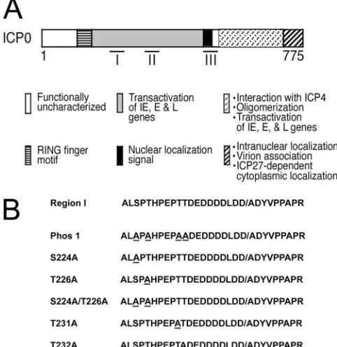
tran-FIG 1Region I phosphorylation site mutations. (A) ICP0 functional domains with three characterized phosphorylation regions (I, II, and III), adapted from reference35. (B) Amino acid sequences of region I and each region I phos-phorylation site mutant. All mutated residues are underlined. The slash in region I indicates the boundary between the second and third exons of ICP0.on November 7, 2019 by guest
http://jvi.asm.org/
[image:3.585.299.540.70.318.2]sient-transfection assays (
36
). To determine which sites in the
Phos 1 mutant form of ICP0 mediate the dissociation of these two
proteins, we transfected Hep-2 cells with plasmids that express
each region I phosphorylation site mutation or the double
muta-tion (
Fig. 1
), and the localization of ICP0 and PML, or of ICP0 and
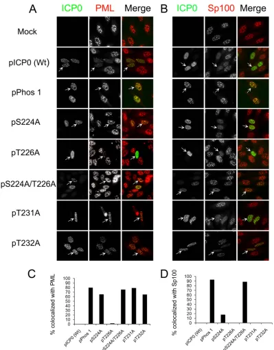
Sp100, was examined by immunofluorescence. As shown in
Fig. 2A
, wild-type ICP0 did not colocalize with PML, dissociating
it from ND10. Of the region I mutant forms that were examined,
only the T226A mutant behaved like wild-type ICP0. As reported
previously (
36
), Phos 1 was unable to efficiently dissociate PML
from ND10 (
Fig. 2A
and
C
). All other region I phosphorylation
site mutant forms of ICP0 (the S224A, S224A/T226A, T231A, and
T232A mutants) were as impaired as Phos 1 in dissociating PML
from ND10. Of these, the T232A mutant colocalized with PML,
FIG 2Region I phosphorylation site mutations and ND10 staining. (A and B) Localization of PML (A) or Sp100 (B) and wild-type (Wt) ICP0 or region I phosphorylation site mutant forms of ICP0. Hep-2 cells were transfected with plasmids expressing Wt ICP0, Phos 1, each region I phosphorylation site mutation, or the double site mutation. Sixteen hours posttransfection, the cells were fixed, stained for ICP0 and either PML (A) or Sp100 (B), and examined by fluorescence microscopy. (C and D) Graphs showing the percentages of ICP0-expressing cells that colocalized with PML (C) or with Sp100 (D). In both experiments, at least 100 ICP0-expressing cells were examined for each form (Wt or mutant) of ICP0.on November 7, 2019 by guest
http://jvi.asm.org/
[image:4.585.93.492.63.573.2]but PML staining appeared to be less intense than that in
mock-transfected cells, indicating that this mutant form of ICP0 may
retain a degree of PML-dissociating activity. In contrast to the
results with PML, all region I phosphorylation site mutants were
capable of dissociating Sp100 from ND10 similarly to wild-type
ICP0, except for the S224A/T226A double mutant, which behaved
like Phos 1 and was unable to disperse Sp100 (
Fig. 2B
and
D
). Our
data show that different phosphorylation residues in region I of
ICP0 regulate the dissociation of PML from ND10 versus the
dis-sociation of Sp100 in Hep-2 cells.
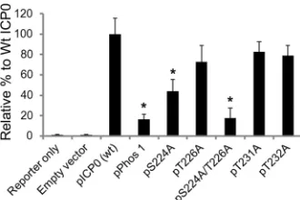
The transactivation activity of ICP0 is regulated by serine
224.
A previous report indicated that the transactivation activity
of ICP0 in cell culture is reduced for Phos 1 (
35
). To test the
transactivation activity of region I phosphorylation site mutant
forms of ICP0, Vero cells were transfected with an HSV-1 VP16
promoter-luciferase reporter construct either alone or in
combi-nation with a plasmid expressing wild-type ICP0 or each region I
mutation. Forty-eight hours posttransfection, cells were lysed and
assayed for luciferase activity, with the wild-type ICP0 assigned a
value of 100%. As expected, Phos 1 was unable to efficiently
in-duce the expression of the VP16 promoter, and its induction
abil-ity was 16% of the level of wild-type ICP0 (
P
⫽
0.029) (
Fig. 3
). Of
all the other mutants tested, only the S224A and S224A/T226A
mutants showed significant decreases in transactivation activity,
with levels that were 43% (
P
⫽
0.02) and 17% (
P
⫽
0.02) of the
level wild-type ICP0, respectively. Induction of the VP16
pro-moter by the T226A, T231A, and T232A mutants was not
im-paired. Thus, S224 contributes to the transactivation activity of
ICP0 in combination with T226 to enable the maximal
transacti-vation function of ICP0. Similar results were observed in two
other cell lines (HeLa and Hep-2 cells) (H. H. Mostafa and D. J.
Davido, unpublished data).
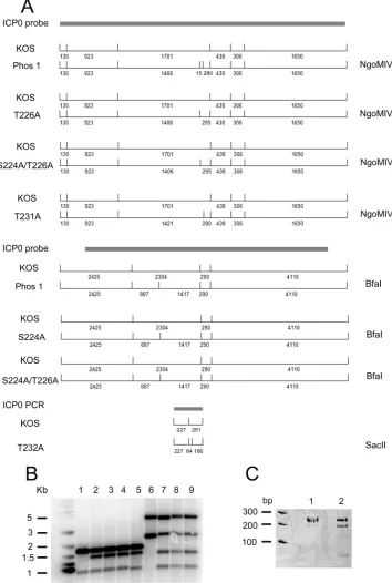
Construction of region I mutant viruses.
In order to
charac-terize the role of region I sites in the context of lytic replication,
our region I mutations were introduced into the viral genomes
as described in Materials and Methods, creating mutant
vi-ruses. The ICP0-null mutant 7134 was used to make each
mu-tant by marker transfer. For each mumu-tant, a marker rescue virus
was made to ensure that any phenotype observed was not due
to secondary mutations in the viral genome. The insertion of
restriction digestion sites during the construction of each
mu-tant helped us identify mumu-tant viruses either by Southern
blot-ting or by PCR (
Fig. 4
).
De novo
replication of S224A and S224A/T226A mutant
vi-ruses is reduced.
Phos 1 showed impaired
de novo
replication in
tissue culture (
36
); consequently, we wanted to determine which
region I site(s) is responsible for this phenotype. To answer this
question, we examined
de novo
replication of region I mutant
viruses. Notably, in the absence of the HSV IE transactivator
VP16, ICP0 transactivating activity is required for high levels of
de novo
replication (
12
). For this experiment, Vero cells were
transfected with infectious viral DNA from wild-type HSV-1
(strain KOS), the ICP0-null mutant 7134, or each
phosphory-lation site mutant. As expected, 7134 replication was reduced
5
⫻
10
5-fold from that of KOS, a finding similar to a previous
report (
36
). The Phos 1 mutant showed a 14-fold decrease, and
of the region I mutants, only the S224A and S224A/T226A
mutants showed significant reductions, of 9-fold and 16-fold,
respectively (
P
⫽
0.009 for both), relative to KOS (
Table 1
).
Taken together, our data indicate that S224 is required for
efficient
de novo
viral replication.
Serine 224 regulates the E3 ubiquitin ligase activity of ICP0.
(i) ICP0 protein stability.
The Phos 1 mutant form of ICP0 was
previously shown to have greater stability than wild-type ICP0
(
36
), indicative of alterations in the E3 ubiquitin ligase activity of
ICP0. To identify sites in region I that contribute to the stability of
Phos 1, we infected HeLa cells in triplicate with KOS, Phos 1, or
each region I mutant. After 4 h of infection, one well was
har-vested, while the remaining two wells were blocked with the
pro-tein synthesis inhibitor cycloheximide for an additional 2 or 4 h
and were subsequently harvested. ICP0 and actin protein levels
were examined by Western blotting. As shown in
Fig. 5
, the Phos
1, S224A, and S224A/T226A mutants were more stable than
wild-type ICP0. The phenowild-type observed for these three mutants
sug-gests that S224 controls the autoubiquitination of ICP0. Similar
results were observed in Hep-2 cells (H. H. Mostafa and D. J.
Davido, unpublished data).
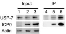
(ii) USP-7 levels.
To confirm that the Phos 1, S224A, and
S224A/T226A mutations impaired the E3 ubiquitin ligase
ac-tivity of ICP0, we examined the protein levels of another target
of ICP0 ubiquitination, USP-7. In this experiment, HeLa cells
were either mock infected or infected with KOS or the 7134,
Phos 1, S224A, or S224A/T226A mutant for 4, 6, or 8 h. The
cells were then harvested, and the protein levels of USP-7,
ICP0, and actin were determined by Western blotting. As
re-ported previously for wild-type HSV-1 (
23
), KOS infection
reduced USP-7 protein levels (
Fig. 6
). In contrast, USP-7
pro-tein levels were comparable over time in mock-infected cells
and cells infected with the 7134, Phos 1, S224A, or S224A/
T226A mutant (
Fig. 6
). Because it has been shown before that
the interaction between ICP0 and USP-7 mediates the
stabili-zation of ICP0 early during lytic infection (
24
), we wanted to
address the possibility that the higher stability of the Phos1
mutant form and/or the higher stability of USP-7 following
Phos 1 infection may be due to alterations in the interaction
between Phos 1 and USP-7. For this experiment, we took
KOS-or Phos 1-infected cell extracts from HeLa cells 4 h
postinfec-tion and determined the extent to which USP-7
coimmunopre-cipitated with ICP0 in these extracts. By accounting for the
amount of Phos 1 that was immunoprecipitated, our data show
that Phos 1 pulled down USP-7 to an extent similar to that for
FIG 3Transactivation activities of region I phosphorylation site mutations.Vero cells were transfected for 48 h with an HSV-1 reporter plasmid (pGL3-VP16; 50 ng) and a plasmid expressing either wild-type (wt) ICP0, Phos 1, one of the region I phosphorylation site mutations, or the double site mutation. Luciferase assays of cell extracts were performed to monitor the transactivating activity of ICP0. *,P⬍0.05 by the Mann-Whitney U test. Error bars indicate the standard errors of the means for all samples.
on November 7, 2019 by guest
http://jvi.asm.org/
[image:5.585.80.245.66.176.2]wild-type ICP0 (
Fig. 7
). Consequently, these data do not
sup-port the hypothesis that an increase in the stability of Phos 1 is
mediated by an increase in its interaction with USP-7. Similar
results were obtained with Hep-2 cells (H. H. Mostafa and D. J.
Davido, unpublished data). Taken together, our data support the
conclusion that the Phos 1, S224A, and S224A/T226A mutants
have impaired E3 ubiquitin ligase activity, and they indicate that
S224 regulates this activity of ICP0.
FIG 4Construction of ICP0 region I phosphorylation mutant viruses. Region I phosphorylation mutant viruses were constructed by marker transfer as described in Materials and Methods. (A) Restriction enzyme digestion patterns, with the fragment lengths (in base pairs), of KOS and region I phosphorylation mutants. Note that the S224A/T226A mutant contains both BfaI and NgoMIV sites. The restriction pattern shown for the T232A mutant is from a PCR product. (B) Southern blots of KOS and the Phos 1, T226A, S224A/T226A, and T231A mutants digested with NgoMIV (lanes 1 to 5, respectively) and of KOS and the Phos 1, S224A, and S224A/T226A mutants digested with BfaI (lanes 6 to 9, respectively). (C) Twelve percent polyacrylamide gel of KOS (lane 1) and T232A mutant (lane 2) PCR products digested with SacII.
on November 7, 2019 by guest
http://jvi.asm.org/
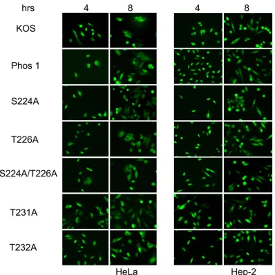
[image:6.585.116.470.70.596.2]The subcellular localization of region I phosphorylation
mu-tants is similar to that of wild-type ICP0.
In order to examine the
possibility that the subcellular localization of these mutant forms
of ICP0 may be altered, thus altering the activities of ICP0, HeLa
and Hep-2 cells were infected with either KOS, Phos 1, or each of
the region I phosphorylation site mutants. The localization of
ICP0 was determined at 4 and 8 h postinfection. All the mutants
showed localization patterns similar to that of wild-type ICP0 in
the two cell lines tested, with primarily nuclear localization at 4 h
postinfection and both nuclear and cytoplasmic localization at 8 h
postinfection (
Fig. 8
).
Serine 224 is required for efficient acute viral replication in
the TG of mice.
We showed previously that Phos 1 replication in
the trigeminal ganglia (TG) during the acute phase of infection
was significantly lower than that of wild-type KOS, with the
max-imal decrease observed at day 5 postinfection (
37
). To test which
residues in region I are required for efficient
in vivo
replication,
CD-1 mice were infected with KOS, Phos 1, each region I
phos-phorylation site mutant, and corresponding marker rescue
vi-ruses. Five days postinfection, mice were swabbed and were then
sacrificed, TG were explanted, and acute viral replication was
monitored by a standard plaque assay. Acute replication in the
eyes on day 5 showed only slight reductions for the S224A mutant
(5-fold [
P
⫽
0.005]) and Phos 1 (11-fold [
P
⫽
0.00012]), whereas
all the other region I mutants replicated to levels comparable to
that of wild-type HSV-1. With regard to productive infection of
the trigeminal ganglia, as expected, 7134 and Phos 1 showed
sig-nificantly lower replication levels (650-fold [
P
⫽
1.2
⫻
10
⫺18] and
11-fold [
P
⫽
2
⫻
10
⫺6], respectively) than that of KOS (
Fig. 9A
).
The replication of the S224A and S224A/T226A mutants was
re-duced by 48-fold (
P
⫽
1.9
⫻
10
⫺11) and 15-fold (
P
⫽
1.8
⫻
10
⫺8),
respectively, whereas the replication of the T226A, T231A, and
T232A mutants (
Fig. 9A
) and of the marker rescue viruses (
Fig.
9B
) was similar to that of KOS. Our results demonstrate that S224
enhances productive infection in the TG of mice.
DISCUSSION
Phosphorylation is a posttranslational modification that regulates
the activities of many viral proteins (
41
,
42
). ICP0 has been shown
to be heavily phosphorylated (
33
), and previous studies from our
lab strongly support the hypothesis that phosphorylation
regu-lates its activities (
35
–
37
). Because four amino acids in region I of
ICP0 have been shown to play an important role in regulating its
activities and viral replication, we performed mutagenesis of a
specific site or sites in this region in order to identify residues that
are required for the activities of ICP0. Specifically, region I has
been shown to be phosphorylated at one or two residues (
35
), and
it has been determined recently by our lab and by other
investiga-tors that S224 and T226 are phosphorylated. Characterization of
FIG 6USP-7 levels observed with region I phosphorylation site mutants. HeLa cells were either mock infected or infected with the KOS, 7134, Phos 1, S224A, or S224A/T226A virus at an MOI of 2 for 4, 6, or 8 h. ICP0, USP-7, and actin protein levels were determined by Western blot analyses. [image:7.585.335.505.65.198.2]FIG 7Interaction of ICP0 and Phos 1 with USP-7. HeLa cells were either mock infected (lanes 1 and 4) or infected with KOS (lanes 2 and 5) or Phos 1 (lanes 3 and 6). Four hours postinfection, ICP0 was immunoprecipitated, and the levels of ICP0, USP-7, and actin were analyzed by Western blotting.
TABLE 1De novoreplication of region I mutantsa
Virus
Titer (% relative to the titer of KOS)
Fold difference from KOSb
KOS 100
7134 0.0002 25⫻105
Phos 1 mutant 6.9 214.5
S224A mutant 11.1 29.05
T226A mutant 79.9 21.25
S224A T226A mutant 6.2 216.1
T231A mutant 128.2 10.78
T232A mutant 130.3 10.77
aVero cells were transfected with infectious viral DNA of wild-type HSV-1 (strain
KOS), an ICP0-null mutant (7134), and region I mutants. At 48 h posttransfection, cells were harvested, and titers of viruses were determined by a standard plaque assay. Mutants whose viral replication was significantly lower than that of KOS were tested with two independent viral DNA preparations. The experiment was repeated five times, and the data presented are the means.
b2, reduction;1, increase.
FIG 5ICP0 stability of region I phosphorylation site mutants. HeLa cells were infected with the indicated viruses at an MOI of 2. Four hours postinfection, cells were harvested, and duplicate samples were treated with cycloheximide (CHX; 100g/ml) and were harvested at 6 and 8 h postinfection. ICP0 and actin levels were determined by Western blot analyses.
on November 7, 2019 by guest
http://jvi.asm.org/
[image:7.585.40.288.78.185.2] [image:7.585.60.266.494.671.2] [image:7.585.367.477.627.684.2]these mutations in the absence of other viral factors indicates that
S224 phosphorylation is important for efficient dissociation of
PML from ND10 (
Fig. 2A
and
C
), and it appears that the
phos-phorylation of either S224 or T226 is sufficient for efficient Sp100
dissociation (
Fig. 2B
and
D
). Interestingly, T231 and T232 also
play an important role in the dissociation of PML (
Fig. 2A
and
C
),
but neither is required for Sp100 dissociation (
Fig. 2B
and
D
),
suggesting a potential structural role for these residues in
dissoci-ating PML from ND10. S224 is also required for optimal levels of
ICP0-mediated transactivation activity in luciferase reporter
as-says (
Fig. 3
). In the context of viral infections, we showed that S224
is important for efficient E3 ubiquitin ligase activity (
Fig. 5
and
6
)
and for viral replication both in tissue culture (
Table 1
) and in the
trigeminal ganglia of mice (
Fig. 9A
). Our data highlight S224,
which is in close proximity to the RING finger motif of ICP0 (
Fig.
1
), as the residue in region I that has the major impact on the
activities of ICP0.
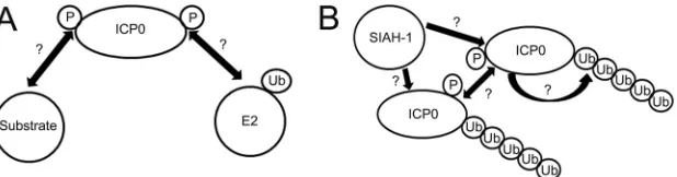
To understand the mechanism by which phosphorylation
regulates the functions of ICP0, particularly its E3 Ub ligase
activity, we propose the hypothetical model shown in
Fig. 10
.
As a RING finger-containing E3 ubiquitin ligase, ICP0 acts as a
scaffold, binding both the E2 ubiquitin-conjugating (Ubc)
en-zyme and its substrates, thereby facilitating ubiquitin chain
transfer and conferring substrate specificity. Given that
phos-phorylation regulates the action of many E3 ubiquitin ligases,
activating (
43
,
44
) or inactivating (
45
) them, it is possible that
the E3 Ub ligase activity of ICP0 is regulated by
phosphoryla-tion, enhancing its contact with a particular substrate or E2
enzyme (
Fig. 10A
). Our data demonstrate that Phos 1 is not
detectably altered from wild-type ICP0 in its interaction with
one of its substrates, USP-7 (
Fig. 7
). Although this finding does
not exclude the possibility that phosphorylation in region I can
affect the binding of ICP0 to other target substrates, it suggests
another possibility, that phosphorylation promotes the
inter-action of ICP0 with a particular E2 Ubc enzyme. In a
prelimi-nary experiment, we examined the colocalization of wild-type
ICP0 or Phos 1 with the E2 enzyme UbcH5a. UbcH5a has been
shown to be one of two E2 Ubc’s of ICP0 that direct the
ubiq-FIG 8Subcellular localization of the ICP0 protein in region I phosphorylation site mutants. HeLa and Hep-2 cells were infected with KOS or one of the region I phosphorylation mutants for 4 or 8 h. The cells were fixed, stained for ICP0, and examined by fluorescence microscopy.on November 7, 2019 by guest
http://jvi.asm.org/
[image:8.585.95.493.63.462.2]uitination of selected ICP0 targets
in vitro
and in cell culture
(
46
–
48
). Our initial results show extensive colocalization
be-tween wild-type ICP0 and UbcH5a, whereas Phos 1 did not
colocalize extensively with UbcH5a (H. H. Mostafa and D. J.
Davido, unpublished data). This suggests that Phos 1 may be
impaired in its interaction with UbcH5a compared to
wild-type ICP0. Ongoing studies will determine the extent of the
role that region I plays in facilitating the interaction of ICP0
with the E2 Ubc enzymes, such as UbcH5a, and will specifically
test the role of the S224 site in facilitating such interactions.
The increased stability of Phos 1 may also be attributed to
alterations in its interactions with the E2 enzymes or the cellular
E3 ubiquitin ligase SIAH-1. In the latter case, the binding of
SIAH-1 to ICP0 has been shown to decrease the stability of ICP0
(
49
). This raises the question of whether SIAH-1 binding by the
Phos 1 mutant form of ICP0 is diminished, resulting in a more
stable protein. This argument is supported by a recent publication
showing that the binding of ICP0 to another cellular E3 ubiquitin
ligase, RNF8, is dependent on ICP0 phosphorylation (
50
).
Inter-estingly, the phosphorylation of ICP0 at an N-terminal residue
specifically directed the degradation of RNF8, whereas the
degra-dation of other ICP0 targets was unaffected. In addition, ICP0, like
its other alphaherpesvirus orthologues (e.g., varicella-zoster virus
ORF61 [
51
]), is capable of autoubiquitination, although whether
this occurs via intramolecular interactions or intermolecular
in-teractions through dimerization (
52
), and the role of
phosphory-lation in either mechanism, is currently unknown. Thus, the
rela-tionships between ICP0 phosphorylation and SIAH-1 binding
and ICP0 phosphorylation and its autoubiquitination remain to
be determined (
Fig. 10B
).
The impairment of PML and Sp100 dissociation was observed
with Phos 1 and other region I phosphorylation site mutations in
the absence of other viral factors in transient-transfection assays
(
Fig. 2
) (
36
) but not during the course of viral infection (
36
; also
H. H. Mostafa and D. J. Davido, unpublished data). These results
indicate that other viral factors and/or the level of their expression
likely compensates for the defect of region I mutants in
dissociat-ing these two ND10-associated proteins. On the other hand,
al-though it has been shown previously that the dispersal of PML and
Sp100 mediated by ICP0 is dependent on the E2 enzyme UbcH5a
in cell culture (
48
), ICP0 was unable to ubiquitinate SUMOylated
PML
in vitro
(
47
). The latter result suggests that other factors are
required for the ICP0-directed ubiquitination of PML (and
po-tentially Sp100). Also, our data show that the regulation of PML by
individual mutant forms of ICP0 differs from their regulation of
Sp100. A similar result was observed in a previous study from our
laboratory in which another ICP0 phosphorylation mutant, Phos
2, could dissociate Sp100 but not PML from ND10 in
transient-transfection assays (
36
). For our region I mutant forms of ICP0,
we hypothesize that distinct cellular factors regulate the
dissocia-FIG 9Acute replication of region I phosphorylation site mutants in the trigeminal ganglia (TG) of mice at day 5 postinfection. CD-1 mice were infected with 2⫻ 105PFU per eye. Five days postinfection, mice were sacrificed; TG were removed and homogenized; and viral titers were determined by standard plaque assays. (A) Replication of region I phosphorylation site mutants. *,P⬍0.05 by Student’sttest. (B) Replication of region I phosphorylation mutant marker rescue viruses. Error bars represent the standard errors of the means. The dashed lines indicate the minimum limit of detection. Eight mice were used per virus.FIG 10Hypothetical models of the mechanism by which the E3 ubiquitin ligase activity of ICP0 is controlled by phosphorylation. (A) Phosphorylation (P) can regulate the interaction of ICP0 with a target substrate or an E2 ubiquitin (Ub)-conjugating enzyme. (B) The ubiquitination of ICP0 is regulated by phosphor-ylation, which enhances intramolecular or intermolecular autoubiquitination or binding to cellular E3 ubiquitin ligases, such as SIAH-1.
on November 7, 2019 by guest
http://jvi.asm.org/
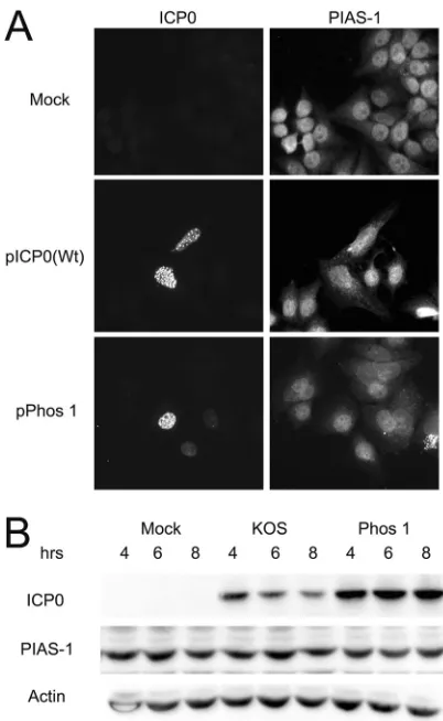
[image:9.585.45.539.63.246.2] [image:9.585.140.449.609.690.2]tion of PML and that of Sp100. In an attempt to determine what
factors could potentially contribute to ICP0-directed PML
disso-ciation, we looked at the staining pattern and protein levels of
PIAS-1, as well as its interaction with ICP0 after transient
trans-fection and intrans-fection. The E3 SUMO-ligase PIAS-1 has recently
been shown to regulate the degradation of PML (
53
), and its loss
results in increased PML stability. Our studies showed that the
expression of wild-type ICP0 or Phos 1 did not influence the
stain-ing of PIAS-1 in transient assays (
Fig. 11A
) or PIAS-1 protein
levels during infection (
Fig. 11B
). We did not detect interactions
between ICP0 and PIAS-1 by coimmunoprecipitation assays
(H. H. Mostafa and D. J. Davido, unpublished data). We propose
that the dissociation of PML directed by ICP0 is not dependent on
ICP0 interacting with PIAS-1.
Many questions remain as to how phosphorylation regulates
the activities of ICP0. For example, how does phosphorylation of
ICP0 differentially regulate its activities during acute infection
in
vivo
? And what are the kinases that contribute to the
phosphory-lation of ICP0? Previous work has shown that the phosphoryphosphory-lation
status of ICP0 is altered along the course of viral replication (
33
,
54
) and is dependent on the viral kinase UL13 (
55
) and cellular
cyclin-dependent kinases (cdk’s) (
34
). Ongoing work in our lab is
aimed at identifying the kinases that phosphorylate and control
the activities of ICP0.
ACKNOWLEDGMENTS
This work was supported by National Institutes of Health grant R01AI72357.
The content of this article is solely the responsibility of the authors and does not necessarily represent the official views of the National Institutes of Health.
We thank Anne Cooper for creating the S224A/T226A mutation, Mat-thew Weitzman for providing results from unpublished data, and mem-bers of the Davido lab and Tom Yankee for discussions related to this project and critical reading of the manuscript.
REFERENCES
1.Roizman R, Knipe DM, Whitley RJ. 2007. Herpes simplex viruses, p 2501–2601.InKnipe DM, Howley PM, Griffin DE, Lamb RA, Martin MA, Roizman B, Straus SE (ed), Fields virology, 5th ed, vol 2. Lippincott Wil-liams & Wilkins, Philadelphia, PA.
2.Roizman B, Kozak M, Honess RW, Hayward G.1975. Regulation of herpesvirus macromolecular synthesis: evidence for multilevel regulation of herpes simplex 1 RNA and protein synthesis. Cold Spring Harbor Symp. Quant. Biol.39:687–701.
3.Stevens JG.1987. Defining herpes simplex genes involved in neuroviru-lence and neuroinvasiveness. Curr. Eye Res.6:63– 67.
4.Cai W, Astor TL, Liptak LM, Cho C, Coen DM, Schaffer PA.1993. The herpes simplex virus type 1 regulatory protein ICP0 enhances virus repli-cation during acute infection and reactivation from latency. J. Virol.67: 7501–7512.
5.Harris RA, Everett RD, Zhu XX, Silverstein S, Preston CM. 1989. Herpes simplex virus type 1 immediate-early protein Vmw110 reactivates latent herpes simplex virus type 2 in an in vitro latency system. J. Virol.
63:3513–3515.
6.Highlander SL, Dorney DJ, Gage PJ, Holland TC, Cai W, Person S, Levine M, Glorioso JC.1989. Identification ofmarmutations in herpes simplex virus type 1 glycoprotein B which alter antigenic structure and function in virus penetration. J. Virol.63:730 –738.
7.Stow ND, Stow EC.1986. Isolation and characterization of a herpes simplex virus type 1 mutant containing a deletion within the gene encod-ing the immediate early polypeptide Vmw110. J. Gen. Virol.67:2571– 2585.
8.Everett RD.1989. Construction and characterization of herpes simplex virus type 1 mutants with defined lesions in immediate early gene 1. J. Gen. Virol.70:1185–1202.
9.Meredith M, Orr A, Elliott M, Everett R.1995. Separation of sequence requirements for HSV-1 Vmw110 multimerisation and interaction with a 135-kDa cellular protein. Virology209:174 –187.
10. Everett RD.1984. Trans activation of transcription by herpes virus prod-ucts: requirement for two HSV-1 immediate-early polypeptides for max-imum activity. EMBO J.3:3135–3141.
11. Cai W, Schaffer PA.1992. Herpes simplex virus type 1 ICP0 regulates expression of immediate-early, early, and late genes in productively in-fected cells. J. Virol.66:2904 –2915.
12. Cai WZ, Schaffer PA.1989. Herpes simplex virus type 1 ICP0 plays a critical role in the de novo synthesis of infectious virus following transfec-tion of viral DNA. J. Virol.63:4579 – 4589.
13. Chelbi-Alix MK, de The H.1999. Herpes virus induced proteasome-dependent degradation of the nuclear bodies-associated PML and Sp100 proteins. Oncogene18:935–941.
14. Everett RD, Freemont P, Saitoh H, Dasso M, Orr A, Kathoria M, Parkinson J.1998. The disruption of ND10 during herpes simplex virus infection correlates with the Vmw110- and proteasome-dependent loss of several PML isoforms. J. Virol.72:6581– 6591.
15. Everett RD, Maul GG.1994. HSV-1 IE protein Vmw110 causes redistri-bution of PML. EMBO J.13:5062–5069.
16. Maul GG, Everett RD.1994. The nuclear location of PML, a cellular member of the C3HC4zinc-binding domain protein family, is rearranged during herpes simplex virus infection by the C3HC4viral protein ICP0. J. Gen. Virol.75:1223–1233.
FIG 11Localization of ICP0 with PIAS-1 and the effect of infection with KOS or Phos 1 on PIAS-1 protein levels. (A) Hep-2 cells were either mock trans-fected or transtrans-fected with plasmids expressing wild-type (Wt) ICP0 or Phos 1. Sixteen hours posttransfection, the cells were fixed, stained for ICP0 and PIAS-1, and examined by fluorescence microscopy. (B) Hep-2 cells were either mock infected or infected with KOS or Phos 1 at an MOI of 2 for 4, 6, or 8 h. ICP0, PIAS-1, and actin protein levels were determined by Western blot anal-yses.
on November 7, 2019 by guest
http://jvi.asm.org/
[image:10.585.60.261.64.391.2]17. Parkinson J, Everett RD. 2000. Alphaherpesvirus proteins related to herpes simplex virus type 1 ICP0 affect cellular structures and proteins. J. Virol.74:10006 –10017.
18. Ching RW, Dellaire G, Eskiw CH, Bazett-Jones DP.2005. PML bodies: a meeting place for genomic loci? J. Cell Sci.118:847– 854.
19. Cho Y, Lee I, Maul GG, Yu E.1998. A novel nuclear substructure, ND10: distribution in normal and neoplastic human tissues. Int. J. Mol. Med.
1:717–724.
20. Zhong S, Salomoni P, Pandolfi PP.2000. The transcriptional role of PML and the nuclear body. Nat. Cell Biol.2:E85–E90.
21. Zhong S, Salomoni P, Ronchetti S, Guo A, Ruggero D, Pandolfi PP.
2000. Promyelocytic leukemia protein (PML) and Daxx participate in a novel nuclear pathway for apoptosis. J. Exp. Med.191:631– 640. 22. Everett RD, Rechter S, Papior P, Tavalai N, Stamminger T, Orr A.2006.
PML contributes to a cellular mechanism of repression of herpes simplex virus type 1 infection that is inactivated by ICP0. J. Virol.80:7995– 8005. 23. Boutell C, Canning M, Orr A, Everett RD.2005. Reciprocal activities between herpes simplex virus type 1 regulatory protein ICP0, a ubiquitin E3 ligase, and ubiquitin-specific protease USP7. J. Virol.79:12342–12354. 24. Canning M, Boutell C, Parkinson J, Everett RD.2004. A RING finger ubiquitin ligase is protected from autocatalyzed ubiquitination and deg-radation by binding to ubiquitin-specific protease USP7. J. Biol. Chem.
279:38160 –38168.
25. Härle P, Sainz B, Jr, Carr DJ, Halford WP.2002. The immediate-early protein, ICP0, is essential for the resistance of herpes simplex virus to interferon-alpha/beta. Virology293:295–304.
26. Lin R, Noyce RS, Collins SE, Everett RD, Mossman KL. 2004. The herpes simplex virus ICP0 RING finger domain inhibits IRF3- and IRF7-mediated activation of interferon-stimulated genes. J. Virol.78:1675– 1684.
27. Melroe GT, DeLuca NA, Knipe DM.2004. Herpes simplex virus 1 has multiple mechanisms for blocking virus-induced interferon production. J. Virol.78:8411– 8420.
28. Melroe GT, Silva L, Schaffer PA, Knipe DM. 2007. Recruitment of activated IRF-3 and CBP/p300 to herpes simplex virus ICP0 nuclear foci: potential role in blocking IFN-induction. Virology360:305–321. 29. Paladino P, Collins SE, Mossman KL.2010. Cellular localization of the
herpes simplex virus ICP0 protein dictates its ability to block IRF3-mediated innate immune responses. PLoS One5:e10428. doi:10.1371 /journal.pone.0010428.
30. van Lint AL, Murawski MR, Goodbody RE, Severa M, Fitzgerald KA, Finberg RW, Knipe DM, Kurt-Jones EA.2010. Herpes simplex virus immediate-early ICP0 protein inhibits Toll-like receptor 2-dependent in-flammatory responses and NF-B signaling. J. Virol.84:10802–10811. 31. Lilley CE, Chaurushiya MS, Boutell C, Everett RD, Weitzman MD.
2011. The intrinsic antiviral defense to incoming HSV-1 genomes includes specific DNA repair proteins and is counteracted by the viral protein ICP0. PLoS Pathog.7:e1002084. doi:10.1371/journal.ppat.1002084.
32. Lilley CE, Chaurushiya MS, Boutell C, Landry S, Suh J, Panier S, Everett RD, Stewart GS, Durocher D, Weitzman MD.2010. A viral E3 ligase targets RNF8 and RNF168 to control histone ubiquitination and DNA damage responses. EMBO J.29:943–955.
33. Ackermann M, Braun DK, Pereira L, Roizman B.1984. Characterization of herpes simplex virus 1 alpha proteins 0, 4, and 27 with monoclonal antibodies. J. Virol.52:108 –118.
34. Davido DJ, Leib DA, Schaffer PA.2002. The cyclin-dependent kinase inhibitor roscovitine inhibits the transactivating activity and alters the posttranslational modification of herpes simplex virus type 1 ICP0. J. Vi-rol.76:1077–1088.
35. Davido DJ, von Zagorski WF, Lane WS, Schaffer PA.2005. Phosphor-ylation site mutations affect herpes simplex virus type 1 ICP0 function. J. Virol.79:1232–1243.
36. Boutell C, Everett R, Hilliard J, Schaffer P, Orr A, Davido D.2008. Herpes simplex virus type 1 ICP0 phosphorylation mutants impair the E3 ubiquitin ligase activity of ICP0 in a cell type-dependent manner. J. Virol.
82:10647–10656.
37. Mostafa HH, Thompson TW, Kushnir AS, Haenchen SD, Bayless AM,
Hilliard JG, Link MA, Pitcher LA, Loveday E, Schaffer PA, Davido DJ.
2011. Herpes simplex virus 1 ICP0 phosphorylation site mutants are at-tenuated for viral replication and impaired for explant-induced reactiva-tion. J. Virol.85:12631–12637.
38. Samaniego LA, Wu N, DeLuca NA. 1997. The herpes simplex virus immediate-early protein ICP0 affects transcription from the viral genome and infected-cell survival in the absence of ICP4 and ICP27. J. Virol.71: 4614 – 4625.
39. Kushnir AS, Davido DJ, Schaffer PA.2010. Role of nuclear factor Y in stress-induced activation of the herpes simplex virus type 1 ICP0 pro-moter. J. Virol.84:188 –200.
40. National Research Council.2011. Guide for the care and use of laboratory animals, 8th ed. National Academies Press, Washington, DC.
41. Rojas S, Corbin-Lickfett KA, Escudero-Paunetto L, Sandri-Goldin RM.
2010. ICP27 phosphorylation site mutants are defective in herpes simplex virus 1 replication and gene expression. J. Virol.84:2200 –2211. 42. Sekhar V, McBride AA.2012. Phosphorylation regulates binding of the
HPV8 E2 protein to host chromosomes. J. Virol.86:10047–10058. 43. Levkowitz G, Waterman H, Ettenberg SA, Katz M, Tsygankov AY,
Alroy I, Lavi S, Iwai K, Reiss Y, Ciechanover A, Lipkowitz S, Yarden Y.
1999. Ubiquitin ligase activity and tyrosine phosphorylation underlie sup-pression of growth factor signaling by c-Cbl/Sli-1. Mol. Cell4:1029 –1040. 44. Vodermaier HC.2004. APC/C and SCF: controlling each other and the
cell cycle. Curr. Biol.14:R787–R796.
45. Debonneville C, Flores SY, Kamynina E, Plant PJ, Tauxe C, Thomas MA, Munster C, Chraibi A, Pratt JH, Horisberger JD, Pearce D, Loffing J, Staub O.2001. Phosphorylation of Nedd4-2 by Sgk1 regulates epithelial Na⫹channel cell surface expression. EMBO J.20:7052–7059.
46. Boutell C, Everett RD.2003. The herpes simplex virus type 1 (HSV-1) regulatory protein ICP0 interacts with and ubiquitinates p53. J. Biol. Chem.278:36596 –36602.
47. Boutell C, Sadis S, Everett RD.2002. Herpes simplex virus type 1 imme-diate-early protein ICP0 and its isolated RING finger domain act as ubiq-uitin E3 ligases in vitro. J. Virol.76:841– 850.
48. Gu H, Roizman B.2003. The degradation of promyelocytic leukemia and Sp100 proteins by herpes simplex virus 1 is mediated by the ubiquitin-conjugating enzyme UbcH5a. Proc. Natl. Acad. Sci. U. S. A.100:8963– 8968.
49. Nagel CH, Albrecht N, Milovic-Holm K, Mariyanna L, Keyser B, Abel B, Weseloh B, Hofmann TG, Eibl MM, Hauber J.2011. Herpes simplex virus immediate-early protein ICP0 is targeted by SIAH-1 for proteasomal degradation. J. Virol.85:7644 –7657.
50. Chaurushiya MS, Lilley CE, Aslanian A, Meisenhelder J, Scott DC, Landry S, Ticau S, Boutell C, Yates JR, III, Schulman BA, Hunter T, Weitzman MD.2012. Viral E3 ubiquitin ligase-mediated degradation of a cellular E3: viral mimicry of a cellular phosphorylation mark targets the RNF8 FHA domain. Mol. Cell46:79 –90.
51. Walters MS, Kyratsous CA, Silverstein SJ.2010. The RING finger do-main of varicella-zoster virus ORF61p has E3 ubiquitin ligase activity that is essential for efficient autoubiquitination and dispersion of Sp100-containing nuclear bodies. J. Virol.84:6861– 6865.
52. Ciufo DM, Mullen MA, Hayward GS.1994. Identification of a dimeriza-tion domain in the C-terminal segment of the IE110 transactivator protein from herpes simplex virus. J. Virol.68:3267–3282.
53. Rabellino A, Carter BJ, Konstantinidou G, Shwu-Yuan W, Rimessi A, Byers LA, Heymach JV, Girard L, Chiang CM, Teruya-Feldstein J, Scaglioni PP.2012. The SUMO E3-ligase PIAS1 regulates the tumor sup-pressor PML and its oncogenic counterpart PML-RARA. Cancer Res.72: 2275–2284.
54. Advani SJ, Hagglund R, Weichselbaum RR, Roizman B. 2001. Post-translational processing of infected cell proteins 0 and 4 of herpes simplex virus 1 is sequential and reflects the subcellular compartment in which the proteins localize. J. Virol.75:7904 –7912.
55. Ng TI, Ogle WO, Roizman B.1998. UL13 protein kinase of herpes simplex virus 1 complexes with glycoprotein E and mediates the phos-phorylation of the viral Fc receptor: glycoproteins E and I. Virology241: 37– 48.
on November 7, 2019 by guest
http://jvi.asm.org/