Pulse oximetry
Amal Jubran
Pulse oximetry is one of the most commonly employed monitoring modalities in the critical care setting. This review describes the latest technological advances in the field of pulse oximetry. Accuracy of pulse oximeters and their limitations are critically examined. Finally, the existing data regarding the clinical applications and cost-effectiveness of pulse oximeters are discussed.
Addresses: Division of Pulmonary and Critical Care Medicine, Veterans Administration Hospital and Loyola University of Chicago Stritch School of Medicine, Hines, Illinois 60141, USA
Correspondence: Amal Jubran, M.D., Division of Pulmonary & Critical Care Medicine, Edward Hines Jr. VA Hospital, Route 111N, Hines, Illinois 60141, USA
Keywords:respiratory monitoring, oxygenation, non-invasive
Received: 20 October 1998 Accepted: 28 February 1999 Published: 18 May 1999
Crit Care1999, 3:R11–R17
The original version of this paper is the electronic version which can be seen on the Internet (http://ccforum.com). The electronic version may contain additional information to that appearing in the paper version.
© Current Science Ltd ISSN 1364-8535
Introduction
The human eye is poor at recognizing hypoxemia. Even under ideal conditions, skilled observers cannot consis-tently detect hypoxemia until the oxygen (O2) saturation is below 80% [1]. The difficulty that physicians have in detecting hypoxemia was recently exemplified in a study of over 14 000 patients being evaluated at the UCLA Emergency Department [2]. Patients were monitored by oximetry but recordings were given to physicians only after they completed their initial assessment. Changes in diagnostic testing and treatment were most likely at an O2
saturation of 89%, and changes were actually less common at lower saturations, probably because the physicians were able to detect evidence of hypoxemia without requiring a pulse oximeter.
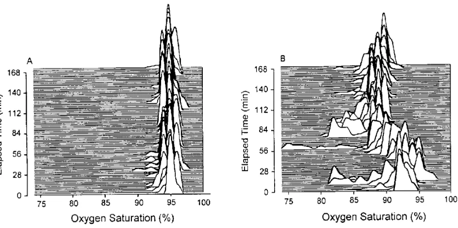
With the proliferation of pulse oximeters in different loca-tions of the hospital throughout the 1980s, several investi-gators demonstrated that episodic hypoxemia is much more common than previously suspected with an inci-dence ranging from 20–82% [3–5] (Fig. 1). The signifi-cance of episodic desaturation on patient outcome is largely unknown [6]. In patients admitted to a general medical service, Bowton et al.[7] found that O2saturation
< 90% of at least 5 min duration occurred in 26% of the
patients. On follow-up over the next 4–7 months, those patients experiencing hypoxemia during the first 24 h of hospitalization had more than a threefold higher mortality than patients who did not desaturate. Although episodic desaturation may simply be a marker of increased risk rather than the direct cause of decreased survival, an increased mortality rate was still observed in patients with episodic hypoxemia when the investigators corrected for severity of illness. Whether or not the early detection and treatment of episodic hypoxemia can affect patient outcome remains unknown.
Principles of pulse oximetry
Pulse oximetry is based on two physical principles: (a) the presence of a pulsatile signal generated by arterial blood, which is relatively independent of non-pulsatile arterial blood, venous and capillary blood, and other tissues; and (b) the fact that oxyhemoglobin (O2Hb) and reduced hemoglobin (Hb) have different absorption spectra [8]. Currently available oximeters use two light-emitting diodes (LEDs) that emit light at the 660 nm (red) and the 940 nm (infrared) wavelengths. These two wavelengths are used because O2Hb and Hb have different absorption spectra at these particular wavelengths. In the red region, O2Hb absorbs less light than Hb, while the reverse occurs
in the infrared region. The ratio of absorbencies at these two wavelengths is calibrated empirically against direct measurements of arterial blood oxygen saturation (SaO2) in volunteers, and the resulting calibration algorithm is stored in a digital microprocessor within the pulse oxime-ter. During subsequent use, the calibration curve is used to generate the pulse oximeter’s estimate of arterial satu-ration (SpO2) [9,10] (Fig. 2). In addition to the digital readout of O2 saturation, most pulse oximeters display a
plethysmographic waveform which can help clinicians dis-tinguish an artifactual signal from the true signal (Fig. 3).
Accuracy
The accuracy of commercially available oximeters differ widely, probably because of the different algorithms employed in signal processing [8]. These algorithms are limited by the range of saturations that can be safely obtained in volunteers, and also the accuracy of the mea-surement standard (CO-oximeter) [11]. Comparison of pulse oximetry with direct CO-oximeter measurements should be reported in terms of the mean difference between the two techniques (bias) and the standard devia-tion of the differences (precision).
In healthy volunteers, oximeters commonly have a mean difference (bias) of < 2% and a standard deviation (preci-sion) of < 3% when SaO2is 90% or above [12,13]. Compara-ble results have also been obtained in critically ill patients
[image:2.671.95.567.138.373.2]with good arterial perfusion [14,15]. Accuracy of pulse oximeters deteriorates when SaO2falls to 80% or less. In healthy volunteers under hypoxic conditions, bias of pulse oximetry varies from –15.0 to 13.1 while the precision ranges from 1.0 to 16.0 [12,16–18]. In a study in critically ill patients, eight out of 13 oximeters had a bias ≥± 5%
Figure 1
Sequential distribution plots of oxygen saturation at intervals of 2 min over a 3-h period in a stable patient (A) and unstable patient (B). The
unstable patient shows episodic desaturations. Published with permission [3].
Figure 2
Red (R) and infrared (IR) scaled alternating current (AC) signals at arterial oxygen saturation (SaO2) of 0%, 85% and 100%. The numeric value of the red-to-infrared (R/IR) ratio can be easily converted to SaO2. Published with permission [10].
S Oa 2 660 nm (R) 940 nm (IR) R/IR
~3.4
1.0
0.43 0%
85%
when SaO2 was < 80% [14]. In a study of 54 ventilator-dependent patients, the accuracy of oximetry deteriorated significantly at low SaO2 values. Bias ± precision was
1.7 ± 1.2% for SaO2 values > 90%, and it increased to 5.1 ± 2.7% when SaO2was ≤ 90% [19].
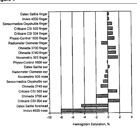
Different probes that are used with a pulse oximeter can also affect the accuracy of SpO2measurements. In patients with poor peripheral perfusion as a consequence of car-diopulmonary bypass surgery, finger probes had lower pre-cision and more readings within 3% of the reference (CO-oximeter) than the other probes. Overall rankings were significantly better for the finger probes than probes on other sites (Fig. 4) [20]. The response time of oximeter probes was assessed by Severinghaus and Naifeh [17] who induced 30–60 s hypoxic plateaus between an SaO2 of 40 and 70% in healthy volunteers. Oximeter probes placed on the ear generally had a much faster response to a sudden decrease in fractional inspired oxygen concentration (FiO2)
than did the finger probes (10–20 versus 24–35 s, repec-tively). Employing hypobaric facility to induce hypoxia in normal volunteers, Young et al.[21] also observed that the response time of the finger probes were slower than the ear probes in response to either a decrease or increase in O2saturation.
Limitations
Oximeters have a number of limitations which may lead to inaccurate readings (Table 1). Pulse oximeters measure SaO2 that is physiologically related to arterial oxygen
tension (PaO2) according to the O2Hb dissociation curve. Because the O2Hb dissociation curve has a sigmoid shape,
oximetry is relatively insensitive in detecting the develop-ment of hypoxemia in patients with high baseline levels of PaO2[11,22].
[image:3.671.83.317.120.312.2]Pulse oximeters employ only two wavelengths of light and, thus, can distinguish only two substances, Hb and O2Hb. When carboxyhemoglobin (COHb) and methemo-globin (MetHb) are also present, four wavelengths are
Figure 3
Common pulsatile signals on a pulse oximeter. (Top panel) Normal signal showing the sharp waveform with a clear dicrotic notch. (Second panel) Pulsatile signal during low perfusion showing a typical sine wave. (Third panel) Pulsatile signal with superimposed noise artifact giving a jagged appearance. (Lowest panel) Pulsatile signal during motion artifact showing an erratic waveform. Published with permission [8].
Figure 4
Pulse oximeter probes placed on the finger, ear, nose or forehead ranked for accuracy in terms of bias under conditions of poor perfusion. Bias of pulse oximeters ranged from 0.2 to 1.7 for finger probes and 0.1 to 8.1 for other probes. Adapted with permission [20].
Table 1
Limitations of pulse oximetry
Shape of oxygen dissociation curve
Carboxyhemoglobin
Methemoglobin
Anemia
Dyes
Nail polish
Ambient light
False alarms
Motion artifact
Skin pigmentation
[image:3.671.351.584.124.342.2]required to determine the ‘fractional SaO2’: i.e., (O2Hb × 100)/(Hb + O2Hb + COHb + MetHb). In the pres-ence of elevated COHb levels, oximetry consistently over-estimated the true SaO2 [23,24] by the amount of COHb present. Elevated MetHb levels also may cause inaccurate oximetry readings [25,26]. Anemia does not appear to affect the accuracy of pulse oximetry: in non-hypoxemic patients with acute anemia (mean Hb, 5.2 ± 0.3 (SE) g/dl), pulse oximetry was accurate in measuring O2 saturation with a bias of only 0.53% [27]. However, in patients with sickle cell anemia presenting with acute vaso-occlusive crisis [28], mean bias of pulse oximetry was 4.5% (in some patients it was as high as 8%), which was significantly greater than in a control group of patients without sickle cell anemia. Severe hyperbilirubinemia (mean bilirubin, 30.6 mg/dl) does not effect the accuracy of pulse oximetry [29].
Intravenous dyes such as methylene blue, indocyanine green, and indigo carmine can cause falsely low SpO2 read-ings [30], an effect that persists for up to 20 min [31]. Nail polish, if blue, green or black, causes inaccurate SpO2
read-ings [32], whereas acrylic nails do not interfere with pulse oximetry readings [33]. Falsely low and high SpO2readings occur with fluorescent and xenon arc surgical lamps [34].
Motion artifact continues to be a significant source of error and false alarms [35–38]. In a recent, prospective study in an intensive care unit (ICU) setting, SpO2 signals accounted for almost half of a total of 2525 false alarms [39] (Fig. 5). In 123 patients recovering from general or spinal-epidural anesthesia, 77% of pulse oximeter alarms were false in nature, which the investigators attributed to sensor displacement, motion artifact, and a decrease in skin perfusion [40]. In this study, the alarm threshold was set at an SpO2of 90% and it is not clear if a minimum dura-tion was specified. A recent study in 647 patients in the recovery room compared the influence of two pulse oximeter lower alarm limit settings (SpO290% = group 90 and SpO285% = group 85) on the incidence of hypoxemia [41]. Although the number of audible alarms was lower in group 85, hypoxic episodes (defined as SpO2≤90% lasting > 1 min) were more common in group 90 than in group 85 (11 versus 6%, respectively). The investigators concluded that decreasing the alarm limit to reduce false alarms may lead to increase in more relevant episodes of hypoxemia.
Various methods have been employed to reject motion artifact but have met with little success [8,42,43]. An inno-vative technological approach, termed Masimo signal extraction technology (SET™; Masimo Corporation, Mission Viejo, California, USA), was recently introduced to extract the true signal from artifact due to noise and low perfusion [44]. This technique incorporates new algo-rithms for processing the pulse oximeter’s red and infrared light signals that enable the noise component, which is common to the two wavelengths, to be measured and
sub-tracted. When tested in healthy volunteers during stan-dardized motion, Masimo SET™ exhibited much lower error rates (defined as percentage of time that the oxime-ter error exceeded 5%, 7%, and 10%) and dropout rates (defined as the percentage of time that the oximeter pro-vided no SpO2 data) than did the Nellcor N-200 and Nellcor N-3000 oximeters (Nellcor Puritan Bennett, Pleasanton, California, USA) for all test conditions [45]. The lowest performance index (defined as the percentage of time that the oximeter’s value was within 7% of the control SpO2value) was 97% for Masimo SET™ compared with 47% for the N-3000 and 68% for the N-200. In 50 postoperative patients, Dumas et al.[46] observed that a pulse oximeter’s alarm frequency was decreased twofold with a Masimo SET™ system versus a conventional oximeter (Nellcor N-200). Improved performance was par-ticularly striking during conditions of gross (non-rhythmic) motion and tremor, when a 22-fold reduction in signal loss over time was observed (Fig. 6).
Inaccurate oximetry readings have been observed in pig-mented patients, but not by all investigators [8]. In 33 healthy black subjects during normoxia and hypoxia, the correlation between SpO2and SaO2was inferior with a Biox IIA oximeter (Ohmeda, Boulder, Colorado, USA) (r= 0.80) than with the older Hewlett-Packard (Waltham, Massa-chusetts, USA) (non-pulse) oximeter (r= 0.94) [47]. In crit-ically ill patients [19], bias ± precision was greater in black patients, 3.3 ± 2.7%, than in white patients, 2.2 ± 1.8%; also,
Figure 5
a bias > 4% occurred more frequently in black patients (27%) than in white patients (11%).
Low perfusion states, such as low cardiac output, vasocon-striction and hypothermia, may impair peripheral perfu-sion and may make it difficult for a sensor to distinguish a true signal from background noise. In cardiac surgery patients experiencing hypothermia and poor perfusion, only two of 20 oximeters (Criticare CSI 503, Criticare Systems, Inc., Milwaukee, Wisconsin, USA; Datex Satlite, Datex Instrumentarium Corp., Helsinki, Finland) pro-vided measurements within ± 4% of the CO-oximeter value [48]. Measurements of SpO2with a Biox 3700 oxime-ter had a bias > ± 4% in 37% of patients receiving vasoac-tive therapy [49].
An under-recognized and worrisome problem with pulse oximetry is that many users have a limited understanding
of how it functions and the implications of its measure-ments. In a recent survey [50], 30% of physicians and 93% of nurses thought that the oximeter measured PaO2. Some clinicians also have a limited knowledge of the O2 -dissoci-ation curve, and they do not recognize that SpO2values in
the high 80s represent seriously low values of PaO2. In the above survey, some doctors and nurses were not especially worried about patients with SpO2 values as low as 80%
(equivalent to PaO2≤45 torr).
Clinical applications
Cullen et al. [51] demonstrated that the introduction of pulse oximetry to areas where anesthesia was adminis-tered decreased the overall rate of unanticipated admis-sions to the ICU. Moller et al. [52] conducted the first prospective, randomized study of pulse oximetry on the outcome of anesthesia care in 20 802 surgical patients. A 19-fold increase in the detection of hypoxemia (defined as an SpO2< 90%) was noted in the oximeter group than in the control group. Myocardial ischemia was more common in the control group versus the oximetry group (26 and 12 patients, respectively); however, pulse oximetry did not decrease the rate of postoperative complications or mortal-ity. In general care units of a university hospital, Bowton et al. [53] reported that 75% of patients had at least one episode of desaturation with SpO2< 90%, and 58% had at least one episode with SpO2< 85%. Despite these events, few nurses, and even fewer physicians, made mention of these hypoxemic episodes in their clinical notes. More-over, the decrease in SpO2 values rarely resulted in a change in respiratory care orders.
Pulse oximetry can assist with titration of FiO2in ventila-tor-dependent patients, although the appropriate SpO2 target depends on a patient’s pigmentation [19]. In white patients, an SpO2target value of 92% predicts a satisfactory level of oxygenation whereas, in black patients, this target may result in significant hypoxemia. While a higher target SpO2 value (95%) avoids hypoxemia in black patients, some will have PaO2values as high as 198 torr (Fig. 7) and,
if receiving a high FiO2to achieve the SpO2target of 95%, O2toxicity may result.
The potential usefulness of pulse oximetry as a screening tool that could supplement or supplant respiratory rate as a ‘pulmonary vital sign’ was investigated [54]. Paired mea-surements of respiratory rate (counted while auscultating breath sounds for 1 min) and SpO2were obtained in over
12 000 adult patients in the triage area of an Emergency Department [54]. The relationship between SpO2and res-piratory rate revealed correlation coefficients of 0.378 to –0.454 with a weighted mean of –0.160, in other words, a weak inverse relationship between SpO2 and respiratory
rate. Overall, only 33% of patients with an SpO2below 90% exhibited an increase in respiratory rate (defined as any rate in the upper five percentile by age). The study
con-Figure 6
[image:5.671.89.324.131.450.2]firmed previous observations [55,56] that respiratory rate alone is not accurate in detecting hypoxemia.
The usefulness of pulse oximetry as a means of screening for respiratory failure defined as PaO2 < 60 mmHg and PaCO2 > 45 mmHg in patients with severe asthma was examined [57]. Respiratory failure occurred in six patients out of 82 (7.3%) with an SaO2> 90% versus only three out of 72 (4.2%) patients with an SaO2> 92% (P< 0.005). The investigators concluded that an SpO2 > 92% suggests that
respiratory failure is unlikely and therefore arterial blood gas measurements are unnecessary when evaluating patients with acute severe asthma. Interestingly, this threshold value of 92% is the same target value that pre-dicted reliably a satisfactory level of oxygenation during titration of FiO2in ventilator-dependent patients [19].
Cost-effectiveness
Bierman et al. [4] reported that fewer arterial blood gas (ABG) samples were obtained in cardiac surgery patients if SpO2data were available to the caregivers. Interestingly, the availability of oximetry data had no effect on the dura-tion of ICU stay, duradura-tion of mechanical ventiladura-tion, or the need for supplemental O2. In an emergency department, a recent report showed that the number of unjustified ABGs (as determined by independent experts) over a 2-month
period decreased from 29% when pulse oximetry was unavailable to 12% when oximetry was available; the number of justified ABGs did not change [58].
Solsona et al.[59] measured the number of blood gas mea-surements in 417 patients admitted to a medical–surgical ICU during a 12-month period in which only two pulse oximeters were available (i.e., control period). They then studied 306 patients admitted over a 9-month period when 12 pulse oximeters were available for the same number of beds (i.e., intervention period). Less frequent use of mechanical ventilation and a slightly lower number of arterial blood samples were observed when pulse oximetry was fully available. Inman et al. [60] examined the effect of implementing pulse oximetry without any specific algorithm for its appropriate use. They studied 148 patients before the implementation of oximetry in their ICU and 141 patients after its implementation. The number of ABG samples decreased from 7.2 to 6.4 per patient per day, a reduction of only 10.3% compared with average reductions of 39% in the previous studies [4,61]. This suggests that, without explicit guidelines, the pulse oximeter was used in addition to, rather than instead of, ABG samples.
Conclusion
Pulse oximetry is probably one of the most important advances in respiratory monitoring. Over the last 15 years, numerous studies have focused on the technical aspects of pulse oximeters and found that these instruments have a reasonable degree of accuracy. This degree of accuracy, coupled with the ease of operation of most instruments, has led to the widespread use of pulse oximetry for moni-toring patients in the ICU. Perhaps the major challenge facing pulse oximetry is whether this technology can be incorporated effectively into diagnostic and management algorithms that can improve the efficiency of clinical man-agement in the intensive care unit.
References
1. Comroe JH, Bothello S: The unreliability of cyanosis in the recogni-tion of arterial anoxemia. Am J Med Sci1947, 214:1–9.
2. Mower WR, Sachs C, Nicklin EL, et al.: Effect of routine emergency department triage pulse oximetry screening on medical manage-ment.Chest1995, 108:1297–1302.
3. Roe PG, Jones JG: Causes of oxyhaemoglobin saturation instabil-ity in the postoperative period. Br J Anaesth1993, 71:481–487. 4. Bierman MI, Stein KL, Snyder JV: Pulse oximetry in postoperative
care of cardiac surgical patients: a randomized controlled trial.
Chest1992, 102:1367–1370.
5. Eichhorn JH: Pulse oximetry monitoring and late postoperative hypoxemia on the the general care floor. J Clin Monit 1997, 14:49–55.
6. Moller JT, Svennild I, Johannessen NW, et al.: Perioperative monitor-ing with pulse oximetry and late postoperative cognitive dysfunc-tion.Br J Anaesth1993, 71:340–347.
7. Bowton DL, Scuderi PE, Haponik EF: The incidence and effect on outcome of hypoxemia in hospitalized medical patients. Am J Med
1994, 97:38–46.
8. Jubran A: Pulse oximetry.In: Tobin MJ (ed). Principles and Practice
of Intensive Care Monitoring.New York: McGraw Hill, Inc.; 1998. pp.
[image:6.671.86.320.120.338.2]261–287.
Figure 7
9. Tremper KK, Barker SJ: Pulse oximetry.Anesthesiology1989, 70: 98–108.
10. Wukitisch MW, Peterson MT, Tobler DR, Pologe JA: Pulse oxime-try:analysis of theory, technology, and practice. J Clin Monit1988, 4:290–301.
11. Ralston AC, Webb RK, Runciman WB: Potential errors in pulse oximetry. I. Pulse oximeter evaluation.Anaesthesia1991, 46:202– 206.
12. Nickerson BG, Sarkisian C, Tremper KK: Bias and precision of pulse oximeters and arterial oximeters.Chest1988, 93: 515–517. 13. Morris RW, Nairn M, Torda TA: A comparison of fifteen pulse
oximeters. Part I: a clinical comparison; Part II: a test of perfor-mance under conditions of poor perfusion.Anaesth Intensive Care
1989, 17: 62–82.
14. Emergency Care Research Institute: Pulse oximeters. Health
Devices 1989, 18:185–230.
15. Webb RK, Ralston AC, Runciman WB: Potential errors in pulse oximetry. II. Effects of changes in saturation and signal quality.
Anaesthesia1991, 96:207–212.
16. Hannhart B, Haberer JP, Saunier C, Laxenaire MC: Accuracy and pre-cision of fourteen pulse oximeters. Eur Respir J 1991, 4:115–119. 17. Severinghaus JW, Naifeh KH: Accuracy of response of six pulse
oximeters to profound hypoxia. Anesthesiology1987, 67:551–558. 18. Severinghaus JW, Naifeh KH, Koh SO: Errors in 14 pulse oximeters
during profound hypoxemia. J Clin Monit1989, 5:72–81.
19. Jubran A, Tobin MJ: Reliability of pulse oximetry in titrating supple-mental oxygen therapy in ventilator-dependent patients. Chest
1990, 97:1420–1425.
20. Clayton D, Webb RK, Ralston AC, Duthie D, Runciman WB: Pulse oximeter probes: a comparison between finger, nose, ear, and forehead probes under conditions of poor perfusion.Anaesthesia
1991, 46: 260–265.
21. Young D, Jewkes C, Spittal M, Blogg C, Weissman J, Gradwell D: Response time of pulse oximeters assessed using acute decom-pression. Anesth Analg1992, 74:189–195.
22. Jubran A, Tobin MJ: Monitoring during mechanical ventilation.Clin
Chest Med 1996, 17:453–473.
23. Barker SJ, Tremper KK: The effect of carbon monoxide inhalation on pulse oximeter signal detection.Anesthesiology1987, 67:599– 603.
24. Buckley RG, Aks SE, Eshom JL, Rydman R, Schnaider J, Shayne P: The pulse oximeter gap in carbon monoxide intoxication. Ann
Emerg Med1994, 24:252–255.
25. Barker SJ, Tremper KK, Hyatt J: Effects of methemoglobinemia on pulse oximetry and mixed-venous oximetry. Anesthesiology1989, 70:112–117.
26. Barker SJ, Tremper KK: Pulse oximetry: applications and limita-tions. Int Anesthesiol Clin 1987, 25:155–175.
27. Jay GD, Hughes L, Renzi FP: Pulse oximetry is accurate in acute anemia from hemorrhage. Ann Emerg Med1994, 24:32–35. 28. Comber JT, Lopez BL: Evaluation of pulse oximetry in sickle cell
anemia patients presenting to the emergency department in acute vasoocclusive crisis.Am J Emerg Med1996, 14:16–18.
29. Chelluri L, Snyder JV, Bird JR: Accuracy of pulse oximetry in patients with hyperbilirubinemia. Respir Care 1991, 36:1383– 1386.
30. Scheller MS, Unger RJ, Kelner MJ: Effects of intravenously adminis-tered dyes on pulse oximetry readings. Anesthesiology1986, 65: 550–552.
31. Saito S, Fukura H, Shimada H, Fujita T: Prolonged interference of blue dye “patent blue” with pulse oximetry readings. Acta
Anaes-thesiol Scand1995, 89:268–269.
32. Cote CJ, Goldstein EA, Fuchsman WH, Hoaglin DC: The effect of nail polish on pulse oximetry. Anesth Analg1989, 67:683–686. 33. Edelist G: Acrylic nails and pulse oximetry [letter].Anesth Analg
1995, 81:882–891.
34. Amar D, Neidzwski J, Wald A, Finck AD: Fluorescent light interferes with pulse oximetry.J Clin Monit1989, 5:135–136.
35. Reich DL, Timcenko A, Bodian CA, et al.: Predictors of pulse oxime-try data failure.Anesthesiology1996, 84:859–864.
36. Moller JT, Pedersen T, Rasmussen LS, et al.: Randomized evaluation of pulse oximetry in 20 802 patients: I. Design, demography, pulse oximetry failure rate and overall complication rate. Anesthesiology
1993, 78:436–444.
37. Runciman WB, Webb RK, Barker L, Curriie M: The pulse oximeter: applications and limitations: an analysis of 2000 incident reports.
Anaesth Intens Care1993, 21:543–550.
38. Lawless ST: Crying wolf: false alarms in a pediatric intensive care unit.Crit Care Med1994, 22:981–985.
39. Tsien CL, Fackler JC: Poor prognosis for existing monitors in the intensive care unit. Crit Care Med1997, 25:614–619.
40. Wiklund L, Hok B, Stahl K, Jordeby-Johnson A: Postanthesia moni-toring revisited: Frequency of true and false alarms from different monitoring devices.J Clin Anesth1994, 6:182–188.
41. Rheineck-leyssius AT, Kalkman CJ: Influence of pulse oximeter lower alarm limit on the incidence of hypoxaemia in the recovery room.Br J Anaesth1997, 79:460–464.
42. Alexander CM, Teller LE, Gross JB: Principles of pulse oximetry: theoretical and practical considerations.Anesth Analg1989, 68: 368–376.
43. Visram AR, Jones RDM, Irwin MG, Bacon-Shone J: Use of two oximeters to investigate a method of movement artefact rejection using plethysmographic signals.Br J Anaesth 1994, 72:388–392. 44. Pollard V, Prough DS: Signal extraction technology:A better
mousetrap? [Editorial].Anesth Analg1996, 83:213–214.
45. Barker SJ, Shah NK: Effects of motion on the performance of pulse oximeters in volunteers. Anesthesiology1997, 86:101–108. 46. Dumas C, Wahr JA, Tremper KK: Clinical evaluation of a prototype
motion artifact resistant pulse oximeter in the recovery room.
Anesth Analg1996, 83:269–272.
47. Zeballos RJ, Weisman IM: Reliability of noninvasive oximetry in black subjects during exercise and hypoxia.Am Rev Respir Dis
1991, 144:1240–1244.
48. Clayton D, Webb RK, Ralston AC, Duthie D, Runciman WB: A com-parison of the performance of 20 pulse oximeters under condi-tions of poor perfusion.Anaesthesia1991, 46:3–10.
49. Ibanez J, Velasco J, Raurich JM: The accuracy of the Biox 3700 pulse oximeter in patients receiving vasoactive therapy. Intensive
Care Med1991, 17:484–486.
50. Stoneham MD, Saville GM, Wilson IH: Knowledge about pulse oximetry among medical and nursing staff. Lancet 1994, 344: 1339–1342.
51. Cullen DJ, Nemeskal AR, Cooper JB, et al.: Effect of pulse oximetry, age, ASA Physical status on the frequency of patients admitted unexpectedly to a postoperative intensive care unit and the sever-ity of their anesthesia-related complications.Anesth Analg1992, 74:181–184.
52. Moller JT, Johannessen NW, Espersen K, et al.: Randomized evalua-tion of pulse oximetry in 20,802 patients: II. Perioperative events and postoperative complications. Anesthesiology1993, 78:445– 453.
53. Bowton DL, Scuderi PE, Harris L, et al.: Pulse oximetry monitoring outside the intensive care unit: progress or problem?Ann Intern Med1991, 115:450–454.
54. Mower WR, Sachs C, Nicklin EL, Safa P, Baraff LJ: A comparison of pulse oximetry and respiratory rate in patient screening.Respir Med1996, 90:593–599.
55. Berman S, Shanks MB, Feiten D, Horgan G, Rumack C: Acute respi-ratory infections during the first three months of life:clinical, radi-ographic and physiologic predictors of etiology.Pediatric Emerg Care1990, 6:179–182.
56. Cherian T, John TJ, Simoes E, Steinhoff MC, John M: Evaluation of simple clinical signs for the diagnosis of acute lower respiratory tract infection. Lancet1988, 8603:125–128.
57. Carruthers DM, Harrison BDW: Arterial blood gas analysis or oxygen saturation in the assessment of acute asthma. Thorax
1995, 50:186–188.
58. Bourdelles Le G, Estagnasie P, Lenoir F, Brun P, Dreyfuss D: Use of a pulse oximeter in an adult emergency department: impact on the number of arterial blood gas analyses ordered.Chest1998, 113: 1042–1047.
59. Solsona JF, Marrugat J, Vazquez A, Masdeu G, Alvarez F, Nolla J: Effect of pulse oximetry on clinical practice in the intensive care unit. Lancet1993, 342:311–312.
60. Inman KJ, Sibbald WJ, Rutledge FS, et al.: Does implementing pulse oximetry in a critical care unit result in substantial arterial blood gas savings?Chest1993, 104:542–546.
![Figure 6of how it functions and the implications of its measure-ments. In a recent survey [50], 30% of physicians and 93%](https://thumb-us.123doks.com/thumbv2/123dok_us/8346830.307872/5.671.89.324.131.450/figure-functions-implications-measure-ments-recent-survey-physicians.webp)

![The Magpie Trial follow up study: outcome after discharge from hospital for women and children recruited to a trial comparing magnesium sulphate with placebo for pre eclampsia [ISRCTN86938761]](data:image/gif;base64,R0lGODlhAQABAIAAAP///wAAACH5BAEAAAAALAAAAAABAAEAAAICRAEAOw==)