i
COMPARATIVE EVALUATION OF CARIES
PREVENTIVE EFFICACY OF RESIN INFILTRANT,
CASEIN PHOSPHOPEPTIDE-AMORPHOUS CALCIUM
PHOSPHATE AND NANO-HYDROXYAPATITE-AN IN
VITRO STUDY
Dissertation submitted to
THE TAMILNADU Dr. M.G.R. MEDICAL UNIVERSITY
In partial fulfillment for the Degree of
MASTER OF DENTAL SURGERY
BRANCH IV – CONSERVATIVE DENTISTRY AND ENDODONTICS
APRIL 2015
RAJAS DENTAL COLLEGE AND HOSPITAL
THIRURAJAPURAM, KAVALKINARU JN – 627 105, TIRUNELVELI
DCI Recognition No. DE-3 (44) – 93/2246, dated 09/11/1993.
ii
CERTIFICATE BY THE GUIDE
This is to certify that this dissertation entitled
“
COMPARATIVEEVALUATION OF CARIES PREVENTIVE EFFICACY OF RESIN INFILTRANT, CASEIN PHOSPHOPEPTIDE-AMORPHOUS CALCIUM
PHOSPHATE AND NANO-HYDROXYAPATITE-AN IN VITRO STUDY” is a
bonafide research work done by Dr. Kingston under my guidance during his postgraduate study period between 2012 – 2015.
This Dissertation is submitted to THE TAMILNADU Dr. M.G.R. MEDICAL UNIVERSITY, in partial fulfillment for the degree of MASTER OF DENTAL SURGERY in CONSERVATIVE DENTISTRY AND ENDODONTICS -BRANCH IV. It has not been submitted partially or fully for the award of any other degree or diploma.
Date : Signature of HOD Signature of the Guide Place : Kavalkinaru
Dr.R.Jonathan, M.D.S Dr. Arvindkumar, M.D.S, Professor and HOD Professor
Department of Conservative dentistry and Endodontics
iii
ENDORSEMENT BY PRINCIPAL/
HEAD OFTHE INSTITUTION
This is to certify that this dissertation entitled
“
COMPARATIVEEVALUATION OF CARIES PREVENTIVE EFFICACY OF RESIN INFILTRANT, CASEIN PHOSPHOPEPTIDE-AMORPHOUS CALCIUM PHOSPHATE AND NANO-HYDROXYAPATITE-AN IN VITRO STUDY” is a
bonafide research work done by Dr. Kingston under the guidance of Dr. Arvindkumar, M.D.S., Professor, Department of Conservative Dentistry and
Endodontics, Rajas Dental College and Hospital, Kavalkinaru, Tirunelveli-627105.
Date : Dr.MARY KUTTY JOSEPH M.D.S,
Place :Kavalkinaru Principal
Rajas Dental College& Hospital, KavalkinaruJn,
iv
ACKNOWLEDGEMENT
I wish to place on record my deep sense of gratitude to my HOD Dr. R. JONATHAN, M D S., for the keen interest, inspiration, immense help and
expert guidance throughout the course of this study as Professor &HOD of the Dept. of Conservative Dentistry and Endodontics, Rajas Dental College and Hospital, Tirunelveli
It is my immense pleasure to utilize this opportunity to show my heartfelt gratitude and sincere thanks to Dr. ARVIND KUMAR, M D S, Professor &Guide, Department of Conservative Dentistry and Endodontics, Rajas Dental College and Hospital, Tirunelveli for his guidance, suggestions, source of inspiration and suggestions for the betterment of this dissertation
My special thanks to Dr.UMA, Senior Lecturer, for her Guidance, advice and suggestions throughout the study.
My sincere thanks to Dr. Bejoy, Dr.Benin, Dr.RamBalaji, Dr.Kannan, Dr.Gnanaseelan, Dr.Prakash Senior Lecturers for their advice and suggestions throughout the study
It is my extreme pleasure to extend my gratitude to my beloved Chairman Dr. Jacob Raja for his valuable support and constant encouragement throughout the period of my study.
v
A sincere appreciation to Dr.Samuel for helping me with statistical analysis and for the timely help and suggestions.
I am grateful to my colleague, Dr. Fazeela ayub for her co-operation and support she offered me during my postgraduate course. I extend my sincere thanks to my senior colleagues Dr.Joan Mathew, Dr.Pramod S Prasad, Dr.Arun Balakrishnan and my junior colleagues Dr.Priya Johnson, Dr.Sherin Leon,
Dr. Ruth and Dr. Aneesha for their kind help and support.
I wish to express my heartfelt thanks to my family members and friends for their constant encouragement, support, help and prayers throughout my course. They have always supported me to achieve my dreams and aspiration. I am extremely thankful to my beloved mother Mrs.Philomena for guiding me through and helping
me to achieve my goals. I am very thankful to my In laws Mr. Johnson and Mrs. Lilly Johnson. I also thank my brother Maxson and my sister Heavensia for
their support throughout my studies.
I dedicate this dissertation to my wife Dr.Janlyn and to my sweet little daughter Jessica Joanna.
I wish to thank all those who helped me directly and indirectly during the course of this study. Above all, I thank GOD Almighty for his blessings and grace all throughout my life in achieving unexpected goals and proceed towards new height of destination.
Place :Kavalkinaru Name : Dr Kingston
vi
LIST OF ABBREVIATIONS USED
( IN ALPHABETICAL ORDER )
ABBREVIATION WORD EXPLANATION
ANOVA Analysis Of Variance
CPP-ACP Casein Phosphopeptide-Amorphous Calcium Phosphate
CLSM Confocal Laser Scanning Microscope CEJ Cemento Enamel Junction
CDC Centre for Disease Control Demin <Demineralization
HA Hydroxyapatite HCl Hydrochloric Acid Nano-HA Nanohydroxyapatite
n Sample Size
OSHA Occupational Safety and Health Administration
p value Probability Value Remin Remineralization
SD <Standard Deviation
TEGDMA Tri Ethylene Glycol Dimethacrylate VHN Vickers Hardness Number
Abstract
vii
Background
White spot lesions are the early signs of demineralization occuring under intact enamel which may or may not lead to the development of caries. Remineralization includes any repair to the crystal lattice in order to bring about a net mineral gain to the enamel subsurface lesion but it does not involve the precipitation of the solid phases onto the enamel surfaces. A new micro invasive treatment method suggested for the management of white spot lesions is the infiltration of a resin into the lesion. The aim of this invitro study is to compare and evaluate the caries preventive efficacy of a resin infiltrant (ICON), Casein phosphopeptide -Amorphous calcium phosphate (GC Tooth mousse) and Nano-hydroxapatite (Aclaim) on non cavitated enamel lesions.
Materials and methods
Abstract
viii
samples with CPP-ACP, nano-HA for a time period of 30days and also after acid challenge for a period of 14 days.
Statistical analysis
The Data were analyzed using Statistical Package for Social Sciences (SPSS) version 20.0. Measures of central tendency such as mean and measures of dispersion like Standard deviation were calculated for all the parameters tested. The Data collected were statistically analyzed using ANOVA and Post hoc bonferroni test was used for comparing intragroups and Tukey test was used to compare Intergroups.
Results
Abstract
ix
Post hoc Tukey inter group comparison Group I showed statistically significant difference which was greater when compared to group II (CPP-ACP) and Group III (nano-HA) where there were no difference.
The mean microhardness after demineralization of enamel samples in Group I (Resin infiltran) is 226VHN, Group II (CPP-ACP) 222VHN, Group III (nano-HA) (207 VHN, Group IV (Control) 215VHN. The pvalue were 0.143 which was not statistically significant. The mean microhardness value after Remineralization of enamel samples is Group I (Resin infiltrant) 316VHN >Group II (CPP-ACP) 282VHN ⩾ Group III
Abstract
x
Conclusion
Within the limitations of this invitro study it can be concluded that resin infiltrant showed higher caries inhibition potential than CPP-ACP and nano-HA. In addition, resin infiltrant showed superior acid resistance compared to CPP-ACP and nano-HA. It has a promising role in the management of early enamel carious lesion. It can be used as an alternative micro invasive approach.
Keywords
xi
TABLE OF CONTENTS
S.NO. CONTENTS PAGE NO.
1 INTRODUCTION 1
2 AIMS AND OBJECTIVES 5
3 REVIEW OF LITERATURE 6
4 MATERIALS AND METHODS 36
5 STATISTICAL ANALYSIS 46
5 RESULTS 47
6 DISCUSSION 65
7 CONCLUSIONS 78
8 SUMMARY 79
9 BIBLIOGRAPHY 82
xii
TABLE OF FIGURES
FIGURE.NO. FIGURES
Fig.1a, 1b Resin infiltrant and CCP-ACP
Fig.2a, 2b Nano-HA and demineralizing solution Fig.3 a, 3 b Straight handpiece and diamond disk Fig.4 a, 4 b Self cure acrylic resin and Oral –b powered
tooth brush
Fig.5 a, 5 b Artificial saliva and pH meter
Fig.6 a, 6 b Weighing balance and confocal laser scanning microscope
Fig.7 a Vickers microhardness tester
Fig.8 a, 8 b 60 maxillary incisor teeth and sectioning of teeth Fig.9 a, 9 b 15 enamel samples each for resin infiltrant and
CCP-ACP
Fig.10 a, 10 b 15 enamel samples each for nano-HA and control
Fig.11 a, 11 b Prepared demineralizing solution and saliva Fig.12 a, 12 b Analysis of samples with CLSM and Vickers
microhardness tester
Fig.13 a, 13 b CLSM images for resin infiltrant Fig.14 a, 14 b CLSM images for CPP-ACP Fig.15 a, 15b CLSM images for Nano-HA Fig 16a, 16 b CLSM images for control
Fig 17a, 17b Microhardness images for Resin infiltrant and CPP-ACP
xiii
LIST OF TABLES
TABLE. NO. DESCRIPTION PAGE NO.
1 Results of Resin infiltrant for confocal group 54
2 Results of CPP-ACP for confocal group 54
3 Results of Nano-HA for confocal group 55
4 Inter group comparison of all the groups for
confocal 56
5 Results of Resin infiltrant for microhardness
group 57
6 Results of CCP-ACP for microhardness group 57
7 Results of Nano-HA for microhardness group 58
8 Results of Control for microhardness group 58
9 Comparison of microhardness results for all
the groups after demineralization 59 10 Comparison of microhardness results for all the
groups after remineralization
59
11 Comparison of microhardness results for all the groups after acid challenge
xiv
LIST OF GRAPHS
GRAPH. NO. DESCRIPTION PAGE NO.
1 Intra group Comparison of results of Resin
infiltrant for confocal group 61
2 Intrar group comparison of results of
CCP-ACP for confocal group 61
3 Intra group Comparison of results of
Nano-HA for confocal group 62
4 Inter group comparison of results for all the
groups for confocal group 62
5 Inter group comparison of results for resin
infiltrant for microhardness group 63
6 Inter group comparison of results of CPP-ACP
for microhardness group 63
7 Intra group comparison of results Nano-HA for
microhardness group 64
8 Inter group comparison of results for all the
INTRODUCTION
1
Enamel carious lesions are characterized by the loss of mineral beneath an apparently intact surface layer. Hence, increased porosity within the carious lesion body causes the characteristic whitish appearance of these lesions. Therefore these lesions are often called as white spot lesions1. White spot lesions are the early signs of demineralization occuring under intact enamel which may or may not lead to the development of caries. It is initiated by the pathogenic bacteria that have breached the enamel layer and by the organic acids which are produced by them. These cause the removal of a certain amount of calcium and phosphate ions which fail to be replaced naturally during the remineralization process2. White spots can also be seen after the removal of orthodontic bands and brackets due to the inadequate oral hygiene resulting from the increased plaque accumulation, due to the high carbohydrate or acid content in the diet and also due to salivary hypofunction3.
INTRODUCTION
2
Deep enamel lesions show a tendency to remineralize only superficially. Consequently, the arrested lesions show a thick and highly mineralized surface layer6 but the underlying lesion body is still porous and the whitish appearance often persists7. Moreover during remineralization stains can be incorporated into the lesion, leading to the formation of brown spots that might be even more unesthetic8. And, also with the deeper penetration of the lesion into the dentin, the remineralization process promoted by the application of fluoride has been seen to have considerable limitations9.
The goal of caries management is therefore to stop or arrest the progression of the lesion. But, remineralization brought about by the topical application of fluoride requires multiple treatment sessions and a strict long term follow up which requires strong motivation and cooperation from the patient but is often seen to be difficult to achieve. In addition, the monitoring systems used for assessing the status and progression of the lesions over time are still being studied and are difficult to apply in every day clinical practice10.
INTRODUCTION
3
In current practice nanohydroxy apatite has been widely used and studied as a biomimetic material both for the reconstruction of demineralized (mineral loss) enamel and also has been seen to be an effective anticaries agent mainly because of its unique potential to bring about remineralization14. The size of the calcium phosphate crystal also plays an important role in the formation of hard tissues and also has a significant impact on its intrinsic properties, solubility and biocompatibility15. A nano-apatite particle can simultaneously repair as well as prevent the initial erosive lesions in enamel compared to conventional hydroxyapatite (HA) crystals which are hundreds of nanometers in length16.
A new micro invasive treatment method suggested for the management of white spot lesions is the infiltration of a resin into the lesion. The resin infiltrant prevents the further progression of the initial enamel caries lesion and by occluding the micro porosities within the lesion as it has a low viscosity. And as it is a light curing resin that can rapidly infiltrate into the porous enamel. The resin completely fills the pores within the tooth thereby replacing the lost tooth structure as well as arresting the caries progression 17. This inhibition of caries progression is achieved by the sequential effects of 15% hydrochloric acid gel applied for two minutes and by the application of a low viscosity tri ethylene glycol dimethacrylate (TEG-DMA) resin which has a sufficiently high penetration coefficient. The unique feature of this technique is that, it is noninvasive, preserves tooth structure and it can be completed in a single visit.
INTRODUCTION
4
studies available in the literature which compare the depth of penetration and the micro hardness of resin infiltrants to other remineralizing agents like casein phosphopeptide amorphous calcium phosphate (CPP-ACP) and nanohydroxyapatite (nano-HA).
AIM & OBJECTIVES
5
AIM
To compare and evaluate the caries preventive efficacy of a resin infiltrant (ICON), Casein phosphopeptide-Amorphous calcium phosphate (GC Tooth mousse) and Nano-hydroxapatite (Aclaim) on non cavitated enamel lesions.
OBJECTIVES
1) To compare the effects of the Resin infiltrant (ICON) and Casein phosphopeptide-amorphous calcium phosphate (GC Tooth mousse) and Nanohydroxyapatite (Aclaim) on the depth of lesion and lesion progression by using confocal laser scanning microscope.
Review of Literature
6
Reynolds et al (1987)19 determined the ability of casein and found that casein and tryptic peptides which prevent enamel demineralization are related to their incorporation into plaque, thereby increasing plaque calcium phosphate and acid-buffering capacity by the phosphoseryl, histidyl, glutamyl, and aspartyl residues and also indirectly through catabolism by plaque bacteria
Reynolds et al (1995)20 stated that casein phosphopeptides (CPP) are produced from a tryptic digest of the milk protein casein by the aggregation with calcium phosphate and purification by ultrafiltration. The CPP have a remarkable ability to stabilise calcium phosphate in solution and substantially increase the level of calcium phosphate in dental plaque. Through their multiple phosphoseryl residues the CPP bind to clusters of amorphous calcium phosphate (ACP) in a metastable solution, preventing their growth to the critical size required for nucleation and precipitation.
Review of Literature
7
increasing pH, which decreased the concentrations of free calcium and phosphate ions and increased the level of CPP-bound ACP. Although most of the remineralizing solutions were supersaturated with respect to the amorphous and crystalline calcium phosphate phases, the solutions were stabilized by the CPP such that spontaneous precipitation of calcium phosphate did not occur. After a ten-day remineralization period, enamel lesions were sectioned, subjected to microradiography, and the mineral content determined by microdensitometry. All solutions deposited mineral into the bodies of the lesions, with the 1.0% CPP-calcium phosphate (pH 7.0) solution replacing 63.9 +/- 20.1% of mineral lost at an averaged rate of 3.9 +/- 0.8 x 10(-8) mol hydroxyapatite/m2/s. The remineralizing capacity was greater for the solutions with the higher levels of CPP-stabilized free calcium and phosphate ions. Remineralization was not significantly correlated with either the CPP-bound ACP of the degrees of saturation for hydroxyapatite, octacalcium phosphate, or ACP. However, remineralization was significantly correlated with the degree of saturation for dicalcium phosphate dihydrate (CaHPO4.2H2O), but his was attributed to the significant correlation of remineralization with the activity gradients from the solution into the lesion of some calcium phosphate ions and ion pairs, in particular the neutral ion pair CaHPO4. The CPP, by stabilizing calcium phosphate in solution, maintain high-concentration gradients of calcium and phosphate ions and ion pairs into the subsurface lesion and this effect high rates of enamel remineralization
Review of Literature
8
enamel were created in extracted human teeth using acidified gels. A range of currently available adhesive materials were then used to infiltrate the porosities. The extent of occlusion of the lesion porosities was determined both qualitatively using light microscopy and quantitatively using a chloronaphthalene imbibition technique. The effect of such treatment upon subsequent exposure to acid gels was also investigated. Results showed that up to 60% of the lesion pore volume had been occluded following infiltration with some of the resin materials and that this treatment was capable of reducing further acid demineralization. The development of such treatment strategies could offer potential noninvasive means of treating early enamel lesions.
Review of Literature
9
penetration time, an intensified etching procedure with ultrasound, and the use of a drying procedure with acetone each showed a positive effect on the fissure penetration depth of the sealant and on the adhesion zone. The combination of these measures improved significantly the quality of the fissure sealing. Penetration depth increased to 92% of the fissure depth. From 95-100% of the total length of the analyzed adhesion zone shows excellent tags of sealant in the conditioned enamel surface
.
Review of Literature
10
CPP-ACP producing an increase in enamel remineralization of 9, 63, 102, and 152%, respectively, relative to the control gum, independent of gum weight or type.
Gray et al (2002)24 conducted a study on the infiltration of resin into white spot caries-like lesions of enamel. The aim of this study was to determine the extent to which caries lesions can be infiltrated successfully with polymerizable resins. This could provide operators with an additional means of managing white spot lesions in high caries risk patients rather than relying on improved plaque control and fluoride application methods. Artificial caries lesions were produced in extracted premolar teeth using an acidified gel. The lesions were infiltrated using two of the resins currently available to the dental profession. The effects of acid etching the surface and of drying the lesion by two methods was also investigated. The degree of resin penetration was evaluated by scanning electron microscopy. The results showed that following a short etch (5s with 36% phosphoric acid), prior dehydration of the lesion with ethanol and the application of multiple layers of bonding resin, lesions could be infiltrated almost completely with organic resin. This approach could offer an alternative approach for the management of uncavitated lesions.
Review of Literature
11
be retained in supragingival plaque and remineralize enamel subsurface lesions in situ when delivered in the form of a mouthrinse or sugar-free gum in randomized, double-blind trials. In the mouthrinse study group, only the CPP-ACP-containing mouthrinse significantly increased plaque calcium and inorganic phosphate levels, and the CPP were immunolocalized to the surfaces of bacterial cells as well as the intercellular matrix. In the chewing gum studies, the gum containing the CPP-ACP, although not containing the most calcium per piece of gum, produced the highest level of enamel remineralization independent of gum-chewing frequency and duration. The CPP could be detected in plaque extracts 3 hrs after the subjects chewed the CPP-ACP-containing gum. The results showed that CPP-ACP was superior to other forms of calcium in remineralizing enamel subsurface lesions
Review of Literature
12
Scheffe's and Bonferroni/Dunn post hoc tests. With a mean penetration depth of 68 +/- 22 microm, tags in demineralized enamel were significantly longer than in other groups (p < or = 0.01). Penetration decreased significantly in remineralized areas or when fluoride was used (p < or = 0.01), but was still significantly deeper than in control sites (p < or = 0.01).
Review of Literature
13
control and CPP-ACP-treated lesions resulted in demineralization underneath the remineralized zone, indicating that the remineralized mineral was more resistant to subsequent acid challenge. The results show that sugar-free gum containing CPP-ACP is superior to an equivalent gum not containing CPP-CPP-ACP in remineralization of enamel subsurface lesions in situ with a mineral that is more resistant to subsequent acid challenge.
Paris et al (2006)28 conducted a comparative study on the progression of sealed initial enamel lesions after exposure to a demineralizing solution in vitro. In each of 54 the bovine enamel specimens three subsurface lesions were created.Two of the lesions were etched with phosphoric acid and sealed with either fissure sealant or with various adhesives (heliobond, resulcinmonobond, excite, solobond M, adper prompt L –pop and fissure sealant helioseal) for 15sec or 30sec,where as one lesion remained as the untreated control. Half of the specimens were covered with nail varnish (baseline) and the other half was reexposed to a demineralizing solution for 14 days (experimental).The specimens were then cut perpendicularly to the surface and infiltrated with a low viscosity fluorescent resin and observed with a confocal laser scanning microscope. For lesions sealed with the fissure sealant and adhesives (1-3) the progression of lesion depth was significantly decreased compared with untreated control, the extended penetration time of 30seconds of adhesives resulted in a reduced lesion progression compared with a penetration time of 15 seconds
Review of Literature
14
into artificial subsurface lesions in bovine enamel. In each of the 54 specimens of bovine enamel, three windows were demineralized for a period of 14 days. Subsequently, two windows were etched with phosphoric acid for 5s in order to degrade the surface layer, whereas one window served as untreated control. The specimens were randomly divided into six groups and a fissure sealant as well as five different adhesives were applied onto the subsurface lesions and allowed to penetrate for either 15 or 30 s. Overlying material was wiped away and the resins were light cured. To visualize the penetrated resins and the remaining pore structures, the specimens were infiltrated with a low viscous fluorescent resin and studied using a Confocal Laser Scanning Microscope (CLSM). For Helioseal, Heliobond, Resulcin Monobond, and Excite an application time of 30s resulted in significantly higher (p < 0.05; t-test) penetration depths (47-105 microm) compared to 15s (29-49 microm.
Review of Literature
15
either once (A) or twice (B). Light-curing followed each application. Half of the area of each specimen window was then covered with nail varnish, and the samples were again stored in the demineralizing solution (pH 5.0; 14 days). The specimens were cut perpendicular to the surface, and both enamel slabs were studied after infiltration using a fluorescent, low viscous resin (VIRIN) and confocal microscopy (CLSM). Lesion depths were calculated (ImageJ) from the surface to that point in the lesion where the grey values clearly changed to a darker grey. After demineralization, mean lesion depths (SD) (14 days) were measured at 105 (21) microm. The second demineralization led to a mean progression of the lesion depths of 52 (31)%. Adper Prompt L-Pop and Solobond M could not significantly prevent lesion progression after a single application (p > 0.05; t-test); however, the second application of Solobond M significantly decreased lesion progression (p < 0.05; t-test). Helioseal, Heliobond, Resulcin Monobond and Excite showed significantly better inhibition of the demineralization compared to the other materials (p < 0.05; Bonferroni). It can be concluded that the penetration of adhesives into initial lesions inhibited a further demineralization in vitro
Paris et al (2007)31 conducted a study on Resin infiltration of natural caries lesions. The aim of this study was to evaluate the penetration of a conventional adhesive into natural enamel caries after pre-treatment with two different etching gels in vitro. Extracted human molars and premolars showing proximal white-spot lesions were cut across the lesions perpendicular to the surface. Corresponding lesion halves were etched for 120 sec with either 37% phosphoric acid gel (H3PO4) or 15%
Review of Literature
16
were observed by confocal microscopy. Mean penetration depths (SD) in the HCl group [58 (37)µm] was significantly increased when compared with those of the H3PO4 group [18 (11)µm] (p < 0.001; Wilcoxon). It can be concluded that etching
with 15% hydrochloric acid gel is a more suitable method than with 37% phosphoric acid gel as a pre-treatment for caries lesions intended to be infiltrated.
Paris et al (2007)32 conducted a study on the Resin infiltration of artificial enamel caries lesions with a experimental light curing resins to prevent enamel lesions from further demineralization, a complete and homogeneous penetration of low-viscosity resins ('infiltrants') should be accomplished. With commercially available adhesives, this goal might not be achieved because of their penetration capabilities. On this note, the Penetration Coefficient (PC) describes the penetrativity of liquids and might be employed to develop optimal infiltrants. Thus, the aim of this study was to compare the penetration abilities of 12 experimental infiltrants (BisGMA/TEGDMA comonomers showing varying PCs) with a commercially available adhesive (Excite, Vivadent). In each of the 156 bovine enamel specimens, four subsurface lesions were created. Three of the four lesions were infiltrated with either the adhesive or one of 12 experimental resins for either 10, 22, or 40 seconds, and subsequently light-cured. Specimens were studied using confocal microscopy and penetration depths were determined. A good correlation between PC and penetration depth was thereby observed (Pearson's correlation coefficient, r=0.820).
Review of Literature
17
carious lesions. The penetration coefficients (PCs) of five adhesives and a fissure sealant as well as 66 experimental composite resins was determined. To establish the resins' PCs the viscosities, surface tensions, and contact angles to the enamel was measured. For the commercially available products PCs from 4.0 to 278.9 cm/s were measured. Four of these materials showed a good correlation to the penetration depths obtained in a previous study. Experimental composites showed PCs from 0.2 to 474.9 cm/s. The addition of ethanol significantly increased the PCs due to a decrease of viscosity and contact angle. Highest PCs were found for mixtures containing TEGDMA, HEMA and 20% ethanol.
Review of Literature
18
removed and half of the remineralized lesion treated with demineralization buffer for 16 h in vitro. The enamel slabs (remineralized, acid-challenged and control) were then embedded, sectioned and subjected to microradiography to determine the level of remineralization. Chewing with gum containing citric acid and CPP-ACP resulted in significantly higher remineralization (13.0 +/- 2.2%) than chewing with either gum containing no CPP-ACP or citric acid (9.4 +/- 1.2%) or gum containing citric acid alone (2.6 +/- 1.3%). The acid challenge of the remineralized lesions showed that the level of mineral after acid challenge was significantly greater for the lesions exposed to the gum containing CPP-ACP.
Ferrazzano et al (2007)35 conducted a study on new strategies in dental caries prevention by experimental study on casein phosphopeptides. 59 samples of dental enamel were divided into 3 groups, which subsequently underwent 3 different chemical treatments: the samples from group I (control group) were preserved in distilled water; the samples from group II were treated with a demineralizing solution for producing artificial caries; the samples from group III underwent the same treatment as group II, but with the addition of CPPs. The effects of these procedures were evaluated by quantitative analysis (change in weight and calcium titration) and qualitative analysis (SEM). Statistical analysis of the results was performed using ANOVA. In presence of CPPs, acid dissolution of human enamel is reduced by over 50% in vitro. Our results demonstrate that CPPs could be a valid preventive system against demineralisation of early enamel lesions.
Review of Literature
19
The arrest of enamel caries lesions by infiltration with low-viscosity light curing resins might be a promising approach of microinvasive dentistry. However, no materials optimized for rapid lesion infiltration ('infiltrants') are commercially available today. The penetration coefficient (PC) of experimental resins has been shown to correlate with penetration speed and, therefore, might be an important feature of infiltrants. The aim of the present study was to evaluate the influence of PC and composition of experimental infiltrants on the progression of enamel lesions in a demineralizing environment. Artificial enamel lesions were prepared in a demineralization solution for 50 days and infiltrated with either one of twelve experimental infiltrants or a commercially available adhesive for 10, 22, and 40 s, respectively. Specimens were cut perpendicularly to the surface and one half of each specimen was exposed to a demineralizing solution for another 50 days, whereas the other half was used as baseline control. Lesion progression was analyzed using confocal microscopy (CLSM) and transversal microradiography (TMR). The square root of the product of PC and application time was negatively correlated with progression of lesion depth (CLSM: r = -0.741; TMR: r = -0.450). Therefore, infiltrants should preferably have high PCs to facilitate inhibition of lesion progression efficiently
Review of Literature
20
an adhesive in vitro. Extracted human molars and premolars showing proximal white spots were cut across the lesions perpendicular to the surface. Corresponding lesion halves were etched for 120 sec with 15% hydrochloric acid gel and were subsequently treated with either an adhesive (PC: 31 cm/sec) or an infiltrant (PC: 273 cm/sec). Specimens were observed by confocal microscopy and transverse microradiography. Penetration depths of the adhesive were significantly lower compared with those of the infiltrant (p < 0.001; Wilcoxon). It can be concluded that resins with higher PCs (infiltrants) show superior ability to penetrate natural lesions compared with resins with lower PCs
Cohrane et al (2008)37 conducted a study on the enamel subsurface lesion remineralization using casein phosphopeptide stabilized amorphous calcium phosphate (CPP-ACP) and amorphous calcium fluoride phosphate (CPP-ACFP).The mineral deposited on the subsurface lesions were analysed using transverse microradiography and electron microscope. Casein phosphopeptide – amorphous calcium fluoride phosphate (CPP-ACFP) solutions produced greater levels of remineralization than CPP-ACP solutions at a pH 5.5 and below.
Review of Literature
21
randomly assigned to five groups and for Group A, a fluoridated toothpaste (1100 ppm) was used as a positive control and in Group B, a non-fluoridated toothpaste was used as a negative control. Tooth Mousse containing CPP-ACP was tested by three different means: as a toothpaste (Group C); as a topical coating (Group D); and (Group E) as a topical coating after treating the sections with the same fluoridated toothpaste as in Group A. The lesion depth decreased significantly by 7 per cent in Group A, 10.1 per cent in Groups C and D, and 13.1 per cent in Group E (Paired t- test, p < 0.05), while in Group B the lesion depth increased significantly by 23 per cent. Based on the data obtained, CPP-ACP containing Tooth Mousse remineralized initial enamel lesions and it showed a higher remineralizing potential when applied as a topical coating after the use of a fluoridated toothpaste
Review of Literature
22
concentration of nano-HA was under 10%, SMH and %SMHR increased with increasing nano-hydroxyapatite concentrations. There were no significant differences between the 10% and 15% groups at different time periods in the pH-cycling. The SEM analysis showed that nano-hydroxyapatite particles were regularly deposited on the cellular structure of the demineralized enamel surface, which appeared to form new surface layers. It was concluded that nano-hydroxyapatite had the potential to remineralize initial enamel lesions. A concentration of 10% nano-hydroxyapatite may be optimal for remineralization of early enamel caries.
Neuhaus et al (2009)40 stated that dental products with casein phosphopeptide--amorphous calcium phosphate-nanocomplexes (CPP-ACP) are used in several tooth products (toothpastes, chewing gums, mouthrinses) and are also used in certain dental restorative material. CPP-ACP containing products are supposed to enhance remineralisation of dental hard tissues and thus might play a major role in the prevention and therapy of initial caries or erosively dissolved enamel.
Review of Literature
23
randomly divided into three groups, each group using one of three types of toothpastes: (a) containing 2% w/w CPP; (b) containing 1,190 mg/kg fluoride as 0.76% sodium monofluorophosphate (SMFP); (c) placebo toothpaste without CPP or fluoride. Students brushed with the given toothpastes for 24 months. Oral hygiene and caries experience was assessed at baseline, 12 months and 24 months. The increments in caries lesions was calculated and analyzed to assess the caries-preventive effect. A significant reduction in the caries increment was observed among students using CPP toothpaste or SMFP toothpaste, compared with the group using the placebo toothpaste. The reduction in caries increment was not significantly different between the CPP and SMFP groups. Oral Hygiene Index score increased from the 12-month to the 24-month examination. It is concluded that CPP can be effectively incorporated into calcium carbonate-based toothpaste and that toothpaste containing CPP is effective in preventing caries. Toothpaste containing 2% CPP seemed to have an efficacy similar to that of a paste containing 1,190 mg/kg SMFP in the prevention of caries.
Review of Literature
24
resins varying in PC and ethanol addition into natural caries lesions using this technique. Extracted human molars and premolars showing proximal white spot lesions (International Caries Detection and Assessment System: code 2) were etched for 2 min using 15% hydrochloric acid gel. After drying, the lesions were stained with tetramethylrhodamine isothiocyanate and 1 of 4 experimental resins (PC63; PC185; PC204; PC391) was applied for 5 min. The materials consisted of bisphenol-A-glycidyl-methacrylate (B), tri-ethylene-glycol-dimethacrylate (T) and ethanol (E) in ratios (B:T:E) of PC63: 25:75:0; PC185: 20:60:20; PC204: 0:100:0; PC391: 0:80:20. Excess material was removed before light curing. The teeth were sectioned perpendicularly to the lesion surfaces and unbound dye was bleached by immersion in hydrogen peroxide. The remaining lesion pores were stained with fluorescein solution. Lesion and penetration depths were analyzed using confocal microscopy (n = 60). At deep lesion sites the percentage penetration of PC204 was significantly higher compared to PC63 and PC391 (p < 0.05; Mann-Whitney test) but only slightly higher than PC185 (p > 0.05). It can be concluded that materials with high PC (infiltrants) are capable of penetrating almost completely into enamel parts of natural caries lesions in vitro. A solvent-free resin mainly consisting of triethylene glycol dimethacrylate seems to be preferable.
Review of Literature
25
water (negative control), crude aqueous extract of G. chinensis (GCE), nano-hydroxyapatite (nano-HA) and GCE with nano-HA. The samples were subsequently evaluated using a microhardness tester, polarised light microscopy (PLM), X-ray diffraction (XRD) and scanning electron microscopy (SEM
Vashisht et al (2010)44 conducted a study on the remineralization of early enamel lesions using casein phosphopeptide amorphous calcium Phosphate: an ex-vivo study. This randomized study was conducted on 10 subjects undergoing orthodontic treatment with premolar extraction as part of their treatment. Artificial white lesions were created with the application of 37% phosphoric acid for 20 mins. Teeth were then divided into two groups: one experimental and the other control. Customised orthodontic band with a window was luted with intermediate restorative material in the experimental group whereas in the control group, band without a window was luted. The casein phosphopeptide amorphous calcium phosphate (GC tooth mousse) paste was then applied on the window region of the experimental group for 3 mins thrice daily after meals for 14 days, whereas no paste was applied in the control group. After 14 days, teeth were extracted and viewed under an SEM. The study groups showed remineralization of the lesions as compared with the control group in most of the samples. Casein phophopeptide could significantly remineralize the artificial enamel lesions in vivo.
Review of Literature
26
analysis(SEM). They found that both CPP–ACP and CPP–ACP with 900 ppm fluoride substantially remineralized the softened enamel, with the CPP–ACP and fluoride combination showing higher remineralization potential than CPP–ACP
Review of Literature
27
subsurface lesions. However, consumption of the sugar confections containing CPP-ACP did not result in lesion progression, but in fact in significant remineralization (regression) of the lesions. Remineralization by consumption of the sugar + 1.0% CPP-ACP confectionwas significantly greater than that obtained with the sugar-free confection.
Huang et al (2011)14 conducted a study on the remineralization potential of hydroxyapatite on initial enamel lesions. Remineralization effect of nano-hydroxyapatite and micro-nano-hydroxyapatite was investigated on demineralized bovine enamel under different PH cycling conditions through surface and cross-sectional microhardness test and polarized light microscopy.Results demonstrated that nano-hydroxy apatite provides better remineralization than micro-hydroxyapatite.Remineralization effect of nano-hydroxyapatite increased when PH was less than 7.0.
Review of Literature
28
ZnCO(3)/n-HAp); E (0.14 wt% amine fluoride); or A (7 wt% pure n-HAp). Differences in mineral loss (ΔΔZ) before and after storage/treatment were microradiographically evaluated. Dentine groups 0, B, BS, and A showed significantly higher ΔΔZ values compared to E (p < 0.05; ANOVA). Enamel ΔΔZ values of group A were significantly higher compared to group E (p < 0.05), whilst no significant differences of these groups could be observed compared to 0, B, and BS (p > 0.05) With the in vitro conditions chosen, toothpastes containing n-HAp revealed higher remineralizing effects compared to amine fluoride toothpastes with bovine dentine and comparable trends were obtained for enamel.
Hamba et al (2011)48 evaluated the effects of casein
phosphopeptide-amorphous calcium phosphate (CPP-ACP) and CPP-ACP with 900 ppm fluoride (CPP-ACPF) pastes on inhibition of enamel demineralization over time, using polychromatic micro-computed tomography (micro-CT). The application of CPP-ACP or CPPCPP-ACPF pastes to sound enamel surfaces resulted in inhibition of enamel demineralization and a better effect was noted for the latter paste
Review of Literature
29
were similarly placed into the mouth and used as controls. SEM analysis revealed a diffuse and homogeneous mineral coating, reducing the surface alterations only in the demineralised specimens treated with synthetic CPPs into the mouth. Results demonstrate that CPPs are able to promote remineralisation of early enamel lesions.
Review of Literature
30
Tahler et al (2012)51 conducted a study on the influence of resin infiltration system on enamel microhardness and surface roughness. Twenty freshly extracted premolars were used. Sound enamel surfaces were treated with a resin infiltrant (Icon) or fissure sealant (Seal-Rite). The average roughness (R a, μm) of the specimens was measured with a profilometer (Surtronic 10 Tylor Hobson). Surface hardness was determined by utilizing Vicker's surface hardness (VHN) with a Micromet II Microhardness tester. Each specimen acted as its own control. Data were analyzed with 2-way analysis of variance (ANOVA), and mean values were compared with independent t-test. All analyses were performed with the SPSS program version 16 (USA). Differences with a P-value of ⩽0.05 were considered statistically significant. Comparison of enamel surfaces before and after application of resin infiltrant revealed no significant differences in surface hardness; however, enamel surfaces treated by infiltrant showed significantly higher VHN (244.0 ± 79.8) values than those treated with fissure sealant (37.5 ± 14.2). Surface roughness did not differ before and after application of either material to sound enamel. Enamel surfaces treated with fissure sealant (5.3 ± 1.4) were significantly smoother than those treated with infiltrant (6.9 ± 2.0. Within the limitations of the study, the results showed that enamels treated with the resin infiltrant showed approximately the same microhardness and surface roughness as sound enamel, indicating that this material might be suitable for the treatment of enamel subsurface lesions.
Review of Literature
31
white spot subsurface lesions compromise esthetics and precede cavitation; therefore, they must be halted. The aim of this study was to evaluate the effect of a caries infiltration technique and fluoride therapy on the microhardness of enamel carious lesions. Subsurface carious lesions were produced in 60 bovine specimens with polished enamel surfaces. The specimens were divided into four groups (n=15), according to the treatment used: ICON, control-immersion in artificial saliva; DF, daily 0.05% fluoride solution; WF, weekly 2% fluoride gel; and IC, resin infiltration (Icon). The specimens were kept in artificial saliva and evaluated for microhardness at five points: baseline, after caries production, after four and eight weeks of treatment, and a final evaluation after being submitted to a new acid challenge. The repeated-measures analysis of variance showed significant differences according to the type of treatment (TREAT; p=0.001) and time of evaluation (EV; p=0.001). The results of the Tukey test were TREAT: CON = 45.18 (±29.17)a, DF = 107.75 (±67.38)b, WF = 83.25 (±51.17)c, and IC = 160.83 (±91.11)d. Analysis of correlation between the TREAT and EV factors showed no significant differences for DF (138.63 ± 38.94) and IC (160.99 ± 46.13) after the new acid challenge. The microhardness results in decreasing order after eight weeks were IC > DF > WF > CON. It was concluded that the microhardness of carious lesions increased with the infiltration of resin, while the final microhardness after a new acid challenge was similar for DF and IC.
Review of Literature
32
Review of Literature
33
Review of Literature
34
Review of Literature
35
MATERIALS AND METHODS
36
The following armamentaria and materials were used in this study Armamentarium
For mounting of enamel samples 1. 60 Human maxillary incisor teeth. 2. 10% formalin
3. Diamond disc
4. Micromotor straight handpiece (NSK,JAPAN) 5. Autopolymerizing resin (DPI)
6. Porcelain cup and spatula
7. LED light curing unit (Satelec mini LED, Aceton group Ltd.,Norwich,UK )
For storing of enamel samples 8. Artificial Saliva
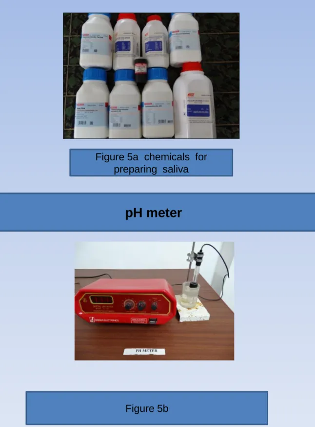
Composition of artificial saliva-(Klimek et al.1982)56
a. Ascorbic acid - 0.002g
b. Glucose - 0.030g
c. Sodium chloride (NaCl) - 0.580g
d. Potassium Chloride (KCl) - 1.270g e. Calcium Chloride (CaCl2 ) - 0.170g
f. Ammonium Chloride (NH4Cl) - 0.160g
g. Sodium thiocyanate (NaSCN) - 0.160g h. Di hydrogen potassium phosphate (KH2PO4) - 0.03g
MATERIALS AND METHODS
37
j. Sodium Hydrogen Phosphate(Na2HPO4) - 0.3g
k. Mucin - 2.7g
L. Distilled water to make up a volume of 1 litre. For demineralizing enamel samples
9. Demineralizing solution (Buskes et al.1985)57 Composition of the demineralizing solution
a) Methylhydroxydiphosphonate - 6µM b) Calcium Chloride Dihydrate (CaCl 2 _2H 2 O) - 3mM
c) Potassium Dihydrogen Phosphate (KH 2 PO 4) - 3mM
d) Acetic acid - 50mM
e) Traces of thymol - 0.2mM
The pH was checked daily and when necessary the pH of the demineralizing solution was adjusted by adding small amounts of either glacial acetic acid or potassium hydroxide solution (10 M )
For checking the pH of demineralizing solution
10. Digisun pH meter (Digisun electronic systems, Hyderabad, India)
For weighing chemicals for preparing artificial saliva and demineralizing solution:
11. Weighing balance (Wensar weighing scales limited, Chennai, India) For brushing of the enamel samples
12. Powered tooth brush (ORAL-B) For Testing of the enamel samples
13.Confocal Laser scanning microscope (Carl Zeiss, Germany)
MATERIALS AND METHODS
38
Materials used in this study
Name Manufacturer Composition
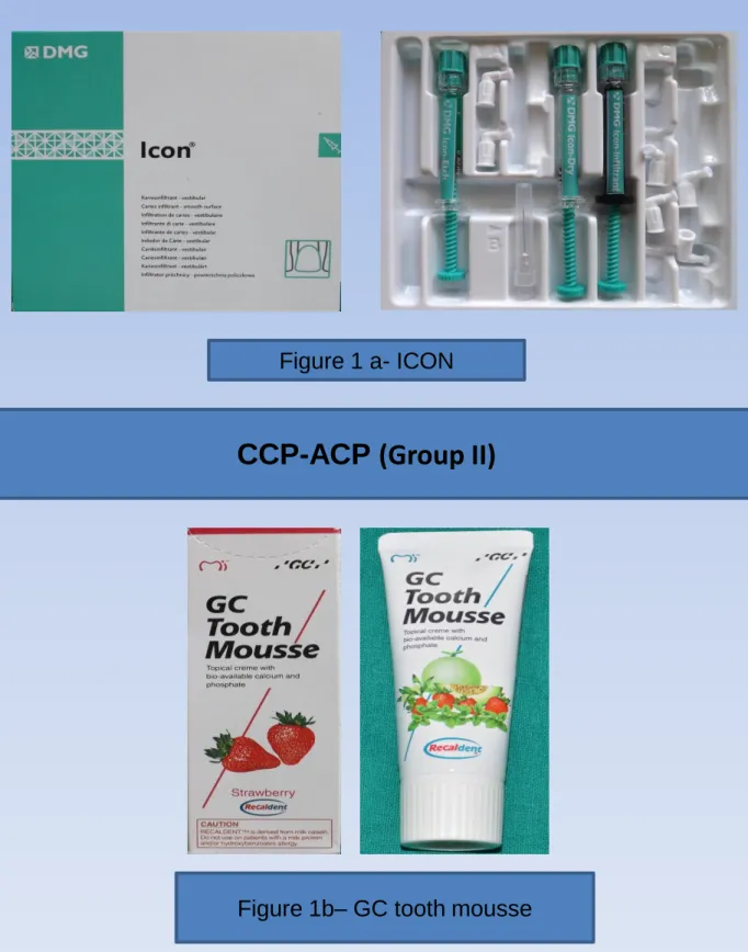
Resin infiltrant (ICON) ICON DMG
(GERMANY)
Icon-Etch: Hydrochloric acid, pyrogenic silicic acid, surface-active substances Icon-Dry: 99% ethanol Icon-Infiltrant:
Methacrylate-based resin matrix,
initiators, additives
CPP-ACP (GC tooth
mousse)
GC Corporation Tokyo, Japan
Glycerol -20.0% Propylene glycol- 2.0% CPP-ACP (casein phosphopeptide - amorphous
calcium Phosphate) -10.0% D-glucitol- 5%
Colloidal silica -2.0% Sodium carboxyl
methylcellulose(CMC-Na)- 2%
Titanium dioxide- 2% Xylitol- 2%
Guar gum <0.1% Phosphoric acid <0.2% Sodium saccharin <0.2% Zinc oxide <0.1%
MATERIALS AND METHODS
39
<0.1%,
Propyl4-hydroxybenzoate 0.1%
Fluoride-900ppm Nano
hydroxyapatite(Aclaim)
Group
pharmaceuticals limited, Mumbai
Nanohydroxyapatite Sorbitol
Glycerin Silica
Purified water
Cocamido Propyl betaine Hydroxyl ethyl cellulose Titanium di oxide
Flavor
MATERIALS AND METHODS
40
STUDY METHODOLOGY Collection of the teeth
60 human maxillary incisors extracted for periodontal reasons were included in this study. Teeth with any visible caries, hypoplastic lesions and white spot lesions were excluded from this study. The teeth were stored in 10% formalin immediately after extraction.
Enamel sample preparation
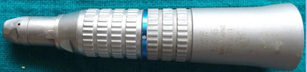
The teeth were thoroughly cleaned of all debris including calculus and tissue debris. Occupational Safety and Health Administration (OSHA) and Centre for Disease Control and Prevention (CDC) recommendations and guidelines were followed during the collection, storage, sterilization and handling of the extracted teeth. Then the radicular portions were removed by decoronating the teeth 2mm coronal to cement enamel junction(CEJ) using a diamond disk (Axis dental, Texas) attached to a slow speed micromotor straight handpiece rotating at 1500rpm. The Sectioning was done at the middle third region of the crown for the 60 samples with approximate dimensions of (5x5x5mm) and then stored in 10% formalin at room temperature of 370c and humidity.
Mounting of the enamel samples
MATERIALS AND METHODS
41
Sample grouping and studyEnamel slabs immersed in demineralizing solution for 14 days
Group I Resin infiltrant n=15 Group II CCP- ACP n=15 Group III Nanohydroxy apatite n=15 Group IV Control n=15
Samples were then subjected to 15%
HCl-etching for 2min, rinsing for
30sec, ethanol dessication for 30
sec, infiltrant applied and light
cured for 40sec.
Untreated enamel samples
were stored in artificial saliva for 30 days Enamel samples
were then brushed with GC
Tooth mousse toothpaste twice
daily for 1 min and stored in artificial saliva
for 30 days
Enamel samples were brushed with
aclaim toothpaste twice daily for 1min and stored in artificial saliva for
30 days
CLSM analysis (10 in each group) group)
Surface microhardness (5 in each group)
60 Enamel slabs
SAMPLE GROUPING AND DESIGN
Surface microhardness and CLSM analysis
Demineralizing solution for 14 days
CLSM analysis (10 in each group)
MATERIALS AND METHODS
42
Demineralization of enamel samples
In order to create the artificial enamel lesions, the samples were demineralized by placing in a beaker containing the prepared demineralizing solution (Buskes et al.1985)57. The study samples were then stored for a period of 14 days all the while maintaining a pH of 5.0 and at 37oC temperature. The pH was checked daily using a pH meter and any variation in pH was corrected by adding either glacial acetic acid or potassium hydroxide solution. The study samples were then randomly divided into four groups containing 15 samples each.
Distribution of samples
The samples were then divided into four groups each containing 15 samples. Group I- Resin infiltrant (ICON) (n=15)
Group II- CPP-ACP (GC Tooth mousse) (n=15) Group III –Nano Hydroxyapatite (Aclaim) (n=15) Group IV –Control (n=15)
Evaluation of microhardness
MATERIALS AND METHODS
43
results were obtained. In order to standardize the enamel samples, 60 samples with VHN in the range of 300.0 to 350.0 was selected for the study. 20 enamel samples (5 in each group) were then evaluated for microhardness after
1) Placing the samples in the demineralizing solution for 14 days
2 ) Exposing the samples to resin infiltrant, CPP-ACP, and nano-HA for 30days. 3) Re-exposing the treated enamel samples to the demineralizing solution for a period of 14 days
Evaluation of penetration
The infiltrated specimens were then observed using a confocal laser scanning microscope equipped with Argon/Krypton laser. The microscope was operated in the fluorescent mode. The excitation light had a maximum wavelength of 560nm and the detected light was passed through a 590 nm long pass filter in order to suppress the reflected and scattered light. The observed layer was then chosen 10µm beneath the specimen surfaces. The microscope setting was kept constant throughtout the investigation period.
MATERIALS AND METHODS
44
Remineralization of enamel samples GROUP I (Resin infiltrant)
15 enamel samples after demineralization were then infiltrated with the resin infiltrant (ICON, DMG) by first etching the enamel samples with 15% hydrochloric acid for 2minutes, after which rinsing with water was done for 30 seconds to remove the etchant. And after drying the sample, ethanol was applied for 30 seconds in order to dessicate the enamel sample and finally to infiltrate it with the resin infiltrant and allowed to remain for 3 minutes and the excess was removed. The curing was done with a LED curing unit for 40 seconds (Satelec mini LED, Aceton group Ltd.,Norwich,UK ). The applicator tip was replaced and allowed to remain for 1 minute and light cured for 40 seconds.
GROUP II (CPP-ACP)
15 enamel samples after demineralization were brushed twice daily for 1 minute using a powered tooth brush(Oral-B) using GC tooth mousse for a time period of 30 days.
GROUP III (Nanohydroxyapatite)
Aclaim tooth paste was applied over the 15 enamel samples after
demineralization twice daily for 1 minute using a powered tooth brush(Oral-B) for 30 days.
Group
IV (Control)The 15 enamel samples were placed in artificial saliva for 30 days and was taken as the control group.
MATERIALS AND METHODS
45
Resin infiltrant (Group I)
Figure 1 a- ICON
Figure 1b– GC tooth mousse
Nano-HA (Group III)
Figure 2a–Aclaim toothpaste
Figure 2b chemicals for
preparing demineralizing
solution
Straight handpiece
Figure 3a – NSK Straight handpiece
Figure 3b
Self cure acrylic resin
Figure 4a
Figure 4b Oral-B
Artificial saliva
Figure 5a chemicals for
preparing saliva
Figure 5b
Weighing balance
Figure 6a Weighing balance for weighing chemicals for
preparing artificial saliva and demineralizing solution
Figure 6b
Vickers microhardness tester
Figure 7a
Figure 10–
Vickers
microhardn
60 Maxillary incisors
Figure 8a
figure 8b
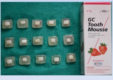
Resin infiltrant (Group I) enamel samples
Figure 9a - Embedded in resin blocks
figure 9b CCP-ACP
Nano-HA (Group III) enamel samples
Figure 10a
figure 10b
Prepared demineralizing solution
Figure 11 a
figure 11 b
CLSM analysis of samples
Figure 12 a
figure 12 b
CLSM images of infiltrated resin for Group I (Resin infiltrant)
Figure 13 a
fig
ur
e
14
–
Vic
ker
s
mi
cro
ha
rd
ne
ss
tes
t
CLSM images after acid challenge for 14 days for Group I
(Resin infiltrant)
CLSM images of Group II (CPP-ACP) after Remineralization
Figure 14 a
Figure 14 b
CLSM images of Group III (Nano-HA) after remineralization
Figure 15 a
Figure 15 b
CLSM images of Group IV (control group) after
demineralization
Figure 16 a
Figure 16 b
Microhardness images for Group I (Resin infiltrant )
Figure 17 a
Figure 17 b
Microhardness images for Group III (nano-HA )
Figure 18 a
Figure 18 b
Microhardness images for Group IV (control)
Statistical Analysis
46
Results
47
Interpretation of results
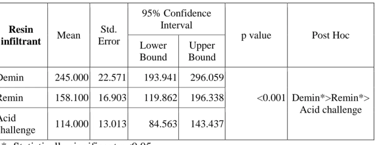
Table 1 shows the intra group comparison for Group I (Resin infiltrant) after demineralization, infiltration, and acid challenge. The mean values after demineralization of enamel samples in the demineralizing solution is 245µm, after infiltrating with resin infiltrant the mean depth of penetration was 158µm. After acid challenge for a period of 14 days the amount of remaining resin infiltrant is 114µm (72%). The p value were <0.001for all the parameters tested. After Bonferroni Post hoc intra group comparison there was statistically significant difference between demineralization, remineralization and after acid challenge.
Table 2 shows the intra group comparison for Group II (CPP-ACP) after demineralization, remineralization and acid challenge. The mean values after demineralization of enamel samples in the demineralizing solution is 246µm. After remineralizing the enamel samples for a period of 30 days, there was a surface remineralization of 28µm. After acid challenge for a period of 14 days the amount of remaining CPP-ACP was 28.8µm (57%). The p value were <0.001for all the parameters tested. After Bonferroni Post hoc intra group comparison there was a statistically significant difference between demineralization, remineralization and after acid challenge.
Results
48
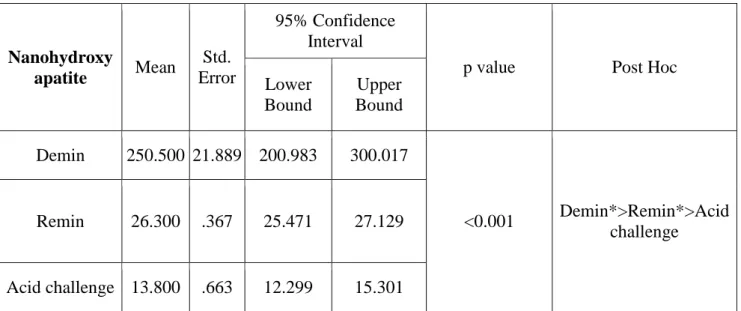
remineralization of 26.3µm. After acid challenge for a period of 14 days the amount of remaining nano-HA was 13.8µm (50%). The p value were <0.001 for all the parameters tested. After Bonferroni Post hoc intra group comparison there was statistically significant between demineralization, remineralization and after acid challenge.
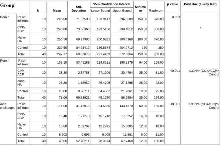
Table 4 shows the inter group comparison for all the groups after demineralization, remineralization and acid challenge for confocal group. The mean values after demineralization of enamel samples in demineralizing solution is 245 µm for Group I (Resin infiltrant), 246 µm for Group II (CPP-ACP), 250 µm for Group III (nano-HA) and 247 µm for Group IV (control). After remineralizing the enamel samples for a period of 30 days, the results are Group I (Resin infiltrant) 158 µm > Group II (CPP-ACP) 28.8µm ⩾ Group III (nano-HA) 26.3µm. After acid challenge for a period of 14 days there was Group I (Resin infiltrant) 114µm (72%) > GROUP II (CPP-ACP) 16.4µm (57%) ⩾ Group IV (nano-HA) 13.8µm (50%) of remaining material. The untreated control group had least resistance with mean of 8 µm. The p value were 0.993 after demineralization, <0.001 after remineralization and after acid challenge for 14 days when comparison was done between all the four groups. After Post hoc Tukey inter group comparison Group I showed statistically significant difference which was greater when compared to group II (CPP-ACP) and Group III (nano-HA) where there were no difference.
Results
49
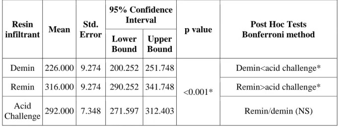
enamel samples is 226VHN. The mean hardness after infiltrating with resin infiltrant increased to 316 VHN. The mean value after acid challenge for 14 days the hardness decreased to 292VHN. The p value were <0.001 for all the parameters. After Post hoc Bonferroni test there was statistically significant difference between demineralization and after acid challenge. There was also significant difference between remineralization and after acid challenge. There was no statistically significant difference between remineralization and demineralization.
Table 6 shows the intra group comparison of surface microhardness for Group II (CPP-ACP). The mean microhardness value after demineralization of enamel samples is 215VHN. The mean value after remineralizing the enamel samples for a period of 30days the hardness increased to 282 VHN. The mean value after acid challenge for 14 days the hardness value decreased to 254VHN. The p value were <0.001 for all the parameters. After Post hoc Bonferroni test there was statistically significant difference between demineralization, remineralization and after acid challenge.
Results
50
Table 8 shows the intra group comparison of surface microhardness for Group IV (Control). The mean microhardness value after demineralization of enamel samples is 207VHN. The mean after remineralizing the enamel samples for a period of 30days in artificial saliva, the hardness increased to 218 VHN. The mean after acid challenge the hardness decreased to 167VHN. The p value were <0.001 for all the parameters. After Post hoc Bonferroni test there was statistically significant difference between demineralization, remineralization and after acid challenge. There was no statistically significant difference between demineralization and acid challenge.
Table 9 shows the inter group comparison of surface microhardness for all the four groups after demineralization. The mean microhardness value after demineralization of enamel samples is Group I (Resin infiltrant) 226VHN, Group II (CPP-ACP) 222VHN, Group III (nano-HA) 207 VHN, Group IV (control) 215VHN. The p value was 0.143 which was not statistically significant.
Results
51
infiltrant) and other 3 groups. Group II (CPP-ACP) and Group III (nano-HA) showed no significant difference.
Table 11 shows the inter group comparison of surface microhardness for all the four groups after acid challenge. The mean microhardness after acid challenge of enamel samples for 14 days is Group I (Resin infiltrant) 292VHN > Group II (CPP-ACP) 254VHN ⩾ Group III (nano-HA) 237VHN > Group IV (control) 167 VHN. The p value was < 0.001 for groups. After Post hoc Tukey test there statistically significant difference between resin Group I (Resin infiltrant) and other 3 groups. Group II (CPP-ACP) and Group III (nano-HA) showed no significant difference.
Graph 1 shows intra group comparison for Group I (Resin infiltrant) after demineralization, remineralization and acid challenge. The different parameters tested demineralixation, remineralization and acid challenge were plotted along x-axis and mean value (µm) were plotted along y-x-axis