Validated and optimized RP-HPLC method for the simultaneous quantification of meloxicam and its
major metabolites in biological fluids with liquid liquid and solid phase extraction technique
Frist Name Middle Name Name Last Academic Degree Affiliation Email Address
Yasar Shah Ph.D Department of Pharmacy, University of Swabi, Swabi [email protected]
Ibrahim Khadra Ph.D Institute of Pharmacy and Biomedical Sciences, Uni [email protected]
Zia Ullah Ph.D Department of Pharmacy, University of Peshawar, P [email protected]
Naila Shahbaz Ph.D Department of Pharmacy, University of Peshawar, P [email protected]
Fazle Khuda Ph.D Department of Pharmacy, University of Peshawar, P [email protected]
Zafar Iqbal Ph.D Department of Pharmacy, University of Peshawar, P [email protected] Aman Ullah Ph.D University of Peshawar [email protected] Lateef Ahmad Ph.D Department of Pharmacy, University of Swabi, Swabi [email protected]
Ismail Ph.D Department of Pharmacy, University of Peshawar, P [email protected]
Muhammad Hassan Ph.D Department of Pharmacy, University of Peshawar, P [email protected]
Abtract
Meloxicam is the most commonly prescribed non-steroidal anti-inflammatory drug. In this study, a simple, rapid and
cost-effective method for the analysis of meloxicam and its major metabolites (5-hydroxy meloxicam and 5-carboxy
meloxicam) in biological fluids (human plasma, urine and saliva) was developed and validated using RP-HPLC
coupled with UV detector. The samples were analyzed by injecting 20 µl into the HPLC system using supelco
analytical C18 (150 mm ×4.6 mm, 5 µm) column, protected by a C18 (30 mm×4.6 mm, 10 µm) Perkin Elmer, guard
column. The mobile phase methanol: TFA (0.05% aqueous solution) in 60:40%v/v was pumped with a flow rate of
1.3 mL/min at ambient temperature and the eluents were checked at 353nm using Piroxicam as internal standard.
Meloxicam and the metabolites were extracted from biological fluids using dichloromethane and the percent
recovery for meloxicam, 5-hydroxy meloxicam and 5-carboxy meloxicam were 98.8%, 97.3%, 97% in plasma, 99%,
5-hydroxy meloxicam and 5-carboxy meloxicam were 3 ng, 10 ng and 8 ng, whereas limit of quantification were 9 ng,
30 ng and 25 ng, respectively. The method was linear over the concentration range of 10 - 2000 ng/mL for
meloxicam, 30 - 1000 ng/mL, 25 - 1000 ng/mL for 5-hydroxy meloxicam and 5-carboxy meloxicam, respectively.
The developed method was validated according to standard guidelines, various experimental parameters and
chromatographic conditions such as mobile phase composition, flow rate, linearity, accuracy, precision, sensitivity
etc. were optimized and were successfully applied for the pharmacokinetic studies in the plasma samples of the
healthy human volunteers.
1. Introduction
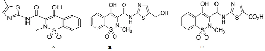
Meloxicam [4-hydroxy-2-methyl-N-(5-methyl-2-thiazolyl)-2H-1,2-benzothiazine-3-carboxamide-1,1-dioxide] a
yellow crystalline powder, with a molecular weight of 351.40 g/mole and is insoluble in water, with a slightly
solubility in organic solvents i.e. methanol and ethanol [1, 2]. Chemical structure of meloxicam and its major
metabolites are presented in Fig-1.
Meloxicam is an NSAID from oxicam group, it preferentially inhibit cyclooxygenase-2 (COX-2) with comparative to
cyclooxygenase-1(COX-1) [3]. Unlike other NSAIDs, meloxicam is gentle on stomach with low ulcer-genic
property, due to lower affinity for COX-1 [4]. It is mainly used in the management of ankylosing spondylitis,
arthritis, rheumatoid arthritis, and other rheumatologic conditions[5].The bioavailability of meloxicam is 89% and
99% of the absorbed drug bind to the plasma protein, the peak plasma concentration is achieved in 5 – 6 hours and
elimination half-life is about 20 hours [6-9].It is metabolized by the hepatic CYP2C9 and to a lesser extent by
CYP3A4 to 5-hydroxy meloxicam which is further converted to 5-carboxy meloxicam [9, 10] that are rapidly
excreted in urine [8]. Whole of administered meloxicam is excreted as metabolites in urine and in feces and only
minimal amounts of parent drug can be detected in urine and in feces [11].
Several methodical approaches have been reported for the analysis of meloxicam in the biological samples, these
techniques include HPLC and LCMS. Reported methods applied advanced detectors like diode array detector and
mass spectrometry [12 -14], which are not readily and easily available. Moreover, mobile phase composition used in
all of the methods is complex, consisting of buffers which can affect the column life and processing time [12, 15,
volumes of plasma without any proper extraction procedure [4, 13]. Not a single method of HPLC for the
simultaneous analysis of meloxicam and its metabolites (5-hydroxy meloxicam and 5-carboxy meloxicam) has been
reported in biological fluids. There is a need to develop a validated, sensitive, easy and rapid method for the
estimation of meloxicam and its metabolites in biological samples, particularly for the pharmacokinetic studies.
In the presented study, suitable extraction procedure has been developed for extraction of meloxicam and its
metabolites from biological samples. The developed method is a novel one with respect to mobile phase composition
and shorter analysis time. The suggested method was then successfully applied for the routine analysis of meloxicam
and its metabolites in pharmacokinetics study of the drug.
2. Experimental
2.1. Chemicals
Meloxicam was provided by Sigma Aldrich, 5-carboxy meloxicam and 5-hydroxy meloxicam was purchased from
Toronto Research Chemicals (Canada), Piroxicam was a kind gift from Medicraft Pharmaceuticals (Peshawar,
Pakistan). The analytical grade HPLC solvents Methanol, Trifluroacetic-acid (TFA) and dichloromethane were
procured from Sigma Aldrich (Oslo, Norway). Freshly, double distilled and deionized HPLC grade water was
obtained from Millipore distillation apparatus (Milford, USA).
2.2. Equipment’s
The analysis were performed using HPLC system (Perkin Elmer Series 200, Norwalk, USA) consisting of a pump,
column oven, UV detector, a vacuum degasser, a rheodyne manual injector with 20 μL loop which is linked to a Pe
Nelson NCI-900. The chromatographic setup was run by a Total Chrome Workstation (version 6.3.1) Perkin Elmer
software for the solicitation and proceeding of data. The column used for analysis of the drug was Supelco C18
reverse phase HPLC column (150mm x 4.6mm ,5µm) and guarded by RP Perkin Elmer C18 (30mm × 4.6mm,
10µm; Norwalk, USA) column. Supel clean SPE cartridges LC-18 (1ml; particle size 56.2µm; Bellefonte, USA) with
the VisiprepTM manifold vacuum were used for solid-phase extraction. A Schimadzu (AX 200) electronic balance,
2.3. Chromatographic conditions
The mobile phase was comprised of methanol: 0.05% TFA (60:40% v/v), pumped at the flow rate of 1.3ml/min.
These solvents were filtered through membrane (0.2µm) and degassed by ultra-sonication. Piroxicam was used as
internal standard and the eluents were monitored at 353 nm.
2.4. Preparation of standard solutions
The standard stock solution of meloxicam (0.5mg/ml) were prepared in methanol containing 20µl of glacial acetic
acid. The standard stock solution of 5-hydroxy meloxicam, 5-carboxy meloxicam and piroxicam (0.5mg/ml) were
also prepared in methanol, DMF (dimethyl-form-amide) and methanol, respectively and further dilutions were
prepared using mobile phase.
The working solutions of meloxicam (10, 50, 100, 250, 500, 1000, 1500, 2000ng/ml) were prepared by diluting the
standard secondary stock solution of meloxicam (10µg/ml). The working standard solutions of 5-carboxy meloxicam
(25, 50, 75, 100, 250, 500, 750, 1000 ng/ml), 5-hydroxy meloxicam (30, 50, 75, 100, 250, 500, 750, 1000ng/ml) and
that of piroxicam (1000ng/ml) were prepared from their respective standard secondary stock solution (10µg/ml) and
were stored at refrigerator temperature.
2.5. Sample collection and preparation
The study was approved by the Ethical Committee of the Department of Pharmacy, University of Peshawar and
informed written assent was attained from the recruited healthy human volunteers, aged between 25 to 35 years and
BMI in the range of 23-30. The volunteers were instructed to avoid any medicine for two weeks prior to the study.
Early morning at fasting condition, a single oral dose of the meloxicam was taken orally with a glass of water (≈ 250
ml) and blood (2ml), urine (20 ml), saliva samples (3 ml) were collected periodically, centrifuged at 3000 rpm for 10
The blank biological fluids (collected before administration of the drug) were thawed at room temperature, at the
time of analysis and spiked with appropriate concentration of working solution of meloxicam (i.e., 10, 50, 100, 250,
500, 1000, 1500, 2000 ng/ml), 5-hydroxy meloxicam (i.e., 30, 50, 75, 100, 250, 500, 750, 1000ng/ml), 5-carboxy
meloxicam (i.e., 25, 50, 75, 100, 250, 500, 750, 1000 ng/ml) and piroxicam (internal standard) to obtain calibration
standards.
2.6. Extraction of meloxicam and its metabolites from biological samples
Liquid-liquid and solid-phase extraction were assessed for the recovery of parent drug and its metabolites from
biological samples.
2.6.1. Liquid-liquid extraction
Biological fluids (200µl) were mixed with internal standard (100µl = 100ng) in eppendorf tube, acetonitrile (600µl)
was added and vortexed for 3min. Then dichloromethane (1100µl) was added, further vortexed for 5min, centrifuged
for 15min at 9000 rpm, organic layer collected and was evaporated under the gentle stream of nitrogen. The residues
were re-dissolved in methanol (2ml) and injected into HPLC for analysis.
2.6.2. Solid phase extraction (SPE)
SPE procedure was also utilized for the biological samples in order to achieve good recovery of analytes and
comparison between the two methods. In SPE, first conditioning of the cartridge was carried out using methanol (1
ml) followed by equilibration with 1ml of water. The biological sample (200 µl) containing internal standard (100
µl), water (3 ml), concentrated HCl (10 µl) were transferred into the cartridge and forced through it by applying
eluate was then dried under the stream of nitrogen gas, final volume was made up to 2 ml with methanol and 20 µl
was introduced into the HPLC system.
2.7. Chromatographic conditions optimizations
Different chromatographic parameters like mobile phase composition ratio, flow rate, wavelengths of detector,
injection volumes, internal standard and extraction solvents were optimized for quantitative determination of the
meloxicam and its metabolites, using RP-HPLC method in an isocratic mode.
2.7.1. Mobile phase composition
A number of organic solvents such as acetonitrile, methanol, trifluoroacetic-acid (TFA), phosphate buffer, pH
adjusted distilled water were tried in different combinations both in gradient and in an isocratic mode for the
determination of the meloxicam and its metabolites. The mobile phase composition which demonstrated good
resolution and separation was selected for analysis.
2.7.2. Flow rate composition
The flow rate significantly affect the sample elution, resolution, separation and peak shape. In this method flow rates
in different ranges i.e. 0.8-1.5ml/min were evaluated and one that gave best resolution was selected as optimum flow
rate.
2.7.3. Detection wavelength
A Perkin Elmer UV lambda (Nowak, USA) was used to determine the maximum wavelength of maximum
absorbance (λmax) of meloxicam. Effect of wave length on analytes response was studied by varying wave length in
2.7.4. Injection Volumes
Different injection volumes in the ranges of 10 µl-50 µl were assessed using rheodyne manual injector.
2.7.5. Internal standard selection
Different internal standards such as naproxen sodium, lumefantrine, diclofenac-sodium, and piroxicam were tried. In
present studies the Piroxicam showed maximum response and better chromatographic properties, therefore it was
selected as internal standard.
2.7.6. Selection of extraction solvent
Various solvents such as diethyl-ether, dichloromethane, acetonitrile and n-hexane were evaluated as extraction
solvents. Optimum percent recovery was achieved with dichloromethane as an extraction solvent.
2.8. Validation of the method
Validation of the developed method was carried out according to ICH guidelines with special emphasis on
specificity, linearity, precision, accuracy, robustness, selectivity, limit of detection (LOD), and limit of quantification
(LOQ) [17].
2.8.1. Specificity
The specificity of developed method was judged from the complete peak resolution of the meloxicam and its
metabolites in mobile phase, spiked biological sample in a 1:1 mixture (i.e. 1 µg/ml of the sample as well as internal
standard).
The linearity was evaluated by scheming calibration curves, at eight concentration level of meloxicam (10, 50, 100,
250, 500, 1000, 1500 and 2000 ng/ml), hydroxy meloxicam (30, 50, 75, 100, 250, 500, 750 and 1000 ng/ml),
5-carboxy meloxicam (25, 50, 75, 100, 250, 500, 750 and 1000 ng/ml) and piroxicam (1000ng/ml). Following analysis,
the ratios of peak area of the analyte and internal standard was plotted as function of the concentration of analyte
using the squares linear least regression equation and the correlation co-efficient.
2.8.3. Accuracy
The accuracy of the developed method was calculated from the percent recovery of the spiked sample. Blank human
biological sample was spiked with analytes of different concentration levels keeping the concentration of I.S.
constant. The similar concentrations levels were also prepared in mobile phase. Samples were then injected in
triplicate and percent recoveries were calculated by the following equation:
Percent recovery = × 100
Where
A = Response ratio of the peak of spiked biological sample (analyte/I.S)
B = Response ratio of the peak of sample in standard solution (analyte/I.S)
2.8.4. Precision
Precision of the developed method was determined by injection and the analysis repeatability. The injection precision
repeatability was determined by injecting biological sample, spiked with 1000ng/ml of meloxicam, 5-carboxy
meloxicam and 5-hydroxy meloxicam for 10 times into HPLC. The peak area and the retention time was obtained
and articulated as mean, standard deviation (±SD), and covariance (% RSD). The precise analysis repeatability of the
method was analyzed by spiking six biological samples with 1000 ng/ml of meloxicam, carboxy meloxicam and
Intermediate precision (i.e. intra-day and the inter-day studies) was determined by injecting plasma sample spiked
with appropriate standard concentration of the analyte and internal standard on 8.0, 16.0 and 24.0 hours for three
successive days. The outcome was depicted as mean, standard deviation as well as covariance and the following
equation was employed to determine the concentration
C = CS DF
Where
x = Peak area of the analyte in biological sample
y = Peak areas of the I.S in biological sample
A = Peak area of the I.S in 1:1 mixture
B = Peak area of the analyte in 1:1 mixture
Cs = Concentration of analyte in the 1:1mixture
DF = is the dilution factor
2.8.5. Sensitivity
The developed method sensitivity was assessed in terms of LOD and LOQ of analytes. The LOD was determined
from the peak of the analyte by the software at which signal to noise ratio is 3 and the LOQ was concentration of the
analyte at which signal to noise ratio is equal to10.
2.8.6. Stability
Stability studies of the meloxicam and its metabolites were assessed in spiked biological samples and standard stock
solution at room temperature (25oC), refrigerator temperature (2-8oC) and at -20oC for one month. Each sample was
then analyzed (n = 3) and the following equation was used to evaluate the stability:
Where
St =Sample stability at that time
So=Initial stability of sample
2.8.7. Robustness/ Ruggedness
The developed method robustness/ruggedness was investigated by bringing deliberately small alterations in
chromatographic conditions, such as composition of the mobile phase (± 2%), temperature of the column oven (±
5°C), mobile phase flow rate (± 0.2 ml/min) and detection wavelength (± 5%).
3. Results and discussion
The developed method for the analysis of meloxicam and its metabolites in human biological fluids using piroxicam
as I.S is novel, fast, economical and sensitive. All the analytes and internal standard were completely resolved.
Different chromatographic conditions and various experimental parameters of the developed method were optimized
and validated according to ICH guidelines. According to literature it is first attempt to analyze the meloxicam and its
metabolites simultaneously in biological samples.
3.1 Sample preparation
Meloxicam is sparingly soluble in methanol, ethanol, and however it was better in methanol that was further
improved by the addition of 20µl of glacial acetic acid to about 10 ml of solution. The stock solution was then diluted
with methanol.
The protein in the plasma samples were precipitated with acetonitrile using three times of the plasma volume. The
extraction of the analytes from the biological samples were carried out with dichloromethane, chloroform, ethyl
acetate, diethyl ether, n-hexane either alone or with different combinations. Dichloromethane showed better percent
recovery in both extraction procedure and was selected for extraction of analytes from the biological samples.
3.2.1. Mobile phase and composition
Different solvents such as acetonitrile, methanol, TFA, water and pH adjusted water were evaluated as mobile phases
in different compositions. First acetonitrile and water (50: 50%v/v) as mobile phase was used, showed good shape
and resolution of the peaks but retention time was not stable. The water was replaced with pH adjusted water in the
range of 2 – 4 and it ends with the same results of stability of retention time. Various ratio of the methanol:
0.05%TFA was evaluated as the mobile phase to achieve better separation and stable chromatogram of the analytes.
The results showed better resolution, peak shape and stability of the methods when methanol and TFA (0.05%) was
used in 60:40% v/v.
3.2.2. Flow rate
The mobile flow rates ranges from 0.8-1.5% were applied; the optimum flow rate 1.3 ml/min was selected that gave
good resolution of the analytes and total elution time was less than 7 min.
3.2.3. Selection of the wavelength
Wavelength 330 nm, 342 nm, 353 nm and 355 nm were evaluated and the best response for meloxicam, its
metabolites and internal standard was observed at 353 nm.
3.2.4. Selection of the internal standard
Various compounds were assessed as internal standard that include naproxen sodium, diclofenac sodium,
lumefantrine and piroxicam. Among these piroxicam showed good resolution, sensitivity, specificity, recovery,
stability, and while other compounds were having long retention time or poor recovery.
3.2.5. Selection of the column
Thermo Quest Hypersil C18 column (250×4.6mm, 5µm; Run-corn, UK), Brownlee Perkin Elmer analytical C18
C18 (250x4.6mm, 5µm) column and Thermo Quest Hypersil C8 (150×4.6mm, 5µm; Run-corn, UK) column were
tried. The best resolution and other chromatographic properties were obtained with supelco discovery analytical C18
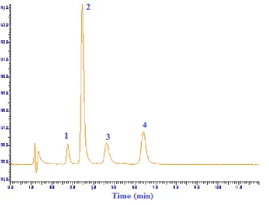
column (150 x 4.6mm, 5µm). The representative chromatogram of the standard sample spiked with meloxicam,
5-hydroxy, 5-carboxy meloxicam and I.S on the supelco/discovery C18 column is shown in Fig. 2.
3.3. Method validation
3.3.1. Linearity
The calibration curves of meloxicam(10-2000 ng/ml), 5-hydroxy meloxicam (30-1000 ng/ml) and 5-carboxy
meloxicam (25-1000 ng/ml) in spiked biological samples demonstrated excellent linearity ( r2 = 0.999) results are
depicted in Table 1 and in Fig. 3.
3.3.2. Selectivity
The selectivity of the method for the meloxicam and its metabolites was determined from a) mobile phase, b) blank
plasma, urine and saliva c) plasma, urine, saliva sample spiked with meloxicam, 5-hydroxy meloxicam, 5-carboxy
meloxicam and piroxicam as I.S, d) plasma, urine, saliva sample spiked with I.S. The Fig. 4, 5, 6 confirms the good
resolution of the peaks and no other exogenous or endogenous peaks interfere with the chromatograms.
3.3.3. Precision
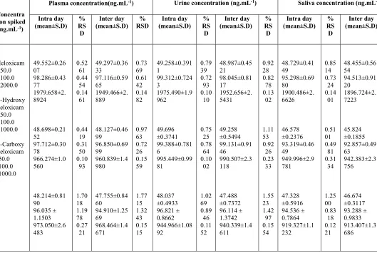
The precision results obtained from the injection repeatability and the intermediate precision (inter-day, intra-day
studies) are summarized in Table 1 and 2. The method demonstrated good precision, the % RSD value for both
inter-day and intra-inter-day which was less than 2%.
The developed method accuracy was assessed from the percent recovery, the mean percent recovery of meloxicam,
5-hydroxy meloxicam, 5-carboxy meloxicam were 98.8%, 97.3%, 97% in plasma 99%, 98.6%, 95.1% in urine and
95.8%, 92.9%, 92.7% in saliva by using liquid-liquid extraction and 92%, 84%, 77.8% in plasma, 93.4%, 87%,
83.1% in urine, 85.5%, 78%, 74% in saliva by using solid-phase extraction showing %RSD value less than 1%,
represents that the developed method was accurate. Percent recovery of all the analytes was higher with liquid-liquid
extraction than SPE.
3.3.5 Sensitivity
The LOD for meloxicam, 5-hydroxy meloxicam and 5-carboxy meloxicam were 3ng, 10ng and 8ng, respectively.
The LOQ were 9ng, 30ng and 25ng, respectively. Chromatograms of LLODs and LLOQs are shown in Fig. 7 and
Fig. 8.
3.3.6 Robustness
The robustness of the developed method was verified by doing minor changes in chromatographic conditions such as
composition of the mobile phase (± 0.3%), flow rate of the mobile phase (± 0.2 ml/min), wavelength (± 3 nm), did
not substantially affect peak shape and the retention time of the sample.
3.3.7 Stability
The spiked plasma samples were stable for I week under all of the conditions; room temperature (25oC), refrigerated
(2 – 8oC) or frozen at (-80oC), while the standard solutions were stable for about a month when stored at 2 – 8oC.
The proposed method is a part of comprehensive clinical study on “Pharmacokinetic drug-drug interaction of
meloxicam, 5-hydroxy meloxicam and 5-carboxy meloxicam with commonly co-prescribed drugs like omeprazole
and fluconazole in healthy human volunteers”. The developed method was successfully applied for the determination
of meloxicam in healthy human volunteers. Pharmacokinetic data was analyzed by using PK summit® software and
will be published somewhere else. In all the analyzed plasma, saliva samples both metabolites 5-hydroxy meloxicam
and 5-carboxy meloxicam concentrations were below the detection limit and thus could not be quantified and were
only quantified in urine. This method was applied to the ongoing clinical trialand can also be employed for the
quantification of meloxicam in various pharmaceutical dosage form as well.
5. Conclusion
The present developed method was optimized and validated for the simultaneous determination of meloxicam,
5-hydroxy meloxicam and 5-carboxy meloxicam in human biological fluids. The method was simple, rapid, easy,
accurate, precise and economical. Various chromatographic conditions were optimized for the analysis of these
analytes according to standard guidelines. The effects of composition of the mobile phase, flow rate, temperature of
the column oven, detection wavelength were studied. In this method meloxicam and its metabolites recovery was
excellent from biological sample by using two step LLE procedures. All the peaks appear within 7 min in a single
chromatographic run. This method can also be employed in various pharmaceutical dosage forms, in biological
matrices and in pharmacokinetic studies.
6. References
[1] M. Yazdanian, K. Briggs, C. Jankovsky and A. Hawi, The “high solubility” definition of the current FDA
guidance on biopharmaceutical classification System may be too strict for acidic drugs. Pharm Res., 21, 293–299
(2004).
[2] An Encyclopedia of Chemical, Drugs and Biologicals, 13th Ed, Merck Research Laboratories, Division of Merck
[3] G. Engelhardt, D. Homma, K. Schlegel, R. Utzmann and C. Schnitzler, Anti-inflamatory, analgesic, antipyretic
and related properties of Meloxicam, a new non-steroidal anti-inflamatory agent with favorable gastrointestinal
tolerance, Inflammation Research., 44, 423-433 (1995).
[4] B. Dasandi, H. Saroj and K. Bhat, LC determination and pharmacokinetics of meloxicam, Journal of
pharmaceutical and biomedical analysis., 28, 999-1004 (2002).
[5] B.J. Gates, T.T. Nguyen, S.M. Setter and N.M. Davies, Meloxicam reapraisal of pharmacokinetics, efficacy and
safety, Experts Opinion on Pharmacotherapy., 6, 2117-2140 (2005).
[6] D. Türck, U. Busch, G. Heinzel, H. Narjes and G. Nehmiz, Effect of food on pharmacokinetics of oral
meloxicam, Clinical Drug Investigation., 9, 270-276 (1995).
[7] V. Hynninen, K. Olkkola, L. Bertilsson, K. Kurkinen, T. Korhonen, P. Neuvonen and K. Laine, Voriconazole
increases while itraconazole decreases plasma meloxicam concentration, Antimicrobial agents and chemotherapy.,
53, 587-592 (2009).
[8] J. Schmid, U. Busch, G. Heinzel, G. Bozler, S. Kaschke and M. Kummer, Pharmacokinetics and metabolic
pattern after intraveinous infusion and oral administration to healthy subjects., 23, 1206-1213 (1995).
[9] D. Türck, U. Busch, G. Heinzel and H. Narjes, Clinical pharmacokinetics of meloxicam,
Arzneimittel-Forschung., 47, 253-258 (1997).
[10] C. Chesne, C. Guyomard, A. Guillouzo, J. Schmid, E. Ludwig and T. Sauter, Metabolism of meloxicam in
human liver involves cytochromes P4502C9 and 3A4, Xenobiotica., 28, 113 (1998).
[11] D. Türck, W. Roth and U. Busch, A review of the clinical pharmacokinetics of meloxicam, Rheumatology., 35,
13-16 (1996).
[12] T. Velpandian, T. Jaiswal, R.K. Bhardwaj and S.K. Gupta, Development and validation of a new high
performance liquid chromatographic estimation method of Meloxicam in biological samples, Journal of
Chromatography B: Biomedical Sciences and Applications., 738, (2000).
[13] A. Medvedovici, F. Albu, C. Georgita, C. Mircioiu and V. David, A non-extracting procedure for determination
of meloxicam in plasma samples by HPLC-Diode array detector, Arzneimittel-Forschung., 55, 326-331 (2005).
[14] H.Y. Ji, H.W. Lee, Y.H. Kim, D.W. Jeong and H.S. Lee, Simulatanious determination of piroxicam, meloxicam
and tenoxicam in human plasma by liquid chromatography with tendon mass spectrometry, Journal of
[15] E. Nemutlu and S. Kir, Method development and validation for the analysis of meloxicam in tablets by CZE, J
Pharm Biomed Anal., 31, 393-400 (2003).
[16] S. Rani, S. Guttikar, R. Rathod, B. Cherian, M. Nivsarkar and H. Padh, Determination of oral meloxicam,
pharmacokinetics parameters in Asian Indian: Comparison with general population, Saudi Pharmaceutical Journal.,
12, 144-149 (2004).
[17] N. A. Epshtein, Validation of HPLC techniques for pharmaceutiacl analysis, Pharm. Chem. J., 38, 212 (2004).
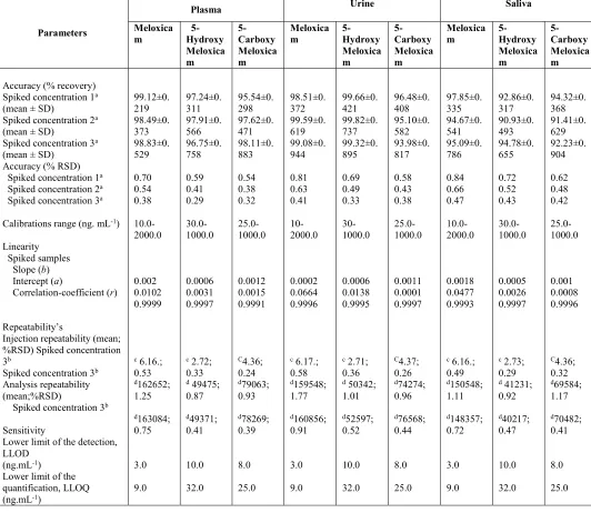
List of Tables:
Table-1: Accuracy, calibration, linearity, repeatability and sensitivity of the current RP-HPLC method.
Table-2: The data of the Intra-day and Inter-days Precision (n = 3).
List of Figures:
Fig. 1. Structures; A: meloxicam, B: 5-hydroxy meloxicam, C: 5-carboxy meloxicam.
Fig. 2. The chromatogram of standard sample spiked with 100 ng/ml of meloxicam (peak -4), 75 ng/ml of each metabolite i.e 5-hydroxy meloxicam (peak-1); 5-carboxy meloxicam (peak-3) and 1000ng/ml of internal standard
(peak-2).
Fig. 3. Calibration curves of (A) meloxicam, (B) 5-hydroxy meloxicam, (C) 5-carboxy meloxicam. Solid line (―)
represents spiked plasma, dashed line (---) represents spiked urine, dotted line (….) represents spiked saliva.
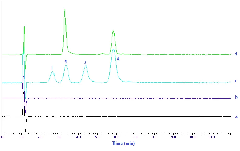
Fig. 4. RP-HPLC chromatograms of different samples, Peak-1; hydroxy meloxicam, 2; internal standard, 3;
5-carboxy meloxicam, 4; meloxicam. Chromatograms a; blank solvent, b; blank plasma, c; plasma sample spiked with
1000ng/ml of meloxicam, 500ng/ml of 5-hydroxy meloxicam, 500ng/ml of 5-carboxy meloxicam and 1000ng/ml of
internal standard, d; plasma sample.
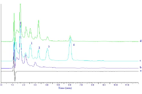
Fig. 5. RP-HPLC chromatograms of different samples, Peak-1; hydroxy meloxicam, 2; internal standard, 3;
1000ng/ml of meloxicam, 500ng/ml of 5-hydroxy meloxicam, 500ng/ml of 5-carboxy meloxicam and 1000ng/ml of
internal standard, d; urine sample.
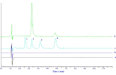
Fig. 6. RP-HPLC chromatograms of different samples, Peak-1; hydroxy meloxicam, 2; internal standard, 3; 5-carboxy meloxicam, 4; meloxicam. Chromatograms a; blank solvent, b; blank saliva, c; saliva sample spiked with
1000ng/ml of meloxicam, 500ng/ml of 5-hydroxy meloxicam, 500ng/ml of 5-carboxy meloxicam and 1000ng/ml of
internal standard, d; saliva sample.
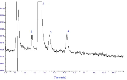
Fig. 7. RP-HPLC chromatogram showing peaks of 5-hydroxy meloxicam (peak-1), 5-carboxy meloxicam (peak-3)
and meloxicam (peak-4) at the lower limit of detection.
Fig. 8. RP-HPLC chromatogram showing peaks of 5-hydroxy meloxicam (peak-1), 5-carboxy meloxicam (peak-3)
Table-2: Accuracy, linearity, calibration, sensitivity, repeatability of the current RP-HPLC method.
Parameters
Plasma Urine Saliva
Meloxica
m 5-Hydroxy Meloxica m 5-Carboxy Meloxica m Meloxica
m 5-Hydroxy Meloxica m 5-Carboxy Meloxica m Meloxica
m 5-Hydroxy Meloxica m 5-Carboxy Meloxica m Accuracy (% recovery)
Spiked concentration 1a
(mean ± SD)
Spiked concentration 2a
(mean ± SD)
Spiked concentration 3a
(mean ± SD) Accuracy (% RSD) Spiked concentration 1a
Spiked concentration 2a
Spiked concentration 3a
Calibrations range (ng. mL-1)
Linearity Spiked samples Slope (b) Intercept (a)
Correlation-coefficient (r)
Repeatability’s
Injection repeatability (mean; %RSD) Spiked concentration 3b
Spiked concentration 3b
Analysis repeatability (mean;%RSD)
Spiked concentration 3b
Sensitivity
Lower limit of the detection, LLOD
(ng.mL-1)
Lower limit of the quantification, LLOQ (ng.mL-1)
99.12±0. 219 98.49±0. 373 98.83±0. 529 0.70 0.54 0.38 10.0-2000.0 0.002 0.0102 0.9999
c 6.16.;
0.53 d162652; 1.25 d163084; 0.75 3.0 9.0 97.24±0. 311 97.91±0. 566 96.75±0. 758 0.59 0.41 0.29 30.0-1000.0 0.0006 0.0031 0.9997
c 2.72;
0.33
d 49475;
0.87 d49371; 0.41 10.0 32.0 95.54±0. 298 97.62±0. 471 98.11±0. 883 0.54 0.38 0.32 25.0-1000.0 0.0012 0.0015 0.9991 C4.36; 0.24 d79063; 0.93 d78269; 0.39 8.0 25.0 98.51±0. 372 99.59±0. 619 99.08±0. 944 0.81 0.63 0.41 10-2000.0 0.0002 0.0664 0.9996
c 6.17.;
0.58 d159548; 1.77 d160856; 0.91 3.0 9.0 99.66±0. 421 99.82±0. 737 99.32±0. 895 0.69 0.49 0.33 30-1000.0 0.0006 0.0138 0.9995
c 2.71;
0.36
d 50342;
1.01 d52597; 0.52 10.0 32.0 96.48±0. 408 95.10±0. 582 93.98±0. 817 0.58 0.43 0.38 25.0-1000.0 0.0011 0.0001 0.9997 C4.37; 0.26 d74274; 0.96 d76568; 0.44 8.0 25.0 97.85±0. 335 94.67±0. 541 95.09±0. 786 0.84 0.66 0.47 10.0-2000.0 0.0018 0.0477 0.9993
c 6.16.;
0.49 d150548; 1.11 d148357; 0.72 3.0 9.0 92.86±0. 317 90.93±0. 493 94.78±0. 655 0.72 0.52 0.43 30.0-1000.0 0.0005 0.0026 0.9997
c 2.73;
0.29
d 41231;
0.92 d40217; 0.47 10.0 32.0 94.32±0. 368 91.41±0. 629 92.23±0. 904 0.62 0.48 0.42 25.0-1000.0 0.001 0.0008 0.9996 C4.36; 0.32 d69584; 1.17 d70482; 0.41 8.0 25.0
Spiked concentration 1= meloxicam: 50ng.mL-1, 5-hydroxy meloxicam: 50ng.mL-1and 5-carboxy meloxicam:
50ng.mL-1; Spiked concentration 2 = meloxicam: 100 ng.mL-1, 5-hydroxy meloxicam: 100ng.mL-1 and 5-carboxy
meloxicam: 100ng.mL-1; Spiked concentration 3 = meloxicam:1000ng.mL-1, 5-hydroxy meloxicam: 1000ng.mL-1and
Table-2: The data of the Intra days and Inter days Precision (n = 3).
Concentra tion spiked
(ng.mL-1)
Plasma concentration(ng.mL-1) Urine concentration (ng.mL-1) Saliva concentration (ng.mL-1) Intra day
(mean±S.D) RS% D
Inter day
(mean±S.D) RSD % (mean±S.D) Intra day RS% D
Inter day (mean±S.D) RS%
D
Intra day (mean±S.D) RS%
D
Inter day (mean±S.D) RS%
Figure 2: The chromatogram of standard sample spiked with 100 ng/ml of meloxicam (peak -4), 75 ng/ml of each metabolite i.e 5-hydroxy meloxicam (peak-1); 5-carboxy meloxicam (peak-3) and 1000ng/ml of internal standard
Figure 4: HPLC chromatogram’s of various samples, Peak-1; hydroxy meloxicam, 2; internal standard, 3;
5-carboxy meloxicam, 4; meloxicam. Chromatogram’s a; blank-solvent, b; blank-plasma, c; plasma spiked sample with
1000ng/ml of meloxicam, 500ng/ml of 5-hydroxy meloxicam, 500ng/ml of 5-carboxy meloxicam and 1000ng/ml of
Figure 5: HPLC chromatogram’s of various samples, Peak-1; hydroxy meloxicam, 2; internal standard, 3; 5-carboxy meloxicam, 4; meloxicam. Chromatogram’s a; blank-solvent, b; blank-urine, c; urine spiked sample with
1000ng/ml of meloxicam, 500ng/ml of 5-hydroxy meloxicam, 500ng/ml of 5-carboxy meloxicam and 1000ng/ml of
internal standard, d; urine sample.
[image:24.612.72.544.58.370.2]
Figure 6: HPLC chromatogram’s of various samples, Peak-1; hydroxy meloxicam, 2; internal standard, 3; 5-carboxy meloxicam, 4; meloxicam. Chromatogram’s a; blank-solvent, b; blank-saliva, c; saliva spiked sample with
1000ng/ml of meloxicam, 500ng/ml of 5-hydroxy meloxicam, 500ng/ml of 5-carboxy meloxicam and 1000ng/ml of
[image:25.612.87.545.85.382.2]Figure 8: HPLC chromatogram showing peaks of 5-hydroxy meloxicam (peak-1), 5-carboxy meloxicam (peak-3)
[image:27.612.84.504.112.377.2]