Human T-Cell Leukemia Virus Type 1-Induced Cell Transformation
Phenotype
Tomonari Shigemura,aMasaaki Shiohara,aMasayoshi Kato,bShuichi Furuta,bKazuko Kaneda,cKazuhiro Morishita,cHiroo Hasegawa,d Masahiro Fujii,eAgnes Gorlach,fKenichi Koike,aTohru Kamatab
Department of Pediatricsaand Department of Molecular Biology and Biochemistry,bShinshu University Graduate School of Medicine, Matsumoto, Nagano, Japan; Division of Tumor and Cellular Biochemistry, Faculty of Medicine, University of Miyazaki, Miyazaki, Japanc; Department of Laboratory Medicine, Nagasaki University Graduate School of Biomedical Sciences, Nagasaki, Japand; Division of Virology, Niigata University Graduate School of Medicine and Dental Science, Niigata, Japane; Experimental and Molecular Pediatric Cardiology, Department of Pediatric Cardiology and Congenital Heart Disease, German Heart Center Munich at the Technical University, Munich, Germanyf
ABSTRACT
Human T-cell leukemia virus type 1 (HTLV-1) is associated with adult T-cell leukemia (ATL) and transforms T cells
in vitro
. To
our knowledge, the functional role of reactive oxygen species (ROS)-generating NADPH oxidase 5 (Nox5) in HTLV-1
transfor-mation remains undefined. Here, we found that Nox5
␣
expression was upregulated in 88% of 17 ATL patient samples but not in
normal peripheral blood T cells. Upregulation of the Nox5
␣
variant was transcriptionally sustained by the constitutive Janus
family tyrosine kinase (Jak)-STAT5 signaling pathway in interleuk2 (IL-2)-independent HTLV-1-transformed cell lines,
cluding MT1 and MT2, whereas it was transiently induced by the IL-2-triggered Jak-STAT5 axis in uninfected T cells. A Nox
in-hibitor, diphenylene iodonium, and antioxidants such as N-acetyl cysteine blocked proliferation of MT1 and MT2 cells. Ablation
of Nox5
␣
by small interfering RNAs abrogated ROS production, inhibited cellular activities, including proliferation, migration,
and survival, and suppressed tumorigenicity in immunodeficient NOG mice. The findings suggest that Nox5
␣
is a key molecule
for redox-signal-mediated maintenance of the HTLV-1 transformation phenotype and could be a potential molecular target for
therapeutic intervention in cancer development.
IMPORTANCE
HTLV-1 is the first human oncogenic retrovirus shown to be associated with ATL. Despite the extensive study over the years, the
mechanism underlying HTLV-1-induced cell transformation is not fully understood. In this study, we addressed the expression
and function of ROS-generating Nox family genes in HTLV-1-transformed cells. Our report provides the first evidence that the
upregulated expression of Nox5
␣
is associated with the pathological state of ATL peripheral blood mononuclear cells and that
Nox5
␣
is an integral component of the Jak-STAT5 signaling pathway in HTLV-1-transformed T cells. Nox5
␣
-derived ROS are
critically involved in the regulation of cellular activities, including proliferation, migration, survival, and tumorigenicity, in
HTLV-1-transformed cells. These results indicate that Nox5
␣
-derived ROS are functionally required for maintenance of the
HTLV-1 transformation phenotype. The finding provides new insight into the redox-dependent mechanism of HTLV-1
transfor-mation and raises an intriguing possibility that Nox5
␣
serves as a potential molecular target to treat HTLV-1-related leukemia.
H
uman T-cell leukemia virus type 1 (HTLV-1) is the first
hu-man oncogenic retrovirus shown to be etiologically
associ-ated with adult T-cell leukemia (ATL) (
1
,
2
). ATL has a poor
prognosis because of its resistance to conventional chemotherapy,
and no effective therapy is currently available for ATL. HTLV-1
infects and transforms human peripheral blood T cells
in vitro
(
3
,
4
), but the precise mechanism of HTLV-1 transformation of T
cells and the development of ATL after HTLV-1 infection are not
fully understood. A HTLV-1 genome pX region-encoded protein,
Tax, is thought to play a central role in activation, proliferation,
and transformation of T cells by transactivating various cellular
genes, including interleukin-2 (IL-2), IL-2 receptor
␣
chain, and
NF-
B (
5–7
). However, advanced ATL cells do not always express
a significant amount of Tax and yet maintain the transformation
phenotype (
8
). This raised the possibility that cellular changes,
including constitutive NF-
B activation, replace Tax functions to
sustain neoplastic features (
8
,
9
). Meanwhile, IL-2 stimulates
nor-mal T-lymphocyte activity and proliferation through the Janus
family tyrosine kinase (Jak)-STAT5 signaling pathway (
10
).
HTLV-1-infected T cells initially grow in an IL-2-dependent
man-ner, but over time, the cells become IL-2 independent (
11
). In
most cases, this transition seems to coincide with acquisition of
constitutive activation of Jak and STAT5 signaling (
12
,
13
), but its
significance in the IL-2-independent growth mechanism remains
Received17 April 2015Accepted16 June 2015
Accepted manuscript posted online24 June 2015
CitationShigemura T, Shiohara M, Kato M, Furuta S, Kaneda K, Morishita K, Hasegawa H, Fujii M, Gorlach A, Koike K, Kamata T. 2015. Superoxide-generating Nox5␣is functionally required for the human T-cell leukemia virus type 1-induced cell transformation phenotype. J Virol 89:9080 –9089.doi:10.1128/JVI.00983-15. Editor:S. R. Ross
Address correspondence to Tohru Kamata, [email protected].
K. Koike and T. Kamata contributed equally to this article.
Copyright © 2015, American Society for Microbiology. All Rights Reserved.
doi:10.1128/JVI.00983-15
on November 7, 2019 by guest
http://jvi.asm.org/
only partly explained. These observations suggest that, to define
the functional role of HTLV-1 in malignant transformation, we
need to understand more of the as-yet-unidentified sequence of
intracellular signals essential for genetic and epigenetic
interac-tions between provirus and host genes.
Accumulating evidence suggests that low levels of reactive
ox-ygen species (ROS) act as second-messenger-like molecules in
multiple cellular processes, including proliferation, apoptosis,
and innate immunity. Superoxide (O
2⫺)-generating NADPH
ox-idase (Nox) family enzymes (Nox1 to Nox5 and Duoxes 1 and 2)
represent a major intracellular source for ROS (
14
,
15
). In fact,
Nox1, Nox2, and Nox4 have been shown to play important
phys-iological and pathophysphys-iological roles in cardiovascular,
pulmo-nary, and renal systems. Nox1 and Nox4 may be linked to
devel-opment of some types of cancers, including prostate and
pancreatic cancers (
16
,
17
). In comparison, the function of Nox5
is poorly understood. Unlike Nox1 to Nox4, Nox5 comprises the
N-terminal EF hand (binding sites for calcium), in addition to the
heme-containing transmembrane and NADPH/flavin adenine
di-nucleotide (FAD)-binding cytoplasmic domains, which are well
conserved among the members of the Nox family and responsible
for electron transfer from NADPH to molecular oxygen (
18
).
There are five variants of Nox5, Nox5
␣
, Nox5

, Nox5
␥
, Nox5
␦
,
and a truncated Nox5S, depending on the splice forms of
N-ter-minal portions (
18
,
19
). Nox5
␣
is present in spleen/lymph node
and Nox5

in testis, while the tissue-specific distribution of
Nox5
␥
and Nox5
␦
is unclear. With respect to cancer
develop-ment, acid-induced Nox5S has recently been implicated in
Bar-rett’s esophageal adenocarcinoma (
20
). However, it is largely
un-known how Nox5 functions in hematopoietic immune cells and
their pathological states.
In the present study, we addressed a functional role of Nox5 in
HTLV-1-transformed T cells. We found that Nox5
␣
is a target
gene of the constitutively active Jak-STAT5 cascade in
IL-2-inde-pendent HTLV-1-transformed cells and that depletion of Nox5
␣
-derived ROS impairs their ability to maintain the HTLV-1
trans-formation phenotype, suggesting the involvement of Nox5
␣
in
HTLV-1 pathogenesis.
MATERIALS AND METHODS
Cell lines and reagents.HTLV-1-infected T-cell lines (MT1, MT2, MT4, and HUT102) (8,21), HTLV-1-uninfected T-cell lines (HUT78, H9, Ju-rkat, Molt-4, and Molt-17) (21), a HTLV-II-infected cell line (Mot) and a Bcr-Abl-positive myeloid leukemia cell line (K562) were maintained in RPMI 1640 supplemented with 10% fetal bovine serum (FBS). Diphe-nyleniodonium (DPI),n-acetyl cysteine (NAC), pyrrolidine dithiocar-bamate (PDTC), AG490, STI-571 (imatinib), and STAT5 inhibitor were purchased from Calbiochem. Rabbit anti-Nox5 antibodies were produced as described previously (22) or purchased from Novus Biologicals. Anti-bodies against phospho-AKT (Thr308), AKT, phospho-extracellular sig-nal-regulated kinase (ERK) (Thr202/Tyr204), ERK, STAT5, and phos-pho-STAT5 (Tyr694) were purchased from Cell Signaling. pGD210-Bcr-Abl was provided by T. Tauchi and pcDNA3.1-myc-STAT5B-CA by K. Ikuta.
Human specimens.The study protocol was approved by the Human Ethics Review Committee of Shinshu University, and all samples were collected after obtaining informed consent from patients. Peripheral blood mononuclear cells (PBMC) from healthy volunteers and ATL pa-tients were purified by Ficoll-Hypaque gradient centrifugation (Amer-sham Bioscience) as described previously (8).
Construction of siRNA and reporter plasmids.Duplex, small inter-fering RNA (siRNA) oligonucleotides for human Nox5 and a nonspecific
control were subcloned into the pSilencer 5.1-HI Retro vector (Ambion) according to the manufacturer’s instructions. siRNAs were designed as follows: 5=-GATCCACTCAAATTCCTCTTCCAGTTCAAGAGACTGG AAGAGGAATTTGAGTTTTTTTGGAAA-3=for siNox5␣and 5=-GATC CGCTCCATAAGGTGGACTTTTTCAAGAGAAAAGTCCACCTTATG GAGCTTTTTTGGAAA-3=for siNox5␣-I. pGL3-Nox5 was constructed as follows: the fragment (⫺1502 to⫺11 from ATG in exon 3) of human Nox5 gene which encompasses two putative STAT binding sequences of TTCCCTTAA (⫺1380 to⫺1382 and⫺733 to⫺725) was PCR amplified from HeLa cell genomic DNA and subcloned into pGL3basic (Promega) at HindIII sites.
Transfection and immunoblotting.Cells were transfected with the indicated vectors or siRNAs utilizing Lipofectamine 2000 (Invitrogen) according to the manufacturer’s protocol. Cell lysates were prepared in radioimmunoprecipitation assay (RIPA) buffer, and immunoblotting analysis was performed using appropriate antibodies as described previ-ously (23).
Isolation of stable transfectants.Packaging cells were transfected with pSilencer 5.1-HI Retro vector carrying siNox5␣ or scrambled siRNAs by using FuGene6 (Roche). The culture supernatant containing viruses was harvested 72 h after transfection. MT1 and MT2 cells were infected with retroviruses according to the manufacturer’s instructions and subjected to selection with 350g/ml puromycin. Cloned cell lines were maintained with 5g/ml puromycin.
Promoter activity assay.Jurkat T cells (4⫻105) were transfected with pGL3-Nox5␣(0.4g) and pGD210-Bcr-Ab1 (0.4g) and treated with STAT5 inhibitor (Calbiochem) for 24 h. Alternatively, MT1 and MT2 cells were transfected with pGL3-Nox5␣(0.4g) together with scrambled or STAT5B siRNAs (Invitrogen). Cells were lysed, and lysates were subjected to promoter activity assay with a reporter assay kit (Promega) according to the manufacturer’s protocol.
Cell growth assay.Cells were grown in the presence or absence of antioxidants and DPI, and live cells were counted.
Measurement of ROS production.Cells were inoculated into 96-well plates and incubated with 200M luminol and 1 U of horseradish perox-idase in Hanks balanced salt solution (HBSS) for 20 min at 37°C as de-scribed previously (24). Luminescence was quantified by the use of a Lu-mat LB9507 Luminometer (Berthold).
Annexin V-PE assay.The annexin V-phycoerythrin (PE) assay (BD Biosciences) was performed according to the manufacturer’s instructions. Briefly, cells were collected and stained with annexin V or Via probe. Cell surface markers were analyzed with a FACScan flow cytometer, using the software program BD FACStation-Data Management System (BD Biosci-ences). Fractions of annexin V-positive cells were estimated.
RT-PCR and real-time PCR.Reverse transcription (RT) was per-formed by the use of a ReverTra Ace kit (Toyobo). Real-time PCR was performed with the specific primers (available upon request) for detection of Nox5 by using TaqMan gene expression (assay Hs00225846_m1; Ap-plied Biosystems) (seeFig. 4B) or for detection of Nox isoforms by using the Mesa green quantitative PCR (qPCR) MasterMix Plus for SYBR assay protocol (Eurogentec) (seeFig. 2). The data were normalized to the ex-pression levels of-actin and calculated by the threshold cycle (⌬⌬CT)
method.
Cell migration assay.Cells were seeded in serum-free media into the top of a Boyden chamber (BD Bioscience). Media containing 5% FBS were added to the bottom of the chamber. After 24 h of incubation, the migrated cells were stained with crystal violet and counted according to the manufacturer’s protocol.
Tumorigenicity assay.NOG mice were obtained from the Central Institute for Experimental Animals (Kawasaki, Japan). Cells (4⫻107)
were suspended in phosphate-buffered saline (PBS) and subcutaneously injected into mice (6 mice for one group) as described previously (25). Mice were sacrificed during the 4-to-5-week follow-up period after inoc-ulation. Tumors were measured with an external caliper, and volume was calculated as described previously (26).
on November 7, 2019 by guest
http://jvi.asm.org/
Immunohistochemistry.Tumor tissues were fixed, and tissue sec-tions were embedded in paraffin and subjected to antigen retrieval in a microwave in Tris-HCl-EDTA buffer (pH 8.0) as described previously (27). Sections were incubated with anti-CD4 and anti-CD25 antibodies (Thermo Fisher Scientific) and subsequently with horseradish peroxidase (HRP)-conjugated secondary antibodies and were counterstained with hematoxylin. 3,3=-Diaminobenzidine was used as a chromogen.
Statistical analysis.Data represent the means⫾standard deviations of results from at least three separate experiments. Statistical analysis of the results from the two groups was performed using Student’sttest. One-way analysis of variance (ANOVA) was performed with two or more groups, followed by Dunnett’s multiple-comparison test or the Bonfer-roni test. Differences withPvalues of⬍0.05 were considered to be statis-tically significant. All statistical analyses were performed with IBM SPSS version 22 software.
RESULTS
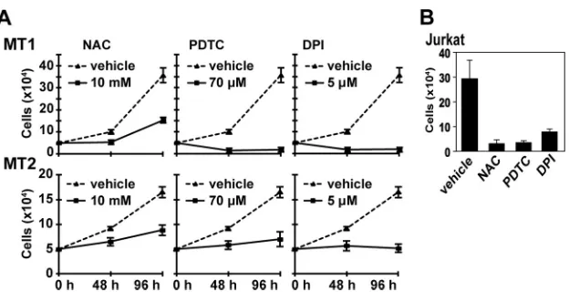
ROS production is required for growth of HTLV-1-infected
cells.
To understand the role of ROS-generating machinery in
HTLV-1-infected T cells, we first examined whether ROS
genera-tion is required for the growth of two HTLV-1-infected T-cell
lines, MT1 and MT2. DPI, a general inhibitor for Nox enzymes
and antioxidants, NAC, and PDTC decreased the growth rate of
cells (
Fig. 1A
). To achieve a similar level of inhibition, much lower
concentrations of these agents were required for uninfected Jurkat
T cells (
Fig. 1B
). This suggests that Nox family genes are involved
in ROS-mediated growth control of MT1 and MT2 cells.
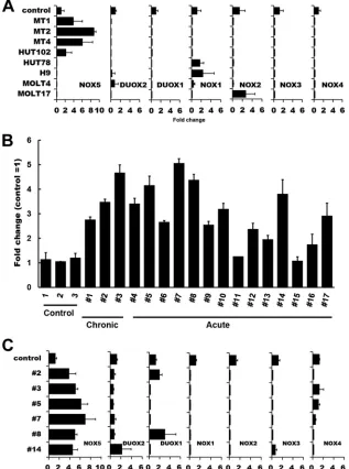
To further explore the nature of the Nox isozymes involved,
the expression of Nox family mRNAs was examined. Real-time
PCR analysis revealed that Nox5 but not other Nox family
mem-bers was expressed in HTLV-1-infected cell lines, namely, MT1,
MT2, MT4, and HUT102 (
Fig. 2A
). In contrast, the Nox5
tran-scripts were not expressed in HTLV-1-uninfected T-cell lines (
Fig.
2A
) or in normal peripheral blood T cells (
Fig. 2B
). Notably, the
levels of Nox5 mRNAs were elevated in 15 of 17 PBMC samples
freshly isolated from ATL patients compared with those in normal
controls (
Fig. 2B
and
Table 1
). No significant correlation was
found between expression of other Nox isoforms and HTLV-1
infection/ATL pathogenesis (
Fig. 2A
and
C
). Thus, it is most likely
that Nox5 is specifically upregulated in both
HTLV-1-trans-formed cell lines and a subset of primary ATL cells.
The Nox5
␣
variant is expressed in MT1 and MT2 cells.
Pre-vious reports described five Nox5 variants—those (
␣
,

,
␥
, and
␦
)
that differ in the sequence of the N-terminal calcium-binding
do-mains and a spliced short variant (Nox5S) lacking this domain
(
18
,
19
). We therefore determined which of the Nox5 variants
exists in MT1 and MT2 cells. RT-PCR together with DNA
se-quencing showed that a primer set (primers a/b) generated a
433-bp fragment encompassing the region between exon 13 and
exon 17 common to Nox5L and Nox5S, whereas another primer
set (primers c/d) generated a 536-bp fragment which corresponds
to the region between exon 3 and exon 6 of Nox5
␣
but not to that
of Nox5
␥
, with an extra 84-bp insertion (
Fig. 3A
and
B
).
More-over, a primer set (primers d/e) specific to Nox5

and Nox5
␦
generated no DNA fragment, indicating that Nox5

, Nox5
␥
, and
Nox5
␦
were not expressed. The data suggest that Nox5
␣
was
ex-pressed but do not exclude the possibility of the presence of
Nox5S. Immunoblotting with antibodies against the COOH-end
region of Nox5 detected 75-kDa Nox5
␣
but not the 60-kDa
Nox5S short variant in the cell lysates. Together, the data
con-firmed that the Nox5 variant in MT1 and MT2 cells is Nox5
␣
(
Fig. 3C
).
Nox5
␣
siRNAs suppress ROS production in MT1 and MT2
cells.
We next examined whether Nox5
␣
-derived ROS are
re-quired for proliferation of MT1 and MT2 cells. To this end, two
siRNAs for Nox5
␣
were tested for knockdown efficiency.
Immu-noblotting analysis with anti-Nox5
␣
antibodies showed that
load-ing with siNox5
␣
decreased the expression of Nox5
␣
proteins
more efficiently than loading with siNox5
␣
-I in MT2 cells (
Fig.
4A
). We therefore used siNox5
␣
to disrupt endogenous Nox5
␣
in
the subsequent experiment. Real-time PCR and
immunoblot-ting demonstrated that the amounts of both Nox5
␣
mRNAs
and Nox5
␣
proteins were decreased in Nox5
␣
siRNA-trans-fected MT1 and MT2 cells (MT1siNox5 and MT2siNox5 cells)
compared with those in scrambled siRNA-transfected cells
(MT1SC and MT2SC cells) (
Fig. 4B
and
C
), which indicates
FIG 1Effects of antioxidants and DPI on proliferation of HTLV-1-infected MT1 and MT2 cells. (A) MT1 and MT2 cells (5⫻104) were cultured in the presence
or absence of the indicated amounts of NAC, PDTC, and DPI. The cell growth was determined at the indicated time intervals. The data represent means⫾ standard deviations (SD) (n⫽3) of the results from four separate experiments. (B) Jurkat T cells (5⫻104) were cultured in the presence of DPI (0.5M), PDTC
(14M), NAC (2 mM), or dimethyl sulfoxide (DMSO) for 48 h, and the cells were counted. The data represent means⫾SD (n⫽3) of the results from three separate experiments. For the data presented throughout panels A and B, statistical analysis was performed with one-way ANOVA, followed by Dunnett’s multiple-comparisonttest.Pvalue for comparisons of chemical treatment versus vehicle,⬍0.05.
on November 7, 2019 by guest
http://jvi.asm.org/
[image:3.585.138.450.63.223.2]that Nox5
␣
siRNAs eliminate the Nox5
␣
transcript from the
cells. Then, the effects of Nox5
␣
siRNAs on intracellular
pro-duction of hydrogen peroxide were examined by a luminol
assay. Catalase-inhibitable ROS generation was significantly
attenuated in MT1siNox5
␣
cells and MT2siNox5
␣
cells
com-pared with control cells (
Fig. 4D
), suggesting the involvement
of Nox5
␣
in ROS synthesis in HTLV1-infected cells. In control
experiments, Nox5
␣
siRNAs did not suppress ROS production in
uninfected H9 cells (data not shown).
Effects of Nox5
␣
siRNAs on both proliferation and cell
sur-vival of MT1 and MT2 cells.
Since growth of MT1 and MT2 cells
was inhibited by DPI, NAC, and PDTC (
Fig. 1
), we investigated
whether the suppression of Nox5
␣
with Nox5
␣
siRNAs blocks cell
proliferation. Both growth in liquid culture (
Fig. 5A
) and colony
formation (
Fig. 5B
) of MT1siNox5
␣
and MT2siNox5
␣
cells were
significantly reduced compared with those of MT1SC and
MT2SC cells, respectively. Immunoblotting with
anti-phos-pho-ERK antibodies indicated that the activated,
phosphory-lated form of ERK, a key growth signal transducer, was
dimin-ished by knockdown of Nox5
␣
(
Fig. 4C
). Neither cell growth
nor Erk/Akt activity was affected in uninfected H9 cells upon
transfection of Nox5
␣
siRNAs (data not shown). These results
FIG 2Analysis of Nox family expression in HTLV-1-infected T-cell lines and ATL PBMC. (A) Total RNAs were extracted from various HTLV-1-infected (MT1, MT2, MT4, and HUT102) and HTLV-1-uninfected (HUT78, H9, MOLT4, and MOLT17) T cells, and levels of mRNA expression of Nox family members were analyzed by real-time PCR. Control data represent normal T cells. One-way ANOVA was performed to determine differences between HTLV-1-infected and -uninfected cell lines. There was a statistically significant difference only in the Nox5 expression data (P⬍0.05 versus control). (B) The levels of Nox5 mRNA expression in ATL primary cells (Table 1) were examined by real-time PCR. CTL (control), normal PBMC. The data represent means⫾SD (n⫽3) of results from three separate experiments. (C) Comparison of levels of Nox isoform expression in ATL patient samples. A total of 6 samples were randomly selected from 17 ATL patient samples which had been analyzed as described for panel B and subjected to the analysis of Nox isoform expression by real-time PCR. Control, normal PBMC.-Actin was used as an internal control. The data represent means⫾SD (n⫽3) of results from three separate experiments. Note that, among the Nox family members, only the levels of Nox5 were increased in the 6 ATL patient samples examined.
on November 7, 2019 by guest
http://jvi.asm.org/
[image:4.585.134.451.69.495.2]suggest that Nox5
␣
-derived ROS, at least in part, sustain
pro-liferation of MT1 and MT2 cells.
Inhibition of cell proliferation is frequently associated with
in-duction of apoptosis. We therefore explored whether ablation of
Nox5
␣
with Nox5
␣
siRNAs leads to cell death. Annexin V labeling
demonstrated that transfection of Nox5
␣
siRNAs into MT1 and
MT2 cells increased apoptosis induction compared with that of
scrambled controls when the transfected cells were serum starved
(
Fig. 5C
) or treated with a chemotherapeutic agent, adriamycin
(
Fig. 5D
). Furthermore, silencing of the Nox5
␣
activity reduced
phosphorylation of AKT, a major cell survival signal mediator
(
Fig. 4C
). These data suggest that Nox5
␣
endows MT1 and MT2
cells with the cell survival activity and that ablation of Nox5
␣
sensitizes the cells to proapoptotic agents. From the therapeutic
viewpoint, it is noteworthy that inactivation of Nox5
␣
enhances
the efficacy of adriamycin.
Inhibition of Nox5
␣
suppresses cell migration.
We next
ex-amined the effects of Nox5
␣
inhibition on the migratory activity
of MT1 and MT2 cells. A migration assay demonstrated that the
numbers of migrating cells were markedly reduced in MT1siNox5
␣
and MT2siNox5
␣
cells (
Fig. 5E
), implying that Nox5
␣
contributes to
motility of HTLV-1-transformed cells.
Nox5
␣
expression is regulated by the Jak-STAT5 pathway.
It
remains to be determined how the Nox5
␣
activity is regulated in
the HTLV-1-transformed cells. Although the Tax viral
transacti-vator is known to affect numerous cell cycle progression genes
(
28
), it seems unlikely that Tax regulates Nox5
␣
expression
be-cause we failed to detect induction of Nox5
␣
expression following
overexpression of Tax (data not shown). Given that Jak1 and Jak3
and their phosphorylation targets, STAT5A and STAT5B, are
stitutively activated in HTLV-1-transformed cells, thereby
con-trolling cellular proliferation (
12
,
13
), we assumed that the
Jak-STAT5 pathway might be coupled to the Nox5
␣
signaling.
RT-PCR analysis showed that treatment of MT1 and MT2 cells
with a Jak inhibitor, AG490, decreased the level of Nox5
␣
mRNAs
(
Fig. 6A
). Similarly, transfection of STAT5B siRNAs blocked
con-stitutive expression of Nox5
␣
(
Fig. 6C
). Immunoblotting analysis
performed with anti-phospho-STAT5B antibodies confirmed
that elevated tyrosine phosphorylation of STAT5B was blocked by
treatment with AG490 (
Fig. 6E
). Moreover, Nox5
␣
was not
up-regulated in an HTLV-II-infected cell line, Mot, in which the Jak/
STAT pathway is not activated (
29
) (
Fig. 6G
). Thus, constitutive
activation of the Jak-STAT5 axis seems to sustain the expression of
Nox5
␣
in HTLV-1-transformed cells.
The Jak-STAT5 pathway also plays a pivotal mediating role in
the IL-2 signaling involved in the growth and survival of T cells,
where ligation of IL-2 to IL-2 receptor

and
␥
chains results in
activation and recruitment of Jak1 and Jak3 and phosphorylation
and nuclear translocation of STAT5A and STAT5B (
30
). We
therefore addressed the activation of Nox5
␣
upon stimulation of
the IL-2 signaling pathway. IL-2 treatment induced expression of
Nox5
␣
in Jurkat T cells, whereas both addition of AG490 and
transfection of STAT5B siRNAs suppressed induction of Nox5
␣
expression by IL-2 (
Fig. 6B
and
D
). Treatment with IL-2 or
phor-bol myristate acetate/phytohemagglutinin (PMA/PHA), which
involves Jak/STAT5, also induced Nox5
␣
expression in normal
PBMC (
Fig. 6H
). The data implied that the activated Jak-STAT5
pathway targets the Nox5
␣
gene in response to IL-2 stimulation.
Because STAT5 is constitutively phosphorylated and activated by
Bcr-Abl tyrosine kinase oncogene, the product of the t (9:22) (q34:
q11) translocation in chronic myelogenous leukemia (CML) cells
(
31
), the expression level of Nox5
␣
was examined in a CML cell
line, K562. Nox5
␣
expression was spontaneously upregulated,
FIG 3Identification of Nox5␣variants in MT1 and MT2 cells. (A) A sche-matic figure shows the configuration of exons in four Nox5 splice variants (␣,
,␥, and␦) belonging to anL-form and in Nox5S, a short form lacking an N-terminal calmodulin-like Ca2⫹binding domain. The exons were numbered
[image:5.585.40.287.77.275.2]based on GenBank sequences as follows:␣,AF353088;,AF325189;␥, AF353089;␦,AF325190. Letters a to e indicate the positions of primers used for PCR analysis as described for panel B. (B) Levels of mRNA expression of Nox5 variants in MT1 and MT2 cells were analyzed by RT-PCR. Primers a, b, c, d, and e were assigned as shown inFig. 3A. PCR products were analyzed by sequencing. (C) Lysates from MT1, MT2, and H9 cells were prepared and subjected to immunoblotting with antibodies against the COOH-end peptide of Nox5.-Actin was used as a loading control.
TABLE 1Clinical characteristics of ATL patientsa
Patient Disease type
No. of WBC (⫻103/l)
Atypical lymphocytes (%)
1 Chronic 15.5 45
2 Chronic 11.9 41
3 Chronic 15.8 65
4 Acute 7.5 ND
5 Acute 41.8 87
6 Acute 15.7 24
7 Acute 26.8 86
8 Acute 8.3 ND
9 Acute 194.5 ND
10 Acute 17.9 20
11 Acute 21.2 49
12 Acute 30.0 56
13 Acute ND ND
14 Acute ND ND
15 Acute 65.6 66
16 Acute 17.8 54
17 Acute 117.2 57
aWBC, white blood cells; ND, not determined.
on November 7, 2019 by guest
http://jvi.asm.org/
[image:5.585.316.525.365.610.2]and this expression was blocked by an Abl-specific tyrosine kinase
inhibitor, STI-571, and, to a lesser extent, by AG490 (
Fig. 6A
).
STAT5B siRNAs also abrogated Nox5
␣
mRNAs (
Fig. 6C
).
Fur-thermore, increased STAT5B phosphorylation was attenuated
upon treatment with AG490 (
Fig. 6E
) and STI-571 (
Fig. 6F
). The
data suggest that Nox5
␣
expression is also upregulated by
Bcr-Ab1 through phosphorylation of STAT5B in CML cells.
Transcriptional activation of the Nox5
␣
promoter by
STAT5.
To further assess the transcriptional regulation of Nox5
␣
by STAT5, we created a luciferase reporter construct carrying the
⫺
1502 to
⫺
11 (positions from ATG) region of the Nox5
␣
pro-moter, which contains two consensus STAT5 biding sequences (
Fig.
7A
). We found that STAT5B siRNAs markedly reduced the Nox5
␣
promoter activity in both MT1 and MT2 cells (
Fig. 7B
), indicating
that STAT5B induces the activation of the Nox5
␣
promoter. The
Nox5
␣
promoter activity was enhanced following overexpression of
Bcr-Abl in Jurkat T cells, whereas this enhancement was diminished
by treatment with a STAT5 inhibitor (
Fig. 7C
). Furthermore,
trans-fection of a constitutively active STAT5B-CA mutant (
32
) enhanced
the Nox5
␣
promoter activity (
Fig. 7D
). This suggests that STAT5
mediates Bcr-Abl-induced activation of the Nox5
␣
promoter, but we
do not formally rule out the possibility that STA5B binds to other sites
distinct from the predicted regions.
Nox5
␣
siRNAs suppress tumor formation by
HTLV-1-trans-formed cells.
To address the contribution of Nox5
␣
to
HTLV-1-induced tumorigenesis, we tested whether HTLV-1-transformed
cells become less tumorigenic due to overexpression of Nox5
␣
siRNAs in a mouse xenograft assay. Scrambled siRNA- and Nox5
␣
siRNA-transfected MT2 cell lines were inoculated subcutaneously
into NOG mice. The animals have been used for successful
en-graftment of HTLV-1-transformed human T cells (
33
). While
scrambled siRNA-transfected cells produced a visible tumor
within a 4-week period, expression of Nox5
␣
siRNAs resulted in a
marked decrease in the tumor growth rate (
Fig. 8
). Both a
leuke-mia-expressing T-cell marker, CD4, and an activating marker,
CD25 (IL-2R
␣
), were expressed in the tumor cells (
Fig. 8
). Thus,
Nox5
␣
siRNAs inhibit the tumorigenicity of MT2 cells, suggesting
an important mediating role of Nox5
␣
-derived ROS in
HTLV-1-dependent transformation.
DISCUSSION
HTLV-1 is the causative agent of ATL, a CD4
⫹T-cell-specific
leukemia. The precise mechanism of the neoplastic growth of
HTLV-1-transformed T cells currently remains unclear. In the
present study, we demonstrated that Nox5
␣
expression is
upregu-lated in HTLV-1-transformed cell lines. Augmented Nox5
␣
-gen-erated ROS are required for proliferation, migration, and survival
of HTLV-1-transformed cells, with accompanying ERK and AKT
phosphorylation cascades. Furthermore, ROS generation by
Nox5
␣
contributes to formation of tumors derived from
HTLV-1-transformed cells in NOG mice. To our knowledge, this is the
first evidence that Nox5
␣
-derived ROS play a critical biological
role in maintenance of the HTLV-1 transformation phenotype.
Identification and characterization of a putative sensor for
Nox5
␣
-generated ROS, which would be crucial in understanding
the Nox5
␣
action, have to await future investigation.
Another major finding is that the Nox5
␣
promoter contains
STAT5 transcription factor binding sequences and that inhibition
of the STAT5 activity by either a Jak inhibitor or STAT5B siRNAs
suppresses Nox5
␣
mRNA synthesis as well as Nox5
␣
promoter
FIG 4Nox5␣siRNA reduces both phosphorylation of Erk and AKT and ROS production. (A) Lysates were prepared from MT2 cells transfected with scram-bled siRNA (SC) or a Nox5-specific siRNA (siNox5␣or siNox5␣-I) and were subjected to immunoblotting with anti-Nox5 or anti--actin antibodies. (B) MT1 and MT2 cell lines stably transfected with Nox5␣siRNA (MT1siNox5␣ and MT2siNox5␣) or scrambled siRNA (MT1SC and MT2SC) were estab-lished. Expression levels of Nox5␣mRNAs were examined by real-time PCR using GAPDH (glyceraldehyde-3-phosphate dehydrogenase) as an internal control. The data represent means⫾SD (n⫽3) of results from three separate experiments. Student’sttest was performed. (C) Expression levels of endoge-nous Nox5␣proteins in the indicated cell lines were determined by immuno-blotting with anti-Nox5 antibodies.-Actin was used as a loading control. Alternatively, phosphorylation levels of Erk and AKT were examined by im-munoblotting with anti-phospho-Erk and anti-phospho-AKT antibodies. (D) Levels of intracellular ROS in MT1siNox5␣, MT1SC, MT2siNox5␣, and MT2SC cells were measured by luminol assay in the presence or absence of catalase (250 U/ml). The data represent means⫾SD (n⫽4) of results from three separate experiments. Statistical analysis was performed with ANOVA, followed by the Bonferroni test.
on November 7, 2019 by guest
http://jvi.asm.org/
[image:6.585.60.269.64.524.2]activity in MT1 and MT2 cells. HTLV-1-transformed cells exhibit
constitutive tyrosine phosphorylation of Jak3 and STAT5 (
12
,
13
),
and lymphocytes isolated from HTLV-1-infected patients display
tyrosine phosphorylation of Jak3, STAT3, and STAT5 (
34
,
35
).
STAT proteins are activated in many human cancers and serve as
oncoproteins by promoting cell proliferation (
36
). These
obser-vations suggest that sustained expression of Nox5
␣
is induced as a
consequence of constitutive activation of the STAT5
transcrip-tional activity by Jak in HTLV-1-transformed cells, contributing
to their growth. Meanwhile, the Jak-STAT5 pathway serves as the
major IL-2 downstream signaling pathway controlling
T-lympho-cyte function, including cell proliferation (
10
). Our data indicate
FIG 5Inhibition of Nox5 suppresses proliferation, cell survival, and migra-tion of MT1 and MT2 cells. (A) Growth rates of indicated cell lines in liquid culture. Cells (5⫻103) were plated, and the numbers of live cells were
deter-mined 48 h later. The cell numbers of MT1SC and MT2SC at 48 h were 1.5⫻ 104and 1.0⫻104, respectively. The data represent means⫾SD (n⫽3) of
results from three separate experiments. Student’sttest was performed. (B) Cells (5⫻103) were inoculated into 6-well plates in 1 ml of MethoCult
(H4230; Stemcell Technologies, Tukwila, WA) containing 10% FBS. Colonies were counted 10 days later. The data represent means⫾SD (n⫽3) of results from three separate experiments. Student’sttest was performed. (C and D) MT1SC, MT1siNox5␣, MT2SC, and MT2siNox5␣cells were serum (FBS) starved for 48 h (C) or treated with 4M adriamycin (Ad) for 48 h (D) using the indicated combinations and subjected to an annexin V-apoptosis assay. The data represent means⫾SD (n⫽3) of results from four separate experi-ments. One-way ANOVA was performed, followed by the Bonferroni test. (E) Cells were plated and subjected to a cell migration assay as described in Mate-rials and Methods. The data represent means⫾SD (n⫽4) of results from three separate experiments. Student’sttest was performed.
FIG 6Nox5␣expression is blocked by inhibition of Jak, BCR-Ab1, and STAT5. (A) MT1, MT2, and K562 cells were treated with 50M AG490 (Jak inhibitor) or 250 nM STI-571 (BCR-Ab1 inhibitor) for 24 h, and levels of Nox5␣expression were examined by RT-PCR. (B) Jurkat T cells were treated with IL-2 (100 ng/ml) in the presence or absence of 50M AG490 for 24 h, and Nox5␣expression was examined by RT-PCR. (C) MT1, MT2, and K562 cells were transfected with STAT5B siRNA or scrambled siRNA, and the levels of expression of Nox5␣, STAT5A, and STAT5B were examined by RT-PCR. (D) Jurkat T cells were transfected with STAT5B siRNAs or with scrambled siRNA and stimulated with IL-2 (100 ng/ml) for 24 h or left unstimulated. The levels of expression of Nox5␣, STAT5A, and STAT5B were examined by RT-PCR. In the experiments whose results are shown throughout panels A to D,-actin was used as an internal control. (E and F) MT1, MT2, and K562 cells were treated with 50M AG490 for 24 h (E); alternatively, HEK293 cells were transfected with Bcr-Abl or control vector and treated with 250 nM STI-571 for 24 h (F). Lysates were subjected to immunoblotting with anti-phospho-STAT5 (Tyr694) and anti-STAT5 antibodies. (G) Nox5␣is not upregulated in HTLV-II-infected T cells. The levels of Nox5␣expression in Mot (HTLV-II-infected T cell line), MT1, and MT2 cells were examined by RT-PCR. (H) Nox5␣expression is induced in IL-2- or PHA/PMA-stimu-lated PBMC. PBMC were treated with IL-2 (100 ng/ml) or PHA (100 ng/ ml)/PMA (5g/ml) for 24 h and subjected to RT-PCR analysis of Nox5␣ expression.-Actin was used as an internal control in the experiments whose results are shown in panels G and H.
on November 7, 2019 by guest
http://jvi.asm.org/
[image:7.585.28.547.64.548.2] [image:7.585.38.295.68.552.2]that IL-2 transiently induced expression of Nox5
␣
in a
Jak-STAT5-dependent manner in non-HTLV1-transformed T cells,
which contrasts with the result of constitutive Nox5
␣
expression
in IL-2-independent MT1 and MT2 cells. Thus, we speculate that
Nox5
␣
functions as an integral component of
Jak-STAT5-medi-ated IL-2 signaling under normal circumstances and that HTLV-1
activates Nox5
␣
by overriding the constitutive Jak-STAT5
signal-ing pathway. Although the modulation by the Tax HTLV-1
trans-activator of the expression of various genes in
HTLV-1-trans-formed cells is well documented (
7
), Tax does not seem to regulate
Nox5
␣
expression, because overexpression of exogenous Tax
failed to induce Nox5
␣
(data not shown). p12
I, the gene product
of the HTLV-1 pX region, has been suggested to be an activator of
STAT5 (
37
). However, overexpression of p12
Iin Jurkat T cells did
not activate the Nox5
␣
promoter activity (data not shown), which
makes it unlikely that p12
Icontrols the transcription of Nox5
␣
.
Further study is required for identification of a HTLV-1 protein(s)
responsible for triggering the Jak-STAT5-Nox5
␣
axis.
Our study also showed that Nox5
␣
was expressed in 88% of 17
ATL patients’ PBMC samples, whereas no Nox5
␣
was detected in
that of the health counterparts. This high incidence indicates that
Nox5
␣
is expressed not only in HTLV-1-infected cell lines but also
in primary ATL cells. The finding suggests some correlation of
Nox5
␣
expression with ATL development, although it is
obvi-ously necessary to study many more cases.
Examining the involvement of Nox5
␣
in other hematopoietic
neoplasms, we found that Nox5
␣
is also upregulated in
Bcr-Ab1-positive CML cells but not Bcr-Abl-negative HL-60 cells (data not
shown) and that Nox5
␣
expression is induced by the Bcr-Ab1
tyrosine kinase oncogene through phosphorylation of STAT5.
Thus, Nox5
␣
participates in the bioactivity of CML as well as that
of HTLV-1-transformed T cells, indicating its wide-ranging roles
in hematologic malignancy. Nox5 was also suggested to be
in-volved in hairy cell leukemia, a chronic B-cell malignancy (
38
).
ROS have been implicated in the regulation of some biological
activities in HTLV-1-infected cells. For example, expression of
ATL-derived factor (ADF), a homologue of thioredoxin (TRX), is
enhanced in HTLV-1-infected cells, and ADF appears to be
in-volved in their autocrine growth (
39
,
40
). ADF seems to transmit
signals by regulating ASK1 via a redox reaction. Since the
interac-tion of TRX-ASK1 is not affected by Nox isozyme-derived ROS
(
41
), it is unlikely that Nox5
␣
participates in the ADF/ASK1
path-way. p13, a HTLV-1-encoded protein located in mitochondria,
also increases ROS generation and thereby induces the death of
HTLV-1-infected cells, contributing to the turnover between
nor-mal and HTLV-1-transformed cells (
42
). However,
Nox-depen-dent ROS generation is not affected by the mitochondrial oxidase
inhibitor rotenone and appears to be functionally different from
mitochondrial ROS production (
14
). Thus, the current study has
significance in the sense that it revealed the alternative impact of
ROS signaling with respect to the biology of HTLV-1 infection.
In conclusion, our findings add a new dimension to the study
FIG 7Activation of Nox5␣promoter activity through STAT5. (A) Schematic structure of the 5=-flanking region of the Nox5␣promoter. The consensus STAT5-binding sites (TTCCCTTAA) are shown in the Nox5␣promoter re-gion (⫺1502 to⫺11 from ATG in exon 3) subcloned into pGL3basic. (B) MT1 and MT2 cells were transfected with pGL3-Nox5␣(⫺1502 to⫺11) together with STAT5B siRNAs or control siRNAs. Lysates were subjected to a reporter assay. The data represent means⫾SD (n⫽3) of results from three separate experiments. Student’sttest was performed. (C) Jurkat T cells were transfected with pGL3-Nox5␣(⫺1502 to⫺11) together with pGD210-Bcr-Ab1 or control vectors and treated with STAT5 inhibitor (250 nM) for 24 h. Lysates were subjected to a reporter assay. The data represent means⫾SD (n⫽3) of results from three separate experiments. One-way ANOVA was performed, followed by the Bonferroni test. (D) Jurkat T cells were cotransfected with pGL3-Nox5␣ (⫺1502 to⫺11) together with the STAT5B-CA mutant or control vector and subjected to a reporter assay. The data represent means⫾SD (n⫽3) of results from three separate experiments. Statistical analysis was performed with Stu-dent’sttest.
FIG 8Tumor formation by Nox5␣siRNA-transfected and control siRNA-transfected cell lines. MT2sc and MT2siNox5␣cells were inoculated into NOG mice, and the growth rate of tumor was monitored by measuring tumor volumes over a 4-week period. The data represent means⫾SD (n⫽6). Student’sttest was performed.P⬎0.05 (versus scrambled data at 4 to 14 days;P⬍0.05 (versus scrambled data at 17 to 28 days). Tumor samples were immunostained with anti-CD4 and anti-CD25 antibodies and counterstained with hematoxylin-eosin (HE).
on November 7, 2019 by guest
http://jvi.asm.org/
[image:8.585.61.265.63.310.2] [image:8.585.76.509.589.685.2]of HTLV-1 neoplastic transformation by highlighting the tyrosine
kinase-based regulation of Nox5
␣
redox signaling and its pivotal
role in the control of proliferation, survival, and motility of
HTLV-1-transformed cells. Because deficiency of Nox5
␣
-depen-dent ROS generation effectively suppresses the transformation
phenotype of HTLV-1-infected cells, our discovery points to an
intriguing possibility that Nox5
␣
could be a potential molecular
target for therapeutic intervention in cancer development.
ACKNOWLEDGMENTS
We declare that we have no conflicts of interest.
We thank T. Tauchi and K. Ikuta for providing Bcr-Abl and STAT5B-CA expression vectors, respectively, Y. Kojima and T. Takeshita for cell lines, K. Sano for technical assistance, and J. Nakayama for valu-able discussion. We are grateful to F. Ushiyama for assistance in manu-script preparation.
Financial support for this work was provided by Grants-in-Aid for Scientific Research for Japan Society for Promotion of Science (22300328 [to T.K.] and 21591357 [to M.S.]) and a Grant on Cancer Research in Applied Areas from the Ministry of Science and Culture of Japan (18012019 [to T.K.]).
REFERENCES
1.Hinuma Y, Nagata K, Hanaoka M, Nakai M, Matsumoto T, Kinoshita KI, Shirakawa S, Miyoshi I.1981. Adult T-cell leukemia: antigen in an ATL cell line and detection of antibodies to the antigen in human sera. Proc Natl Acad Sci U S A78:6476 – 6480.http://dx.doi.org/10.1073/pnas .78.10.6476.
2.Poiesz BJ, Ruscetti FW, Gazdar AF, Bunn PA, Minna JD, Gallo RC.
1980. Detection and isolation of type C retrovirus particles from fresh and cultured lymphocytes of a patient with cutaneous T-cell lymphoma. Proc Natl Acad Sci U S A77:7415–7419.http://dx.doi.org/10.1073/pnas.77.12 .7415.
3.Miyoshi I, Kubonishi I, Yoshimoto S, Akagi T, Ohtsuki Y, Shiraishi Y, Nagata K, Hinuma Y.1981. Type C virus particles in a cord T-cell line derived by co-cultivating normal human cord leukocytes and human leu-kaemic T cells. Nature294:770 –771.http://dx.doi.org/10.1038/294770a0. 4.Popovic M, Lange-Wantzin G, Sarin PS, Mann D, Gallo RC. 1983. Transformation of human umbilical cord blood T cells by human T-cell leukemia/lymphoma virus. Proc Natl Acad Sci U S A80:5402–5406.http: //dx.doi.org/10.1073/pnas.80.17.5402.
5.Grassmann R, Berchtold S, Radant I, Alt M, Fleckenstein B, Sodroski JG, Haseltine WA, Ramstedt U.1992. Role of human T-cell leukemia virus type 1 X region proteins in immortalization of primary human lym-phocytes in culture. J Virol66:4570 – 4575.
6.Yoshida M.2001. Multiple viral strategies of HTLV-1 for dysregulation of cell growth control. Annu Rev Immunol19:475– 496.http://dx.doi.org/10 .1146/annurev.immunol.19.1.475.
7.Hall WW, Fujii M. 2005. Deregulation of cell-signaling pathways in HTLV-1 infection. Oncogene24:5965–5975.http://dx.doi.org/10.1038/sj .onc.1208975.
8.Mori N, Fujii M, Ikeda S, Yamada Y, Tomonaga M, Ballard DW, Yamamoto N.1999. Constitutive activation of NF-kappaB in primary adult T-cell leukemia cells. Blood93:2360 –2368.
9.Sun SC, Yamaoka S. 2005. Activation of NF-kappaB by HTLV-I and implications for cell transformation. Oncogene24:5952–5964.http://dx .doi.org/10.1038/sj.onc.1208969.
10. Leonard WJ, O’Shea JJ.1998. Jaks and STATs: biological implications. Annu Rev Immunol16:293–322.http://dx.doi.org/10.1146/annurev.immunol.16.1 .293.
11. Sun SC, Maggirwar SB, Harhaj EW, Uhlik M.1999. Binding of c-Rel to STAT5 target sequences in HTLV-I-transformed T cells. Oncogene18:
1401–1409.http://dx.doi.org/10.1038/sj.onc.1202430.
12. Migone TS, Lin JX, Cereseto A, Mulloy JC, O’Shea JJ, Franchini G, Leonard WJ.1995. Constitutively activated Jak-STAT pathway in T cells transformed with HTLV-I. Science269:79 – 81.http://dx.doi.org/10.1126 /science.7604283.
13. Xu X, Kang SH, Heidenreich O, Okerholm M, O’Shea JJ, Nerenberg MI.1995. Constitutive activation of different Jak tyrosine kinases in
hu-man T cell leukemia virus type 1 (HTLV-1) tax protein or virus-transformed cells. J Clin Invest96:1548 –1555.http://dx.doi.org/10.1172 /JCI118193.
14. Lambeth JD.2007. Nox enzymes, ROS, and chronic disease: an example of antagonistic pleiotropy. Free Radic Biol Med43:332–347.http://dx.doi .org/10.1016/j.freeradbiomed.2007.03.027.
15. Bedard K, Krause KH. 2007. The NOX family of ROS-generating NADPH oxidases: physiology and pathophysiology. Physiol Rev87:245– 313.http://dx.doi.org/10.1152/physrev.00044.2005.
16. Kamata T.2009. Roles of Nox1 and other Nox isoforms in cancer devel-opment. Cancer Sci 100:1382–1388. http://dx.doi.org/10.1111/j.1349 -7006.2009.01207.x.
17. Block K, Gorin Y.2012. Aiding and abetting roles of NOX oxidases in cellular transformation. Nat Rev Cancer12:627– 637.http://dx.doi.org/10 .1038/nrc3339.
18. Bánfi B, Molnar G, Maturana A, Steger K, Hegedus B, Demaurex N, Krause KH.2001. A Ca(2⫹)-activated NADPH oxidase in testis, spleen, and lymph nodes. J Biol Chem276:37594 –37601.http://dx.doi.org/10 .1074/jbc.M103034200.
19. Cheng G, Cao Z, Xu X, van Meir EG, Lambeth JD.2001. Homologs of gp91phox: cloning and tissue expression of Nox3, Nox4, and Nox5. Gene
269:131–140.http://dx.doi.org/10.1016/S0378-1119(01)00449-8. 20. Fu X, Beer DG, Behar J, Wands J, Lambeth D, Cao W. 2006.
cAMP-response element-binding protein mediates acid-induced NA-DPH oxidase NOX5-S expression in Barrett esophageal adenocarci-noma cells. J Biol Chem281:20368 –20382.http://dx.doi.org/10.1074 /jbc.M603353200.
21. Sugamura K, Fujii M, Kannagi M, Sakitani M, Takeuchi M, Hinuma Y.
1984. Cell surface phenotypes and expression of viral antigens of various human cell lines carrying human T-cell leukemia virus. Int J Cancer34:
221–228.http://dx.doi.org/10.1002/ijc.2910340213.
22. BelAiba RS, Djordjevic T, Petry A, Diemer K, Bonello S, Banfi B, Hess J, Pogrebniak A, Bickel C, Gorlach A.2007. NOX5 variants are func-tionally active in endothelial cells. Free Radic Biol Med42:446 – 459.http: //dx.doi.org/10.1016/j.freeradbiomed.2006.10.054.
23. Mitsushita J, Lambeth JD, Kamata T.2004. The superoxide-generating oxidase Nox1 is functionally required for Ras oncogene transformation. Cancer Res64:3580 –3585.http://dx.doi.org/10.1158/0008-5472.CAN-03 -3909.
24. Komatsu D, Kato M, Nakayama J, Miyagawa S, Kamata T. 2008. NADPH oxidase 1 plays a critical mediating role in oncogenic Ras-induced vascular endothelial growth factor expression. Oncogene27:
4724 – 4732.http://dx.doi.org/10.1038/onc.2008.102.
25. Dewan MZ, Terashima K, Taruishi M, Hasegawa H, Ito M, Tanaka Y, Mori N, Sata T, Koyanagi Y, Maeda M, Kubuki Y, Okayama A, Fujii M, Yamamoto N.2003. Rapid tumor formation of human T-cell leukemia virus type 1-infected cell lines in novel NOD-SCID/gammac(null) mice: suppression by an inhibitor against NF-kappaB. J Virol77:5286 –5294. http://dx.doi.org/10.1128/JVI.77.9.5286-5294.2003.
26. Janik P, Briand P, Hartmann NR. 1975. The effect of estrone-progesterone treatment on cell proliferation kinetics of hormone-dependent GR mouse mammary tumors. Cancer Res35:3698 –3704. 27. Shi SR, Shi Y, Taylor CR.2011. Antigen retrieval
immunohistochemis-try: review and future prospects in research and diagnosis over two de-cades. J Histochem Cytochem 59:13–32.http://dx.doi.org/10.1369/jhc .2010.957191.
28. Sun SC, Ballard DW.1999. Persistent activation of NF-kappaB by the tax transforming protein of HTLV-1: hijacking cellular IkappaB kinases. On-cogene18:6948 – 6958.http://dx.doi.org/10.1038/sj.onc.1203220. 29. Mulloy JC, Migone TS, Ross TM, Ton N, Green PL, Leonard WJ,
Franchini G.1998. Human and simian T-cell leukemia viruses type 2 (HTLV-2 and STLV-2(pan-p)) transform T cells independently of Jak/ STAT activation. J Virol72:4408 – 4412.
30. Lin JX, Migone TS, Tsang M, Friedmann M, Weatherbee JA, Zhou L, Yamauchi A, Bloom ET, Mietz J, John S, Leonard WJ.1995. The role of shared receptor motifs and common Stat proteins in the generation of cytokine pleiotropy and redundancy by IL-2, IL-4, IL-7, IL-13, and IL-15. Immunity2:331–339.http://dx.doi.org/10.1016/1074-7613(95)90141-8. 31. Shuai K, Halpern J, ten Hoeve J, Rao X, Sawyers CL.1996. Constitutive
activation of STAT5 by the BCR-ABL oncogene in chronic myelogenous leukemia. Oncogene13:247–254.
32. Burchill MA, Goetz CA, Prlic M, O’Neil JJ, Harmon IR, Bensinger SJ, Turka LA, Brennan P, Jameson SC, Farrar MA.2003. Distinct effects of
on November 7, 2019 by guest
http://jvi.asm.org/
STAT5 activation on CD4⫹and CD8⫹T cell homeostasis: development of CD4⫹CD25⫹regulatory T cells versus CD8⫹memory T cells. J Im-munol171:5853–5864.http://dx.doi.org/10.4049/jimmunol.171.11.5853. 33. Ito M, Hiramatsu H, Kobayashi K, Suzue K, Kawahata M, Hioki K, Ueyama Y, Koyanagi Y, Sugamura K, Tsuji K, Heike T, Nakahata T.
2002. NOD/SCID/gamma (c) (null) mouse: an excellent recipient mouse model for engraftment of human cells. Blood100:3175–3182.http://dx .doi.org/10.1182/blood-2001-12-0207.
34. Takemoto S, Mulloy JC, Cereseto A, Migone TS, Patel BK, Matsuoka M, Yamaguchi K, Takatsuki K, Kamihira S, White JD, Leonard WJ, Waldmann T, Franchini G.1997. Proliferation of adult T cell leukemia/ lymphoma cells is associated with the constitutive activation of JAK/STAT proteins. Proc Natl Acad Sci U S A94:13897–13902.http://dx.doi.org/10 .1073/pnas.94.25.13897.
35. Tomita M, Kawakami H, Uchihara JN, Okudaira T, Masuda M, Mat-suda T, Tanaka Y, Ohshiro K, Mori N.2006. Inhibition of constitutively active Jak-Stat pathway suppresses cell growth of human T-cell leukemia virus type 1-infected T-cell lines and primary adult T-cell leukemia cells. Retrovirology3:22.http://dx.doi.org/10.1186/1742-4690-3-22. 36. Bowman T, Garcia R, Turkson J, Jove R.2000. STATs in oncogenesis.
Oncogene19:2474 –2488.http://dx.doi.org/10.1038/sj.onc.1203527. 37. Nicot C, Mulloy JC, Ferrari MG, Johnson JM, Fu K, Fukumoto R,
Trovato R, Fullen J, Leonard WJ, Franchini G.2001. HTLV-1 p12(I) protein enhances STAT5 activation and decreases the interleukin-2 re-quirement for proliferation of primary human peripheral blood mononu-clear cells. Blood98:823– 829.http://dx.doi.org/10.1182/blood.V98.3.823.
38. Kamiguti AS, Serrander L, Lin K, Harris RJ, Cawley JC, Allsup DJ, Slupsky JR, Krause KH, Zuzel M.2005. Expression and activity of NOX5 in the circulating malignant B cells of hairy cell leukemia. J Immunol
175:8424 – 8430.http://dx.doi.org/10.4049/jimmunol.175.12.8424. 39. Tagaya Y, Maeda Y, Mitsui A, Kondo N, Matsui H, Hamuro J,
Brown N, Arai K, Yokota T, Wakasugi H.1989. ATL-derived factor (ADF), an IL-2 receptor/Tac inducer homologous to thioredoxin; pos-sible involvement of dithiol-reduction in the IL-2 receptor induction. EMBO J8:757–764.
40. Wakasugi N, Tagaya Y, Wakasugi H, Mitsui A, Maeda M, Yodoi J, Tursz T.1990. Adult T-cell leukemia-derived factor/thioredoxin, pro-duced by both human T-lymphotropic virus type I- and Epstein-Barr virus-transformed lymphocytes, acts as an autocrine growth factor and synergizes with interleukin 1 and interleukin 2. Proc Natl Acad Sci U S A
87:8282– 8286.http://dx.doi.org/10.1073/pnas.87.21.8282.
41. Mochizuki T, Furuta S, Mitsushita J, Shang WH, Ito M, Yokoo Y, Yamaura M, Ishizone S, Nakayama J, Konagai A, Hirose K, Kiyosawa K, Kamata T.2006. Inhibition of NADPH oxidase 4 activates apoptosis via the AKT/apoptosis signal-regulating kinase 1 pathway in pancreatic cancer PANC-1 cells. Oncogene25:3699 –3707.http://dx.doi.org/10.1038 /sj.onc.1209406.
42. Silic-Benussi M, Cavallari I, Vajente N, Vidali S, Chieco-Bianchi L, Di Lisa F, Saggioro D, D’Agostino DM, Ciminale V.2010. Redox regulation of T-cell turnover by the p13 protein of human T-cell leukemia virus type 1: distinct effects in primary versus transformed cells. Blood116:54 – 62. http://dx.doi.org/10.1182/blood-2009-07-235861.
on November 7, 2019 by guest
http://jvi.asm.org/