0022-538X/89/020555-09$02.00/0
Copyright C)1989,American Society for Microbiology
Activation of
Human
Papillomavirus
Type
18
Gene Expression by
Herpes
Simplex Virus Type
1Viral Transactivators
and
a
Phorbol Ester
DAVID GIUS1AND LAIMONIS A. LAIMINS12.3*
Committee on Virology,1 Department of Molecular Genetics and Cell Biology,2 and Howard Hughes MedicalInstitute,3 The University of Chicago, Chicago, Illinois 60637
Received 4 August1988/Accepted 26 October 1988
Several viral trans-activators and a tumor promoter were examined for the ability to activate human
papillomavirustype18(HPV-18)geneexpression. A plasmid containing the HPV-18 noncoding region placed upstream of the chloramphenicol acetyltransferase reporter gene was cotransfected with different herpes
simplex virus type 1 (HSV-1) genes into several cell lines. Both HSV-1 TIF and ICPO activated HPV-18
expression; however,activationbyTIFwasobservedonlyinepithelial cells,whileICPO stimulatedexpression inawide varietyof cells. The element activatedbybothTIFandICPOwasmappedtoa229-base-pair fragment
which also contains an HPV-18 epithelial cell-preferred enhancer. The inclusion of a papillomavirus E2
trans-activator withTIF and ICPO further increased HPV-18 expression. In contrast, the HSV-1 ICP4 and ICP27genes, aswellasthe human T-cell lymphotropic virustypeI andhumanimmunodeficiency virustype 1tatgenes, werefoundtohavenoeffectonHPV-18 expression. In transientassays,theaddition ofthetumor promoter 12-0-tetradecanoylphorbol-13-acetate (TPA) also activated HPV-18 expression. The region of
HPV-18activatedbyTPAwaslocalizedtoasequencewhichishomologoustootherTPA-responsive elements.
Papillomaviruses are a group of small double-stranded
DNA viruses thatinduce benign epithelial lesions. Asmall
group of papillomaviruses is, however, involved in the
etiology of malignant lesions (8, 13, 33). Human papilloma-virus (HPV) types 16, 18, and 31 have been found inover 80% of cervical tumors, suggesting that these types play a causative role (13, 27, 36, 48). In malignant lesions, HPV DNA is integrated into the host genome, while in benign lesions the virusexistsas amulticopy episome (37, 38). HPV
integration often interrupts theEl orE2openreading frame
(ORF),withresultantdisruptionof E2expression (37, 39).In cervicaltumorcelllines,mostof thetranscripts arederived
from the E6-E7 region (4, 38), which contains a major
transformingfunction (6, 23,26).
Papillomaviruses contain multiple elements that act to
regulate transcription during the viral life cycle. The
papil-lomavirus E2 gene product is a strong trans-activator of bovine papillomavirus and human papillomavirus gene
expression (11, 18, 41).The E2proteinactivatesexpression by binding to the sequence ACCG(N)4CGGT, which is present four times in HPV-18 and six times in bovine papillomavirus type 1 (2, 10). Inthe absence of E2
expres-sion, HPV-18transcription iscontrolled by otherelements, including a constitutive enhancer (Fig. 1) (11, 15, 42, 43).
The constitutive element stimulatesgene expressionin sev-eral cell lines but shows apreference for cells ofepithelial
origin (15, 42).
Infectionby HPV-16and HPV-18maynotbe sufficient for
development of cervicalcancer,andasecondeventmaybe
required (45). Early studies have shown an association between thepresenceof both HPV andherpes simplexvirus
(HSV) genomes in cervical tumor cells (25, 34). These observations led us to examine whether the HSV TIF
* Correspondingauthor.
(trans-inducingfactor), ICPO, ICP4, orICP27gene canalter the expression of HPV-18 (5, 14, 32). These HSV genes activate theexpression of both HSV(30)andnon-HSV(32)
promoters. In this report, we show that the HSV TIF late
(gamma) gene can activate expression ofthe HPV-18
non-coding region in epithelial cell lines, such as HeLa and
SCC-13. In addition, the ICPO immediate-early gene is
capable ofactivatingHPV-18expression inavariety of cell
lines, including HeLa, SCC-13, CV-1,and NIH 3T3.
Activation ofHPV-18 expression is not limited to HSV
viral cofactors and isalso sensitive to chemicalagents. We demonstrate that the HPV-18 promoter is activated by 12-0-tetradecanoylphorbol-13-acetate (TPA)in transient as-saysperformedinHepG2cells. The DNAsequence respon-sible for TPA induction waslocalizedtoa30-base-pair (bp)
fragmentof the HPV-18noncoding region,which containsa
TPA-responsive element (TRE). Furthermore, addition of TPA to a transformed NIH 3T3 cell line that expresses
integrated copies of the HPV-18 E6-E7 region from the HPV-18promoterresulted inanincrease inexpression,with acorrespondingchange in themorphologically transformed state of the cells.
MATERIALS ANDMETHODS
Cell culture. African green monkey kidney CV-1 cells,
18:8 cells (6), NIH 3T3 cells, and HeLa cells were main-tained in Dulbecco modified Eagle medium supplemented
with10%fetal calfserum(FCS)and 40,ugofgentamicinper ml. SCC-13 cells, a human squamous carcinoma cell line obtained from Elaine Fuchs, weremaintained in E medium
(47).
Plasmids and DNA preparation. Plasmids pA10CAT2, pTKCAT, and pSV2CAT are enhancer tester and control vectors(3, 16),while pCAT3misa promotertesterplasmid
thatcontains the cat gene butno promoter sequences (16).
555
on November 10, 2019 by guest
http://jvi.asm.org/
El
E2A . I NCR rE7 El l
BR RRBS
IE6
ATGACTAAG
c TRE
a . am
B R RTIC R TAT
GCTTGCATAACTATATCC
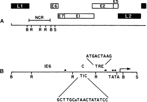
FIG. 1. (A) Genomic organization of HPV-18, w
region designated byopenboxes and thelateregion (
solid boxes. The genomic organization is according Danos (10). Restriction sites (B, BamHI; R, RsaI; indicated. NCR, Noncoding region. (B) Expanded HPV-18 noncodingregion, representedbyaBamHI-S
Thelocationsofthe putative E2-bindingsitesareindic
darkenedrectangles. ThepreviouslyidentifiedE6
enh;
(IE6) and theconstitutive enhancer element (C) arei(
The E6-E7 promoterTATA box is shown, andan ar
the transcription initiation site, which has beenmapl
(38, 42, 44). The sequence GCTTGCaTAACTATAT(
the HPV-18 sequence with homology to the HS-cis-actingDNA sequence involved in TIF-mediatedi]
The sequence ATGACTTAAG represents the HPV
which contains7ofthe 8 bp ofthe TRE consensuss
Plasmids p18CATa, pl8IE6a, and pl8Ca have
ously described (15). Briefly, these plasmids entire HPV-18 noncoding region (p18CATa) or
fragments of this region(pl8IE6aand pl8Ca)
pla
BgII site of pA1OCAT2 (see Fig. 2). p18RB
structed by placingthe 242-bp RsaI-BamHI fraj
noncoding region into the BglII site of pAlOCA
p18CAT1221 consists ofa 1,221-bp BamHI-Sp
containing the entire HPV-18noncoding region the BglII site of pCAT3m (see Fig. 2). p18
constructed by synthesizing two complementa
30-bp oligonucleotides (GATCTATAATATGI GTGCATACA) which were annealed and pla4
BamHI site of pTKCAT. pTKIE6 and pTKC structed byplacingthe 388-bpIE6fragment an
C fragment into the BamHI site ofpTKCAT,
(see Fig. 5). TheHSVgeneexpression plasmid
from Bernard Roizman and have been descrit
elsewhere (22). Briefly, pRB3522 expresses the
geneORFcontainedinaSall F'-to-SalIJ'fragn
a 216-bp BglII-SacI fragment of the human mel
Ila promoter in pUC9. pRB420 expresses the
ORF from its natural promoter contained wi
kilobase SacI-HpaI fragment cloned into the
pUC18. pRB419expressesthe HSV-1ICP27ger
its natural promoter and is contained within a
BamHI-SacI fragment cloned into pUC13. p
presses the HSV-1(F) ICP4 gene
ORF
frompromoterand contains the entire ICP4geneOR]
bp upstream of the transcription initiation sit
downstream of its polyadenylation sequence
pUC18. Plasmidpc59 containsbovinepapilloma
5 (BPV-1) E2 cDNA under control of the simian virus 40 ] *
(SV40)
early promoter and was agift from P. Howley(41).
Thehuman immunodeficiency virustype1 and human T-cell lymphotropic virus type I tat expression vectors (pSVETA
and pHTLVtat-1,respectively) were obtained fromRichard
Gaynor (9, 35) and Rudy Pozzatti (28),
respectively.
pl8PEpolyA
wasconstructed byplacinga3.5-kilobasePstI-EcoRI
fragment of HPV-18 into thePstI-EcoRI
sites ofpML2 (6).
Transfections and CAT assays. Transfections were by
calcium phosphate-mediated precipitation with a total of30
p,g
ofDNAaddedto106cellsper100-mm-diameter dish.ForoA
B S cotransfectionexperiments,
10,ug
ofchloramphenicol
ace-tyltransferase (CAT) testerDNAwastransfected with20
,ug
ofcarrier DNA (pUC9 orITSVx DNA, which gave identicalresults) or 20 p.g of a trans-acting gene expression vector. Cells were incubated with precipitates overnight and
har-vith
the early vested 24 h later. Cell extracts wereprepared,
and CATdesignated by assays were
performed
aspreviously
described(16).
Theto Cole and amount of
acetylation
was determinedby
counting
theS,
SphI)
are acetylated and nonacetylated forms separated byascendingview of the thin-layer chromatography.
,phI
fragment. Transient assays involving TPA (Sigma Chemical Co.) (3):ated bysmall were performed following calcium phosphate-mediated
pre-ancerelement
cdentified
(14).cipitation
with 4pLg
ofDNA added toHepG2
cells cultured-rowindicates in
0.5%
FCS for4h,
followedby glycerol
shock. After 36h,
ped
by others TPAwasaddedto the cellsataconcentration of240ng/ml,
XC
represents and cells were harvested 5 h later. CAT assays wereper-V-1 TIC, the formedin
duplicate
andrepeated
at least three times. nduction (24). Addition of TPA to NIH 3T3 and 18:8 cells. NIH 3T3 and r-18 sequence 18:8 cells were plated at a concentration of106
cells per equence (3). 100-mm-diameter plate in Dulbecco modified Eagle medium supplemented with 10% FCS and 40,ug
of gentamicin perml. The cells were allowed to attach, and 24 h laterfreshmedium been previ- was added. Control cells were cultured without TPA while contain the two plates each of NIH 3T3 and 18:8 cells received 160 or smaller sub- 240 ng of TPA per ml.iced into the The 18:8 cells were plated at a concentration of
106
cells,S
was con- per 100-mm-diameter plate and incubated with or without gmentofthe 240,ug
of TPA per ml for 16 h. Total cellular RNA wasLT2. Plasmid isolated by the guanidium isothiocyanate method (6), and 20 )hI fragment
,ug
was analyzed by Northern (RNA) blots.cloned into
IETPA was RESULTS
ry synthetic
kCTAAGCT
trans-Activation of the HPV-18 promoter by HSV gene ced into the products. Transient assays were performed with plasmids ' were con- containing the CAT gene as a reporter gene to determine d the 229-bp whether any HSV-1 gene products are capable ofactivating respectively HPV-18 expression (16). In these assays, activation of the s were a gift HPV-18 promoter, contained within a 1,221-bpBamHI-SphI
)ed in detail fragment
(pl8CAT1221),
was used as an indicator of gene HSV-1 TIF expression (Fig. 2). The transcription initiation site for thenent
fused to HPV-18 E6-E7 promoter has been mapped to the region near tallothionein the ATG of the E6ORF
(44). p18CAT1221 wascotrans-ICPO gene fected into CV-1 cells with either carrier DNA or the HSV-1
ithin a 4.35- trans-acting genes
TIF,
ICPO, ICP4, andICP27.
In theSacl site of presence of carrier DNA, the CAT activity induced by
le
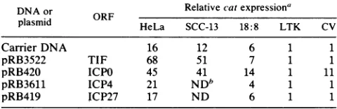
ORF from p18CAT1221 was identical to that seen with pCAT3m (Table 2.4-kilobase 1), a promoterless CAT plasmid (Fig. 2). However, when)RB3611 ex- p18CAT1221was cotransfected with
pRB420
(expressing the its natural ICPO gene), a 10-fold increase in expression was observed. F from -330 No activation ofp18CAT1221 was observed with pRB3522 e to 130 bp (expressing theTIF
gene), pRB3611 (expressing the ICP4 s cloned in gene), or pRB419 (expressing theICP27
gene) (Table 1). ivirus type 1 These experiments were also performed in HeLacells,
a Bon November 10, 2019 by guest
http://jvi.asm.org/
[image:2.612.65.305.77.244.2]IE6 C RBS
B R R R B S
-1037-836 -448 -219 +23 184
CATGene pCAT3M -1037 HPV-18Promoter r-o- 184
--. ---i p18CAT122
SV40 Promoter
X pA1OCAT2
+23
D -j p1 8CATa
-836 -448
-_J p181E6a
-448_--219-- - -I | I p18Ca
-219 +23
_ p18RBS
FIG. 2. Schematic diagram illustrating the constructs used to localize the TIF- and ICPO-responsive sequences in the HPV-18
noncodingregion. Plasmid pCAT3m contains only thecatgeneand
was used to test for promoter activity. p18CAT1221 contains a
1,221-bp BamHI-SphI fragment cloned into the BglII site of
pCAT3m from +184to-1,037bp relativetothe E6-E7transcription
initiation site. PlasmidpAlOCAT2 contains the SV40 21-bprepeats
driving expression of the cat gene but lacks any enhancer
se-quences. Plasmids p18CATa, pl8IE6a, p18Ca, and p18RBS are
derivativesofthepAlOCAT2construct (14).p18CATacontains the
1,060-bp BamHI fragment from -1,037 to 23 bp upstream ofthe
E6-E7 transcriptioninitiationstartsite.p181E6acontainsthe388-bp
RsaI fragment from -448 to -836 bp. pl8Ca contains the 229-bp
RsaI fragmentfrom -219to -448 bp. p18RBScorntains the242-bp
RsaI-BamHI fragment from +23to -219bp.
cervical tumorcelllinethatexpressesthe HPV-18 E6 andE7
ORFs(38). In thepresenceof carrier DNA, the CATactivity induced by p18CAT1221 was 16-fold higher than that seen
with pCAT3m (Table 1). The activation ofpl8CAT1221 expression inthesecellsisduetoanepithelialcell-perferred
enhancer element (15, 43). However, when p18CAT1221
was cotransfected with pRB3522 (expressing TIF)
expres-sion increased to 68-fold, while the pRB420 (expressing
ICPO) expression increased to 45-fold (Table 1). No activa-tion ofp18CAT1221wasobservedwithpRB3611 (expressing ICP4) or pRB419 (expressingICP27). Todeterminewhether
TABLE 1. trans-Activation of theHPV-18 promoter
(pl8CAT1221)by HSV-1TIF, ICPO,ICP4, andICP27
DNA or Relative catexpressiona
plasmid HeLa SCC-13 18:8 LTK CV
CarrierDNA 16 12 6 1 1
pRB3522 TIE 68 51 7 1 1
pRB420
ICPO 45 41 14 1 11pRB3611 ICP4 21 NDb 4 1 1
pRB419 ICP27 17 ND 6 1 1
aRelativecatexpressionwasnormalized pCAT3mlevels. Assays
performed at 40 h posttransfection. Results represent the average fold activation of at least three separate transfections performed in duplicate.
Results from individual transfections varied by less than 25%. Ten
micro-grams of testerplasmidwastransfected with 20,ugofeithercarrierorHSV DNA.
b ND,Notdetermined.
TIF
activation of HPV-18 is epithelial cell dependent,p18CAT1221 was also tested in SCC-13 cells, a squamous
cell carcinoma cell line lacking HPV-18 sequences. When
p18CAT1221 was cotransfected with carrier DNA into
SCC-13 cells, a 12-fold increase in expression was observed.
When cotransfected with pRB3522 (TIF) and pRB420
(ICPO), increases in expression to 51-fold and 41-fold,
re-spectively, were observed. Activation of HPV-18 expression was similar to that observed in HeLa cells, suggesting that
E6 and E7 do not contribute to HSV trans-activation.
The sameexperimentswererepeated in NIH 3T3 cells and
the NIH 3T3-derived cell line 18:8, which constitutively express the E6-E7 region of HPV-18. The results obtained with these two cell lineswereidentical and indicate that only
ICPO can activate HPV-18 expression in rodent fibroblasts
(Table 1). In addition, these results confirm that the E6 and
E7gene productsdonotact synergistically with HSVgenes toactivate HPV-18expression. Tocomparethe levels ofTIF
and ICPO gene-induced activation on HPV promoters with
that seen with HSV promoters, TIF and ICPO expression
plasmids (pRB3522 and pRB420) were cotransfected with
plasmids containing the ICP4 gene promoter or the tk promoterplacedupstreamof thecatgene(12). The TIFgene wasobserved to increase expression of the ICP4gene9-fold,
while the ICPO gene increased tk expression 4.5-fold. Wealso examined whether other viral genes can alter the
expressionofp18CAT1221. Human immunodeficiency virus type 1 and human T-cell lymphotropic virus type I contain
viral genes which activate the expression of their host promoters(28, 35). The human immunodeficiency virus type
1and human T-cell lymphotropic virustype Itatexpression
plasmids (pSVETA and pHTLVtat-1, respectively) were
cotransfectedwith p18CAT1221 into HeLa and CV-1 cells to
evaluate their effects on HPV-18 expression. No activation
of p18CAT1221 by pSVETA or pHTLVtat-1 was observed
(data not shown).
Localization of the cis-acting DNA activated by the TIFand ICPO genes. The HPV-18 sequences responsive to the TIF
and ICPO genes were localized by using a second series of constructs with heterologous promoters. When the TIF (pRB3522) or ICPO (pRB420) gene was cotransfected with
p18CATa, which contains the entire HPV-18 noncoding region placed upstream of the SV40 early promoter in the
antisense orientation (Fig. 2), levels of activation similar to
that seen with the homologous HPV promoter (13-fold and 16-fold, respectively) were observed. These results suggest that the HSV-responsive sequences in the noncoding region arecapableofactivating heterologous promoters evenwhen
placed in the antisense orientation.
Tofurtherlocalize the responsive sequences in the
HPV-18 noncoding region, we used plasmids which contain sub-fragmentsof thisregion cloned upstreamof pAlOCAT2 (Fig. 2). Twoadjoining RsaI fragments of 388 and 229 bp and an
RsaI-BamHI 242-bp fragment werecloned into the BglII site
ofpAlOCAT2to formpl8IE6a,pl8Ca, andp18RBS, respec-tively (Fig. 2).pl8IE6a contains an element that previous
studies suggest may be trans-activated by the HPV-18 E6
ORF, whilepl8Ca contains an epithelial cell-preferred
en-hancer element (15, 42). Plasmidp18RBS contains the
prox-imal 242 bp oftheHPV-18 E6and E7promoter. These three
recombinant plasmids collectivelycontain theentire HPV-18 noncoding region (Fig. 2).
In cotransfection experiments withpl8IE6a or p18RBS
and the TIE gene expression plasmid (pRB3522), no change
in the level ofexpression was observed in HeLacells (Fig. 3). The sixfold activationpl8IE6aof seen in these cells is
-1037
-01=
on November 10, 2019 by guest
http://jvi.asm.org/
[image:3.612.63.303.591.669.2]El(-)
* ICPO
40-
20-p18CAT1221 p18RBS p18Ca p181E6a
FIG. 3. Activationof HPV-18 expressionby the HSV TIFand ICPOgeneproducts. TransfectionswereperformedinHeLacells,a
human cell line derived fromanadenocarcinoma of thecervix,with
plasmid p18CAT1221, pl8IE6a, pl8Ca, or p18RBS. Cells were
transfected with 10 p.g ofpAlOCAT2, pSV2CAT, orthe HPV-18
expression tester plasmid and either 20 p.g of carrier DNA (-),
HSV-1 TIF,orICPO.The dataforp18RBS,pl8Ca,andpl8IE6aare
presentedaspercentacetylation normalizedto thatofpAlOCAT2. The bar graphrepresentstheHPV-18expressionvectorstransfected withcarrier, HSV-1 TIF, orHSV-1 ICPO DNA. Results represent theaverage fold activationofat least three separate transfections performed in duplicate. The results of individual transfections
varied by less than 25%. Assays were performed as previously
described (15).
due to an E6-responsive element which was previously
localized to this fragment (Fig. 1) (15). Cotransfection of pl8Ca with carrier DNA resulted in a 21-fold level of
activation, which increased to75-fold with pRB3522, local-izing the TIF-responsive sequences to this region (Fig. 3). When the ICPO gene expression plasmid (pRB420) was cotransfected with pl8IE6aorp18RBS,nochangein
expres-sion was observed. However, when it was cotransfected
with pl8Ca, an increase in expression from 21-fold with
carrier DNA to 50-fold with pRB420 was observed. These
experiments wererepeatedin SCC-13, CV-1, and 18:8cells,
and similar results were observed (Table 2). Cotransfection
TABLE 2. trans-Activation of plasmid pl8Ca by HSV-1 TIF andICPO
DNA or Relative catexpression'
plasmid ORF HeLa SCC-13 CV-1 18:8
CarrierDNA 21 20 1 8
pRB3522 TIF 75 50 1 NDb
pRB420 ICPO 50 35 10 17
a Relative cat expression was normalized topAlOCAT2 levels. Assays
were performed at 40 h posttransfection. Results represent the average activation ofat least three separate transfections performed in duplicate. Results from individual transfections varied by less than25%. Ten
micro-gramsoftesterplasmidwastransfectedwith 20 ,ug ofeithercarrier orHSV
DNA.
bND,Notdetermined.
of pA1OCAT2 with pRB3522 or pRB420 was observed to
have no effect on the expression (data not shown). We therefore localized the TIF- and ICPO-responsive elements
tothe229-bpRsaI fragmentcontained in pl8Ca.
E2trans-activation in thepresence of HSV geneproducts.
The HPV-18 noncoding region contains a conditional
en-hancerelement that is inducible in trans by the BPV-1 E2 ORF (15). The BPV-1 E2 gene product isatrans-activator equivalent to or better than the HPV-18 E2 gene product (43). To examine whether any additive interaction occurs
between theseviraltrans-acting proteins,aplasmid express-ing the BPV-1 E2 cDNA (pc59) was cotransfected with p18CATa andeither pRB3522 or pRB420 in HeLa cells. E2
exhibitsacomplexpatternofpositiveandnegative effectson
the E6-E7 promoter included in p18CAT1221 (43;
unpub-lished observations). We sought to simplify our study by concentratingonthe activation effects of E2 in the enhancer tester plasmid p18CATa(Fig. 2).
Transfection of p18CATa into HeLa cells resulted in a
13-fold increase in expression above that of pAlOCAT2,
which increased to 64-, 43-, and 86-fold with cotransfection with pRB3522 (TIF), pRB420
(ICPO),
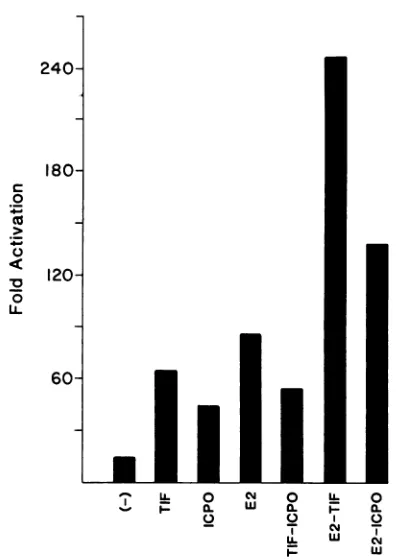
or pc59 (E2),respec-tively (Fig. 4). When p18CATawas cotransfected with pc59
and pRB3522 an increase in expression to 246-fold was obtained, while with pc59 and pRB420 an increase in expres-sion to 138-fold was observed. These results suggest a
possible additive effectbetween the E2 gene product and the HSV TIF and ICPO genes. However, both the TIF (6-fold;
unpublished observation) andICPO (3- to 10-fold; 39a) genes activate the expression of pSV2CAT but not pA1OCAT2 in transient assays. Therefore, since the synthesis of E2 is under control of the SV40 early promoter in pc59, we cannot exclude the possibility that the additive effect of the TIF or ICPO gene and E2 on HPV expression is, at leastin part, an indirect effect. When p18CATa was cotransfected with pRB3522 and pRB420 together, an increase in expression to 54-fold was observed (Fig. 4). This result is similar to the activation by the TIF gene alone, suggesting that no coop-erative effect exists between these two HSV trans-acting genes and HPV-18 expression.
Activation of HPV-18 expression in the presence of TPA. Phorbol esters induce the expression of several cellular and viral genes, including c-myc, BPV-1, and SV40 (1, 17, 19). To determine whether TPA activates HPV-18 expression, transient assays were performed in HepG2 cellstransfected with the HPV-18 promoter plasmid (pl8CAT1221) and treated with TPA. HepG2 cells are very sensitive to TPA induction in transient assays (3). pSV2CAT expression in-creased from 86-fold without TPA to 506-fold with TPA, representing 5.9-fold by TPA induction (Fig. 5) (3, 19).
Without TPA, p18CAT1221 was expressed 2.6-fold above pTKCAT,which increased to8.0-fold with TPA,
represent-ing 3.1-fold induction byTPA.
Tolocalize theTRE, theIE6 and Cregions of the HPV-18
noncoding regionwere placed upstream of the heterologous
pTKCAT plasmid and testedwithTPA. pTKCAT was used
becausepA1OCAT2 contains a TRE and isactivated by TPA
(3). Plasmids pTKIE6a and pTKCa were transfected into
HepG2cells andfound to be unresponsive to TPA induction (Fig. 5). This localizes the TRE to the remaining 403-bp RsaI-SphI region. Examination of this region revealed a sequence which contains 7 of the 8 bp of the TRE consensus sequence located 95 bp upstream of the E6-E7 promoter TATA box (TGACTAAG) (Fig. 1) (3, 10). To examine whether this sequence is responsive to TPA, a 30-bp oligo-nucleotide containing the HPV-18 TRE was cloned into the
on November 10, 2019 by guest
http://jvi.asm.org/
18(
a
0
120-
60-U. 0 N 0 U- 0
I-. 0. W a. P- a. U. W
,l
- - Cq
I- Wt
FIG. 4. Effect ofE2andTIF orICPOonactivationofHPV-18. Transfectionswereperformed in HeLa cells withp18CATa, which containsthe HPV-18noncoding region withina1.1-kilobase BamHI fragment placed in theBglII site ofpAlOCAT2 in the antisense orientation. Cells weretransfected with (i) 10 ,ugofp18CATaand carrierDNA(20,ug) orcarrierDNA (10 ,ug)withTIF,ICPO,or E2 (10,ug) or(ii)E2(10pg)with plasmids expressing either TIF or ICPO (10 ,ug).
HPV-18 TPAExpression Vectors
BamHI site of pTKCAT to create pTK-TPA (Fig. 5). When pTK-TPA wastransfected into HepG2 cells, an increase in expression of 3.4-fold above that seen with pTKCAT was
observed, which in the presenceofTPAincreased to
14.4-fold, demonstrating4.2-fold TPA induction (Fig. 5). We have designated this element IETPA, for a TPA-inducible ele-ment. We cannot exclude the possibility that additional TREs which lack sequence homology to previously identi-fied TREs are presentin the 403-bp RsaI-SphI fragment.
TPA-induced effects on cells that stably express HPV-18.
While NIH 3T3 cells thatexpress pl8PEpolyA(18:8 cells, e.g.) grow in soft agar and form tumors in nude mice, they exhibit minimal morphological transformation (6) and are unable to formfoci in tissue culture. In contrast, when the corresponding region of HPV-16 is expressed from a strong heterologouspromoter, morphological transformation is ob-served (26). We sought to investigate whether a TPA-induced increase in HPV-18 expression could produce sim-ilar resultsin 18:8 cells. TPA (240 ng/ml) was added to NIH 3T3 and 18:8 cells, and the cells were monitored for 5 days. Thecells were maintained in 10% FCS, in contrast to the
0.5% FCS usedin TPA trans-activation experiments, since
18:8 cellscultured inlow serum with TPA become rounded anddetach after36 h and NIH 3T3 cellsgrowpoorly in low serum. NIH 3T3 cellswithorwithoutTPAdisplayedsimilar morphologies and did not form foci (Fig. 6). When TPA was added to 18:8cells, theyappeared to beveryrefractile and foci were apparent after 2 days, while at lower concentra-tions foci appearedafter 4 days (Fig. 6).
To determine whether TPA increased expression ofthe
HPV-18E6-E7region, 18:8 cells were examined by North-ernanalysiswith or without TPA. Cells were incubated ata
concentrationof 240 ,ug of TPA per ml and were harvestedat
16 h. A roughlyfourfold increase in E6-E7 expression was
Relative CATConversion
Sequence -TPA +TPA Induction
-1037 -836 -448-219 23184 CAT
I I ,
-J ATTAGTCAG 86 506 5.9
J ATGACTAAG 2.6 8.0 3.1
pTK-TPA
pTKCa
-172 -146
-448 -219 __ _s
ATGACTAAG 3.4 14.4 4.2
1l
-836 -448
pTKIE6a ' --
[image:5.612.82.280.69.349.2]-ATGACTCAG
FIG. 5. Constructs usedtotestfor TPAinduction of HPV-18 and relativeamountof TPA induction. PlasmidpfKCAT (3) containsthe
HSVthymidine kinasegenepromoterplacedupstreamofcat,whilepSV2CAT (15)contains theSV40promoterand the72-bptandemrepeats placedupstreamofcat.p18CAT1221containsa1,221-bp BamHI-SphI fragmentfrom+184to-1,037 bpupstream of the E6-E7transcription
startsitecloned upstreamofcat. pTK-TPAcontainsa30-bpsynthetic oligomer containingthe HPV-18TREplacedinto theBamHIsiteof
pTKCAT. pTKIE6a andpTKCa contain the 388- and 229-bp RsaI fragments placed into the BamHI site ofpTKCAT. The underlined sequencesrepresent the promotersequencesofSV40andHPV-18 thatarehomologousto the8-bpconsensusTRE sequence from thehuman
metallothionein Ila promoter (3). Cells were transfected with 4 Fgoftester DNA, andassays were performed as previously described.
Relativecat expression wasnormalized tothe level ofpTKCAT. Results representthe average fold activation of at least three separate transfectionsperformed induplicate.Results from individual transfections variedbyless than 25%.
pTKCAT pSV2CAT
CAT
p18CAT1221
Ion November 10, 2019 by guest
http://jvi.asm.org/
[image:5.612.128.492.464.643.2]560 GIUS AND LAIMINS
FIG. 6. MorphologyofNIH3T3cells(BandD)and 18:8 cells, whichexpresstheHPV-18 E6-E7region(A andC),after 5days of induction with240 ngof TPA per ml (A and B)andwithout TPA(Cand D).Magnification, x135.
observed in 18:8 cells incubated with TPA (Fig. 7). These
results suggestthat TPA increases the expression ofstably integrated copiesof HPV-18,resultingin increased
morpho-logical transformation of 18:8 cells.
A
B
C
28S-
18S-*: .:
_
.. _
*: F
...
FIG. 7. Northern analysis of HPV-18 expression in 18:8 cells incubatedwithout(lane A)orwith (lane B) 240 jLgof TPApermlfor
16handNIH 3T3cells (lane C). A 20-jig sample of total cellular RNAwas run on aformaldehyde-agarose gel, blottedto nitrocellu-lose, andprobed witha1,578-bp HpaII fragment from the HPV-18
E6-E7region. The positions of 18S and 28S rRNAsareindicated.
DISCUSSION
HPVinfection alone is probably not sufficient for
induc-tion of cervical
carcinogenesis,
anda secondevent may berequired. As an example, epidemiological studies have shown that HPV-16-orHPV-18-infectedwomenwhosmoke
have a 5- to 10-fold-higher rate ofcervical cancer than do
infectedwomenwho donotsmoke(45).Thepossible role of
a second event has also been shown experimentally in HPV-16- and HPV-18-mediated transformation of primary rodent cells. While HPV-16 and HPV-18 sequences are
capable ofimmortalizing rodent cells, they were unable to
fully transformthese cells(6, 23, 26). Instead, HPV-16 and HPV-18 require the presence of a cooperating oncogene, such as ras, for complete cellular transformation (26; M.
Bedell, personal
communication).
Inthe present study, weexamined whether other viral orchemical factors can
acti-vate HPV-18 gene expression and may act as cooperating
agents intransformation.
The ability of one virus to augment the expression of another virus or rescueit fromalatentstate haspreviously
beenshown. Adenovirus and HSV canactivatethe expres-sionofthehumanimmunodeficiencyvirus type 1and human T-cell lymphotropic virus type I long terminal repeats,
respectively(9,31). While HSVinfection ofgenitalepithelia probablydoesnotresult inmalignancy,studies suggest that it mayact as acofactor. This issupported by the presenceof
both HSV and HPV DNA sequences inasmall percentageof cervicaltumorcells(25, 34). Cervical cellsarepermissiveto
both HSV andpapillomavirus infections, and thepossibility
J. VIROL.
on November 10, 2019 by guest
http://jvi.asm.org/
[image:6.612.76.535.75.380.2] [image:6.612.135.226.503.668.2]exists that both virusescanbetranscriptionally active in the
samecell(21, 34, 48). HSVgene productshave been shown to code for trans-acting factors which activate the
immedi-ate-early genesof HSV(5) and non-HSV promoters(14, 32)
andincludeTIF,astructural component ofthevirion, ICPO, ICP4, and ICP27 (22, 31). In thisstudy, weconcentrated on HSV-1 viral trans-activators, but we suspect that similar
results would be observed with HSV-2. This similarity is
supported by studiesindicatingthat ICPOs from HSV-1 and HSV-2 are functionally interchangeable during productive viral infectionand actby identical mechanisms (40).
In our studies, ICPO and TIF were found to activate HPV-18 expression. Activation by TIF appeared to be cell type specific and limited to epithelial cell lines, HeLa and SCC-13. Theresponsiveelements werelocalized toa229-bp RsaI fragmentof the HPV-18 noncoding region, which also contains an epithelial cell-preferred enhancer (15, 42).
Ex-amination of this fragment revealed an 18-bp region (GCT-TGCaTAACTATATCC)with ahigh degreeofhomologyto the HSV-1 consensus sequence (TIC; trans-induction cis
site)that is activatedbyTIF(Fig. 1) (22).Smallvariationson the TIF consensus sequencehave been foundin
nonherpes-virusgenes which are responsiveto TIF (32). The celltype specificity displayed by TIF is surprising, since TIF acti-vates HSV promoters in a variety of cell types, including
fibroblasts(22). This suggests that for HPV-18
trans-activa-tion, TIF may interact with transcriptional regulatory pro-teins that are present only in epithelial cells. Activation of
HPV expression by TIF and ICPO was observed when the
HPV-responsive elements were located upstream of their
homologous promoter as well as when they were placed in the antisense orientationupstreamoftheheterologousSV40 early promoter. The ability of TIF and ICPO to activate
expression in these latter constructs suggests that these
elements act in a manner similar to that of other mobile
inducible elements.
Activation ofHPV-18expression byICPOwasobservedin
epithelial (HeLa and SCC-13) and nonepithelial (CV-1 and NIH 3T3) cells. Interestingly, the region of HPV-18 acti-vatedbyICPO is thesame229-bp fragmentactivatedbyTIF. No cis-acting DNA sequence has been identified for ICPO, andprevious studiessuggestedthat ICPOactedas a nonspe-cific trans-acting gene (32). Since TIF and ICPO activate HPV-18 expressionin different celltypes, their mechanisms
of trans-activation are probably different. When TIF and ICPO werecotransfected simultaneously with p18CATa, no additive effect on expression was observed. This suggests
that either one HSV-encoded factor has priority over the other or while TIF and ICPO may function by different
mechanisms, theymayactuponthesame saturated site.We are attempting to separate the HSV TIF- and
ICPO-respon-sive sequences fromoneanotherto determine whetherthey
aredistinct elements. Incontrast, when either TIFor ICPO wascotransfected withE2, thereappearedto be anadditive
effecton HPV-18 expression.
Phorbol esters act topotentiatethe effects ofcarcinogens
at subtumorigenic concentrations (46). TPA activates the
transcriptionof several viral and cellulargenesby a mech-anisminvolvingthe activation ofprotein kinaseC (3, 7, 19,
29, 49). When TPA was added to HepG2 cells transfected
withp18CAT1221, anincrease inexpressionof 3.1-foldwas observed above that of cells transfected in the absence of TPA.TPA inductionexperimentswereperformedinHepG2
cells because they are extremely sensitive to TPA trans-activationin transient assays (19, 20). The HPV-18 respon-sive element was localized to a 30-bp region (designated
IETPA)
which contains a sequence similar topreviously
identified TPA-inducibleelements
(3,
24).
Thelevel of TPA induction of HPV-18 isslightly
less than that observed withother
promoters
that contain TRE sequences and may be duetoa1-bp
mismatchatposition
six oftheconsensusTRE(3).
Addition ofTPAtoNIH 3T3cells
expressing
the HPV-18E6-E7
region
from theirhomologous
promoter
(18:8
cells)
resulted in an increase in
steady-state
RNA levels and achange
intheir cellularappearance.When TPAwasaddedto18:8cells
they
appeared
morerefractile and formed foci after2
days,
while atlower concentrations fociappeared
after4days.
Untreated 18:8 cells and TPA-treatedNIH 3T3 cellsappeared
tobe similartoNIH3T3 cells. Similarchanges
inmorphology
wereobservedwhen HPV-16geneswereover-expressed
fromastrong
long
terminalrepeat promoter
(26).
We cannot,
however,
excludethepossibility
that TPA alsoactivates additionalcellulargenesthatmay
contribute
tothis alteredphenotype
(46).
Inpreliminary
experiments,
cotrans-fection of ICPOand
pl8PEpolyA
intoNIH 3T3cellsresulted in ahigh frequency
of focusformation,
suggesting
thatdifferent viral genomes also
cooperate
in the induction ofmorphological
transformation(Gius,
unpublished
data).
Sev-eral formsof chemical and viralcofactorsmaythusinfluence theHPV-induced transformationstateby
altering
papilloma-virus
expression.
Malignant
transformation is amultistep
process whichmay result from a wide
variety
of cellularabnormalities,
carcinogenic
agents,
orviral infections. Each is(or
perhaps
many
are)
required
to induce a normal cell to becomeneoplastic.
The events thattrigger
the transition from abenign
to amalignant
HPV-induced lesion remainpoorly
defined and may involve various forms of
transcriptional
activation. An increase in HPV-18 geneexpression
may be the second event necessary to induce amorphological
change
fromanormal celltype
toatransformedphenotype.
In this
study,
we demonstrated thattwo HSV genes and aphorbol
ester can stimulate HPV-18expression
and thusmay
play
a role in thedevelopment
of HPV-18-inducedmalignancies.
ACKNOWLEDGMENTS
This workwas
supported by
the HowardHughes
MedicalInsti-tute. D.G.was
supported by
Public HealthServicegrantGM 07183from the National Institutes of Health.
WethankBernard Roizman for the HSV-1
expression
vectorsand forhelpful
discussions; and Ira Blitz, Steven Grossman, JeffHubenthal-Voss, Katherine H. Jones, andMary Bedell for assis-tanceandcomments.
LITERATURE CITED
1. Amtmann, E.,andG. Sauer.1982.Activation of
non-expressing
bovine
papillomavirus
genomesby
tumor promoters. Nature(London)
296:675-677.2.
Androphy,
E.J.,D.R. Lowy,andJ. T. Schiller.1987. Bovinepapillomavirus
E2 trans-activatinggeneproduct
binds tospe-cific sites in
papillomavirus
DNA. Nature(London)325:70-73.3. Angel, P., M. Imagawa, R. Chiu, B.Stein, R.J. Imbra,H. J.
Rahmsdorf,C.Jonat,P.Herrlich,and M. Karin.1987.Phorbol ester-induciblegenescontainacommonciselement
recognized
byaTPA-modulated
trans-acting
factor. Cell 49:729-739.4. Baker,C. C., W. C. Phelps,V. Lindgren, M.J. Braun,M.A.
Gonda, and P. M.Howley. 1987. Structural and
transcriptional
analysis
of humanpapillomavirus
type16 sequencesincervical carcinomacell lines. J. Virol.61:962-971.on November 10, 2019 by guest
http://jvi.asm.org/
5. Batterson, W., and B. Roizman. 1983. Characterization of the herpes simplex virion-associated factor responsible for the induction ofagenes. J. Virol. 46:371-377.
6. Bedell,M. A., K. H. Jones, and L. Laimins. 1987. The E6-E7
region of human papillomavirus type 18 is sufficient for transformation ofNIH 3T3 and Rat-1 cells. J. Virol. 61:3635-3640.
7. Blumberg,P. M.1985.Invitro studiesonthe mode of action of thephorbolesters, potenttumorpromoters. Crit. Rev. Toxicol. 9:153-197.
8. Boshart, M., L. Gissmann, H. Ikenberg, A. Kleniheinz, W. Scheurlen,and H. zurHausen. 1984. Anewtype of papilloma-virusDNA, its presenceingenital cancerbiopsiesand in cell lines derivedfrom cervicalcancer. EMBOJ. 3:1151-1157. 9. Chen, I. S. Y., N. P. Cann, A. Shah, and R. Gaynor. 1985.
Functionalrelationship between HTLV-IIIxand theadenovirus EIAproteinsintranscriptionalactivation.Science 230:570-573. 10. Cole, S. T., and 0. Danos. 1987. Nucleotide sequence and
comparative analysis of the human papillomavirus type 18 genome.Phylogenyofpapillomavirusesand repeatedstructure
of the E6 and E7 geneproducts.J. Mol. Biol.193:599-608. 11. Cripe, T. P., T. H. Haugen, J. P. Turk, F. Tabatabai, P. G.
SchmidIII,M.Durst,L.Gissmann,A.Roman,and L. P. Turek. 1987.Transcriptional regulationofthehumanpapillomavirus-16
E6-E7promoterbyakeratinocyte-dependent enhancer,andby
viral E2 trans-activator and repressor gene products:
implica-tionsfor cervicalcarcinogenesis. EMBOJ.6:599-608. 12. DeLuca, N.A.,and P. A. Schaffer.1985.Activationof
immedi-ate-early, early, and late promoters by temperature-sensitive and wild-type forms of herpes simplex virus type 1 protein
ICP4. Mol. Cell. Biol. 5:1997-2008.
13. Durst, M.,L.Gissmann,H.Ikenberg,and H.zurHausen.1983. A papillomavirus DNA from a cervical carcinoma and its
prevalencein cancerbiopsy samples fromdifferentgeographic regions.Proc. Natl. Acad. Sci. USA80:3212-3215.
14. Everett, R. D., and M. Dunlop. 1984. trans-Activation of plas-mid-borne promoters by adenovirus and several herpesgroup viruses. Nucleic Acids Res. 12:5969-5977.
15. Gius, D.,S. Grossman,M. A.Bedell, and L. A. Laimins. 1988. Inducible and constitutive enhancer domains in thenoncoding regionofhumanpapillomavirus type 18. J. Virol. 62:665-672. 16. Gorman, C.,L.F. Moffat,and B. Howard. 1982. Recombinant
genomes which express chloramphenicol acetyltransferase in mammaliancells. Mol. Cell. Biol.2:1044-1051.
17. Greenberg,M.E.,and E. B. Ziff.1984.Stimulation ofNIH 3T3 cellsinducestranscriptionof the c-fosproto-oncogene. Nature
(London)311:433-438.
18. Hirochika, H.,T.R.Broker,and L. T. Chow. 1987.Enhancers andtrans-actingE2transcriptionalfactorsofpapillomaviruses. J. Virol.61:2599-2606.
19. Imbra, R. J., and M. Karin. 1987. Phorbol esterinduces the
transcriptional stimulatory activity of the SV40 enhancer. Na-ture (London)323:555-559.
20. Imbra,R.J.,and M.Karin.1987. Metallothionein gene expres-sion is regulated by serum factors and activators of protein
kinase C. Mol. Cell. Biol. 7:1358-1363.
21. Kawana, T.,K.Kawagoe,K.Takizawa,J.T.Chen,T.
Kaqagu-chi, andS. Sakamoto. 1982. Clinical andvirologicalstudies on
femalegenital herpes. Obstet. Gynecol.60:456-461.
22. Kristie,T.M.,andB. Roizman. 1988. Differentiationand DNA contact points of host proteins binding at the cis site for virion-mediated induction ofao genesofherpes simplexvirus 1. J. Virol. 62:1145-1157.
23. Laimins,L.A., M. A. Bedell, K. H. Jones, and J. A. Long. 1987. Transformationof NIH3T3 andprimaryratembryo fibroblasts
by humanpapillomavirus type 16 and 18. CancerCells(Cold
Spring Harbor)5:201-207.
24. Lee, W.,P.Mitchell, and R. Tjian. 1987. Purified transcription factor AP-1 interacts with TPA-inducible enhancer elements. Cell 49:741-752.
25. Macnnab, J. C. M., M. B. Walkinshaw, M. B. Cordiner, and J.B. Clements. 1986. Human papillomavirus in clinically and
histologicallynormal tissue ofpatients with genitalcancer. N.
Engl.J. Med. 315:1052-1053.
26. Matlashewski, G., K. Osborn, A. Murray, L. Banks, and L. Crawford. 1987. Transformation of mouse fibroblasts with HPV-16 DNAusingaheterologouspromoter, p. 195-200. Cold SpringHarbor Laboratory, Cold Spring Harbor, N.Y. 27. McCance, D. J. 1986. Human papillomaviruses and cancer.
Biochim. Biophys. Acta 823:195-205.
28. Nerenberg,M.,S. H.Hinrichs,R.Reynolds,G.Khoury,and G. Jay. 1987. The tat gene of HTLV-1 induces mesenchymal
tumorsintransgenicmice. Science 237:1323-1329.
29. Nishizuka,Y.1984. Therole ofproteinkinase C in cell surface signal transduction and tumor promotion. Nature (London) 308:693-698.
30. O'Hare, P., and G. S. Haywood. 1985. Three trans-acting regulatory proteins ofherpes simplex virus modulate immedi-ate-early geneexpressionin apathway involving positiveand negative feedback regulation. J. Virol. 56:723-733.
31. Ostrove,J. M.,and K. I.Berns. 1980. Adenovirusearlyregion lb gene functionrequiredtorescuethe latent adeno-associated virus. Virology 104:502-505.
32. Ostrove, J. M., J. Leonard, K. E. Weck, A. B. Rabson, and H. E. Gendelman. 1987.Activationof the human immunodefi-ciency virus by herpes simplex virus type 1. J. Virol. 61: 3726-3732.
33. Pater, M. M., and A. Pater. 1985. Human papillomavirus type 16 and 18 sequences in carcinoma cell lines of the cervix. Virology 145:313-318.
34. Prakash, S.S., W. C. Peeves, G. Sisson, M. Brenes, J. Godoy, S. Bacchetti, R. C. de Britton, and W. R. Rawls. 1985. Herpes simplexvirus2 and humanpapillomavirus type 16 incervicitis, dysplasia, and invasive cervical carcinoma. Int. J. Cancer 35:51-57.
35. Rosen, C. A., J. G. Sodroski, W. C. Goh, A. I. Dayton, J. Lippke, and W. Haseltine. 1986. Post-translational regulation accounts for the trans-activation of the HTLV-III. Nature (London) 319:555-559.
36. Schneider-Gaedicke, A., and E. Schwarz.1986. Differenthuman cervicalcarcinoma cell lines show similar transcription patterns of human papillomavirus type 18 early genes. EMBO J. 5: 2285-2292.
37. Schneider-Maunoury, S., 0. Croissant, and G. Orth. 1987. Integration of humanpapillomavirus type 16 DNA sequences: a possible early event in the progression of genital tumors. J. Virol. 61:3295-3298.
38. Schwarz, E., U. K. Freese, L. Gissmann, W. Mayer, B. Roggen-buck, A. Stremlau, and H. zur Hausen. 1985. Structure and transcription of human papillomavirus sequences in cervical carcinoma cells. Nature(London) 314:111-114.
39. Schwarz, E., A. Schneider-Gaedicke, and H. zur Hausen. 1987. Human papillomavirus type-18 transcription in cervical carci-nomacell lines and in human cell hybrids. Cancer Cells (Cold Spring Harbor)5:47-53.
39a.Sekulovich, R. E., K. Leary, and R. M. Sandri-Goldin. 1988. The herpes simplex virus type 1 a protein ICP27 can act as a trans-repressor or a trans-activator in combination with ICP4 and ICPO. J. Virol. 62:4510-4522.
40. Smith, C. A., and P. A. Schaffer. 1987. Mutants defective in herpes simplex virus type 2 ICP4: isolation and preliminary characterization. J. Virol. 61:1092-1097.
41. Spalholz, B. A., Y. C. Yang, and P. M. Howley. 1985. trans-Activationofabovinepapillomavirustranscriptional regulatory elementby the E2 geneproduct. Cell 42:183-191.
42. Swift, F. V., K. Bhat, H. B. Younghusband, and H. Hamada. 1987. Characterization of a cell type-specific enhancerfoundin the human papillomavirus type 18 genome. EMBO J. 6:1339-1344.
43. Thierry, F., A. G. Carranca, and M. Yaniv. 1987.Elements that control the transcription of human papillomavirus type 18. CancerCells(ColdSpring Harbor) 5:23-32.
44. Thierry, F., J. M. Heard, K. Dartmann, and M. Yaniv. 1987. Characterization ofatranscriptional promoterofhuman papil-lomavirus 18 and modulationof its expression by simian virus 40 and adenovirusearly antigens. J. Virol. 61:134-142.
on November 10, 2019 by guest
http://jvi.asm.org/
45. Trevathan, E., P. Layde, L. A. Webster, A. B. Adams, B. B. Benignu, and H.Ory. 1984.Cigarette smoking anddysplasia and carcinomain situ of the uterine cervix. J. Am. Med. Assoc. 250:499-502.
46. Weinstein, I. B., L. S. Lee, P. B. Fisher, A. Mufson, and H. Yamasaki. 1979. Actionofphorbolestersincellculture: mim-icry of transformation, altered differentiationandeffectsoncell
membranes. J. Supramol. Struct. 12:195-208.
47. Wu, Y., and W. Rheinwald. 1981. Anewsmall(40 KD)keratin filamentprotein made by somecultured human squamouscell carcinomas.Cell 25:627-636.
48. zurHausen, H. 1985. Genital papillomavirus infections. Prog. Med. Virol. 32:15-21.
49. zurHausen,H., M. T. Nilsson, B. Kallin, and G. Klein. 1987. Persisting oncogenic herpesvirus induction bytumorpromoter.
Nature(London) 272:373-375.