“TO STUDY THE ROLE OF DIFFUSION-WEIGHTED MRI IMAGING IN
PREDICTING THE RESPONSE TO CONCURRENT CHEMORADIOTHERAPY IN
PATIENTS WITH NASOPHARYNGEAL CARCINOMA”
DEPARTMENT OF RADIOTHERAPY CHRISTIAN MEDICAL COLLEGE
VELLORE 632004
DISSERTATION SUBMITTED IN PARTIAL FULFILLMENT OF MD BRANCH RADIOTHERAPY
EXAMINATION MAY 2018
TAMIL NADU DR. M.G.R MEDICAL UNIVERSITY CHENNAI - 600032.
This is to certify that the dissertation titled “TO STUDY THE ROLE OF DIFFUSION-WEIGHTED MRI IMAGING IN PREDICTING THE RESPONSE TO CONCURRENT
CHEMORADIOTHERAPY IN PATIENTS WITH NASOPHARYNGEAL
CARCINOMA”is a bonafide work done by Dr Koti Krishna Amulya, Post Graduate Student in the
Department of Radiotherapy, Christian Medical College, Vellore during the period from April 2016 to March 2018 and is being submitted to The Tamil Nadu Dr M. G. R Medical University in partial fulfilment of the MD Branch Radiotherapy examination conducted in May 2018.
Guide
Dr. Simon P Pavamani Professor
This is to certify that the dissertation entitled “TO STUDY THE ROLE OF
DIFFUSION-WEIGHTED MRI IMAGING IN PREDICTING THE RESPONSE TO
CONCURRENT CHEMORADIOTHERAPY IN PATIENTS WITH
NASOPHARYNGEAL CARCINOMA” is a bonafide work done by Dr. Koti. Krishna Amulya,
Post Graduate Student in the Department of Radiotherapy, Christian Medical College, Vellore during the period from April 2016 to March 2018 and is being submitted to The Tamil Nadu Dr. M. G. R Medical University in partial fulfilment of the MD Branch IX Radiotherapy examination conducted in May 2018
Head of the Department Principal Dr Selvamani B
This is to certify that the dissertation entitled “TO STUDY THE ROLE OF
DIFFUSION-WEIGHTED MRI IMAGING IN PREDICTING THE RESPONSE TO CONCURRENT
CHEMORADIOTHERAPY IN PATIENTS WITH NASOPHARYNGEAL
CARCINOMA” is a bonafide work done by Dr. Koti. Krishna Amulya, Post Graduate Student in
the Department of Radiotherapy, Christian Medical College, Vellore during the period from April 2016 to March 2018 and is being submitted to The Tamil Nadu Dr. M. G. R Medical University in partial fulfilment of the MD Branch IX Radiotherapy examination conducted in May 2018.
Contents
ACKNOWLEDGEMENT ... 12
AIM ... 13
OBJECTIVE ... 13
HYPOTHESIS ... 13
INTRODUCTION ... 14
REVIEW OF LITERATURE ... 17
Epidemiology ... 17
Anatomy of nasopharynx ... 21
Risk Factors ... 22
Clinical presentation ... 25
Diagnostic Evaluation ... 26
Staging of the Disease ... 31
Treatment of Non-Metastatic Nasopharyngeal malignancies ... 33
Role of Intensity Modulated Radiation Therapy (IMRT)... 35
Lacunae in Current Knowledge and Need for Treatment Response Assessment... 36
Role of Functional Imaging Modalities in response assessment ... 37
Rationale for Choosing Diffusion Weighted MRI in our study ... 42
Diffusion Weighted Magnetic Resonance Imaging – Biophysical basis ... 44
Treatment response assessment using Diffusion Weighted MRI ... 46
Diffusion Weighted MR Imaging in Nasopharyngeal carcinoma: A Predictive Biomarker ... 49
MATERIALS AND METHODS... 51
RESULTS ... 57
Social Demographics ... 57
Complete Responders: ... 73
Complete Responder Vs Partial Responder... 79
1. Mean Baseline ADC: ... 79
2. Visual Restriction: ... 80
3. Rate of change in ADC values ... 81
Correlations ... 87
Sensitivity and Specificity ... 89
DISCUSSION ... 91
LIMITATIONS OF THE STUDY ... 96
FUTURE DIRECTIONS ... 97
CONCLUSIONS ... 98
REFERENCES ... 99
ANNEXURES ... 103
Annexure 1 : Patient Information ... 103
Annexure 2: Certificate of Consent ... 105
Annexure 3: Clinical Research Form ... 106
ACKNOWLEDGEMENT
I would like to express my heartful gratitude to all those who have directly or indirectly helped
in conducting this study. To commence with, I thank God, Almighty for bestowing upon me
good health, courage and inspiration.
I express my sincere and deepest gratitude to my guide, Dr Simon Pavamani, for his untiring
efforts, patience, constant support and scientific inputs. His continual inspiration, helped me
in all the time of research and writing of this thesis. It would have not been possible to
complete this study without his encouragement. I am inspired by his zeal and enthusiasm for
research.
I would like to my co-guide Dr Aparna, Professor, Department of Radiology for all the efforts
and time that she had put in for providing me with the necessary data.
I thank my co-guide Dr Rajesh I, Professor, Department of RT for his able guidance and for
having the faith in me.
I thank Dr Selvamani, Professor and Head of the Department, Radiotherapy and all the RT
Consultants for their concern, care and encouragement.
I take the opportunity to thank all the senior residents and fellow Post Graduates special
mentions to Dr Solly for helping me out in carrying out this study.
I am thankful to Dr Visalakshi, Department of Biostatistics, for her assistance during the
statistical analysis of this research work. I am also thankful to Radiology technicians for
accepting my study requests and extend all their co-operation and support to help in carrying
out this study
I wish to thank all my patients for their co-operation in this study.
And finally, last but by no means least, I thank my wonderful husband, my loving mother and
AIM
“To study the role of diffusion weighted MRI imaging in predicting the response to concurrent
chemoradiotherapy in patients with nasopharyngeal carcinoma
OBJECTIVE
Primary Outcome: To study the role of diffusion weighted MRI derived parameters i.e. ADC
as an imaging biomarker in nonmetastatic nasopharyngeal carcinoma.
Secondary Outcome: Validating the role of diffusion weighted in detection and
characterization of nasopharyngeal cancers as well as for monitoring the response to
radiotherapy and chemotherapy.
HYPOTHESIS
Diffusion weighted imaging plays a key role in prediction of treatment response to
chemo-radiotherapy in patients diagnosed with nasopharyngeal malignancies
INTRODUCTION
According to the Globocan fact sheet, worldwide there are approximately 60,896 new
nasopharyngeal cases and a reported of 35,756 deaths annually. In India, there are about 3947
cases being reported annually with nasopharyngeal malignancy.
Carcinomas of Nasopharynx are known to be radio-responsive and are treated using radiation
therapy with or without chemotherapy. The standard of care for the treatment of patients with
non-metastatic nasopharyngeal cancer is radiotherapy with or without chemotherapy. Based
on the stage, they receive radiation alone or concurrent chemoradiotherapy followed by
adjuvant chemotherapy. Some patients receive chemotherapy prior to Radiation to decrease
the size of the tumor or lymph nodes especially if they lie close to critical structures in the head
& neck region e.g. optic apparatus brainstem & the spinal cord. However, nearly in 7%–13%
of cases residual disease persists after treatment. Moreover, the appearance of local or distant
relapse determines a less favorable prognosis for these patients. Salvage treatment options are
with limited success. Thus, it is essential to be able to predict and monitor the therapeutic
response of patients with Carcinoma Nasopharynx. Hence attempts are being made to predict
response to radiation therapy with various functional and metabolic imaging.
Anatomic imaging (CT and MR imaging) is focused on diagnosing and/or characterizing the
disease, defining its local extent, and evaluating distant spread. While the accurate assessment
of the biologic status of the cancer (cellularity, growth rate, response to chemoradiation
therapy) with functional imaging modalities like dynamic contrast enhancement MRI,
diffusion weighted MRI, FDG PET can be essential for prognostication, planning therapy, and
techniques can thus provide a more comprehensive evaluation of the patient helping in better
understanding of the pathophysiology of the disease and allowing better evaluation of
treatment strategies.
This study is mainly focused on evaluating the role of Diffusion-weighted Magnetic Resonance Imaging (DW MRI) in predicting early response to chemoradiation in nasopharyngeal malignancies. DW MRI explores the random motion of water molecules in the tissue. Water molecules are in a constant random movement called the Brownian motion. The Diffusion
Weighted signal in biologic tissues is derived from the motion of water molecules in the
intracellular space, the extracellular space, and the intravascular space. The degree of
restriction to water diffusion in biologic tissue is inversely correlated to the tissue cellularity
and the integrity of cell membranes. The motion of water molecules is more restricted in tissues
with a high cellular density associated with numerous intact cell membranes (e.g., tumor
tissue). The lipophilic cell membranes act as barriers to motion of water molecules in both the
extracellular and intracellular spaces. By contrast, in areas of low cellularity or where the
cellular membrane has been breached, the motion of water molecules is less restricted. A less
cellular environment provides a larger extracellular space for diffusion of water molecules, and
these molecules may also freely.
Effective anticancer treatment results in tumor lysis, loss of cell membrane integrity, increased
extracellular space, and, therefore, an increase in water diffusion. Impediments such as cell
membranes, organelles, and macromolecules interferes with the free movement of water
molecule, this diffusion in biological tissue is quantified by means of an APPARENT
Malignant tissues have more densely packed cells as compared to their nonmalignant
counterparts. Thus, the “diffusion” of water molecules is restricted due to high cellularity in
malignant tissue. With radiation therapy, there is cell death and increased water movement
across tumor and thus increased diffusion which in turn is expressed in increase in ADC values.
Therefore, as treatment progresses, the ADC values are expected to rise as the tumour cells
respond to the chemoradiation. Thus, the serial monitoring of the ADC values before radiation,
during radiation and at 6 weeks after radiation will help in correlating the ADC values of the
responders and non-responders. We chose the time periods for assessment as
1. Pre-RT: Prior to initiation of radiation therapy
2. Week 1: At 1 week of radiation therapy
3. Follow up: At 4-6 weeks after completion of RT
This response evaluation can be utilized for tailoring treatment for patients in the future.
Therefore, patients showing response utilizing Diffusion weighted imaging may not need
REVIEW OF LITERATURE
Epidemiology
Nasopharyngeal carcinoma(NPC) is a rare malignancy worldwide but is seen in high incidence
in Sothern China, Hongkong and South-east Asia. These areas are termed “High-risk” or
“Endemic” for NPC. According to the Globocan fact sheet, worldwide there are approximately
60,896 new nasopharyngeal cases and a reported of 35,756 deaths annually.
According to the Indian data there are about 3947 cases being reported annually with
[image:17.595.58.541.413.670.2]nasopharyngeal malignancy(1)
Table 2: The INDIAN Incidence, mortality and 5year prevalence of nasopharyngeal cancers – adopted from the Globocan
Though NPC is a rare malignancy in the Indian subcontinent, the North-eastern tribes are
reported to be an intermediate risk population with an average incidence of 4.3 per 100,000
people/year(2). NPC is often misdiagnosed and underreported. Hence the actual incidence can
be higher than reported. Highest incidence in our country is reported from the Imphal district
of Manipur, closely followed Manipur and Sikkim.
ESTIMATED BURDEN – 2020
Anatomy of nasopharynx
The nasopharynx is a tubular space situated below the base of the skull. It is a part of the
Waldeyer ring. It is lined by stratified squamous epithelium and ciliated epithelium. NPC is
commonly developing from the lateral wall of nasopharynx, particularly at the fossa of
Rosenmuller.
Figure 7: Sagittal section of human head showing Nasopharynx (Highlighted in yellow)
BOUNDARIES OF NASOPHARYNX
Anterior Posterior nares and posterior nasal septum
Posterior C1 and C2 vertebral bodies
Retropharyngeal space
Superior Basi-sphenoid and Basi-occiput
Inferior Anteriorly: Soft palate
Posteriorly: Communicates with Oropharynx via Nasopharyngeal
isthmus
NEUROVASCULATURE
Arterial supply Ascending pharyngeal Artery
Facial Artery
Ascending cervical artery
Venous drainage Pterygoid and Pharyngeal Plexus
Lymphatic drainage Lateral lymphatic channels Lateral retropharyngeal, deep
cervical and posterior triangle nodes
Medial lymphatic channels Median retropharyngeal nodes
Nerve innervation Sensory – V & IX cranial nerve branches
Motor – Pharyngeal plexus
Synonyms for NPC: The first description of NPC as “Skull base tumor” dates to 1845. In 1921,
it was termed “lymphoepithelioma” by Regaud and Schmincke.
Risk Factors
The risk factors most frequently associated with nasopharyngeal carcinoma are ethnicity,
smoking and Epstein Barr Virus (EBV) infection.
Ethnicity – Nasopharyngeal carcinoma (NPC) is a very common neoplasm in Southern
Chinese and south-east Asians. China along with southeast Asia is considered an endemic
region for NPC. Southern Chinese who have migrated to non-endemic regions are found to
they continue to have higher incidence rate compared to other ethnic groups residing in the
same geographical regions.(3) Ethnicity shadows genetic susceptibility.
Genetic Predisposition – There has been much debate over genetic predisposition in
nasopharyngeal malignancies. A review of Genome-wide association studies (GWAS) showed
association of immune related genes with NPC. DNA repair genes, Classical and Non-classical
HLA genes are found to have a strong association with NPC in the Chinese (4). The association
in the non-endemic region needs to be proven yet.
Familial aggregation of NPC has also been reported.
Epstein Barr virus infection – EBV infection is a well-known causal factor in NPC. Epstein-
Barr virus (EBV) infection has been linked with the non-keratinising subtypes of
nasopharyngeal cancers. High pre-treatment plasma EBV DNA titres correlate with higher
stage of disease and persistent EBV DNA titres after treatment are associated with poor
prognosis. EBV DNA is now an established biomarker for prognosticating outcomes after
treatment.
Smoking – Smoking tobacco products (cigarettes, cigars, pipes) is an important risk factor for
the development of NPC. It is a major factor in development of NPC in non-endemic areas.
Association of smoking was found with well differentiated histology rather than an
undifferentiated histology. Furthermore, those with > 30 pack-years had double the incidence
of NPC than those with < 30 pack-years. From a recent Metanalysis, the pooled risk estimate
for nasopharyngeal malignancy associated with each 1-pack-year increment of smoking was
Alcohol — Like smoking, alcohol intake was also found to have increased the risk of well
differentiated NPC. The quantitative relationship is however complex. Results from a
Systematic review showed a J shaped pooled dose response curve indicating that risk of NPC
decreases up to 15 drinks/week and thereafter increases with further increase in alcohol intake.
Confounding factors like smoking and salted fish could have led to such a conclusion.(6)
Occupational exposure — Multiple other occupational or environmental toxins have been
studied for potential relationship with NPC. Formaldehyde, wood dust and fumes are known
carcinogens(7).
Radiation — Prior irradiation for either malignant or benign disease has been linked to thyroid
cancer, salivary gland tumors, squamous cell cancers, and sarcomas. Although this
relationship appears to be real, its relevance in NPC is unclear.
Diet — Some dietary factors may have a role in protecting individuals from head and neck
cancer, while others may increase susceptibility for specific diseases. Salted fish and
Clinical presentation
Nasopharyngeal malignancies can spread through local, lymphatic and hematogenous
pathways. Symptoms and its severity varies according to the site and extent of involvement.
Owing to its proximity to base of skull and sinuses, it can often present with cranial nerve
involvement. Common symptoms secondary to local spread are described below as a flow
diagram
Lymphatic spread assumes great significance in NPC as nasopharynx has a rich lymphatic
network. The retropharyngeal group of lymph nodes are believed to be the first echelon nodes
and the risk of involvement of deep cervical nodes unilaterally or bilaterally is the highest in
NPC in comparison to any other head and neck tumor. As high as 70% of patients present with
nodal involvement at presentation and lymph node enlargement is the most common clinical
Distant metastases are not very uncommon in NPC. The common sites of involvement are
bone, liver and lung. Nodal involvement and the site of lymphadenopathy predicts distant
metastatic potential.
Diagnostic Evaluation
NASOPHARYNGOLARYNGOSCOPE (NPL): The flexible fibreoptic
Nasopharyngolaryngoscopy is a very valuable tool that can visualise Nasopharynx and
structures around it
It helps in
- Identifying the site and extent of the disease
- Biopsy and
- Stage the disease.
BIOPSY: The histopathology is the gold standard for diagnosis of NPC. ls called squamous
cells. The WHO classification of NPC began in 1978 and has had multiple revisions since then.
The most recent classification in 2005 divides NPC into three histological subtypes:
1. Keratinizing squamous cell carcinoma
- Well differentiated / Moderately differentiated / Poorly differentiated
2. Non-Keratinizing carcinoma
- Undifferentiated / Differentiated
Non-keratinizing carcinoma is the most common (~90%) variant in endemic areas (high risk
areas). Keratinizing is relatively rare and is seen in high incidence in patients with smoking
and alcohol abuse. Basaloid is the rarest variety (~1%). Association with EBV was found in
Non-keratinizing and Basaloid types.
IMAGING: The anatomy of nasopharynx presents structural and functional variations,
making precise diagnosis and staging a challenging task. NPL scopy will not be able to exactly
stage disease, Example: Intracranial extension which is identified on cross-sectional imaging
may be missed in a routine NPL scopy. There is a greater need for precise staging of the disease
as survival varies with each stage and so is the treatment planning.
Cross-sectional imaging was revolution since its invention in the mid seventies. Anatomical
imaging techniques such as Computerized Tomography (CT) and Magnetic resonance imaging
(MRI) provide morphological information while the evaluation of biological characteristics of
tumors is by the functional imaging modalities.
1. Computed Tomography: CT can identify tumor in the nasopharynx based upon
anatomic distortion and/or specific tumor enhancement. In general, tumors enhance
more than normal structures except for mucosa, extraocular muscles, and blood vessels.
Compared with MRI, CT provides greater spatial resolution, and can be performed
faster, thereby eliminating motion artefact, and it is better for the evaluation of bone
destruction such as skull base erosion seen in T3 disease. Modern multidetector CT
technology allows scanning to be performed with slice thickness less than 1 mm. Slice
thickness of 3 mm is generally optimal.
• Primary site — For primary site, contrast-enhanced CT can help determine the extent
of the para-pharyngeal space, sinuses and skull base invasion. The modern technology
of dual energy and multispectral CT has demonstrated improved accuracy for assessing
bone invasion compared with conventional CT. Asymmetry is a feature suggestive of
malignant process in nasopharynx in the initial stages. However, clinical relevance of
such asymmetry is difficult to interpret on CT because of inflammatory process,
lymphoid tissue and secretions in nasopharynx. Presence of minor asymmetry is often
mistaken for disease and results in false positivity. A modified Valsalva manoeuvre
which opens collapsed lateral pharyngeal recess can be tried during CT. Also,
occasionally CT may fail to depict an early cortical erosion of the skull base that is
better depicted on MRI.
• Regional nodes — Imaging by CT or MRI is complementary to the clinical
examination for the staging of the neck lymph nodes. CT evaluation of regional lymph
nodes primarily relies upon size criteria as well as the appearance of lymph nodes to
differentiate involved from uninvolved lymph nodes. The use of size criteria alone
results in frequent false positive and false negative assessment of regional nodes. CT is
also highly sensitive for detection of extracapsular spread of tumor.
Pathologic lymphadenopathy is usually identified radiologically as a node greater than
10 to 11 mm in minimal axial diameter or presence of central necrosis. In general,
criteria based on measurement of minimal axial diameter are considered the most
accurate, effective and probably most reproducible. Other features pointing towards
lymph nodal involvement by malignancy include rounded shape, loss of normal fatty
hilum, and increased or heterogeneous contrast enhancement. Although CT is superior
to clinical examination, the implementation of size criteria and presence of central
differentiate between reactive and normal nodes. This is a critical issue since
microscopic or occult lymphadenopathy is very common in NPC which is known for
its high lymphatic spread.
1. Magnetic Resonance Imaging(MRI) : MRI provides superior soft tissue definition compared with CT and can often provide information that is complementary to CT.
Moreover, there is no use of non-ionizing radiation with MRI. Most important imaging
sequences for include non-contrast enhanced T2-weighted images and contrast T2 and
T1 sequences. Images in axial and coronal plane are the most useful. For general
purpose, slice thickness should be no more than 5 mm. Some applications, such as
evaluation of skull base and perineural spread, may require thinner slice thickness,
typically around 3 mm.
• Primary site – As the soft tissue is better seen on MRI than CT, it adds to the
information generated from CT. MRI is also better than CT for discriminating tumor
from mucus and in detecting bone marrow invasion. MRI is superior to CT for
evaluation of perineural spread, para-pharyngeal extension, skull base invasion, and
intracranial extension. MRI may also provide additional benefits compared with CT in
the evaluation of the base of tongue involvement and parotid glands.
• Regional nodes - Fat surrounding the nodes decreases their prominence on the MRIs.
Fat-suppressed contrast-enhanced sequences are the optimal sequences for detecting
nodal metastasis. The retropharyngeal nodes, which are the echelon nodes, are easily
detected on MRI.
tissues. PET images are created by detecting these emissions by an array of detectors
and then using reconstruction techniques to create a three-dimensional image. The most
commonly used agent is 18F-flourodeoxyglucose (FDG), which is taken up into cells
in different concentrations depending on the relative metabolism of different tissues. It
is specific for tumors because metabolic rates are very high in many tumors.
Imaging of the primary tumor site and regional lymph nodes with PET is limited by its
poor spatial resolution, which can make it difficult to localize the anatomic location of
the FDG uptake. These issues can be at least partially addressed with integrated
PET-CT imaging, in which PET and PET-CT are performed sequentially during a single visit on
a hybrid PET-CT scanner. The images are then co-registered using fusion software,
enabling the physiologic data obtained on PET to be localized according to the anatomic
CT images.
• Primary site - Historically, CT images obtained from integrated PET-CT scanners had
lower spatial resolution compared with dedicated CT scanners. This problem is now
being overcome by new generation of PET-CT scanners that offer volumetric CT
capability. Thus, currently PET appears to be at least as sensitive and specific as CT
and MRI in detecting primary head and neck tumors.
• Regional nodes - More importantly, PET is superior to both CT and MRI for detecting
regional nodal metastases, as well as second primary tumors(10) Data indicates
that PET/CT is accurate for detecting occult cervical nodal metastases, although it does
not have the sensitivity to replace neck dissection. However false negatives of PET may
be seen in lymph nodes less than 5 mm, necrotic or cystic lymph nodes, and tumors of
• Distant metastases -Its main utility is in finding distant metastases, unknown primary
lesions, and synchronous second primary tumors.
An updated Metanalysis comparing CT Vs MRI Vs PET-CT showed the following results(11)
CT MRI PET CT
Sensitivity Specificity Sensitivity Specificity Sensitivity Specificity
Local site – T staging 0.84 0.80 0.95 0.76 0.85 0.91
Nodal site – N staging
0.92 0.93 0.88 0.95 0.88 0.95
Authors have concluded by stating that MRI was more accurate than CT or PET-CT in local
staging and fairly comparable in nodal staging. PET-CT is preferred for detecting occult neck
nodes, metastatic disease, synchronous second primaries and recurrences.
Staging of the Disease
Currently we are following the 7th edition of AJCC/UICC staging system. However, the 8th
edition which would come into effect from January 2018 has major modifications in NPC
staging. We had followed the 7th edition in our study.
The 7th and 8th editions are both presented below:
7th EDITION AJCC STAGING T
stage
8th EDITION AJCC STAGING
Tumor confined to Nasopharynx or extension to oropharynx / nasal cavity
Extension to:
- Para-pharyngeal space
T2 Extension to:
- Para-pharyngeal space
- Pterygoid muscle (Medial / lateral)
- Prevertebral structure
Invading:
- Skull base
- Paranasal sinuses
T3 Invading:
- Skull base
- Paranasal sinuses - Pterygoid structures - Cervical vertebrae Any invasion into
- Infratemporal fossa
- Masticator space
- Hypopharynx - Orbit
- Intracranial extension - Cranial nerve involvement
T4 Any invasion into: - Parotid gland
- Beyond lateral surface of lateral pterygoid
- Hypopharynx - Orbit
- Intracranial extension - Cranial nerve involvement Changes are highlighted in Bold Italics
Changes are highlighted in Bold Italics
7th EDITION AJCC STAGING Nodal 8th EDITION AJCC STAGING
< 6 cms in greatest dimension, above supraclavicular fossa
- Unilateral metastases, in cervical lymph nodes (or)
- Unilateral / Bilateral metastases in retropharyngeal nodes
N1 < 6 cms in greatest dimension,
above the caudal border of cricoid cartilage
- Unilateral metastases, in cervical lymph nodes (or)
- Unilateral / Bilateral metastases in retropharyngeal nodes
< 6 cms in greatest dimension, above supraclavicular fossa
- Bilateral cervical nodes
N2 < 6 cms in greatest dimension,
above the caudal border of cricoid cartilage
- Bilateral cervical nodes
>6 cms in greatest dimension (and / or) N3 >6 cms in greatest dimension (and / or)
In the supraclavicular Below the caudal border of cricoid
7th EDITION AJCC STAGING STAGE
GROUP
8th EDITION AJCC STAGING
T1 N0 M0 I T1 N0 M0
T1 N1 M0
II
T1 N1 M0
T2 N0/1 M0 T2 N0/1 M0
T1/2 N2 M0
III
T1/2 N2 M0
T3 N1/2 M0 T3 N1/2 M0
T4 N0/1/2 M0
IV A
T4 N0/1/2 M0
Any T N3 M0
Any T N3 M0 IV B Any T Any N M1
Any T Any N M1 IV C No longer exists
Treatment of Non-Metastatic Nasopharyngeal malignancies
EARLY DISEASE (T1 N0 M0): Radiotherapy alone is treatment for early disease. According
to Song et al. use of chemotherapy has not shown additional benefit in stage I NPC(12). In
another study, the 10-year disease specific survival, recurrence free survival and distant
metastasis free survival rates were all over 95% with radiation alone(13). Guidelines by the
European Society for Clinical Oncology -ESMO (14), the Spanish Society of Medical
Oncology -SEOM(15) and National Comprehensive Cancer Network - NCCN(16) suggest RT
alone is an optimal modality of therapy for T1N0M0 NPC.
LOCALLY ADVANCED DISEASE (T1, N1–3; T2–4, Any N, M0): Addition of
chemotherapy both concurrently and as adjuvant was found to survival advantage in locally
(SWOG) showed increased survival with concurrent chemoradiotherapy (CRT) followed by
adjuvant chemotherapy compared to radiotherapy alone in stage III and IV carcinoma
nasopharynx patients(17). The benefit combined therapy in Stage III and IV is undebated.
Stage II NPC has remained an enigma for most clinicians with conflicting evidence on addition
of chemotherapy to radiation. Most studies on CRT excluded stage II NPC. Cheng et al. (18)
showed that disease-free survival in stage II NPC with CRT is equal to that of patients with
stage I disease with radiation alone. While SEOM(15) recommends radiation alone for stage
II, both ESMO(14) and NCCN(16) recommend CRT.
Metanalysis of 2138 stage II NPC patients by Cheng Xu et al. had shown chemoradiation to
have significantly higher overall survival and locoregional relapse free survival compared to
RT alone(19). But there was no difference in distant metastases free survival. Moreover, the
equivalence of RT alone with chemoradiation was seen when IMRT was used in subgroup
analysis. Also grade 2/3 toxicities were lesser in IMRT group compared to chemoradiation
group. This lead the authors to the conclude that chemoradiation was superior to 2D
radiotherapy but the same does not hold true for IMRT in stage II NPC.
ROLE OF INDUCTION / NEOADJUVANT CHEMOTHERAPY:
A
pooled dataanalysis was conducted by Chua et al. of two phase III studies (n=784). It showed that the
addition of platinum-based induction chemotherapy to radiation had caused decrease in relapse
by 14.3%. The cancer-related deaths reduced by 12.9 percent at 5 years. However, there was
no improvement in overall survival (61.9 % Vs. 58.1 %, p=0.092). This could be attributed to
more late intercurrent deaths in both the groups(20)
Other benefit of Induction chemotherapy is downsizing tumor which could be very useful in a
making the dose delivery to tumor a challenge without limiting the organs at risk (OAR) dose.
In our institution, we follow induction/neoadjuvant chemotherapy whenever there is high
tumor bulk or when the lesion is close to critical OARs.
Role of Intensity Modulated Radiation Therapy (IMRT)
Tumor control for carcinoma nasopharynx highly correlates with the tumor dose. In a
retrospective review of 85 patients with NPC, an improved local control was seen when the
tumor received > 67 Gy(21). In another series having 118 patients, the improved tumor control
was not just due to higher doses delivered to tumor, but also due to improvements in technical
accuracy(22). As nasopharynx is surrounded by many critical structures, accuracy in dose
delivery is very important in dose escalation.
Intensity-modulated radiation therapy allows very high dose to be delivered to the tumor while
sparing the surrounding critical structures. These critical structures include the salivary glands,
optic apparatus, brainstem, spinal cord and the auditory apparatus. In addition, IMRT can
deliver a higher dose per fraction which yields a biologically more effective dose to the tumor.
Therefore, IMRT offers a significant advantage over conventional radiotherapy. Nasopharynx
is a site where IMRT can play a key role in improving the therapeutic ratio which can translate
into better tumor control. Several studies have proved that IMRT results in improved survival
in NPC along with reduced long term toxicities(23) (24) (25) (26) (27). A high dose can be
given to the nasopharynx while sparing critical normal structures such as the brainstem, the
spinal cord, the optic chiasm, and salivary glands.
At our institute, non-metastatic nasopharyngeal malignancies are treated with IMRT and we
Lacunae in Current Knowledge and Need for Treatment Response Assessment
Response prediction in Nasopharyngeal cancers is very essential as it helps in:
DETECTING EARLY RELAPSES: Although NPC is very radiosensitive, in nearly 7%–
13% of cases residual disease persists after treatment(28). Moreover, the appearance of local
or distant relapse determines a less favorable prognosis for these patients. Salvage treatment
with radiosurgery or brachytherapy has been shown to give excellent tumor control in residual
disease(29). Salvage surgery was also proved to achieve long-term survival in patients with
local persistent or recurrent NPC who could achieve long-term survival(30). Thus, it is
essential to be able to predict and monitor the therapeutic response of patients with NPC.
After treatment, local findings of clinical examination may be unreliable as nasopharyngeal
site is not completely accessible at clinical examination. The residual tumor may be
sub-mucosal making it unsuitable for biopsy. Moreover, CT imaging may be equivocal or
inconclusive for detection of residual NPC due to the presence of diffuse soft-tissue
inflammation and edema post radiation therapy at primary site which is often reported as
merely an increase or decrease in soft tissue thickening. False-positive findings in CT
regarding local persistence of disease detection are mainly correlated with inflammatory
changes of the nasopharyngeal mucosa and sub mucosa. Nodal persistence also signifies a
poorer prognosis if not detected and appropriately treated with salvage neck dissection. MR
imaging is reported as more sensitive in depicting retropharyngeal lymph nodes(31),(32) which
are the most frequent lymph node sites affected and not identifiable at clinical examination.
Thus, CT imaging is inferior in detecting residual disease both at the primary site and neck
Functional imaging techniques using CT, MRI and positron emission tomography (PET) are
increasingly being studied for the evaluation of tumors for staging, planning treatment
volumes, assessment of response etc. These techniques exploit pathophysiological changes that
occur within tumors such as altered blood flow, increased glucose metabolism, hypoxia and
cellularity for tumor detection, monitoring of treatment response and to detect relapsed disease.
1. TAILORED TREATMENT: The entire treatment strategy for locally advanced
nasopharyngeal cancer has been majorly drawn from the Intergroup 0099 study. With time,
the focus has shifted from ‘Cure alone’ to ‘Cure with better quality of life’. The intent of
treatment is to achieve cure with least toxicities. Chen L et al. in a well conducted phase 3
randomized trial, found that adjuvant cisplatin and 5 FU (Fluorouracil) chemotherapy did
not significantly improve failure-free survival following concurrent chemoradiotherapy
(33). In the current era, the whole concept of adjuvant chemotherapy is being questioned
for those small cohorts of patients with complete response following chemoradiotherapy.
This would potentially prevent physicians form unnecessarily subjecting those ‘Complete
responders’ to adjuvant chemotherapy and its toxicities. This hypothesis however needs to
be proven through a well conducted study. Before planning any study on de-escalation
therapy, it is important to have:
1. An accurate predictive tool for response evaluation to therapy
2. Stringent follow up
And our study is directed to find if Diffusion Weighted MRI is that predictive tool in question.
Role of Functional Imaging Modalities in response assessment
Various Functional imaging modalities have been described in literature, a few are described
1. Dynamic Contrast Enhanced Imaging
2. Diffusion Weighted MR Imaging
3. CT Perfusion Study
4. FDG PET
5. Bold Imaging
6. Imaging in Hypoxia
1. Dynamic Contrast-enhanced Imaging
Dynamic contrast-enhanced MR imaging is a non-invasive technique that helps characterize
the microvasculature, thereby providing markers specific to perfusion, permeability of blood
vessels, and the volume of extracellular space.
Newbold et al demonstrated a statistically significant correlation between various dynamic
contrast-enhanced MR imaging parameters, particularly Ktrans (which represents the
permeability of blood vessels) and pimonidazole staining, an exogenous marker for
hypoxia.(34) A pilot study done by Donaldson et al, supported the theory that more-hypoxic
tumors have poorer vascular function, resulting in VEGF expression to increase blood flow to
the tumor.(34) Hypoxia is known to limit the efficacy of radiotherapy and some chemotherapy
agent. Non-invasive assessment of tumor micro vascularity with dynamic contrast-enhanced
MR imaging could be incorporated in RT planning of Nasopharyngeal tumors, potentially
allowing the selection of treatment strategies to combat radiation resistance associated with
2. Diffusion-weighted Magnetic Resonance Imaging (DW MRI)
Diffusion-weighted MR imaging is also another non-invasive imaging technique that
facilitates tissue characterization on the basis of the molecular motion of water molecules.
Diffusion is quantified by using the Apparent Diffusion Coefficient values (ADC). The ADC
is inversely correlated with cellularity meaning the more cellular the tumor is, lesser is the
space for water molecule motion leading to its restriction for diffusion in the tumor tissue. This
limited diffusion of water molecules in a very cellular tumor tissue is represented by a lower
ADC value. Subsequently, with treatment as the tumor gets kills, it becomes less cellular and
there is higher degree of diffusion of water molecules in the tumor tissue now which would be
represented as increase in the ADC value.
The increased density of cells within the primary tumor or malignant lymph nodes
reduces their ADC at diffusion-weighted MR imaging.
Study by Kim et al included 33 patients with head and neck SCCs, the change in ADC
was used as a marker of tumor response 1 week after the commencement of chemoradiation
therapy. Change in tumor ADC after 1 week of treatment had a high sensitivity and specificity
for identifying patients who would have a partial or complete response to treatment. The
pre-treatment ADC value of complete responders was significantly lower (P < 0.05) than that from
partial responders. A significant increase in ADC was observed in complete responders within
1 week of treatment (P < 0.01), which remained high until the end of the treatment(37). Dirix
et al evaluated the usefulness of diffusion-weighted MR imaging for radiation therapy planning
after 4 weeks of radiation therapy. This finding suggests that diffusion-weighted imaging
would be useful for early identification of responders to treatment.(38)
Multiple studies have been done in head and neck tumors evaluating the role of DW
MRI as biomarker for response prediction and they had excluded Nasopharyngeal
cancers(NPC). Hence there is a need for such studies in NPC.
3. Perfusion CT
Perfusion CT is based on the passage of iodinated contrast material through a region of
interest to produce changes in attenuation, which may be used as markers of microvascular
blood flow. A kinetic model analysis of these changes in attenuation allows the derivation of
several physiologic parameters, including blood flow (BF) or perfusion, blood volume (BV),
mean transit time (MTT), and permeability(39)
CT perfusion has been studied in patients with head and neck SCC for the diagnosis and
characterization of disease and the prognostication and evaluation of its response to treatment.
The development of new blood vessels (i.e. Neo-Angiogenesis), which is an adaptive response
to hypoxia in the tumor, is an indirect marker that is demonstrated on perfusion CT as an
increase in perfusion, BV, MTT, permeability, or a combination these. The results from
numerous studies support the hypothesis that tumors with low perfusion have greater levels of
hypoxia and therefore exhibit more resistance to treatment.
4. FDG PET:
FDG-PET imaging relies on transport of radiolabelled glucose into the cell, thus providing an
Treatment Response Assessment
FDG-PET/CT can be used to assess treatment response in tumors, with most studies indicating
that the efficacy of treatment not be assessed fully for at least 8–10 weeks after completion due
to treatment (Chemotherapy or Radiation) induced inflammation. It is generally accepted that
FDG-PET be performed 10–12 weeks after treatment completion to reduce the false-positives
Evaluation for Residual or Recurrent Disease
FDG-PET/CT is one of the most accurate non-invasive modalities presently available for
differentiating posttreatment changes from residual or recurrent disease. While there may be
false-positives (brown fat, asymmetric muscular activity, inflammation from infection or
previous therapy) or false-negatives (small tumor size, low glycolytic activity), the sensitivity
and specificity of FDG-PET/CT has been fair (85% and 91%, respectively)
5. Blood Oxygen Level Dependent Imaging (BOLD Imaging)
BOLD imaging, also known as intrinsic susceptibility–weighted MR imaging, is a functional
imaging technique that is primarily used to evaluate brain activity triggered by exercise or
other external stimuli. In recent years, it also has been used as a hypoxia-specific imaging
technique. Contrast at BOLD imaging depends on the quantity of paramagnetic
deoxyhaemoglobin within red blood cells, which generates an MR signal. BOLD uses a T2
-sensitive sequence during oxygen inhalation to detect an increase in signal resulting from the
reduced paramagnetic effect of a reduction in the blood deoxyhaemoglobin within a cancer.
As with all functional MR techniques, BOLD presents challenges; the signal is not purely the
result of oxygenation, the effects are short lived, and signal changes are small and may be
difficult to reproduce.(40) However, despite these difficulties BOLD has been used
carbogen inhalation and shows promise for tailoring treatment for hypoxic cancers in the
future. The use of BOLD MR imaging in patients with Nasopharyngeal cancer still under
development, and further research must be performed before the technique may be validated
and standardized to ensure reproducibility.
6. Other Modalities of Hypoxia Imaging
Hypoxia represents a negative prognostic factor for radiation treatment of Nasopharyngeal
malignancies and is associated with significant resistance to chemoradiation. Hypoxic cells are
resistant to cytotoxic effects of both chemotherapy and ionizing radiation. They require
radiation doses much higher than those in the same cells under normoxic condition to achieve
the same level of cell kill. Hence, there is growing need in diagnosing hypoxic areas before
treatment in the hope of applying novel treatment strategies which might overcome resistance
to conventional chemoradiation.
Potential tumor hypoxia imaging agents include 18F-FMISO and copper 60
(II)-diacetyl-bis(N4-methylthiosemicarbazone). 18F-FMISO is bound to cell constituents under hypoxic
conditions with the level of hypoxia demonstrated by 18F-FMISO before treatment. Treating
patients with hypoxic primary tumors with additional cytotoxin is expected to result in
significantly lesser local failures than treating patients with chemotherapy alone. Biologic
imaging for hypoxia detection can be crucial for prognostication and tailored therapy.
Rationale for Choosing Diffusion Weighted MRI in our study
1. GOOD ACCURACY WITH MRI
As stated earlier, MRI is a better imaging modality than CT or PET in detecting tumor at
primary site and retropharyngeal nodes(11). A study by Comoretto et al compared MRI and
in nasopharyngeal cancers(41). It concluded that MR demonstrated a trend toward higher
accuracy than did FDG PET/CT (PPV of 87.1% Vs 85.2%, NPV of 96.9% Vs 86.1%, Overall
accuracy of 92.1% Vs 85.7%) in depicting residual NPC at the primary tumor site and fairly
well in detecting residual at nodal site (PPV of 83% Vs 95%, NPV of 95% Vs 97%, Overall
accuracy of 91% Vs 96%).
2. DWI MRI – MORE ECONOMIC
Serial monitoring of response evaluation would need a minimum of imaging the patient thrice.
PET would be much expensive compared to DWI MRI.
Cost of different functional imaging in the institute where this study was conducted:
PET CT – 3 x 22,000= Rs 66,000.00
Functional MRI – 3 x 2265 =Rs 6,795.00
MRI is cheaper with equivalent results as seen with PET.
3. HIGH FALSE POSITIVITY WITH PET IN THE IMMEDIATE POST RT PERIOD
PET imaging poses challenge during immediate post RT period, study performed too early
may lead to false positive as well as false negative results. Post-treatment inflammation results
in false positivity with resultant reduction in the specificity. Possible mechanism for false
negative studies has been attributed to radiation induced vascular damage, which temporarily
prevents concentration of radiotracer in the viable tumor cells(42). To strike a balance between
misguiding PET results when study was done early and the drawbacks of imaging late, an
interval of 12 weeks after completion of radiation is usually recommended. However, PET
cannot be used for identifying complete response immediately following RT. MRI would be
4. BETTER TUMOR DELINIATION IN RADIOTHERAPY PLANNING
MRI and PET CT images are often used to guide oncologist in delineating the tumor volume
on the planning CT. However, tumor margins are indistinct on PET images because of
movement, partial volume effects, and the lower spatial resolution of PET. The lesion
appearance is influenced by the quality of the PET scanner, the reconstruction algorithm, and
the image display threshold. Without standardization, apparent variations in tumor dimensions
occur, making it difficult to ensure uniformity of contouring among different clinicians and
facilities(43). MRI provides excellent soft tissue contrast resolution and helps in tumor and
critical structure delineation for radiotherapy planning.
Diffusion Weighted Magnetic Resonance Imaging – Biophysical basis
Diffusion Weighted Imaging(DWI) explores the random motion of water molecules in the
tissues. Water molecules are in a constant random movement called the Brownian motion. This
uninhibited motion of water molecules is due to free diffusion. However, the movement of
water molecules in tissues is restricted as their motion is limited ad modified by interactions
with cellular membranes and macromolecules(44)
The DWI signal in biologic tissues is derived from the motion of water molecules in the
intracellular space, the extracellular space, and the intravascular space. The degree of
restriction to water diffusion in biologic tissue is inversely correlated to the tissue cellularity
and the integrity of cell membranes. The motion of water molecules is more restricted in tissues
with a high cellular density associated with numerous intact cell membranes (e.g., tumor
tissue). The lipophilic cell membranes act as barriers to motion of water molecules in both the
extracellular and intracellular spaces. By contrast, in areas of low cellularity or where the
cellular environment provides a larger extracellular space for diffusion of water molecules, and
these molecules may also freely(45)
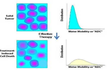
Figure 8: Relationship between change in cellular density following an effective therapy and the
corresponding distribution of water diffusion values within tumor. The mean diffusion value of
tumor increases early following the loss of cellular density.
Measuring Water Motion - Apparent Diffusion Coefficient (ADC)
The moving water molecules acquire different phase information from the first gradient, but because
of their motion, their signal will not be completely rephased by the second gradient, thus leading to
a signal loss. Hence, the motion of water molecules is detected as attenuation of the measured signal
intensity at DWI. The degree of water motion has been found to be proportional to the degree of
[image:45.595.63.497.168.454.2]The sensitivity of the DWI sequence to water motion can be varied by changing the gradient
amplitude, the duration of the applied gradient, and the time interval between the paired
gradients. On clinical MR scanners, the diffusion sensitivity is easily varied by changing the
parameter known as the “b value,” which is proportional to these three factors. When the b
value is changed, it is usually the gradient amplitude, rather than the duration or time intervals
between gradients, that is altered.
Treatment response assessment using Diffusion Weighted MRI
Radiation therapy with chemotherapy as induction or concurrent or adjuvant is the standard of
care for Carcinoma Nasopharynx. However, not all patients respond to chemoradiation
therapy. Thus, it is important to develop prognostic imaging biomarkers that can accurately
predict treatment outcome before initiation of treatment. These imaging biomarkers may help
in stratifying patients who would benefit from chemoradiation therapy from those who would
not. With the use of Diffusion Weighted MRI (DW MRI), the ADC measurements will be able
to predict the response of tumor to chemotherapy and radiation treatment
Effective anticancer treatment results in tumor lysis, loss of cell membrane integrity, increased
extracellular space, and, therefore, an increase in water diffusion. Impediments such as cell
membranes, organelles, and macromolecules interferes with the free movement of water
molecule, this diffusion in biological tissue is quantified by means of an APPARENT
With the use of Diffusion Weighted MRI (DW MRI), the ADC measurements will be able to
predict the response of tumor to chemotherapy and radiation treatment. Two major factors in
predicting response are:
1. Baseline ADC values
2. Magnitude of serial change in ADC values
BASELINE ADC VALUES: Cellular tumor having low baseline ADC values respond better
to chemotherapy or radiation treatment than tumors with high baseline ADC values. One
possible explanation is that tumors with high baseline ADC values are likely to be more
necrotic than those with low values. Necrotic tumors frequently are hypoxic, acidotic, and
poorly perfused, leading to diminished sensitivity to chemotherapy and to radiation therapy.
Studies in cerebral gliomas and breast cancers have also shown that an early increase in the
ADC after commencing treatment was predictive of better treatment outcome.(46)
MAGNITUDE OF SERIAL CHANGE: Diffusion weighted images with quantitative ADC
(Apparent diffusion Coefficient) values have shown promise in predicting response to chemo
radiation by assessing pre-treatment ADC values and monitoring their magnitude of change
serially during treatment at different time frames. This diffusion restriction of water molecules
can be assessed quantitatively by using ADC by obtaining two or more images with different
gradient duration and amplitudes (which are referred to as different b values).
The term apparent refers to the dependence and modulation of these
coefficients/diffusion processes on multiple factors in tissues such as restriction in closed
spaces, tortuosity etc. ADC values are calculated automatically by the software and then
different tissues. Then, by use of a dedicated workstation, ADC measurements are recorded
for a given region by drawing regions of interest (ROIs) on the ADC map. An ADC of a tissue
is expressed as a value in first decimal place x10-3 in units of mm2/sec (Eg: 1.1x10⁻³
mm2/sec).
Rate of change of ADC values during the course of treatment have also been
predictive of response to chemo radiation. Increase in ADC values as seen on subsequent scans
with ongoing chemo radiation reflects treatment response as opposed to an inital rise followed
by a fall in ADC values which would indicate no response to chemo radiation.
Hence at the start of treatment, tumors are anticipated to be densely cellular, posing a
restricted diffusion with a diffusion histogram reflective of this. Shortly after initiation of the
treatment, there is breakdown of cellular membranes, which is detectable with a rise in the
ADC values. Finally, after the completion of an effective treatment, tumor death leading to
further breakdown of cells leading to a further rise in the ADC values. However, the poor or
non-responders would not show any significant rise in the ADC values during the course of
their treatment owing to the less cellular breakdown resulting in poor response to the treatment
to Chemoradiotherapy. These tumors may show an initial rise in ADC values followed by a
drop in the same. Thus, the serial monitoring of the ADC values at baseline, 1st week, 4th
week and at 6 weeks follow up will help in correlating the ADC values of the responders and
non-responders. If the ADC values are found to help in predicting treatment response, this
valuable outcome could be utilized for tailoring the treatment care for this group of patients in
Diffusion Weighted MR Imaging in Nasopharyngeal carcinoma: A Predictive Biomarker
Studies conducted in squamous cell carcinoma of head and neck to establish DW MRI as
predictive biomarker for response assessment have shown promising results. However, they
have excluded Nasopharyngeal malignancies as it forms a distinct entity among the Head and
Neck malignancies (37) (47) (48) (49). Studies on DW MRI in Carcinoma Nasopharynx are
very limited. Two studies looked at response assessment using DWI-MRI in patients
Carcinoma Nasopharynx undergoing chemoradiation. They had showed similar results as seen
with other sites in Head and Neck suggesting the usefulness of DW MRI for response
prediction in Nasopharynx. Jinsheng Hong, et al proved role of magnetic resonance
diffusion-weighted imaging in predicting radiosensitivity in NPC (50) (51). Guo-Yi Zhang et al. showed
that pretreatment ADCs could be used as a new pretreatment imaging biomarker of response
to neoadjuvant chemotherapy in nasopharyngeal malignancies (52).
There has been no consensus as to an ideal follow-up time for imaging during and post
chemoradiation. In a paper published on consensus of DWI imaging as a biomarker in cancers;
it was emphasized that if DWI is used as a biomarker, imaging follow-up should be at a time
before changes in size become appreciable (53). Study by King AD et al, showed that ADC
post-treatment was a marker for local failure. Serial change in ADC was an even stronger
marker (54) . Long term time frames may normalize ADC values as liquefactive necrosis
resolves and becomes replaced by fibrosis. However, no uniform consensus has been
formulated regarding an ideal time for follow. All patients routinely undergo a CT scan at the
end of Chemoradiation (i.e. at 4- 6 weeks) to assess response to treatment. As the lesion may
be sub mucosal, the NPL scopy may not show a growth. CT scan may show a response which
reduction in the size of the nodes. An MRI with DWI and increase in ADC values may in turn
give additional evidence of response.
The eventual goal of this line of research is to use DW imaging and its parameters like ADC
values at the various stages of treatment to prospectively identify patient who would benefit
from concurrent chemoradiotherapy and classify them as responders and nonresponders. This
valuable information can be further utilized for the modification of the treatment process, such
MATERIALS AND METHODS
Place of study: Christian Medical College, Vellore, Tamil Nadu, India
Department Registry: Department of Radiotherapy in our Institute, on an average has 14-17
patients of Carcinoma Nasopharynx treated per year
Study Design: Prospective Study
Study Type: Observational
Inclusion Criteria: Patients with biopsy proven nonmetastatic carcinoma nasopharynx and
fulfilling the following were be asked to enroll in the study after informed consent.
1. Female or male patients aged 18-70 years
2. ECOG Performance Status of 0 to 2
3. Histologically confirmed carcinoma (Keratinizing, Non- keratinizing or Basaloid)
4. Non- metastatic disease
5. No previous chemotherapy or radiotherapy for any neoplasm/tumor
6. Weight > 35kgs
7. Adequate bone marrow function: WBC > 4000/mm3; Absolute Neutrophil count > 2000/cc
platelets > 100,000 mm3
8. Estimated creatinine clearance > 50ml/min/1.73m2
9. Total bilirubin ≤ 1.5 times upper limit of normal (ULN), SGOT and SGPT ≤ 2 times ULN
Exclusion Criteria:
2. Pregnant or breastfeeding mother
3. Contraindication to MRI like MRI incompatible pacemaker / aneurysm clip
Institutional Review Board Approval and Funding:
Institutional Review Board (IRB) approval and fund grant was obtained prior to the
commencement of the study (IRB No 10259 dated 05.09.2016) (attached in Appendix 4)
Sample Size Calculation:
The required sample size to show the proportion of response of about 80% with a precision
of 15% was found to be 27 subjects who underwent chemoradiotherapy. However due to
logistic issues and limited study period, we decided to include 20 patients. (Ref: Jinsheng
Hong et al. Value of Magnetic Resonance Diffusion-Weighted Imaging for the prediction of
Radiosensitivity in Nasopharyngeal Carcinoma. Otolaryngology – Head and Neck Surgery
149(5) 707–713) with 95% confidence limits.
Single Proportion - Absolute Precision
Expected proportion 0.8 0.8
Precision (%) 1 10 15
Desired Confidence level (1- alpha) % 95 95
Required sample size 61 27
Reference: Lemeshow S, Hosmer DW, Klar J, Lwanga SK. Adequacy of Sample Size in Health Studies.John Wiley and Sons, 1990.
Sampling and Consent: The prospective study patients were seen in the outpatient department
of Department of Radiotherapy, Christian Medical College Hospital from September 2016 to
September 2017. All patients who fulfilled the inclusion criteria were included in the study.
No specific sampling strategy was employed to enrol the patients. The baseline data was
entered in a numbered proforma (Annexure 3). Informed written consent was obtained from
the patient/ patient relatives prior to the baseline diffusion weighted MRI imaging. The consent
form along with the Patient Information Sheet is (attached in Annexure 1 and 2).
Treatment Protocols: Patients diagnosed to have Carcinoma Nasopharynx fulfilling the
criteria were assessed based on the clinical examination findings Nasopharngolaryngeal scopy
findings and a baseline CT scan and the suitable patients would be subjected to Diffusion
weighted MRI imaging which is a non-invasive stud requiring less than 6 minutes to acquire a
baseline ADC value. Subsequently the patients received their planned therapy with radical
radiotherapy with 66-70 Gy in 33-35 fractions with concurrent 3 weekly Cisplatin spanning
over 6 -7 weeks. As part of the study, ADC values were measured at baseline, 1 week, and
4-6 weeks of follow up were compared and analysed for their changes to determine the response
MRI was repeated for evaluation of treatment response. Patients receiving induction
chemotherapy were also included in the study.
Data acquisition: MRI imaging was performed on a 3T Philips Achieva scanner with a neck
coil. An axial T2HR sequence will be done in the neck from ventricles to clavicle with TE: 90;
TR: 3500 and slice thickness of 5mm and slice gap of 5.6 mm. Matrix size used was 362 x
292. DWI images were acquired with b values of 0 and 1000s/mm2 with slice thickness of
5mm.
Data analysis: ADC values are calculated automatically by the software and then displayed
as a parametric map that reflects the degree of diffusion of water molecules through different
tissues. Then, by use of a dedicated workstation, ADC measurements are recorded for a given
region by drawing regions of interest (ROIs) on the ADC map. The DW MR and conventional
T2-weighted images were reviewed by a senior radiologist. The radiologist blinded to the
treatment outcome. Regions of interest (ROI) were placed on the primary lesions and target
metastatic lymph nodes including most of the substantial area and avoid the necrotic area.
Metastatic lymph nodes were chosen as per imaging criteria (a minimal axial diameter of 10
mm threshold, including most of the substantial area and avoiding the necrotic area. ADC
values were calculated using different b-values, which measures the degree of diffusion
weighting applied. A useful rule of thumb is to choose the b value such that (b X ADC) is
nearly equal to 1. ADC values were derived from the ROI’s drawn over the metastatic nodes
and tumor using a free handed drawn setting. Both mean and minimum ADC value were taken
into consideration. The difference between Complete Response (CR) and Partial Response
(PR)/ non- responders was assessed by Mann Whitney U test. ROC analysis was done to
Outcome
1. Primary Outcome: To study the role of diffusion weighted MRI derived parameters i.e.
ADC as an imaging biomarker in non-metastatic nasopharyngeal carcinoma.
2. Secondary Outcome/s: Validating the role of diffusion weighted in detection and
characterization of nasopharyngeal cancers as well as for monitoring the response to
CRT.
Timing of DW MRI acquisition:
A series of 3 diffusion weighted MRI scans were obtained serially at
1. Pre- RT: Prior to initiation of radiotherapy
2. Week 1: At 1 week after initiation of Radiation
Detailed diagrammatic Algorithm of the study:
Patients diagnosed Non-Metastatic Nasopharyngeal malignancies recruited after biopsy, staging & informed consent
Suitable for Radical Radiotherapy or Concurrent chemoradiotherapy
Along with the baseline CT scan a DW MRI imaging is done to calculate the ADC value
Initiation of treatment
Baseline ADC vales compared with the serially monitored ADC values are
measured at baseline(Pre-RT), at 1 week of RT and at 4 to 6 weeks follow up after RT
Clinical examination, NPL Scopy/ RNE and a CT scan at 4 to 6 weeks after
completing entire treatment including adjuvant chemotherapy (if any) for assessment of treatment response.
No evidence of residual disease – Complete Responders
Asymptomatic patient with doubtful significance of residual disease – Partial Responders
Any evidence of residual disease then patient is categorized as - Non-responder
Patient to be kept on follow
Wait for 6 weeks more Repeat CT/PETCT to rule out the post radiotherapy changes
RESULTS
Social Demographics
20 patients with histologically proven Carcinoma Nasopharynx were screened and 4 were
excluded after reviewing the inclusion criteria. One patient had defaulted treatment. 2 other
patients were enrolled into the study at the time of this write up, However, they were included
for analysis. 13 were enrolled in this prospective study after informed consent. Pre-treatment
diagnostic examinations included Endoscopy, biopsy and baseline contrast-enhanced CT in all
patients.
Flow chart: Consort diagram explaining the recruitment of patients for the study 20 Patients with histologically diagnosed Carcinoma Nasopharynx
were screened for the study
4 Patients with Metastatic disease were excluded
16 Patients meeting the inclusion criteria were invited to participate in the study
1 Patient had not consented as she sought