CopyrightC) 1987, AmericanSocietyfor Microbiology
Isolation and Characterization of Recombinants
between Attenuated
and
Virulent
Aphthovirus Strains
ANA T. GIRAUDO, ADRIAN SAGEDAHL, INGRID E. BERGMANN, JOSE L. LA TORRE,
AND EDUARDOA. SCODELLER*
Centro de Virologia Animal, Serrano 661, 1414 Capital Federal, Argentina Received 18 June 1986/Accepted 20 October 1986
Aguanidine-resistantmutantof the attenuated strain of aphthovirustype01strain Camposand theoriginal
wild-type strainwerecrossedto generaterecombinant viruses. Two independently derived recombinant viruses
were isolated. One isolate (RI) contained the P1 (structural proteins)generegionof attenuated strain and P3
(polymerase precursor) generegion ofthe wild-type strain. The other isolate (RII) had a genomicstructure
complementarytothat of RI, this is,P1 ofthe wild-type strain and P3ofthe attenuated virus. Recombinant
RIIinheritedsomein vitro phenotypic markers thatwerecharacteristic of theattenuatedstrain,whereas the
RIrecombinanthadinvitro behavior thatwassimilartothatof the wild-type strain. The data obtainedsuggest
that thepolymeraseprecursor(P3) oftheattenuated strain (01Campos)could be involved in the determination
of the attenuated phenotype for fetal bovine kidney cellsand,eventually, forcattle.
Although attenuated strains of polioviruses and aphtho-viruses (foot-and-mouth disease virus [FMDV]) have been
used for large-scale vaccination, up to few years ago little
was known about the molecular basis that determines the
attenuation processinpicornaviruses.
In recent years, however, work done by researchers in
several laboratories has allowed the accumulation of
consid-erable knowledgeonthe nature of the attenuation process in
poliovirus. Alterations locatedinthe capsid proteins of the
Sabin I strain have been suggested to play a major role in
determining the attenuated phenotype (1, 2, 3). Recently, a
recombinant poliovirus strain was constructed from
infec-tious cDNAclones ofneurovirulent (Mahoney) and
attenu-ated (Sabin I) strains. Analysis of the growth properties of
the recombinant viruses in tissue culture has allowed the
correlation of specific mutations that are located in the
genomicsegmentthatencodescapsid protein VP1and part
ofcapsid protein VP3 with in vitro phenotypic markers of
neurovirulence (14). Evidence has also been reported ofa
correlation between neurovirulence and asingle nucleotide
change in the 5' end ofthe noncoding region ofthe Sabin
type IIIpolio vaccinegenome (9).
Ithasalso been postulatedthatvariationsin the isoelectric
points of capsid proteinsVP1 andVP3between virulent and
lessvirulentstrains oftheTheiler murineencephalomyelitis
virus could be related to the differences in their degree of
virulence (22).
The involvement of capsid protein modifications in virus
attentuation has also been reported for other virus-host
systems(8, 18, 23, 24).
Inthecaseofaphthoviruses, littlework has been doneon
the mechanism(s) of attenuation. Harris and Brown (11)
havereported that in virus strain SAT1 limited changes in
thefingerprintof thegenomic RNA and ashortening ofthe
poly(C)-richtract areassociatedwith the processof
attenu-ation. Dawe and King (7) have reported a correlation
be-tweenalterationsin
capsid protein
VP1andmousevirulenceforthe FMDV-SAT1 strain.
Morerecently, wehave described
(21)
some biochemicaldifferences betweena strainof
aphthovirus
type01Campos
*Correspondingauthor.
(O1C-O/E)attenuated for cattle and the wild-type strain(O1C)
from which it was derived. Changes in the viral structural
and nonstructural proteinsand in the lengthofthe
poly(C)-rich tract were found, but no correlation between the bio-chemical differences and the attenuated phenotype was
established(21).
To assesswhich regions of theO1C-O/Estrain are
respon-sible forthe attenuatedphenotype,weobtained recombinant
aphthovirus strains that carry different genomic regions of
thevirulentand attenuated strains.
Herewe report the biochemicalcharacterization and the
determination ofsomephenotypic markers,intissue culture,
oftwo such recombinants. It was found that recombinant
viruses that carry theP3/ABCD (P100)region of the genome
ofthe attenuated strain show in vitro phenotypic markers
similartothose of theparental attenuated strain.
MATERIALSANDMETHODS
Cells. Baby hamster kidney (BHK-21) cells were
culti-vated in Eagle minimum essential medium as described
previously (5).
Bovine kidney (BK) cells were obtained as primary
cul-tures from the Instituto Nacional de Tecnologia
Agro-pecuaria, BuenosAires, Argentina. Subsequentpassages(2
to 4) were performed in our laboratory with the same
medium as described above for BHK-21 cells, except that
fetal bovine serum (GIBCO Laboratories, Grand Island,
N.Y.)was used instead ofregular calfserum.
Virusstrains. Theoriginal01 Campos strainwasobtained
from tongue epithelium after bovine passages,
replicated
seventimes in BHK-21cells, and then plaque
purified;
oneclone (01C) was selected and was used
throughout
theexperiments.
The guanidine-resistant strain
(O1C-O/Egr)
was derivedfrom an attenuated strain ofthe FMDV 01
Campos
strain(O1C-O/E)
(21) asfollows. The01C-O/E
strain waspassaged
twice in BHK-21 cells in the presence of600,ugof
guanidine
hydrochloridepermlfor12to 24
h;
after the appearance ofcytopathic
effect bottleswerefrozen and thawed. This virusstock was plated in the absence of
guanidine,
and severalplaques were isolated. The
guanidine-resistant
cloneswereassayed by -titrationin the presenceorabsence of the
drug.
419
on November 10, 2019 by guest
http://jvi.asm.org/
TABLE 1. Plaque assay of the parent strains and theprogeny obtained by the yieldtestof the01C x 01C-O/Egrcrossunderrestrictive
and nonrestrictive conditions
Progeny virus titer(PFU/ml) in: Frequencyofpresumptive
Infecting viruses BHK-21 cells BK cells recombinants and
mutantsa Without guanidine Withguanidine Withoutguanidine Withguanidine
o1C 3.0 x 10⁸ 5.0 x 10⁴ 1.5 x 10⁸ 2.5 x 10³ 8 x 10⁻⁶
01C-O/Egr 2.5 x 10⁸ 1.5 x 10⁸ 1.0x 10⁴ 2.0 x 10⁴ 8 x 10⁻⁵
O1C x O1C-O/Egr 2.5 x 10⁸ 1.5 x 10⁸ 1.5 x 10⁸ 1.0 X 10⁵ 4 x 10⁻⁴
aRatio of infectious particles able to form plaques inBKcellsinthepresence of800,ugofguanidinehydrochlorideperml to totalinfectiousparticles.
Those clones showing the same titer under both conditions
wereselected.
Virus plaque titration. A total of 0.4 ml of serial 1:10
dilutions ofthe virus to be titrated was used to infect, in
duplicate, monolayersofBHK-21cells(5 x 10⁶)orBKcells
(3 x 10⁶) in glass bottles (21 cm2). Following adsorption
monolayers were overlaid with 6.0 ml of Eagle minimum
essential medium containing 40 mM HEPES
(N-2-hydroxyethylpiperazine-N'-2-ethanesulfonic acid) buffer
(pH 7.6) and1% karayagum(4). After 48 hofincubation at
37°C cellmonolayers were stained with0.2% crystal violet
andplaqueswerequantitated.
Clone isolation. The same procedure describedabove for
virustitrationwasusedfor cloneisolation,exceptthatin this
case karaya gum was replaced
by
1.2% agar(Oxoid Ltd.,
London, England)and 2% fetal bovineserum wasaddedto
theoverlay. Plaqueswerevisualized aftermonolayerswere
stained withneutral red. Thecentersofwell-isolatedplaques
werepunched out to isolatethe virus.
Generation and isolation of recombinants. Two different
methods wereused togenerate and isolate recombinants.
(i)Conventional yield test. Monolayersof BHK-21 cells(5
x 106) in 21-cm2 flasks were coinfected with both parent
viruses at 10 PFU per cell. Control cultures were also
inoculated with either parent at the same multiplicity of
infection usedforthecoinfections. After 30 minof
adsorp-tion at 37°C, the unadsorbed virus was thoroughly washed
off with salineand maintenance mediumwas added.
The infected cultureswere incubatedat 37°Cfor 7h, and
at that time a marked cytopathic effect was seen. The
cultures were frozen and thawed once and clarified to
discardcell debris. Virus in each samplewasquantitated by
plaque titration under both restrictive and nonrestrictive
conditions (see below).
Virusfromthecoinfection thatwerecapable ofproducing
plaques under restrictive conditions was amplified by one
passage in BHK-21 cells, clonedtwiceundernonrestrictive
conditions, and amplified againin roller bottles.
(ii) Infectious center method. Theinfectious center method
was essentially that described by McCahon and Slade (17).
Plaques that were picked under restrictive conditions were
amplified in BHK-21 cells. From each of these isolations,
differentclones were obtained by plaque purification.
Labeling and preparation of cell extracts and of a
p56a-enriched fraction. Labeling with
[35S]methionine
andimmu-noprecipitation of viral-induced polypeptides in
BHK-21-infected cells, analysis of polypeptides on 10%
polyacrylamide gels, and isolation of viral
polymerase-enriched fraction were performed as described previously
(21).
Analysis of virion polypeptides by electrofocusing. Labeled
virionproteins that were obtained after acetone precipitation
ofpools ofpurified virions were suspended in
electrofocus-ing sample bufferand separated in nonequilibrium pH
gra-dients (NEPHGE) on slab gels (20 by 13 by 0.15 cm).
Electrofocusing conditions were carried outby the method
of O'Farrell et al. (19), as modified by King and Newman
(13).
Analysis of RNase Tl-resistant oligonucleotides by
one-dimensional gel electrophoresis. Preparations of 32P-labeled
RNA from the cytoplasm of infected cells, digestion with
RNaseTi, andelectrophoretic analysisof the digests were
performed asdescribedpreviously (15).
Kinetics of RNA synthesis. To determine the kinetics of
RNAsynthesis,24-well flat bottomdisposable plates(Nunc,
Roskilde,Denmark) containing5 x 105BHK-21cellsor2.5
x
105
BK cells per well were used. Monolayers wereinfected with the viruses that were to be compared at an
inputmultiplicityof infection of 10 PFU/ml. After 30 min of
adsorption, the unadsorbed viruswasremoved and 0.4 ml of
mediumcontaining5 pLgofactinomycinD(SigmaChemical
Co., St. Louis, Mo.)wasadded. This mediumwasreplaced
after 30 minbymediumcontaining5 ,ug ofactinomycinD per
mlplus 15 ,uCiof[3H]uridine (30.6 Ci/mmol; NewEngland
Nuclear Corp., Boston, Mass.) per ml. Incorporation was
stopped byadding 1 mlof SDS-NET buffer(100mMNaCl,
50 mM Tris hydrochloride [pH 7.4], 1 mM EDTA, 0.05%
sodium dodecyl sulfate [SDS]) at the desired time. The
radioactivity of the trichloroacetic acid-insoluble material
wasdetermined. To evaluate thebackgroundofactinomycin
D-resistant nucleoside incorporation in BK cells,
mock-infected wells were assayed ateach time point.
RESULTS
Phenotypic properties of parental strains. To determine
appropriatemarkersfortheisolation and characterization of
recombinant strains, bothparental strains O1C andO1C-O/E
and a guanidine-resistant mutant derived from the latter
(O1C-O/Egr;
seeabove)weretitrated inBHK-21and BKcellsin the presenceorabsence of800 ,gof guanidine
hydrochlo-ride per ml.
Allthree strains grewequallywell in BHK-21 cells, which
is a permissive system for these viruses (Table 1). The
replication of strains O1C and O1C-O/E, however, was
stronglyinhibited by the presence of guanidine in the same
cells, whereas mutant O1C-O/Egr grew equally well in the
presenceor absenceofthe drug.
BKcells are a semipermissive system for the attenuated
virus strains; hence, in these cells only the wild-type strain
(O1C) reached titers similar to those obtained in BHK-21
cells, whereas strain O1C-O/E and the guanidine-resistant
mutantstrain derived from it(O1C-O/Egr)replicated poorly in
these cells. Strain O1C-O/Egr displayed its
guanidine-resistant phenotype,as wasthe casein BHK-21 cells. With
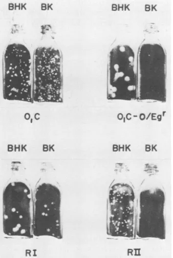
regard to plaque morphology, the wild-type strain (O1C)
elicited clear plaques that were ofsimilar size in both BK
andBHK-21cells(Fig. 1).InBHK-21 cells strainO1C-O/Egr
on November 10, 2019 by guest
http://jvi.asm.org/
[image:2.612.64.564.97.168.2]produced plaques thatwereconsiderablylarger than thoseof
strain 01C; however, the border of the plaques were not
clear, as was the case in the wild-type strain. Plaques
produced by 01C-0/Egr in BK cells were small and ill
defined. Identical plaque featureswerealsodisplayed bythe
guanidine-sensitive, attenuated strain 01C-O/E (data not
shown).
Generation and isolation of recombinantviruses. Based on
the phenotypic features of strains 01C and 01C-O/Egr, we
usedasselective markersthe ability of 01C-0/Egrtogrowin
thepresenceof 800 ,ug of guanidine hydrochlorideperml and
thecapacity of strain01Ctogrow athigh titers in BK cells.
The recombination experiments were carried out as
de-scribedaboveby usingasrestrictive conditions theplating of
progeny virus in BK cells in the presence of 800 ,ug of
guanidine hydrochloride per ml. The data obtained on
titration ofprogeny produced by the yield testaregiven in
Table 1. The proportion of plaques developed under
restric-tive conditions was fivefold higher in the progeny from
mixed infection than in theprogenyobtained frominfection
with 01C-0/Egr alone (recombination frequency, ca. 4 x
10-4).
The10plaques thatwereformed under the conditionsdescribed above by the progeny of mixed infection were
picked up, amplified as described earlier, and investigated
with respect to their resistance to guanidine and to their
capacity togrowinBK cells. All 10 cloneswere guanidine
resistant; and 7 of them exhibited high titers in BHK-21 or
BK cells,as wasexpected for recombinants.
The other three clones replicated in BK cells at titers
similartothat of strain01C-0/Egr; this observation could be
an indication that these clones are truly parental strains.
Further biochemical analysis of these viruses supported this
assumption. The seven clones with the phenotypic
charac-teristicsexpected for recombinant viruseswere selected for
biochemical characterization to assess whether they were
truerecombinants (see below).
BHK BK BHK BK
ofc O,C-O/Egr
BHK BK BHK BK
RI
RII
FIG. 1. Representativeplaque morphologyofparentand
[image:3.612.313.554.95.170.2]recom-binant strainsonBHK-21 and BK cells.
TABLE 2. Growthcharacteristicsof recombinants RI and RIIin BHK-21andBKcellsa
Titers(PFU/ml) in thefollowing
Cell recombinants:
RI RII
BHK-21 2.0 x 10⁷ 2.0 x 10⁸
BHK-21with guanidineb 2.0 x 10⁷ 1.0 x 10⁸
BK 2.0 x 10⁷ 5.0x10⁵
BKwithguanidineb 2.0x 10⁷ 2.5 x 10⁶
a The virus plaque titration assay used was the karaya gum overlay method.
bGuanidine(800 ,ug/ml) was included in theoverlay medium.
Alternatively, recombinants were obtained by using the infectious center assay reported by McCahon and Slade (17).
By thisprocedure five plaques were isolated under
restric-tive conditions, and viruses recovered from each isolation
were amplified in BHK-21 cells. Biochemical analysis of
viruses recovered from four of the plaques indicated that
they were indistinguishable from the parental strains,
whereas viruses recovered from a fifth plaque elicited the
biochemical characteristics of recombinants viruses (see
below). It is well known that this methodproduces mixed
populations of viruses rather than single homogeneous
strains(17);therefore,recombinants recovered from the fifth
plaque were cloned in BHK-21 cells, and several clones
were recovered. Viruses from two of the plaques were
guanidine resistant and elicited titers of105 PFU/ml in BK
cells. Although these features are quite similar to those of
the01C-0/Egr strain, the plaques produced by these clones
in BK cells were well defined and small; hence, they were
clearly distinguishable from the typical ill-defined, small
plaques produced by the 01C-O/Egr parent strain in this
system. Therefore, these clones wereselected for
biochem-ical characterization.
Representative clones derived from both recombination
methods were titrated in BHK-21 and BK cells.
Recombi-nant I (RI) is a representativeof the presumptive
recombi-nantclones selected in theyieldexperiment,whereas
recom-binant II (RII) is a clone that was selected by using the
infectious center assay. RI elicited a high titer in both
cellular systems(Table 2),as wasexpectedforastrain(O1C)
with a phenotype like that of the wild type; on the other
hand, the titer of RII in BK cellswas ca. 3 log units lower
than that in BHK-21cells. Thisbehavior is similartothat of
the attenuated parent strain (01C-0/Egr).
Thefeatures of theplaques induced byboth recombinants
(RIandRII) in BKandBHK-21cellsareshown inFig. 1. In
BHK-21cells RIproduces plaquesthatareindistinguishable
from those produced by strain
01C-0/Egr,
whereas theplaques producedin BKcells, althoughsmaller than those of
BHK-21 cells, show a similar morphology. On the other
hand, in BHK-21 cells RII produces plaques that were
indistinguishable from those of strain 01C, but in BK cells
RII elicited very small
(pinpoint)
clearplaques. Therefore,taking into account differences between titers in BK and
BHK-21cells, RIwould bethoughttohaveawildtype-like
behavior. On the contrary RII showed an attenuatedlike
behavior,
according
tothetiters obtained in BK and BHK-21cells.
Biochemicalanalysis ofputative recombinants. To screen
theputative
recombinants,
weusedasnonselective markersbiochemical differences between strains 01C-0/E and 01C
which mapped on the viral
capsid (P1)
andpolymerase
precursor(P3)
regions
ofthe viral genome(21).
Inaddition,
,,*
on November 10, 2019 by guest
http://jvi.asm.org/
[image:3.612.90.259.451.703.2](i 4.
0' 0 ~0
0-K
Ki
t.
' oko0?
-ft
=VP3
alCs
-* -
i_ _ sVP,
FIG. 2. Analysisofviral structuralproteins ofparentand recom-binant virusesbyNEPHGE. Forstraindesignations, see text.
thepoly(C)-richtract,which is locatednearthe5' endof the
viral genome of the attenuated strains, is considerably
shorterthan that of thewild-type strain from which it was
derived. Although thesetraitshave beendefinedby
compar-ing 01C with 01C-0/E, these differences in P1, P3, and the
poly(C) sequences were maintained when 01C was
com-pared with01C-0/Egr(data notshown).
From the 10 clones obtained by theyield method, 3 were
phenotypically andbiochemically indistinguishable fromthe
parent strain01C-0/Egr, whereas the other 7clones elicited
phenotypesthat were predictedfor recombinant viruses.
Biochemical analysis of the 7 putative recombinants
showed that all of them had identicalcapsidprotein
compo-sitioninNEPHGE andSDS-polyacrylamidegel
electropho-resis (data not shown). One of the recombinants (RI) was
&i\4' C'
K0 (p00
l0'
,0'1>'N,1
Kan- --Am _ _ m Pco
- - -- P88
a
-_-- a U p72selected as being representative of this group and was
analyzed inparallel with RII and the parent strains.
The pattern of structural (capsid) proteins of the viruses
analyzed by NEPHGE (see above) is shown in Fig. 2. The
migrationpattern of VP1-VP2 and VP3 of RI is
indistinguish-able from that of the attenuated parent (01C-0/Egr) and is
clearly different from the pattern ofproteins elicitedby the
wild-type parent strain 01C. In the case ofRII, which was
obtained by the infectious center assay, the pattern of
structuralpolypeptides is indistinguishable from that of the
wild-type strain but clearly different from that of the
atten-uated parent.
Analysis of virus-induced polypeptides (see methods) by
SDS-polyacrylamide gel electrophoresis shows that RI
ac-quired the polymerase precursor P3 (P100) that is
character-istic of the wild-type strain, whereas RII inherited P3 of its
attenuated parent (Fig. 3).
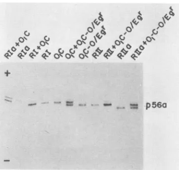
Analysis of p56a of theparental strains and both
recom-binants by NEPHGE (21) shows that RII inherited an RNA
polymerase polypeptide that was indistinguishable from that
of theattenuated parental strain01C-0/Egr, whereasp56a of
RI was similar to that of the wild-type strain 01C (Fig. 4).
Not all of the recombinant viruses recovered showed this
trait, however. Two of those clones that carry RNA
poly-merases thatresembled neither parent are shown in Fig. 4.
As suggested by McCahonetal. (16), thesevariations could
be due to recombinationevent(s) within the codingregion of
that protein.
The results shown in Fig. 2 to 4 demonstrate that both
strains that were analyzed in this study are true
recombi-nants. RI carries the P1 region of the genome of the
attenuatedparent and the P3region of the wild-type strain,
whereas RIIcontains the reverseofRI, in that it carries P1
of the wild-type strain and P3 of the attenuated parent.
Further evidence was obtained by genomic analysis ofRI
and RII and their respectiveparents by RNase Ti maps on
one-dimensional gels. It was found that RI carries theshort
poly(C)-rich tract that is characteristic of the attenuated
strain (21), whereas RII inherited the poly(C)-rich tract of
thewild-typestrain(datanotshown). These results suggest
that besides the P1 region, RI possibly carries all the
Ko°so 0°'s 2
C,}0S°
C, x0'0 KpI
4qmw
Mq11qpp1
p am p56a.
'- - - _ p56a
__m mm -_ VPo
______*m mm.e
1 40_
q&MW4_a
]JPI-3_a _ _ _
FIG. 3. Polyacrylamide gel electrophoresis of virus-induced
polypeptides in BHK-21 cells infectedwith parental and
recombi-nant viruses; monolayers were pulse-labeled, and viral-specific
proteinswereisolated and analyzedasdescribedin thetext.
FIG. 4. Analysisofp56aof recombinants and parental strains by NEPHGE. RIais a recombinant variant representing one of the
seven clones obtained in the yield assay. Rlla is a recombinant variant recovered by further plaque purification of the original isolate obtainedbytheinfectiouscenter assay (see the text).
on November 10, 2019 by guest
http://jvi.asm.org/
[image:4.612.132.247.76.246.2] [image:4.612.121.261.473.681.2] [image:4.612.360.537.506.674.2]sequences upstreamof theP1region or at least up to the 5'
endofthe poly(C)-rich tract of the attenuated strain.
Viral RNA synthesis in BHK-21 and BK cells. Strain
01C-0/Egr and its parent strain 01C-0/E are deficient in RNA
synthesis in cells from bovine origin because they reach at
most 10% ofthe level of RNA synthesis obtained in strain
01C (data not shown). Therefore, we investigated whether
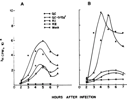
this trait was inherited by RI or RII. The kinetics of RNA
synthesis werestudiedin BHK-21 and BK cells. The
kinet-ics of RNA synthesis induced in BHK-21 cells by the
parental, RI, and RII strains are shown in Fig. SA. The
attenuated strain 01C-0/Egr reached roughly 50% of the
levelsachieved in thewild-typestrain, which wasthe same
as that achieved in its parental strain 01C-0/E (data not
shown). RIandRIIdiffer greatlyin the synthesis of specific
viral RNA induced in BHK-21-infected cells. RI reached
synthesis rates
similar
to those reached in the attenuatedstrain,whereas RIIelicitedan RNAsynthesisratesimilarto
that ofthe wild-type strain.
Ontheother hand,in BKcells(Fig. SB)thehighestvalues
were reached by the wild-type strain, and as stated above,
01C-0/Egr reached values that were 10% ofthose obtained
with strain 01C. The rate ofRNA synthesis in BHK cells,
which was highest in RII, fell dramatically in BK cells,
showing values similar to those of01C-0/Egr. The rate of
RNA synthesis of RI increased considerably in BK cells,
showing RNA synthesis rates similarto those ofthe
wild-typestrain. These results suggest that thefailureto
synthe-sizeRNAinBKcellswasinheritedbyRIIratherthanby RI,
in spite ofthefact that synthesis in BHK-21 did notreflect
thisfact.
The rate ofRNA synthesis in BK cells was also
estab-lished for other recombinants
(RIa
and RIIa; Fig. 4) thatcarrypolymeraseswithchargesthat aredifferentcompared
with their parental strains. It was found that RIa has a
slightly lower rate of RNA synthesis than RI, whereas
recombinantRllareached values slightly higherthan those
ofRII(datanotshown).No otherbiochemicalor
biological
differencesbetween RI-RII andRIa-RIIa,
respectively,
weredetected. Therefore, it seems that theonly difference found
between viruses thatcarry
parental
orunique p56awasthatofadifferentrateofRNA
synthesis
in BK cells.DISCUSSION
Wehave
recently
describedthebiochemicalcharacteriza-tion of a host range mutant of FMDV serotype 01 strain
Campos
thatwasattenuatedfor cattleand thatwasobtainedthroughadaptationonserialpassagesofthe
wild-type
strainin chickenembryos. Theattenuated strain showeda
short-enedpoly(C)-richtractandseveralchangesin structural and
nonstructuralproteins.Theinvolvement ofthosechangesin
the determination of the attenuated
phenotype, however,
was notestablished.
Recently, it has been demonstrated (12, 16) that in
aphthovirus intra- and
intertypic reconmbinants
can beob-tained at
high
frequencies
in thelaboratory.
Therefore,
recombinants between
wild-type
andattenuated strainswereconstructed to ascertain which parts of the attenuated
genomewererelevant forattenuation. Of 10 clones isolated
by
theyield method,
7were consideredtobetruerecombi-nantsbecause
they
containedtheP1region
ofthe01C-0/Egr
genome and the P3
region
ofthe01C genome. The fact thatvirusesofthe RIgroup inherited the
poly(C)-rich
tractof theattenuated strain is additional evidence that supports the
idea that these viruses are truerecombinants.
A
10r
8-IV
0.
U
.-.OAc
oOA,-O/Egr h- RI
Mo
Rck
*--a Mock
B
HOURS AFTER INFECTION
FIG. 5. Kinetics ofRNAsynthesisof recombinants andparental
viruses in BHK-21 (A)and BK(B)cells. Fordetails,seethetext.
Basedonbiochemicaland
biological
datapresented here,
itcanbe
postulated
that attenuatedviruses inwhichtheP3regions
have beenreplaced by
anequivalent region
ofthewild-type
strain(O1C)
have also been relieved of itscon-straint(s)
togrowinBKcells.Based onthe restrictive conditions used for
selection,
itwas
expected
thatonly
those recombinantswith thecapacity
togrowat
high
titers inBKcells would beisolated.By
usingthe infectious center assay,
however,
a recombinant(RII)
withalimited
capacity
togrowin BK cells(like
theattenu-ated
strain)
wasisolated. Thiswas notsurprising,
consider-ing
the differences between the methods usedtoobtaintherecombinants. In the
regular yield
assay, the virusespro-duced in BHK-21 cells were
plated
in BK cellsplus
guanidine,
sothatplaques
wereproduced by
asingle
infec-tive virion. On the other
hand,
in theinfectious
centerassayBHK-21 infected cells were
plated
1 h afteradsorption
onthe BKcell
monolayer,
andplaques
wereproduced by
theprogenyoftheseinfectedBHK-21cells.
By
thenatureof thismethod,
itislikely
thatmore thanone recombinantcanbefound in the same
plaque (17).
Ifrecombinant RI and RIIwere
produced
in the samecell,
theoriginal
plaque
fromwhich the
virus(es)
was recovered would contain bothrecombinants.
Taking
intoaccountthatthevirus(es)
in thisplaque
wasamplified
in BHK-21 cellsbefore
itwasclonedand that recombinants ofthe RII type reached a titer that
was 10timeshigher than that in RI in these cells
(Table 2),
itis understandable that clone RII can be
preferentially
se-lectedafter
cloning.
Biochemical
analysis
oftherecombinantobtainedby
thismethod demonstrated
that,
indeed,
RII carried P1 and thepoly(C)-rich
tractofstrain01C
and P3ofstrain01C-0/Egr.
With
regard
toothergeneral
characteristics oftherecombi-nants,itwasfound that RI grew
equally
wellin BHK-21 andBK cells
(Table 2),
produced
large-sized plaques
in bothsystems, andwasabletoinduceanRNA
synthesis
level inBKcells similartothatofthe
wild
type.On the otherhand,
thetiter of RII in BHK-21 cells was3
log
unitshigher
thanthat in BK
cells,
but in thissystem
the recombinant elicitedverysmall
(pinpoint) plaques,
and therateof RNAsynthesis
reflectedthatofits attenuatedparent
01C-O/Egr.
Taking
allthis into account, one could assumethat RII shares the in
on November 10, 2019 by guest
http://jvi.asm.org/
[image:5.612.308.552.69.259.2]vitro markers of attenuation of strain
01C-0/Egr.
We havepreviously postulated (21)
that theincreasedelectrophoretic
mobility
ofP100 could be duetothedeletion(s)
thatdevelops
during
the processofattenuation; therefore,
it istempting
tospeculate
that thoseputative
deletedregions
could bein-volved in thedetermination ofthe attenuated
phenotype.
Ifattenuation lies outside of the
capsid proteins,
then itwould be feasible
by
means of recombination to introducethe
noncapsid
part ofthe genome of an attenuated donorstrain,
which isresponsible
forthe attenuatedphenotype,
innewemergent variants without
altering
theantigenic
struc-tureofthesenewstrains. Thetime
required
todothiswouldbe
considerably
shorter than the time necessarytodevelop
anattenuated virus
by
theclassicalprocedures. Approaches
of this type have beenshowntobeuseful in the construction
of new attenuated strains in influenza virus
(25).
We arecurrently
testing
thishypothesis using intertypic
recombina-tion.Major
determinants ofattenuation were located in the 5'halfof the viral genome,
specifically,
in thecapsid region
ofSabin type I
(1, 2, 3)
andin the5'-noncoding region
ofSabintype III
(9).
Recently, however,
Omata et al.(20)
haveshownthat
multiple
determinantsofattenuation reside in thepoliovirus
Sabintype I genomeatlocations other than in thecapsid region.
Evidence forothervirus families also existsthat
points
thefact that structural
proteins
areimportant
determinants ofvirulence
(8,
18, 22, 23, 24).
In this
study
anoncapsidal region
of the viral genomeappeared
toplay
amajor
role indetermining
the in vitrophenotypic
markers of attenuation.It appears,
however,
that with the in vitro markers ofattenuation that are inherited
together
withP3, plaque
fea-tures appear to be
mainly
linked to the inheritance of P1(compare
theplaques
of recombinants andparental
strains inFig.
1).
Although
it has been shown that BK cells are agood
system for
testing
viruses thatareattenuatedforcattle andthat all theknownattenuatedviruses reflecttheir features in
this cell
(6, 10;
PabloAuge
deMello, personal
communica-tion),
we mustkeep
in mind that the definitiveproof
ofattenuation will be
provided by
resultsofpathogenicity
testsin
cattle,
whicharecurrently
in progress.ACKNOWLEDGMENTS
Wethank A. M. Sadir andA. Zabal(INTA) forprovidingPBK cells and C. Ricarte and E. Yampolsky for excellent technical assistance.
This workwassupported by ConsejoNacional deInvestigaciones
Cientificas y Tecnicas and by Instituto Cientifico Paul Hermanos
(Bs. As., Argentina). I.E.B. isarecipientofaHumboltFellowship.
LITERATURE CITED
1. Agol,V.I.,S.G.Drozdov,M.P.Frolova,V. P.Grachev,M.S.
Kolesnikova,V.G.Kozlov,N. M.Ralph,L.I.Romanova,E. A.
Tolskaya, and E. G. Viktorova. 1985. Neurovirulence of the
intertypic poliovirus recombinant v3/al-25: characterization of
strainsisolated from thespinal cordof diseased monkeys and evaluation of the contribution of the 3' half of the genome. J. Gen. Virol. 66:309-316.
2. Agol, V. I.,S. G.Drozdov, V. P.Grachev, M. S. Kolesnikova,
V.G.Kozlov,N. M.Ralph,L. I.Romanova,E. A.Tolskaya, A.
V.
Tyunfanov,
and E. G. Viktorova. 1985. Recombinantsbe-tween attenuated and virulent strains of poliovirus type 1: derivationandcharacterization ofrecombinants withcentrally locatedcrossoverpoints. Virology 143:467-477.
3. Agol, V.I.,V. P. Grachev, S. G.Drozdov, M. S.Kolesnikova, V. G.Kozlov, N. M.Ralph, L. I.Romanova,E. A.Tolskaya,A. V. Tyufanov, and E. G. Viktorova. 1984. Construction and properties ofintertypicpoliovirus recombinants: first approxi-mation mapping of the majordeterminants of neurovirulence. Virology 136:41-55.
4. AugedeMello,P. 1976.Plaquereduction neutralizationtestfor the assay of antibodies againstfoot-and-mouth disease. Bull. Centro Panamericano Fiebre Aftosa 21-22:30-34.
5. CostaGiomi, M. P., I. E. Bergmann, E. A. Scodeller, P.Auge de Mello, I. Gomes, and J. L. La Torre. 1984.Heterogeneity of the polyribocytidylic acid tract in Aphtovirus: Biochemical and biological studies of viruses carrying polyribocytidylic acid tractsof differentlengths. J. Virol. 51:799-805.
6. Crandell, R.A., and I. Gomes. 1970. Further studiesonmarkers of foot-and-mouth disease virus. Bull. Off. Int. Epiz. 73:521-533.
7. Dawe, P. S., and A. M. Q. King. 1983. Point mutations in polypeptide VP1 of foot-and-mouth disease virus affectmouse
virulenceand BHK-21 cellpathogenicity.Arch. Virol. 76:117-126.
8. Dietzschold, B.,W. H. Wunner,T.J.Wiktor, A. D.Lopes, M. Lafon, C. L. Smith, and H. Koprowski. 1983. Characterization of an antigenic determinant of the glycoprotein that correlates withpathogenicityof rabies virus. Proc.Nati. Acad. Sci. USA 80:70-74.
9. Evans, D. M. A., G. Dunn, P. D. Minor, G. C. Schild, A. J. Cann, G. Stanway, J. W. Almond, K. Currey, andJ. V. Maizel Jr. 1985. Increased neurovirulence associated with a single nucleotide change in a noncoding region of the Sabin type 3 poliovaccinegenome. Nature (London) 314:548-550.
10. Gomes, I., and R. A. Crandell. 1969. Marker studies of foot-and-mouth disease virus. Bull.Off. Int.Epiz. 71:835-846. 11. Harris,T.J. R.,and F. Brown.1977. Biochemicalanalysisof a
virulentand an avirulentstrain of foot-and-mouthdisease virus. J. Gen. Virol. 34:87-105.
12. King,A. M.Q., D. McCahon, K.Saunders, J. W. I.Newman, and W. R. Slade. 1985. Multiple sites of recombination within the RNAgenome of foot-and-mouth disease virus. Virus Res. 3:373-384.
13. King, A. M. Q., and J. W. I. Newman. 1980. Temperature sensitive mutants of foot-and-mousedisease viruswith altered structuralpolypeptides. I. Identificationby electrofocusing. J. Virol. 34:59-66.
14. Kohara, M.,T. Omata,A. Kameda,B. L. Semler, H. Itoh, E. Wimmer,and A. Nomoto. 1985. Invitrophenotypicmarkersof
a poliovirus recombinant constructed from infectious cDNA clonesof theneurovirulentMahoneystrain and the attenuated Sabin 1 strain. J. Virol. 53:786-792.
15. La Torre, J. L., B. 0. Underwood, M. Lebendiker, B. M. Gorman,and F. Brown. 1982.Applicationof RNase Tione-and two-dimensionalanalysestotherapid identification of foot-and-mouth diseaseviruses. Infect.Immun. 36:142-147.
16. McCahon, D.,A. M.Q. King,D. S.Roe, W. R. Slade, J. W. I. Newman, and A. M. Clearly. 1985. Isolation andbiochemical characterizationofintertypic recombinants offoot-and-mouth diseasevirus. Virus Res.3:87-100.
17. McCahon,D.,andW. R. Slade.1981.Asensitive method forthe
detection andisolationofrecombinants offoot-and-mouth
dis-easevirus. J. Gen.Virol.53:333-342.
18. Mettenleiter, T. C., N. Lukacs, and H. J. Rhiza. 1985. Pseudorabies virus avirulent strains fail to express a major
glycoprotein.J. Virol.56:307-311.
19. O'Farrell,P. Z.,H. M. Goodman,and P. H. O'Farrell. 1977. High resolution two-dimensional electrophoresis of basic as wellas acidicproteins. Cell 12:1133-1142.
20. Omata, T.,M.Kohara,S.Kuge, T. Komatsu, S. Abe, B. Semler, A. Kameda, H. Itoh, M. Arita, E. Wimmer, and A. Nomoto. 1986. Genetic analysis of the attenuation phenotype of
poliovirustype 1. J. Virol. 58:348-358.
21. Parisi, J. M.,M.P.CostaGiomi, P. Grigera, P. Auge de Mello, I. E. Bergmann, J. L. La Torre, and E. A. Scodeller. 1985. Biochemical characterization ofanaphthovirus type 01 strain
on November 10, 2019 by guest
http://jvi.asm.org/
Campos attenuated for cattle by serial passages in chicken embryos. Virology 147:61-71.
22. Rozhon, E. J., J. D. Kratochvil, and H. L. Lipton. 1985. Comparisonofstructuralandnonstructural proteinsofvirulent andlessvirulentTheiler'svirusisolates using two-dimensional gelelectrophoresis. Virus Res.2:11-28.
23. Spriggs, D. R., and B. N. Fields. 1982.Attenuated reovirustype
3 strains generated by selection of haemagglutinin antigenic
variants.Nature(London)297:68-70.
24. Stanley, J., S. J. Cooper, and D. E. Griffin. 1985. Alphavirus neurovirulence: monoclonal antibodies discriminating wild-type fromneuroadapted Sindbis virus. J.Virol. 56:110-119. 25. Tian,S. F.,L. X. Zhang, andB. R. Murphy. 1984.
Character-ization ofthegenotypeand levelofattenuationofaninfluenza
reassortant virus produced by mating the Xia-ts donor virus withA/Beijing/79(H1N1) wildtypevirus. Vaccine 2:189-192.