0022-538X/89/093844-08$02.00/0
Copyright ©1989,American SocietyforMicrobiology
Identification
of the Latency-Associated Transcript
Promoter
by
Expression of
Rabbit
Beta-Globin
mRNA in
Mouse
Sensory Nerve
Ganglia
Latently
Infected with
a
Recombinant Herpes Simplex Virus
ANTHONY T.
DOBSON,'
FARHADSEDERATI,'
GAYATHRI DEVI-RAO,2 W. MICHAEL FLANAGAN,2 MICHAEL J. FARRELL,3JACK G.STEVENS,'
EDWARD K. WAGNER,2 AND LAWRENCE T. FELDMANl3* Department ofMicrobiology andImmunology'
andMolecularBiologyInstitute,3 University of California, LosAngeles,LosAngeles, California 90024, and Departmentof MolecularBiologyandBiochemistry, University ofCalifornia,
Irvine, Irvine, California 927172 Received 21 March 1989/Accepted 8 June 1989
The herpes simplex virus type 1 latency-associated transcript (LAT) is expressed as a major species in latentlyinfected mouseneurons. Previous sequence analysis revealedno obvious promoter elements near the 5'endoftheLAT,but aTATAbox and otherpotentialpromoter elements were found 700 base pairs upstream. Arecombinant virusinwhichtherabbitbeta-globingene wasinsertedimmediatelydownstream of the TATA boxexpressedglobinmRNA and did not express the LAT. A second recombinant virus, in which this TATA box wasremoved, wasnegative forLATexpression in a latent infection. The location of the LAT promoter suggested that RNA upstream ofthe LAT was synthesized and degraded during latent-phase transcription. Lowlevelsof this RNAwereobserved byin situhybridization.In otherexperiments,RNA from aproductive infection was used to detect a transcript extending from the LAT promoter to a polyadenylation signal approximately 8.5kilobases downstream. These data suggest that the LAT may be processed from a larger transcriptionunitwhichbegins distaltothe TATA box 700 basepairsupstreamofthe LAT and extends toa polyadenylation signal almost 5kilobasesdownstreamofthe 3' end of the LAT.
Herpes simplex virus (HSV) can produce latent infections of the peripheral and central nervous systems of humans and
experimental animals (J. G. Stevens, Microbiol. Rev., in press).Transcripts from only one region of the viral genome are readily detectable during such an infection, and these have beencollectively referredto as thelatency-associated transcripts (LATs) (1, 2, 5, 14, 15, 19, 20, 21, 25, 26). Two colinear forms of the LAT have been described, one is an
apparently unspliced 2.1-kilobase (kb) transcript while the othercontainsan approximately600-base intron (15, 24, 25, 27). Two laboratories have also described a third, slightly smaller species (16, 27). Interestingly, stable LAT is not
polyadenylated and is largely confined to the nucleus of latently infected neurons.
The region surrounding the LAT has been sequenced in threestrains of HSV type1(HSV-1): KOS(M), 17syn+, and F (12, 24, 28). Comparison of sequence data with primer extension andS1nuclease analysis has positioned the 5' end
ofthe LAT within the sequenceAGGT, which is a potential 5' splice signal (13). The region just upstream of this site
contains no obvious promoter elements such as TATA or CAATbox homologies, although such homologies and two
potential SPl-binding sites are found approximately 700 bases further upstream of this point (12, 28) and have been noted by others (27, 28).
These data areconsistent with one of two possible models. (i) The LAT is transcribed from an unusual promoter
con-tiguouswith its 5' end, or (ii) the LAT is transcribed from a conventional promoter element, and the stable species ob-served arederived from an unstable primary transcript.
* Correspondingauthor.
MATERIALS AND METHODS
Establishment oflatent infection in mice and processing of spinal ganglia. Six-week-old outbred Swiss-Webster mice
(Simonson Laboratories, Gilroy, Calif.) were used in all
experiments.Theywereinoculated in eachrearfootwith
108
PFU of HSV-1 strain KOS(M)or with recombinant viruses of this strain. The derivation of this virus strain and the methods used for inoculation have been described previ-ously (20, 22). At 3 or more weeks later, after latent
infections had beenestablishedin lumbosacralspinalganglia
(7, 18),gangliawereremoved from theanimals,quickfrozen in liquid nitrogen, and either sectioned on a cryostat or storedat -70°C until used.
RNA isolation and fractionation. RNA was isolated from latently infectedganglia by theguanidinium
isothiocyanate-hotphenol methodasdescribed previously(20). RNAfrom a productive infection of rabbit skin cells infected at a multiplicity of 10 PFU per cell at 6 h postinfection was isolated as described previously (20, 22).
Poly(A)--
andpoly(A)+-containing
RNA was selected byoligo(dT)-cellu-lose (Collaborative Research, Inc., Waltham, Mass.) chro-matography (3). RNA was size fractionated by electropho-resis with1.2%agarosegels containing 6% formaldehydeas describedpreviously (25); RNAwas transferredby
electro-phoresisontonylon membranes (GeneScreen;NewEngland NuclearCorp., Boston, Mass.).
Analysis of RNA. HSV-1 DNAfragments wereuniformly labeled by random hexamer priming with
[cx-32PjdCTP
(3Ci/mol; AmershamCorp., ArlingtonHeights, Ill.)(24). Syn-thetic oligomer probes were 5'-end labeled to a specific activity of5 x 105 cpm/ng,using
[ry-32P]ATP
(7Ci/mol; ICN Biochemicals, Irvine, Calif.) and bacteriophage T4 kinase (Bethesda Research Laboratories, Inc., Gaithersburg, Md.) asdescribed by Maniatis etal. (10).3844
on November 10, 2019 by guest
http://jvi.asm.org/
LATENT EXPRESSION OF GLOBIN 3845 RNA transfer blots were hybridized in a volume of 5 ml
containing 4 x
107
cpm (Cerenkov) of radiolabeled DNAin the presence of 50% formamide-0.4 MNa+-0.1 M HEPES (N-2-hydroxyethylpiperazine-N'-2-ethanesulfonic acid) (pH8.0)-0.005
M EDTA-Denhardt solution containing 100 g of denatured calf thymus DNA per ml at49°C for 40 h. Details and procedures for washing have been described previously (24). For hybridization with synthetic oligomer probes, blots with the same amount of radioactivity were incubated at 480C in 6x SSC (lx is 0.15 M NaCl plus 0.015 M sodiumcitrate-lOx Denhardt solution-0.5% sodium dodecyl
sul-fate-50 mMNa2HPO4(pH 6.5) for 16 h. Blots were washed as described previously (25). Primer extension experiments were carried out as described previously, using unlabeled primers and
[a_-32P]dCTP
in the extension mix (24). Exten-sion products were fractionated on a denaturing 10% acryl-amide gel containing 7 M urea.RNase protection experiments. The basic methods de-scribed in Current Protocols in Molecular Biology (4) were used for RNase protection assay of RNA. The RNA probe was made by using a pGEM1 template with Promega Biotec T7 polymerase, following the instructions supplied. Probe was labeled by using 100 ,uCi of [32P]UTP (800 Ci/mM; Amersham) per
,ug
of template DNA and 15 U of enzyme. The template was prepared by cloning the 207-base-pair (bp)PstI
piece containing the LAT promoter sequence into pUC, determining the proper orientation by sequencing, and then cloningthe entireEcoRI-HindIll fragment into pGEM. The template was linearized at the EagI site to get a 113-base full-length transcript (73 bases of which is HSV-specific sequence and 40 bases of which is polylinker and the T7 start site) and some premature termination product. The full-length probe was fractionated on a 6% acrylamide gel-8 M urea sequencing gel and eluted for hybridization. A total of 106 cpm of purified RNA probe was hybridized withpoly(A)+
RNA from 2 x 10⁶ infected cells (10R,g)
or poly(A)- RNA from 106infected cells (40jig)
in 50,ul of80% formamide-40 mM PIPES [piperazine-N,N'-bis(2-ethane-sulfonic acid]) (pH 6.4)-400mMNaCl-1 mM EDTA at56°C for 16 h. The mix was then digested by the addition of 300,ul of 40,ug
of RNase A (Sigma Chemical Co., St. Louis, Mo.) per ml-2,ug
of RNase T1 (Sigma) per ml in 10 mM Tris (pH7.5)-300
mM NaCl-5 mM EDTA for 30 min at 30°C. The digestion mix was then treated with 50 pLg of proteinase K (Sigma) at37°C
for 15min and phenol-chloroform extracted. RNase-resistant material was fractionated on a 12% acryl-amide-8 M urea sequencing gel, using DNA size standards. Because RNA migrates at a different rate than single-stranded DNA, we used 273-, 113-, and 54-base RNA transcripts as calibration size markers.In situ hybridization methods. Appropriate cloned DNA fragments were labeled by nicktranslation,using35S-labeled deoxynucleotides to specific activities of108 cpm/pg of DNA (20). Each probe was used on cryostat-cut sections of lumbosacral spinal ganglia taken from latentlyinfected mice. Slide preparationsandRNA-specific hybridization reactions were modifications of those described by Haase et al. (6). After a 2-week exposure, the slides were developed, stained with Giemsa, and examined.
Construction of recombinant viruses. Rabbit beta-globin virus was made by inserting acopy ofthe rabbitbeta-globin gene into the HSV-1 genome approximately 700 bases up-stream of the 5' end of the LAT at anEagI site 76 bases 5' of the PstI site (Fig. 1). This positioned the beta-globin cap site 26 bases 3' of the potential TATA box. The location of this construction was confirmed by DNAsequencing; the
v- (D
(0 CD (0 CD
A
B
- m r',
0 (D 0
F4,',t a)
0 0 0
(%JC' ClJ
I I I
P P Hp Hp S
- (A-)
OD OD CDCO 0
= = OD cm
zzZ,,,,, -
_-(D- 0
P P Hp
A 118,665-118,866
P P Hp Hp S
No transcriDt
FIG. 1. Recombinant HSV-1 with altered ability to express the HSV-1 LAT during latent infection. The region correspondingtothe HSV-1 internal long repeat
(IRI)
is shown; these structuresarealso seen in the terminal long repeat (TR,). (A) The positions ofthe spliced and unspliced poly(A)- nuclear stable LAT species are shown. Nucleotide numbers refer to the 17syn+ strain(11). Restric-tion sites are: P, PstI; Hp, Hpal; and S, Sall. The previously reported 5' end of the LAT is at base 119,463. The TATA box homology discussed in the text is between bases 118,775 and 118,781. (B) Structure of the globin-containing recombinant virus. The rabbit beta-globin gene was clonedfrom aPvuII site 10bases5' of the cap site through 300 bases 3' of its polyadenylation signalto aHindIll site within pBR322 (23). This fragment was inserted into the EagI site at base 118,794. The expression ofpoly(A)+ globin RNA during latency is indicated in parentheses.The correctsplicing patterns were assumed from the fact that the size of the RNA comigrated with authentic globin mRNA (seetext). (C)Structureof the PstI deletion virus which expresses no transcript during the latent phase of infection.sequence at the 5' insertion site was (HSV) TATA AAAGCGGGGGCGCGGC/CTGCTGCTTAC (globin). The globin gene contains three exons, a polyadenylation signal, and approximately 300 bases of DNA downstream of the polyadenylation signal (23). The beta-globin gene was in-serted into the PstI fragment cloned into pUC19, and then the PstI fragment was cloned into alargerfragment encom-passingmuchofthe HSV-1restriction fragment SalI F.This clone was then used to make recombinant viruses by cotransfection with KOS(M) virion DNA as described pre-viously (8). Viruses were screened in 96-well plates by hybridizing 50 pL1 of the supernatant with radioactively labeled globin DNA in a dot blot assay. This virus was plaque purified three times by this assay, and six plaques were analyzed by Southern blot analysis. Two viruses showed a complete isomerization ofglobin to both sides of the repeat region, and one of these was used for the experiments in this report.
The PstI deletion virus (Fig. 1) was made by deletion of the 200-bp PstI fragment from a subclone of HSV-1 restric-tion fragment Sall F, and this DNA fragment was cotrans-fected with KOS(M) DNA which had a small BstEII frag-ment deleted. Recombinantviruseswere screened forrescue of this smalldeletion, andgenomestructures wereconfirmed by the absence of the PstI fragment. By Southern blot analysis, this virus wasalso shown tocontain the deletion in both long repeat regions and to have recovered the BstEII fragment.
VOL. 63,1989
on November 10, 2019 by guest
http://jvi.asm.org/
[image:2.612.317.557.67.244.2].. .'i
.K...
G K G K<
Globin
Probe
Globrn
Probe-.1
FIG. 2. Hybridization in situ of "S-labeled beta-globin and
HSV-1 DNA probes to RNA in latently infected murine spinal
ganglia. Probeswith themappositionsnoted,preparedand
hybrid-izedtotissue sectionsasdescribed in Materials andMethods,were
used to detect virus-encoded RNA in ganglionic neurons. (A)
Ganglia latently infected with the recombinant globin virus and
probedwith thePvull-Hindlllfragmentof thebeta-globin plasmid. (B)Ganglialatentlyinfected with the recombinantglobinvirus and
probedwith ATD19,a347-basefragment entirelywithin the stable
LAT RNA sequences (119,628 to 119,975). (C) Ganglia latently
infectedwith wild-type virus andprobedwith ATD 19.
FIG. 3. Northern blot analysis of RNA isolated from mouse
sensory ganglia latently infected with HSV-1 KOS(M) and the globin-containing recombinant HSV-1. Ganglia RNAwas fraction-ated into poly(A)+ and poly(A)-fractions, size fractionated, blot-ted,andhybridized with 32P-labeled RNA homologoustobases 1to
1200 of the sequence of the rabbit beta-globin gene (23; Globin
Probe)ortobases119,292to119,972 in HSV-1 (LAT Probe). These latterareboundedbyanSphI andaKpnI site in the KOS(M) strain.
Exposurewasfor 16 hat-70°C,using intensifyingscreens.Lanes:
G, RNA from gangliaof mice latently infected with recombinant virus (lanes ii, iv, vi, and viii); K, RNA from mice latentlyinfected with the KOS(M) strain of virus (lanes iii, v,vii, and ix); R, RNA fromaganglion ofarabbitlatently infected with the McKraestrain of virus (lanei).
RESULTS
Latent-phase expression of poly(A)+ globin RNA from a
recombinant virus. The location of the HSV-1 LATrelative totherecombinant virusesused inthisstudy is shown inFig. 1. In the first recombinant virus, a genomic copy ofthe rabbit beta-globin gene was inserted immediately
down-streamof the TATA box. As shown inFig. 1B,thisinsertion positionedthecapsite of betaglobin26bpdownstreamfrom the putative TATA box. If the TATA element is used to specify the start oftranscription in a latent infection, this virus should initiatetranscription nearthe beta-globin cap.
This virus was used to establish latent infections inmouse
ganglia by footpad inoculation. In situhybridization of the latently infected ganglia revealed the presence of globin
transcripts inthecytoplasm (Fig. 2A). Nodetectable hybrid-izationwasobserved with the LATprobes (Fig. 2B).
Thesehybridization signals were confirmed by Northern
(RNA) blot analyses oflatently infected ganglia. A rabbit beta-globin transcript, approximately 600 bp in size, was
observed in the poly(A)+ fraction (Fig. 3, lane vi). This transcript comigrated with authentic rabbit beta-globin mRNA from rabbitreticulocytes (datanotshown). Thus,the genomiccopyof thisgenewasapparently correctly spliced,
polyadenylated, andtransported to thecytoplasm. A small amount of globin RNA was also seen in the poly(A)-fraction (lane iv); thiscould be due to inefficient fractiona-tionortothepresenceofsomepoly(A)-globin RNA. LAT
expressionwas observed only in gangliafrom mice latently
infected with wild-type virus (lane iii); there was no LAT
detectable in the latent infection withtheglobin-containing recombinant virus(lanesiiandviii). As reportedpreviously,
3846
LAT Probe
A()
G K
LAT
Probe A()
f"' 't,
I.A
Aell.,
'rIV
'^''
i:,,
'i;on November 10, 2019 by guest
http://jvi.asm.org/
[image:3.612.66.304.51.619.2] [image:3.612.324.563.53.258.2]LATENT EXPRESSION OF GLOBIN 3847
/.
A
64-5, --i
D._
5-j.
F-'
5.2
-2;)-
I
J. ,...,
-6000
A+
A-118,757 -18,774 pro be
A±
A-pr I
probe
211
.Nm
C At
FIG. 4. Expression ofpoly(A)+-containing globin RNA in cells productively infected with the globin-containing recombinant virus. (A)Northernblots of 5 ,ug of poly(A)+ and 40 ,ug of poly(A)- RNA hybridized with an oligonucleotide homologous to HSV-1 sequences 5'of theglobin insert (lanes i andii)orwith a probe homologous to bases66to 85of the published rabbit globin sequence. The 600-base globin mRNA-sized transcript is shown, and the location of a smaller,aberrantly processed transcript is indicated (?) (lanes iii and iv). Exposure was for8 hat -70°C with intensifying screens. (B) Primerextension. The sizes of extension products, using a primer homologous to bases 40 to 61 of the globin sequence and poly(A)+ RNAfrom cultured cellsproductively infected with the recombinant globin containing virus, are shown in lane iii. A lane containing material obtained by using uninfected cell RNA (lane ii) is also shown. The size markers (lane i)arederived fromHaeIII-digested pBR322DNAwhich was then end labeledby theaddition of kinase. no poly(A)+ RNA homologous to the LAT probe was seen
from such latent infections (lane ix). We do not know the sourceof the higher-molecular-weight hybridization in lane
vi, it may be unspliced beta globin.
Two experimentswithpoly(A)+ and
poly(A)-
RNAfromcellsproductively infectedwith theglobin-containing
recom-binant virus were performed to confirm that transcription initiatednearthe expected capsite. First,Northern blots of
RNA were hybridized witholigonucleotide probes comple-mentary to a region 32 bases upstream of the site of the
globin gene insertion andto twoprobes complementary to
globinmRNA(probe G-1 covered bases 40 to 61 and probe G-2 covered bases 66to 85 of thepublishedsequence [23]). Examples of data are shown in Fig. 4A, in which no
hybridization was seen with the upstream HSV-1-specific probe (lanes i and ii), whereas a poly(A)+ RNA migrating
with a rate corresponding to that of 600 bases was seen with
either probe G-1 (notshown) orG-2 (lane iii). It was clear that not all the globinRNAwas correctly processed during productive infection since variable amounts of smaller
poly(A)+ species were also seen in productive infection of cultured cells but not in latently infected ganglia. Aprimer
complementary to aregion 30bases 3' of the insertion site didnothybridizetotheglobinRNA(datanotshown). These dataindicated that theglobin transcript initiated withinthe regionof -32 and +40 bases of the nominal cap site and is terminated within theinserted sequences.
Primer extension experiments were then carried out by
usingprimer G-1 to more precisely locate the transcription
".
.Jt ..4 \... I..
II
B
-^ .
[image:4.612.62.292.72.280.2]I.. t
FIG. 5. Hybridization in situ of35S-labeled HSV-1 DNA to RNA from murine spinal ganglia latently infected with the PstI deletion recombinant virus and with wild-type virus. (A) Ganglia latently infected with thePstI deletion virus and probed with ATD 19. (B) Ganglia latently infected with HSV-1 strain KOS(M) and probed withATD19.
startof thepoly(A)+ globinRNAexpressedin productively infected cultured cells. Extension of the primer to the nominal globincap would produceanextension product61 bases inlength.As shown inFig. 4B,an RNAproductofthis size was obtained. Therefore, the poly(A)+ globin mRNA expressedduringa productive infection starts atthe globin cap site.
To confirm that the TATA element688bp upstreamofthe stable LAT RNA was needed fortranscriptionof theLAT, asecond recombinant virus wasconstructed. In this virus, the TATA box andapproximately 100bponeither side were deleted (Fig. 1C). This virus was used to establish latent infectionsinmice.Transcription of theLAT wasreducedto background levels in this virus as observed by in situ hybridization (Fig. 5A andB). This resultconfirms thatthe LAT transcript requires an element from this region for maximaltranscription rate.
Detectionof RNAproximaltothe TATA box. To search for evidence of unstable LAT RNA between the TATA boxand the 5' end of the stable LAT, two additional DNA probes VOL. 63, 1989
I
on November 10, 2019 by guest
http://jvi.asm.org/
[image:4.612.318.559.74.455.2]J.VIROL. 3848 DOBSON ET AL.
A
¢
N.
B
¶4f
I
cj<
r
4
,.l
Ie;.I s
hD :+ s ,. #4
. RttsW
-FIG. 6. Hybridization in situ of35S-labeled HSV-1 DNA fragments to RNA inganglia latently infected with wild-type HSV-1 strain KOS(M). (A) ProbePstI-PstI(bases 118,665to 118,866), (B)probeATD 17 fromPstl-SphI(bases 118,866to119,269), (C) probeATD19 (bases 119,628to 119,975), and (D)probeSphl-SacI (bases 124,465to125,046).
were made and used for in situ hybridization of latently
infected ganglia. Hybridization with the 200-bp PstI frag-mentwasweaklypositive (Fig.6A). Thisregioncontains the TATA box and 90 bases of RNA 3' of the expected cap.
Hybridization with a probe for the next 420bases (PstI to SphI)was largely negative, althoughan occasional positive neuron could be observed (Fig. 6B). These regions were compared with the LAT region which is very strongly positive (Fig. 6C) and with the region downstream of the LAT(SphItoSstI)whichwasalsopositive(Fig. 6D). While it is difficult to quantitate the level of transcripts in these three regions, the upstream 700-base region has approxi-mately fivefold fewer sites of hybridization than the LAT anda signal intensityperhaps 1/10 to 1/20 that of the LAT. The downstreamregion has perhaps twofold fewer sites than theLAT, withasignalintensity about one-fourth that of the LAT.Thus, the viral RNAs from different regions surround-ing the LAT appear to have very different RNA stabilities.
Thedetection of viral RNA from the region upstream of the LAT in a latent infection is further evidence that the pro-moter element functioning in a latent infection is far up-stream of the stable LAT RNA. Although this region has been scoredasnegative by Wechsleret al. (27), thepossible
presence of very faint signalsfrom thisregion is mentioned intheirdiscussion.
Detection ofalargepoly(A)+ transcript whichcontainsthe LAT inproductive infection. Theprecedingdatademonstrate that LATexpression requiressequencesatleast 700bases 5'
of the start site of the stable species. Such a situation
suggests that a primary transcript is expressed from this
point through the LAT andterminates somewhere 3' ofthe stable species. We have
previously
described the presence of weak in situ hybridization signals extending past the 3' endof the stable LAT RNA (2, 15, 24). Intheory,the RNApolymerase should continue transcription through the LAT until it reaches apolyadenylation signaland T-rich attenua-tion sites (9). The nearestpolyadenylation signalis approx-imately8.5kb downstreamof theTATAbox. This isacross thejointregion and just 55 bases tothe left of the
polyade-nylation signal for the ICP4 geneontheoppositestrand(11). Figure6Dshowsanin situhybridizationwithaprobeinthis
region. Weak signals are obtained for probes extending beyond the 3' end of the LAT and endingin the vicinityof thepolyadenylation signal.
A transcript extending from nearthe TATA box to near the polyadenylation site noted on the sequence of HSV-1
n
4
jr.
'. 'I .W:.W
V4.
on November 10, 2019 by guest
http://jvi.asm.org/
[image:5.612.67.564.72.453.2]o0
cco
CM ccC
co-0
-ac
cc -cDO
=-r"
o
-\
CM-O.
l,I
tI
i.
' 11 IV ' v tvi tvl
B.
t CO
-(0
-QcAa
|-IE- I
co
10,
Hpo Sal
f'1 I1
Bcl BamHI
N) 0)Lt)
Jt -! -!
*r-acuteonly A ICPO
acute/ ;, I
latent lotent only acute/
latent(?)
A ICP4
4-50b
500bp
Stable (A-)
nuclear
(-Spliced Species.? A
[image:6.612.126.484.59.461.2]ii iii ivv
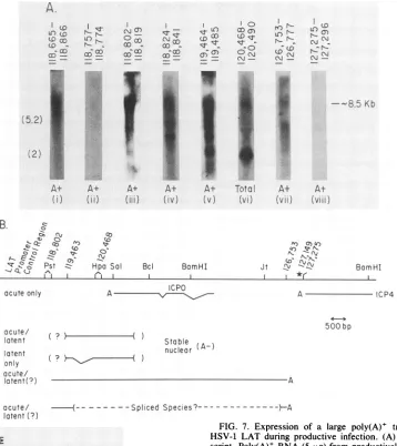
FIG. 7. Expression of a large poly(A)+ transcript containing
HSV-1 LAT during productive infection. (A) Limits of the
tran-script. Poly(A)+ RNA(5 ,ug) fromproductively infected cells was
fractionated, blotted, andhybridized with the probes shown above the appropriate lanes. Lane i was hybridized with labeled PstI
fragment while the otherswere hybridized with synthetic
oligonu-cleotides. Lane vi contained 40 ,ug of total infected-cell RNA in ordertoestimate the relativeabundance of the 8.5-kbtranscript. (B) Proposed scheme for the expression of the LAT from the primary transcript. The transcripts identified in the present report and previouslyareshown. Theexistence ofsplicedpoly(A)+ speciesis inferred fromthecomplexity ofpoly(A)+ transcriptsseenwiththe
hybridization probes used. Uncertaintyconcerning therelationship between the 5'endof stablenuclearpoly(A)- LAT and the poly(A)+ primary transcript is indicated. The location of oligonucleotide probes hybridizing to the primary transcript, the polyadenylation signalatthe3' end ofICP4, and the location of the oligonucleotide
3' ofthis, whichdoes nothybridize tothe primary transcript, are
notedby the sequencenumbers. (C) RNase protection analysisof the 5' end ofthe long poly(A)+ transcript. As described in the
experimental section, RNase-resistant material formedby hybrid-ization of the73-base RNAprobe thatspanstheEagI-to-PstI sitesof HSV-1 andnonspecific linkerRNA was hybridizedwithpoly(A)+
andpoly(A)-RNA from infected cultured cells andwasfractionated onadenaturingacrylamide gel. Lane i contains DNA size markers from HaeIII-digested pBR322. The migration of two RNA size
markers of 113 and 54 bases is different from these DNA markers and is shown inparenthesesandindicatedasRNA-M. Theposition
of themajor protected speciesfrompoly(A)+ RNA(lane iii)
corre-spondstoabout63 bases. Lanevshows the material obtained when
theprobewashybridizedwith50,ugof total RNA from uninfected
rabbit skin cells. 3849
;)
cc)r
LC)
r-(0
r-tcM
CM,
-j
A.
o 27CO
0
--co
co
-(5.2)
'k2) ;.
A+
()I
CO
A+ (Viii)
BomHl
LL
I I Z
Z °.. z zZ
acute!
latent(?)
r
C.2
(113)---l123/41|
104 89 1 (-'63) 80(1
64
I
(54)--57.^ 51 *
on November 10, 2019 by guest
http://jvi.asm.org/
wasdetected in the poly(A)+ fraction of RNA isolated from productively infected cultured cells, andrepresentative data are shown in Fig. 7A. Hybridization of a Northern blot of poly(A)+ RNA with radiolabeled DNA that spans the PstI sites indicates the presence of atranscript migrating with a sizecorresponding to 8.5 kb (lane i).
Oligonucleotide probes were used to define thelocation of
this transcript on the HSV-1 genome, and the location of eachprobe is indicated in theappropriate lanes in the figure. It should be noted that the sequences for the KOS(M) and 17syn+ strains covering these probes are all identical except for position 118,807 in which there is a G-C pair in the latter sequence and a C-G in the former. Northern blots which werehybridized with oligonucleotide probes homologous to a region 5' of the putative cap site (lane ii) and 3' of the polyadenylation site (lane viii) did notreveal this large RNA species; however, all oligonucleotides within this region of the genome did (lanes iii to vii). Other smaller poly(A)+ species were also seen by using the positive
oligonucleo-tides, which suggests that this long transcript was variably processed to a number of spliced poly(A)+ species; how-ever, this finding was not investigated further at this time. Finally, a Northern blot of total infected-cell RNA was hybridized with a probe homologous to the stable
poly(A)-LATspecies in order to estimate the relative abundance of the large poly(A)+ transcript (lane vi). It is clear that the transcript is significantly less abundant than is LAT in productive infection.
The precise 5' end for transcription of the LAT was determined for RNA from a productive infection with wild-type virus. RNase protection experiments showedan RNA product of 63 to 64 bp, using a probe from the EagI site upstream of the cap site to the downstream PstI site and
poly(A)+ RNA (Fig. 7C). Smaller bands may reflect cryptic
startsites. A possible cap site would be in the sequence ATC 24 bp downstream of the TATAbox (11).
Taken together, the data suggest that the stable LAT transcript which is detected in latently infected neurons is derived from an 8.5-kb transcription unit (Fig. 7B). From this transcription unit a number of different nuclear RNA species have been observed. These species are present in different concentrations within the nucleus.
DISCUSSION
The presentcommunicationdemonstrates that insertionof
the rabbit beta-globin gene into HSV-1 immediately down-stream of a potential Pol II promoter sequence 700 bases 5' of the start of the stable nuclear
poly(A)-
LATresults in the expression of cytoplasmic poly(A)+ globin RNA in latently infected neurons. This insertion results in the loss of all detectable LAT expression in the latent phase ofinfection.In a productive infection, this mRNA starts at the normal mRNA cap site. Our results indicate that stable LAT is derived from a primary transcription unit starting approxi-mately 700 bases 5' of its previously reported start. In further experiments, we have confirmed the existence of the primary transcript and shown that at least some transcription extends 8.5 kbfrom the start to a polyadenylation signal near the 3' end of ICP4. Although we have been unable to detect such a transcript in Northern blots of acute or latently infected neurons, the weak in situ hybridization signals seen with probes spanning the LAT and its flanking sequences reported by us and others suggests that this transcription unit operates in neuronal infection with HSV-1 (2, 15, 24).
Previous work with an HSV variant (X10-13) which has a
spontaneous deletion of sequences that include the newly characterizedLAT promoterhas clearlydemonstrated that neither the expression of stable LATs nor of the less abundant RNA 3' of these species is required for the establishmentormaintenanceof the latentphase of infection in mouse neurons(8). Although not different fromwild-type
virus in establishment ormaintenance, virus X10-13 reacti-vates poorly when induced in the rabbit eye model (T. J. Hill, F. Sederati, E. K. Wagner, and J. G. Stevens, manu-script inpreparation). In additiontothis in vivoreactivation, there is some evidence thatLATexpression affects in vitro reactivation (17). Thus, LAT expression does have an im-portant role in HSV latency. It is not clear that this role involves the expressionofaprotein,but the datapresented
here suggest that the900-base openreadingframepreviously
noted in the stable LAT species is not the only candidate protein whichcould be expressed.
Fromtheseconsiderationsand from the datapresentedin this report,we are nowabletomake severalnewstatements about theexpression of LATsduringalatent infection.First, it is likely that transcription of LATs is regulated by con-ventional Pol II promoter elements. Second, stable LAT is probably processed from a much larger primary transcript
thatispolyadenylated inproductiveinfectionatleast.Third,
the facts that stable LAT is flanked by potential splice signals and is nuclear
poly(A)-
speciessuggestthat the LAT may be an intron. It is possible that a large part of the regulation of thistranscriptionalunit isat theposttranscrip-tional level.RNAsplicing,polyadenylation,RNAtransport, and RNA stability may all play a role in regulating the products from thistranscriptionalunit.
If the product of the LAT is a protein or a number of
proteins, then an importantgoal becomes the identification ofexonsthat encode theseproteins. Sincestable,
cytoplas-mic, poly(A)+ globin mRNA can be expressed from a recombinant HSV genome during a latent infection, the
instability of LAT mRNAsmustbe dueto the
properties
of the RNA expressed. Thus, it may be possible to devise methods of stabilizing such a species. We are currentlyinvestigating such apossibility.
Finally, thereis considerable interest inusingHSV-1 as a vector for gene transfer to neurons. The beta-globin virus used in these experiments fairly faithfully expresses a
for-eign gene product stably in neurons in vivo. The lack of
expression of other lytic genes during latency and the stability of HSV-1 in latent neurons may make this an attractive system.
ACKNOWLEDGMENTS
We thank M. Rice and V. Dissette for helping with technical details. We thank Ken Izumi for the use of viral DNA used to
constructthe PstI deletion virus.
This work was supported by grant MV-351 to L.T.F. from the American Cancer Society and by Public Health Service grant A106246 from the National Institutes of Health to J.G.S. and E.K.W. Further support was provided by Public Health Service grant CA11861, American Cancer Society grant MV372, the UC Irvine Focused Research Program in the Molecular Biology of Eukaryotic Viruses (E.K.W.), and Multiple Sclerosis grant RG-1647-Al (J.G.S.). A.T.D. was supported by molecular sciences training grant program grant GM08042. W.M.F. is a predoctoral traineeofacarcinogenesis traininggrant(T32-CA-09054). M.J.F.is
a predoctoral trainee of the Genetics Training grant National Re-search Service Award GM-07104. L.T.F. is a recipient offaculty research award FRA-340 from the American CancerSociety.
on November 10, 2019 by guest
http://jvi.asm.org/
LATENT EXPRESSION OF GLOBIN 3851 LITERATURE CITED
1. Croen, K. D., J. M. Ostrove, L. J. Dragovic, J. E. Smialek, and S. E. Straus. 1988. Latent herpes simplex virus in human trigeminal ganglia. Detection of animmediate-early gene "anti-sense" transcript by in situ hybridization. N. Engl. J. Med. 317:1427-1432.
2. Deatly, A. M., J. G. Spivack, E. Lavi, and N. W. Fraser. 1987. RNA from an immediate-early region of the type 1 herpes simplex virus genome is present in the trigeminal ganglia of latently infected mice. Proc. Natl. Acad. Sci. USA 84:3204-3208.
3. Draper, K. G., G. B. Devi-Rao, R.H. Costa, E. D. Blair, R. L. Thompson, and E. K. Wagner. 1986. Characterization of the genesencodingherpes simplex virus type 1 and type 2 alkaline exonucleasesand overlappingproteins. J. Virol. 57:1023-1036. 4. Gilman, M. 1989. Preparation and analysis ofRNA, p. 4.7.1-4.7.8. In F. Ausubel, R. Brent, R. Kingston, D. Moore, J. Seidman, J. Smith, and K. Struhl (ed.), Current protocols in molecular biology. Wiley-Interscience, New York.
5. Gordon,Y.J., B. Johnson, E.Romanowski, and T. Arullo-Cruz. 1988.RNAcomplementarytoherpes simplex virus type 1 ICPO demonstrated inneurons of humantrigeminal ganglia. J. Virol. 62:1832-1835.
6. Haase, A., M. Brodie, L. Stowring, and H. Blum. 1984. Detec-tion of viral nucleic acids by in situ hybridization. Methods Virol.7:189-226.
7. Hill, T. J. 1985. Herpessimplexviruslatency, p. 175-240. In B. Roizman (ed.), The herpesviruses, vol. 3. Plenum Publishing Corp.,New York.
8. Javier, R. T., J. G. Stevens, V. B. Dissette, and E. K. Wagner. 1988. A herpes simplex virus transcript abundant in latently infectedneurons isdispensable for establishment of the latent state. Virology 166:254-257.
9. Logan, J., E. Falk-Pedersen, J. E.Darnell, and T. Shenk. 1987. Apoly(A) addition site andadownstreamtermination regionare required for efficient association oftranscription byRNA poly-meraseII inthemouse betamaj-globingene. Proc. Natl. Acad. Sci. USA84:8306-8310.
10. Maniatis, T., E. F. Fritsch, and J. Sambrook. 1982. Molecular cloning: alaboratory manual,p. 109-121. Cold SpringHarbor Laboratory, ColdSpring Harbor, N.Y.
11. McGeoch, D. J., M. A. Dalrymple, A. J. Davison, A. Dolan, M.C. Frame, D. McNab, L. J.Perry, J. E.Scott,andP.Taylor. 1988.ThecompleteDNAsequenceofthelongunique regionin the genome of herpes simplex virus type 1. J. Gen. Virol. 69:1531-1574.
12. McGeoch, D. J., A. Dolan, S. Donald, and D. H. K. Brauer. 1986.CompleteDNAsequenceoftheshortrepeatregioninthe genome ofherpes simplex virus type 1. Nucleic Acids Res. 14:1727-1745.
13. Padgett, R. A.,P.J. Grabowski, M. M.Konarska, S.Seiler,and P. Sharp. 1986.Splicing ofmessenger RNA precursors. Annu. Rev.Biochem. 55:1119-1150.
14. Puga, A., and A. L. Notkins. 1987. Continued expressionofa
poly(A)+transcript of herpes simplexvirus type 1 intrigeminal
gangliaoflatently infected mice.J. Virol.61:1700-1703. 15. Rock, D. L., A. B. Nesburn,H. Ghiasi, J. Ong, T. L. Lewis, J. R.
Lokensgard, and S. L. Wechsler. 1987. Detection of latency-related viral RNAs in trigeminal ganglia of rabbits latently infected with herpes simplex virus type 1. J. Virol. 61:3820-3826.
16. Spivack, J. G., and N. W. Fraser. 1987. Detection of herpes simplex virus type 1 transcripts during latent infection in mice. J. Virol. 61:3841-3847.
17. Steiner, I., J. G. Spivack, R. P. Linette, S. M. Brown, A. R. MacLean, J. H. Subak-Sharpe, and N. W. Fraser. 1989. Herpes simplex virus type 1 latency associated transcripts are not essential for latent infection. EMBO J. 8:505-511.
18. Stevens, J. G., and M. L. Cook. 1971. Latent herpes simplex virus in spinal ganglia ofmice. Science 173:843-845.
19. Stevens, J. G., L. Haar, D. Porter, M. L. Cook, and E. K. Wagner. 1988. Prominence of theherpes simplex virus latency-associated transcript in trigeminal ganglia from seropositive humans. J. Infect. Dis. 158:117-123.
20. Stevens, J. G., E. K. Wagner, G. B. Devi-Rao, M. L. Cook, and L. Feldman. 1987. RNAcomplimentaryto aherpesvirus alpha gene mRNA is predominant in latently infectedneurons. Sci-ence235:1056-1059.
21. Stroop, W. G., D. L. Rock, and N. W. Fraser. 1984.Localization ofherpes simplexvirusin thetrigeminalandolfactorysystems of the mouse central nervous system during acute and latent infectionsby in situhybridization. Lab. Invest. 51:27-38. 22. Thompson, R. L., M. L.Cook, G. B. Devi-Rao, E. K.Wagner,
andJ. G. Stevens. 1986. Functional and molecularanalyses of theavirulentwild-type herpessimplexvirus type 1 strain KOS. J. Virol. 58:203-211.
23. Van Oogen, A., J. van den Berg, N. Matei, and C. Weissmann. 1979. Comparison of total sequence of a cloned rabbit beta-globin gene and its flanking sequences with a homologous mousesequence. Science 206:337-344.
24. Wagner, E. K., G. B.Devi-Rao, L. T.Feldman,A. T.Dobson, Y. F.Zhang, W. M.Flanagan,andJ. G.Stevens. 1988.Physical characterization of the herpes simplexviruslatency-associated transcriptin neurons. J.Virol. 62:1194-1202.
25. Wagner, E. K., W. M. Flanagan, G. Devi-Rao, Y. F. Zhang, J. M. Hill, K. P. Anderson, and J. G. Stevens. 1988. Theherpes simplexviruslatency-associated transcriptisspliced duringthe latentphase of infection.J. Virol. 62:4577-4585.
26. Wechsler,S.L.,A. B.Nesburn,R.Watson,S.Slanina,and H. Ghiasi. 1988. Finemappingof themajor latency-relatedRNA of herpes simplex virus type I in humans. J. Gen. Virol. 69: 3101-3106.
27. Wechsler, S. L., A. B.Nesburn,R.Watson,S. M.Slanina,and H. Ghiasi. 1988. Fine mapping ofthe latency-related gene of herpes simplex virus type 1: alternative splicing produces dis-tinctlatency-related RNAscontaining openreadingframes. J. Virol. 62:4051-4058.
28. Wechsler, S. L.,A. B. Nesburn, J. Zwaagstra, and H. Ghiasi. 1989. Sequence of the latency-related gene ofherpes simplex virus type I. Virology 168:168-172.
VOL.63,1989