0022-538X/90/010207-08$02.00/0
Copyright © 1990, American Society for Microbiology
Mutational Analysis of
Human
Papillomavirus
Type
16 E7
Functions
SUMIE WATANABE, TADAHITO KANDA, HIRONORI SATO, AKEMI FURUNO,AND KUNITO YOSHIIKE*
Department of Enteroviruses, NationalInstitute ofHealth, Kamiosaki, Shinagawa-ku, Tokyo 141,Japan Received19 June1989/Accepted17September 1989
The human papillomavirus type 16 E7 gene encodes a nuclear oncoprotein (98 amino acids [AAs] long) consisting of three regions: regions1 (AAs 1 to20) and2 (AAs 21 to40), which show high homologytothe
sequences of conserved domains 1 and 2, respectively, of adenovirus ElA; and region 3 (AAs 41 to 98)
containingtwometal-binding motifs Cys-X-X-Cys (AAs 58to61 and 91to94). Weconstructed AA deletion
(substitution) mutantsandsingle-AAsubstitution mutantsof E7 placed under thecontrolofthesimianvirus
40promoterand examinedtheirbiologicalfunctions. Stable expression of E7 protein inmonkey COS-1cells required almost the entire length of E7 and was markedly lowered by the mutations in region 3.
Transactivation oftheadenovirus E2 promoterin monkeyCV-1cells was loweredbythe mutations. Itwas abolished by changing Cys-24toGly and markedly decreased byamutationatHis-2oratthemetal-binding
motifs in region 3.Focal transformationofrat3Y1 cells by E7 waseliminatedbychanging His-2 toAspor
Cys-24 to Gly and was greatly impairedby changing Cys-61 or Cys-94 to Gly. The transforming function
survived mutationsatLeu-13 and Cys-68anddeletion ofAsp-Ser-Ser (AAs 30to32). The datasuggestthat
regions 1 to 3 are requiredfor its functions and that the metal-binding motifs in region 3are required to
maintainastable orfunctionalstructureof the E7 protein.
Humanpapillomavirus type16 (HPV 16) (5) isbelievedto causecervicalcancers(for review,seereference 47), and its open readingframe, E7, encodesasmall nuclear(33) onco-protein composed of 98 amino acid (AA) residues (35). Expression of the E7genecaninduce cell DNA synthesis in
serum-starved, contact-inhibited rat 3Y1 cells (32). The E7
geneis immortalizing and transforming for primaryratcells (17, 39) and establishedlines ofmouseandratcells (15, 28) andiscapable oftransforming primary human fibroblasts in cooperation with the E6gene (42). The E7gene can
trans-activate the adenovirus E2promoter(28). The E7 protein is nuclear when the E7-expressing monkey COS-1 cells are examined by theimmunofluorescence method (33), although it is found in the cytoplasmic fraction upon subcellular fractionation (33, 37). The functions and localization of E7 resemble those of adenovirus ElA and simian virus 40 (SV40) large-T, well-studied viral nuclear oncoproteins (26, 41). It was recently reported that, like ElA and SV40 T
antigens, HPV 16 E7 protein is able to bindto the retino-blastomageneproduct (6).
The nucleotide sequences of HPV DNAs enable us to
deduce the AAsequencesof E7 and otherHPVproteins (4, 34, 35). From the structural features,the HPV 16E7protein
canbetentatively divided into three regions (Fig. 1). Region
1 (the N-terminal 20 AAs) and region 2 (AAs21 to40) show high homologyto partsof adenovirus ElA(26,28). AAs 6to
20 and AAs 21to40 of HPV 16 E7 resemble AAs41to56(in conserved domain 1) and AAs 121 to 139 (in conserved
domain2), respectively, ofadenovirus 5ElA,both of which
constitute the essential regions for the ElA transforming function(21, 26, 27, 45). Region 3,theC-terminal half(AAs 41to98), containsapossible zinc fingerstructure(1, 8).Two
metal-binding motifs, Cys-X-X-Cys, are separated by a
stretch of 29AAs (Fig. 1).Like HPV 16, HPV 18(3, 4)and
HPV6(34) show similarE7structuralfeatures (Fig. 1). Inthisstudy,wepreparedvarious HPV 16 E7mutantsby introducing deletions into the E7gene andby site-directed
*Correspondingauthor.
mutagenesis and examined theircapabilities to exert
trans-activating and transforming functions. Recently, Edmond and Vousden (7) reported a point-mutational analysis of HPV 16 E7protein.
MATERIALS AND METHODS
Plasmids. Plasmidsused inthis studywerepSV2-E7P (15), pSV2-0 (40), pE2CAT (14), pEJ6.6 (36), pSV2neo (38), and pSVneo-E6 (15). Plasmid pSV2-E7Pexpresses the HPV 16 E7 gene controlled by the SV40 promoter. It contains an
HPV16 DNAfragment, nucleotides (nt) 554to874(spanning the entirecoding region forE7), inserted attheHindIIl site into vectorpSV2-0 containing SV40 transcriptional
regula-toryelements(promoter-enhancer, intron, and polyadenyla-tion site). Mutants of HPV 16 E7 were constructed from
pSV2-E7P. In pE2CAT, which was used as a reporter of transactivationby E7, the bacterialchloramphenicol acetyl-transferasegeneisplaced under the control of the
adenovi-rus 5 E2 promoter. Plasmid pEJ6.6, which was used in
cotransfection for determination of the immortalizing func-tion of E7, contains the activated ras oncogene. Plasmid pSV2neo expresses the neomycin (G418) resistance gene under thecontrol of the SV40promoter. Fordetermination
of the transforming function of E7 for WI38 human
fibro-blasts, pSVneo-E6wasused forcotransfectionwithmutated
pSV2-E7 plasmids. PlasmidpSVneo-E6 containsanHPV 16
E6 (nt 25 to 657) transcription unit (15) inserted into
pSV2neo at the BamHI site. Escherichia coli HB101 was used for propagation of all plasmids except pEJ6.6, for which E. coli DH-1wasused.Preparation and purificationof
plasmids weredonebystandard methods(22).
Cells. Monkey COS-1 (9)and CV-1 celllines, therat3Y1
cell line (18), primary baby rat kidney (BRK) cells, and
primary human fibroblasts, W138 (13), were used in this
study. The COS-1 cell line, which is an African green
monkey kidney-derivedCV-1 cell line transformedby origin-minus SV40 DNA, is constitutively expressing SV40 T
antigenandcansupport replicationoftheplasmidswiththe
SV40 DNA replication origin. COS-1 cells, grown in
Dul-beccomodifiedEaglemedium with 10% fetalbovine serum, 207
on November 10, 2019 by guest
http://jvi.asm.org/
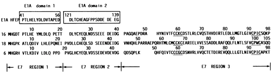
ElA domain 1 ElA domain 2
41 56 121 139
ElA HFE PTLHELYDLDVTAPE DLTCHEAGFPPSDDE DE EG
10 20 30 40
16 MHGDT PTLHE YMLDLQ PETT DLYCYEQLNDSSEEE DEIDG PAGQAEPI
10 20 30 40 50
18 MHGPK ATLQDIV LHLEPQNEI PVDLLCHEQLSD SEEENDEIDG VNHQHLP
10 20 30 40
6 MHGRH VTLKDIV LDLQ PPD PVGLHCYEQLVDSSEDEVDEVDG QDSQPLK
E7 REGION 1 E7 REGION 2 * a
50 60 70 80 90 98
'DRA HYNIVTFCCKCDSTLRLCVQSTHVDIRTLEDLLMGTLGIVCPICSQKP
60 70 80 90 100 105
OARRAEPQRHTMLCMCCKCEARIELVVESSADDLRAFQQLFLNTLSFVCPWCASQQ
50 60 70 80 90 98
QHFQIVTCCCGCDSNVRLVVQCTETDIREVQQLLLGTLNIVCPICAPKT
E7 REGION 3 -
1
FIG. 1. AAsequencesofHPV16,18, and6E7proteins.Thesequencesofadenovirus 5ElAand HPVsarebasedonthepublished DNA
sequences (4, 34, 35, 41). LikeHPV16, HPV18DNA has beencloned fromacervicalcarcinoma(3) and its E7 is transforming (44). The E7
proteins are dividedintothreeregions. Regions1and2showhomologytotheAAsequencesinconserveddomains1and2, respectively, of
adenovirusElA(28). Region3containstwometal-binding motifsCys-X-X-Cys(underlined).
wereusedtodetectproduction of E7 protein by immunoflu-orescence staining (33) andimmunoprecipitation (33). CV-1 cells, growninDulbecco modified Eagle mediumwith 10% fetalbovineserum, wereusedtodetermine the
transactivat-ing function of E7. Rat cell line 3Y1, grown in minimum
essentialmedium with 10%fetal bovine serum,wasusedto measuretheE7functiontoinduce focal transformation(16). Primary BRK cells were prepared from 5-day-old Fischer rats. The cells were grown in Dulbecco modified Eagle
medium with 10% fetal bovine serum and were used at passage 3 for determination of E7 immortalizing function
(23). WI38 primary human fibroblasts, the gift of Y. Doi
(JapanPoliomyelitis Institute),were usedat 25 to 30 popu-lationdoublings fordetermination of E7transforming
func-tion,asdescribed previously (42).
Antisera. Rabbit antisera raised against bacterial fusion
proteinHPV 16 lac-E7 were used for immunofluorescence
detection (33) and immunoprecipitation (33) of E7protein. Preparation andcharacterization of HPV 16 lac-E7 protein and anti-lac-E7 sera were described previously (33). For
immunofluorescence staining, antiserawere extensively in-cubated at4°C withacetone-powdered Africangreen mon-key kidney tissue before use. For immunoprecipitation,
antisera wereusedwithout suchtreatment.
Constructionofmutantsby deletion. We introducedDNA deletionsintotheE7genebyutilizing convenient restriction sites. Restriction endonucleases (AccI, HpaII, Hinfl, MnlI,
NcoI, and SspI), other enzymes, and linkers (EcoRI and
HpaI)werepurchased from Takara Shuzo Co., Ltd., Kyoto,
Japan, and New England BioLabs, Beverly, Mass., and
wereusedasrecommended by the suppliers. The nucleotide sequence around the mutation site (and of the E7 coding region for themutantsaccidentally made)wasdetermined by
theM13-dideoxymethod (25, 31) withanM13sequencing kit (Takara Shuzo) for each mutantconstructed.
Forconstruction ofmutants729, 747, 774, 7SS, 7DSS, and 54/61, the shorterHindlll fragment (HPV 16nt 554to 874)
was isolated from pSV2-E7P by gel electrophoresis and
circularized by ligation. The E7 minicircles were digested
with appropriate restriction enzymes, blunt ended by
Kle-now, and, withorwithout insertion ofappropriate linkers,
recircularized byT4 ligase. The mutated and recircularized DNAsweredigested withHindIII andreinserted into pSV2-0atthe HindlIl site ina sense orientation.
Mutantswithdeletions ofC-terminal AAs of the E7gene
(790, 791, 795,and 796)wereconstructedinadifferentway.
The shorter NcoI fragment was isolated from HPV 16
pSV2-E7P, which hastwoNcoIsites:onewithin the E7gene
near its C-terminal end and the other within the SV40 transcriptional control
region.
The isolated NcoIfragment
was digested with exonuclease III, blunt ended with
Si
nuclease, andaddedwithHindllllinkersatboth ends. This product was digested with HindlIl. The
Hindlll-digested
DNAretainedthe intactN-terminal end ofthe E7 gene and the exonuclease III-digested, truncated C-terminal end of the E7 gene. The final
Hindlll-digested
fragments were reinserted intopSV2-0 at theHindlll site in a sense orien-tation.Construction of mutants by site-directed mutagenesis. A single base at a specific site in the HPV 16 E7 gene was replaced with another base by the gapped duplex DNA method (19),
using
Mutan-G(Takara Shuzo),asite-directed mutagenesis system. The shorterHindIlI
fragment from pSV2-E7P (HPV 16 DNA nt 554 to 874) was cloned into M13. Forannealing,thecomplementary17-merscontaining asinglebase substitutionin the middleweresynthesized
in amodel 381ADNAsynthesizer
(Applied Biosystems,Foster City, Calif.).Themutantswereselectedonthebasis ofDNA sequence (31) around the site of intended mutation. For confirmation,theentireE7codingregionwassequenced
for each selected mutant by the dideoxy method (31). The E7HindlIl
fragments withmutations, isolated from replicative forms, were inserted into pSV2-0 vectors in a sense orien-tation. Inmutants7H02Dand 7L13S,Catnt565andTat nt599 were replaced with G and C, respectively. In mutants 7C24G, 7C61G, 7C68Gand7C94G,T's at nt631, 742, 763, and 841werereplacedwithG's,respectively. Mutant7CC/G wasadouble mutant made from mutants7C61G and 7C94G. Detection of E7 protein by immunofluorescence staining. COS-1 cells on cover slipswere transfected by the DEAE-dextran method (24) with pSV2 plasmids containing E7 or
mutated E7 DNAs (1
jig
of DNA per cover slip). At 48 h after transfection, the transfected cells were processed asdescribed previously (33) with the anti-lac-E7 serum, and fluorescent cellswere scored.
Detection of E7 protein by immunoprecipitation. COS-1 cells (60-mm dish) were transfected by the DEAE-dextran method(24) with pSV2plasmidswith mutated E7 (4 ,ug) and
were labeled 48 h later with 0.2 mCi of
[35S]methionine-cysteinemixture
(Trans35S-label;
ICNRadiochemicalsInc., Irvine, Calif.) for 1 h. Then the labeled cultures were subjectedto subcellularfractionation, as described by Bar-bosa and Wettstein (2). Samples of cytoplasmic fractionswereanalyzedasdescribed previously (33). In chase exper-iments, the labeled cultures were maintained for 60 min in
on November 10, 2019 by guest
http://jvi.asm.org/
[image:2.612.74.556.84.212.2]medium without35S-labeledAAsbefore subcellular fraction-ation.
Analysis of RNA. Total RNA extracted from the pSV2 plasmid-transfected COS-1 cultures 48 h after transfection was analyzed by the Northern (RNA) blot method (20) as described previously (42). RNA on membrane filters was probed with HPV 16 E7 DNA fragment (nt 554 to 874) labeledwith 32Pbynick translation (30).
Assay for transactivation by E7. CV-1 cells (60-mm dish) were transfected with pE2CAT (5 ,ug) and pSV2-E7P or pSV2 plasmids containing mutated E7 (5 ,ug) by the calcium coprecipitation method (11) under conditions used previ-ously (43). Chloramphenicol acetyltransferase activities were assayedfor
[14C]-chloramphenicol
(Amersham Inter-national plc, Buckinghamshire, England) with the cell ex-tracts prepared 48 h after transfection, as described by Gormanet al. (10).Assayfor focal transformation of rat 3Y1 cells by E7. The capacityofpSV2-E7Por pSV2plasmids with mutated E7 to induce focal transformation was examined in rat 3Y1 cells, an
immortalized
normal cell line, as described previously (16). Plasmids (10 ,ug) were transfectedto subconfluent rat 3Y1 cells (30-mm dish) by the calcium coprecipitation method(11).Fociwere scored 4 to 5 weeks after transfection with cultures stained with5% Giemsa.Assay forimmortalization ofprimary ratcells by E7.The immortalizing function was assayed as described by Mat-lashewski etal. (23). Primary BRKcellswerecotransfected with pEJ6.6,pSV2plasmidswithmutatedE7, andpSV2neo (5 ,ug eachfor a 60-mm dish culture) bythe calcium copre-cipitation method(11). Some 3 to 4 weeks aftertransfection, the cultures (resistant to G418 [Sigma Chemical Co., St. Louis, Mo.])werefixed withmethanol and stained with 5% Giemsa. Foci of the cells characteristic of those cotrans-formedby E7 and activatedras werescored.
Assay for E7 transforming function for human cells. The
ability
of the E7genetotransform human cells (to extend the life span) in cooperation with theE6 gene wasexamined inW138
primary human fibroblasts (42). Plasmids (with or without mutation) pSV2-E7 and pSVneo-E6 (5 ,ug each) werecotransfected toW138
cells bythe calcium coprecipi-tation method(11). TheG418-resistantcellsweremaintained andprocessedessentially
as described by Pirisi etal. (29). Cultures containing cells surviving beyond 65 population doublingswereregardedastransformation positive(42).RESULTS
AAdeletion(substitution) mutants of HPV 16 E7.Deletion ofasmallsegmentfromHPV16 E7 DNA
generated
aseries offrameshift mutants and AA deletion(substitution)
mu-tants. Figure 2 is a schematic
representation
of the AA sequences of these mutants estimated from their DNA sequences. Mutants 729, 747, 774, 790, 791, 795, and 796 were frameshift mutants. These mutants encode truncated E7proteins with
the N-terminal 29to96AAsfollowed
by
ashortstretchofAAsequence
originating
fromframe-shifted sequencesof eitherHPV 16orSV40DNA. Mutants7DSS,
7SS, and 54/61 encodemutated E7
proteins
withashort AA deletion(substitution)
inthemidsection.The pSV2 plasmids
containing
these mutated E7 genes (placedundercontrol oftheSV40promoter)weretested for abilities to induceproduction
of E7protein (assayed
by
immunofluorescence) in
monkey
COS-1 cells(33),
to acti-vate expression ofchloramphenicol
acetyltransferase
from the adenovirus E2promoter inmonkey
CV-1 cells(28),
to20 40 60 80 AAS 100
l ~c c c
5861 9194
16E7 1l
Rl R2 R3
29 (35) 729
47 (55) 747
74(76)
774 1
90 (95)
790
91(96)
791
-95(100)
795
-96(101) 796
29 33 98
'7rCCI/uL:l r 7SS i:
54/61 E
98
3033
-Er-53 62 98
-]: I I
FIG. 2. Schematic representation of E7 deletion mutants of HPV 16. HPV 16wild-type E7 (98 AAs) is indicated by 16E7. Regions1, 2,and 3(Fig. 1) are indicated by Rl, R2, and R3, respectively. C's in R3 indicate cysteines in metal-binding motifs. Open rectangles represent AA sequences originating from E7 protein, and the number above themshows the end AA from E7. Filled rectangles representAAsequencesfrom fame-shifted DNA sequences and the number in parentheses shows the total number of AAs of mutant proteins. Filled rectangles for HPV 16 represent Glu-Arg-Arg-Arg-Met-Lys (in 729),Arg-Thr-Glu-Pro-Ile-Thr-Ile-Leu (747), Gly-Cys (774), Lys-Leu-Gly-Ser-Leu (790, 791, and 795),
Ala-Cys-Gly-Ser-Leu(796), Glu (7DSS), Arg (7SS), and Pro-Asn-Ser(54/61). induce focal transformation of rat 3Y1 cells (16), to trans-form primary BRK cells in conjunction with activated ras (23), and to extend the life span of human WI38 cells in cooperation with the HPV 16 E6 gene(42). Table 1 summa-rizes the E7functions of the HPV 16 E7 deletion mutants shown inFig. 2.
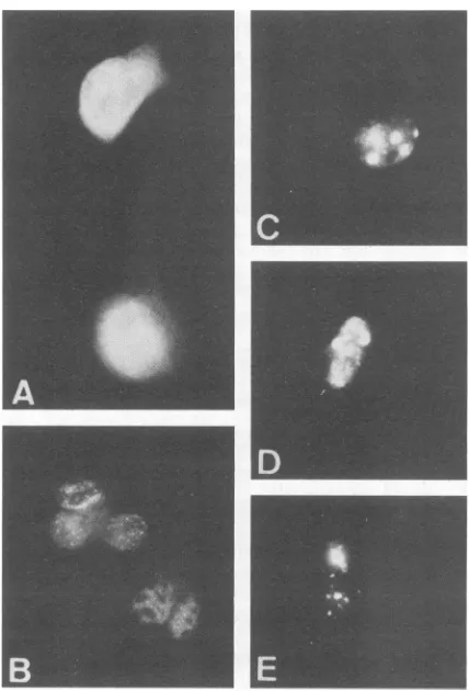
Steady-state expression of the mutated HPV 16 E7 from the SV40 promoter was examined inCOS-1 cellsby immu-nofluorescence staining (Fig. 3 and Table 1). As described previously (33), E7-producingcells wererecognizedasthose withfluorescent nuclei stainedwithrabbit anti-lac-E7serum 48 h after transfection. Comparison of the frameshift mu-tants729, 747,774, 790, 791, 795,and796 showsthat,for full orstableexpression,almost the entirelengthof E7(95 of 98 AAs)wasrequired.Probable mutantpolypeptidesof 729(35 AAs long) and 747 (55 AAs long) were undetectable by immunofluorescence. The 774 protein (76 AAs long) was detected in a markedly reduced number ofcells, and the positive nuclei with the presumably truncated 774 protein appeared
differently
underaUVmicroscope.
Whereasmost oftheE7-positive nuclei,
like SV40-T- oradenovirus E1A-positivenuclei,containedimmunofluorescent dots with vari-able density (Fig. 3A and B), the 774 E7-positive nuclei contained condensed masses ofimmunofluorescence (Fig. 3C, D, andE). Theseabnormally stained nuclei constituted about 0 to 3% of thepositive cells in thewild-type (wt) E7 transfection and about30% in transfection with mutant790 or791.Deletion ofAsp-Ser-Ser
atAAs 30to32orSer-SeratAAs 31 to32
(in
mutants 7DSS and7SS)
did notaffect the level ofE7-positivecells in the transfected COS-1 cultures much, but deletion of AAs 54 to 61(in
mutant 54/61)on November 10, 2019 by guest
http://jvi.asm.org/
[image:3.612.318.541.81.301.2]TABLE 1. Functions of HPV 16 E7deletion mutants
Transformingactivity for:
a Steady-stateE7protein Transactivation(%)for
Plasmid COSSin - cells
(no.
)b adenovirus E2 promoter in Rat3Y1 BRK HumanW138CV-1 cells' (no.offoci)d (immortalization (transformation
CV-1cellsC
~~~~~~~~efficiency)'
efficiency)fpSV2-0 0 1.0 0 0/2 0/2
pSV2-E7P 1.00 9.2 1.00 4/4 4/4
pSV2-729 0 0.9 0 0/4 NT9
pSV2-747 0 1.0 0 0/4 NT
PSV2-774 0.01 1.7 0 0/4 0/4
pSV2-790 0.16 1.7 0.01 NT 0/4
pSV2-791 0.18 1.6 0 0/4 1/4
pSV2-795 0.77 4.5 0.77 4/4 NT
pSV2-796 0.98 5.5 0.68 4/4 NT
pSV2-7DSS 1.12 4.0 0.34 4/4 2/2
pSV2-7SS 1.34 4.2 0.18 4/4 2/2
pSV2-54/61 0.14 3.4 0 0/4 2/4
a AA sequencesof the protensencodedby theseplasmidsareshown inFig.2.
bTransfected COS-1 cell cultures were stained with anti-lac-E7 48hafter transfection. Number ofimmunofluorescence-positivecellswerenormalizedtothat
inpSV2-E7P-transfected cultures, whosepositive cells constituted about 0.5%. Average of fourexperiments.
cAverageof twoexperiments. Thechloramphenicol acetyltransferaseactivities(percentconversiontoacetylatedform perprotein content)ofpE2CATwith
each mutant werenormalized to that withpSV2-0, which isanontransactivatingbackbonevector.
d Average of threeexperiments. Number of fociwerenormalizedtothat ofpSV2-E7P.Efficiencyof focal transformationbypSV2-E7Pwas66foci per10,ug
ofDNA.
e Primary BRK cells were cotransfected byE7and activatedras,and cultureswith foci of the cotransformed cellswerescoredasimmortalizationpositive.
Immortalization efficiency is expressed as a ratio of cultures with colonies of transformed cellstoculturesinitially replated.
fPrimary human fibroblastsW138werecotransfectedwith mutated E7andHPV 16 E6(pSVneo-E6). Transformationefficiencyisexpressedasaratio of
cultures with transformed cellstoculturesinitially replated (42).
g NT,Nottested.
decreasedthenumber ofE7-positive nuclei. Deletionof AAs 92through 98 (inmutants 790 and 791) drastically lowered the steady-state E7protein expression.
Capacity to activate chloramphenicol acetyltransferase expression fromtheadenovirusE2 promoter wasexamined with pSV2 plasmids containing the mutated E7 (Fig. 2) in monkey CV-1 cells. Cell extractsprepared from cultures48 h after transfection were measured for ability to convert chloramphenicol toacetylated forms; the relative activities (compared with backbone vector pSV2-0 and wt E7) are shownin Table 1.Thetransactivating functionwaslowered byall mutations shown inFig. 2,butit was lessaffected by the deletion ofAAs 96 to 98 at theC terminusor AAs 30 to 31thanbythe otherdeletions.
Transforming functions of the mutated E7 genes were tested inrat3Y1cells,primaryBRK cells, andW138primary human fibroblasts. The E7 gene controlled by the SV40 promoter is capable by itself of focal transformation of immortalized rat 3Y1 cells (15). pSV2 plasmids with the mutantE7 genes (Fig. 2) were transfected into3Y1 cells, and foci ofthetransformed cells were scored 4 to 5 weeks later (Table 1). Comparison of the frameshift mutants (729, 747, 774, 790, 791, 795,and796)indicatesthat,forefficient focal transformation, almost theentire length of the E7 polypep-tide (95 of 98 AAs; in mutant 795) was required. Transfor-mationby mutantswith 90 to 91 AAs was drastically reduced (mutants 790 and 791). Whereas deletion of AAs 30 to 32 (in mutant7DSS) andAAs 31 to 32 (in mutant 7SS) decreased focal transformation, deletion of AAs 54 to 61 (in mutant 54/61) totally abolished the transforming function for 3Y1 cells. Mutants whose activity for focal transformation sur-vivedshowed animmortalizing function, which was assayed astransformingactivity for primary BRK cells after cotrans-fection with plasmids with the activated ras (Table 1). Mutants7DSS and 7SS extended the life span ofW138cells, in cooperation with the E6 gene. Mutant 791 and mutant 54/61 lacking AAs 54 to 61 were weakly transforming for
W138
cells.FIG. 3. Immunofluorescence staining of monkey COS-1 cells expressing HPV 16 E7protein. Cellsweretransfectedwith pSV2-E7P (A, B) or pSV2-774 (C, D, E) and stained indirectly with anti-lac-E7 serum2days after transfection.
on November 10, 2019 by guest
http://jvi.asm.org/
[image:4.612.337.551.382.697.2]16E7
7H02D
7L13S
7C24G
7C61G
7C68G
7C94G
I
20
40
60
80
AAS 100
I I I C C I C C 1
5861
91 94
I
Jl-lt
lill
I
Rl
R2
R3
His-2--AsD
Leu-13--.Ser
Cys-24-.Gly
Cys-61-Gly
I
I
I
Cys-68-pGly
I
IF
1:
Cys-94--.mGly
I
I'
Cys-611y
CyG-94-GGly
I
I
7CC/G
L
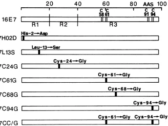
FIG. 4. Schematic representation of AA sequences of HPV 16 E7 single-AA substitution mutants. Wild-type E7 protein (98 AAs long) is asgiven in the legend to Fig. 2. Vertical bars in open rectangles indicate the positions of replaced AAs. Conversion of AAs is indicated above the rectangles representing mutated E7 proteins.
Single-AA substitution mutants of HPV 16 E7. Replace-mentof the single nucleotideat nt 565, 598, 631, 742, 763, or 841 by site-directed mutagenesis was intended to generate the AA substitution mutants shown in Fig. 4. His-2 was replacedwith Asp in the protein encoded by 7H02DDNA. Leu-13 was replaced with Ser in 7L13S protein. Cys-24, Cys-61,
Cys-68,
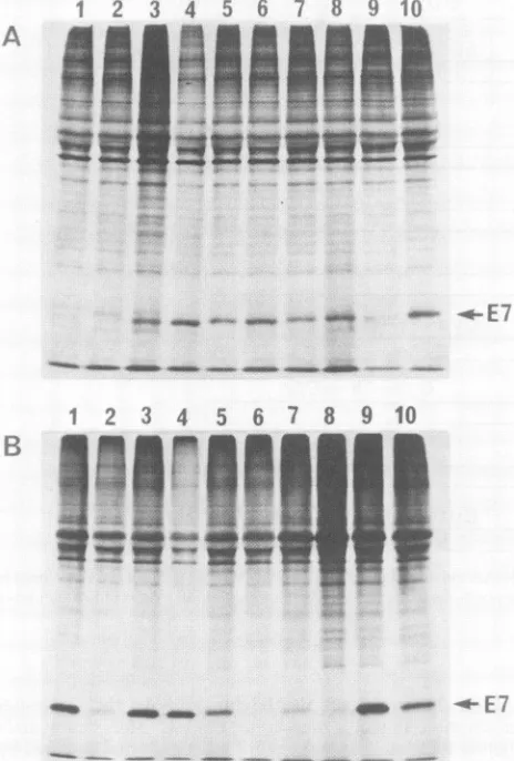
andCys-94 were each replaced with Gly in 7C24G, 7C61G, 7C68G, and 7C94G proteins, respectively. Themutant7CC/G DNA was constructed from 7C61G and 7C94GDNAs.Some of the E7 deletionmutants(774,54/61,and 791) and the single-AA substitution mutants shown in Fig. 4 (all mutantswereplaced under the control of theSV40promoter in
pSV2-0)
were transfected into monkey COS-1 cells, in which production of E7 mRNA and E7 protein was exam-ined48 hafter transfection. Total RNAfromthetransfected cells was electrophoresed and subjected to Northern blot analysis. E7-specific RNA of 1.4 kilobases in length was detectable in all cases (data not shown). The transfected cultureswerelabeledwith35S-AAs for1 handsubjected to subcellular fractionation. Cytoplasmic fractions, in which theradiolabeled E7proteinisfound almost exclusively (33, 37),wereallowedto react withanti-lac-E7,and theresulting immunocomplexes wereelectrophoresed in sodium dodecyl sulfate gels (Fig. 5A). Except for 774 and 54/61, 19S E7 protein bands were detected in all cases. The level of radioactive E7(darkeningofthenegatives) wassignificantly lower in pSV2-7C94G-transfected cells than in the other positive cases. Chase experiments (Fig.SB)
revealed that the7H02D proteinwas significantlymorestable thanwtE7. The 791 protein seemed to beslightlyless stable thanwtE7 protein.The low levelsof 7C94G and 791proteins probably reflectthe instability ofthese proteins, because E7mRNAlevels wereallsimilar to one another(datanotshown).
Table 2 summarizes the E7 functions of the single-AA substitution mutants shown in Fig. 4. The steady-state nuclear E7 protein expressed in COS-1cells and detected by immunofluorescence staining tended to be less affected in themutationsin theN-terminal (regions 1 and 2) than in the C-terminal (region3) half. Thecapacitytotransactivate the adenovirus E2 promoter inCV-1 cells was lowered in all of the mutants. It virtually disappeareduponchangingCys-24 to Gly, but was less affected by changes of Leu-13' and Cys-68 thanby other changes. The ability to induce focal transformation ofrat3Y1 cellswas eliminatedbychanging His-2 toAsp and Cys-24to Gly.Thetransforming capacity was somewhat lowered in the mutants when Leu-13 or Cys-68waschangedtoSerorGly, respectively.Theactivity was greatly lowered by the other changes (Cys-61 and Cys-94 toGly).
Two mutantsbehaveddifferentlyintransformation func-tion. TheabilitytotransformhumanWI38 cells(in cooper-ationwith E6) survived thechange of His-2 to Asp, which eliminatedtheabilitytotransformrat3Y1 cells. The trans-formingfunction forW138cellswasabolishedbythechange of Leu-13 toSer, whichlowered thetransforming activityfor rat 3Y1 cells only slightly. The change of Cys-24 to Gly, however,abolishedthetransformingactivity forbothratand human cells. The transforming function ofE7for both rat and human cells survived the mutation at Cys-68. The transforming activitiesfor human cells in the othermutants (Cys-61 and Cys-94) were lowered to undetectable levels. Mutant7CC/G seemedtobe
weakly
transforming
for W138 cells.DISCUSSION
The 98-AA-long HPV 16 E7 protein consists of three regions(Fig. 1). Regions1and 2 show
high
homology
totheon November 10, 2019 by guest
http://jvi.asm.org/
[image:5.612.135.479.81.340.2]L3
5 6 7 8
9
10
- - ~~~~- t
FIG. 5. Immunoprecipitation of E7 proteins from COS-1 cells
transfected with HPV16 E7 mutants. PlasmidpSV2-E7Pexpresses
wild-type E7 protein. AA sequences of mutant E7 proteins are
shown inFig.2and 4.(A)Cells transfectedwithpSV2-774 (lane 1), pSV2-54/61 (lane 2), pSV2-791 (lane 3), pSV2-7H02D (lane 4), pSV2-7L13S (lane 5), pSV2-7C24G (lane 6), pSV2-7C61G (lane 7), pSV2-7C68G (lane 8), pSV2-7C94G (lane 9),andpSV2-E7P (lane 10)
werelabeled with15S-AAsfor 1 hat48 haftertransfection. Labeled
cells weresubjected tosubcellular fractionation (2), and the cyto-plasmicfractionsweremixed with anti-lac-E7 serum. Immunocom-plexes were electrophoresed in a sodium dodecyl sulfate-12% polyacrylamide gel. (B) Duplicate transfected cultures were pre-pared: one(lanes 1, 3, 5, 7, and9) wasprocessedasdescribed for panel A;theother(lanes 2, 4, 6, 8,and10)waschasedfor60min and then processed in the same way. Cultures were transfected with pSV2-791 (lanes1 and2), pSV2-7H02D (lanes3 and4), pSV2-7C94G (lanes5and6), pSV2-7CC/G (lanes7and8), andpSV2-E7P (lanes
9 and 10).
AA sequencesof conserved domains 1 and2, respectively,
of adenovirus ElA, both of which are essential for ElA
transformationfunctions (21, 26, 27, 45). Region 3 contains
two metal-binding motifs, Cys-X-X-Cys. Among the three regions, the AAsequencesofregion2inHPV 16, 18, and 6
are most highly conserved (Fig. 1). In this study, we con-structed frameshift mutants, deletion mutants (Fig. 2), and single-AA substitution mutants of HPV 16 E7 (Fig. 4) and
examined theirbiologicalfunctionsexpressed under control
of the SV40promoter (Tables 1 and 2; Fig. 3 and 5). Our studyrevealed some essential andnonessential AAs in the
[image:6.612.67.299.89.432.2]E7proteinfor itsfunctions.
TABLE 2. Functions ofsingle-AAsubstitutionmutants of HPV 16 E7
Transactiva- Transforming activity for:
Steady-state tion(%) for
Plasmida E7protein adenovirus HumanW138 in COS-1 E2promoter Rat3Y1
(transfor-cells(no.)b in CV-1 (no. offoci)d mation
cellsc efficiency)e
pSV2-0 0 1.0 0 0/2
pSV2-E7P 1.00 9.2 1.00 4/4
pSV2-7H02D 0.98 1.8 0 2/4
pSV2-7L13S 0.43 5.5 0.59 0/4
pSV2-7C24G 0.48 1.2 0 0/4
pSV2-7C61G 0.15 2.7 0.02 0/4
pSV2-7C68G 0.16 5.2 0.32 3/4
pSV2-7C94G 0.11 2.5 0.02 0/4
pSV2-7CC/G 0.11 1.7 0.01 1/4
aAA sequencesoftheproteinsencodedby these mutants areshown in
Fig.4.
bAverage of threeexperiments.Sameasfootnoteb,Table1.
Averageoftwoexperiments.Sameasfootnote c, Table1.
d Average of three experiments. Sameasfootnoted, Table1.
Humanfibroblastswerecotransfected with mutatedE7andpSVneo-E6.
Transformation efficiency was expressedas aratioof cultures with
trans-formed cellstoculturesinitiallyreplated(42).
Stability of the E7 protein expressed in monkey cells appearedto be affectedby someof the mutations. Because the mRNA levels in the transfected cells were not much different among the cultures transfected with various
mu-tants,it islikelythat themutantswithloweredefficiency in inducing steady-state nuclear E7 (Tables 1 and 2) and a
lowered levelof labeled E7protein (Fig. 5)producemutant proteins thatareless stable thanwtE7protein. The muta-tionsinregion3appearedtodecrease thestabilityof the E7 proteinmorethanthose in regions 1and 2 did.Clearly,the mutationsaffecting Cys-X-X-Cys motifs decreasedthe prob-ablestabilityof E7 proteinand its activities. It may be that themetal-binding motifs, especiallythatatAAs 91 to94 of HPV 16, areimportantfor the E7 protein toform a stable
structure by binding zinc. Unlike the other mutations, the changeofHis-2toAspcaused extensionofthehalf-lifeofE7 protein (Fig. 5). Itis unclear atpresentwhether or not the stability of mutant protein varies with the species from whichthecellsexpressing E7proteinoriginate.
The E7 transactivating function for the adenovirus E2 promoter in monkey CV-1 cells was lowered to various degrees bythemutationsexamined in thisstudy.Expression of the full activity (of wt-E7) seemed torequire the entire length of E7. In region 1, the His-2 mutation markedly lowered the transactivating function, but the Leu-13 muta-tion didso less markedly. Inregion2, theCys-24 mutation eliminated thetransactivatingfunction, which survived (with loweredactivity)thedeletion of Asp-Ser-Ser (AAs 30 to 32).
Inregion 3, the Cys-68 mutation, which is unrelated to the metal-binding motif,lowered thefunctionless markedly than the otherCys mutations in the metal-binding motif did (in
mutants790, 791,7C61G,and 7C94G). Thus, all regions are required for efficienttransactivating function, and in region 3 themetal-binding motifs seem tobe important.
The transactivating function of the mutants for the E2 promoter wasalso tested in rat 3Y1 cells (data not shown). Theactivityin 3Y1 cells wasabout1/10 of that in CV-1 cells. Althoughthe datafluctuatedfrom experiment to experiment (especially for mutants with low activity), the activities in 3Y1 cells tendedto parallel those in CV-1 cells.
All three regions of E7 were found to contain AAs susceptible tothe mutations thateliminate orgreatly lower
A
2
3
4
L-i P"
r
t,
;Ai 4w"!*
Iftm.&w
"W
on November 10, 2019 by guest
http://jvi.asm.org/
thecapacity to transform rat 3Y1cells (Tables 1 and2). In region 1, His-2 was essential, but Leu-13 was nonessential for transformation of rat cells. In region 2, Cys-24, which was essential for E7 transactivation function, was also essential for transformation of rat cells. Asp-Ser-Ser (AAs 30 to 32) and Ser-Ser (AAs 31 to 32) were nonessential, but deletion of these amino acids (7DSS and 7SS) lowered efficiency of transformation of rat cells. In region 3, the transforming capacityseemed to be especially susceptible to destruction of metal-binding motifs. Mutations involving Cys-61 and Cys-94 (790, 791, 7C94G, 54/61, and 7C61G) drastically lowered orabolished transformation ofrat cells, but the transformation function survived mutation between the two metal-binding motifs (7C68G). Thus, all three re-gions of E7 protein appear to be required for efficient transformation by E7.
From the AA sequence homology, regions 1and 2 ofE7 areexpectedtofunction like adenovirus ElA domains1and 2, respectively. Like ElA(21, 26, 27,45), bothregions1and 2ofE7 seem to berequired for transformation. In fact, the results obtained withthe mutants in region 2 are similar to those obtained with the ElA mutants in domain 2 (45). Cys-24 ofE7andCys-124 of ElAareboth essentialforthe respective transformation function. Like Ser-132 of ElA, Ser-31 and Ser-32 ofE7 are nonessential but necessaryfor efficient transformation. Since ElA domains 1 and 2 are known to contain the binding sites for cellular target pro-teins, including retinoblastoma gene product (12, 46), E7 regions1 and 2 areexpectedtoplay similarroles (6), which should be examined with the mutantsin future studies.
Whereas the immortalizing function (transformation in conjunction with activated ras) for primary ratcells paral-leledthe transforming function forrat 3Y1 cells, the trans-formation function (in cooperation with the E6 gene) for humanWI38 cellsdidnot agreewiththatforratcells insome mutants in region 1. Changing His-2 to Asp abolished the transforming function forratcells,but notthe
transforming
function for human cells. Changing Leu-13 to Ser, on the other hand, did notaffect the
transforming
function forrat cells much, but eliminated the function for W138 cells. It may be that the difference in host range ofthese mutants results from the difference between the twospecies
of cellular target proteins binding toregion
1 (6, 12,46).
Transformation of WI38 cells was barely affected
by
the change of Cys-68toGly(7C68G)
and survived deletion of54 to61 AAs(mutant54/61).Mutants791 and7CC/Gseemedto beweaklytransforming (Tables1and3).Thestability
of the mutantproteins (astothe roleofthemetal-binding
motif)in humanfibroblasts remains tobeinvestigated.
Unlike289-AA-long
ElA,
therewas noclearseparation
of transactivating and transforming functions between the re-gions within 98-AA-longE7. Probably because the E7 pro-tein is small, thefunctional sitesmay beclosely
distributed oroverlappingonanE7molecule,
or amutationmayreadily
affect the entire molecular structure.
Region
3 of E7 isprobably
required
tomaintainastableorfunctionalstructureof theprotein. Mutations in
region
3tendedto lowerthe E7 protein level detected by immunofluorescencestaining
and immunoprecipitation.Thebiological
functionswerereduced when themetal-binding motifsinregion
3 were modified orremoved. Although the present
study
and therecentstudy
by Edmonds and Vousden (7) have revealed a few of the essential and nonessential AAs in each
region
of the E7 protein for itsfunctions,
more studies are needed for full elucidation ofthe relation between the structureand func-tion ofthis interestingprotein.
ACKNOWLEDGMENTS
This workwassupported byagrant-in-aidfrom the Ministryof Health and Welfare forthe Comprehensive 10-Year Strategy for Cancer Control andbya cancerresearch grant from theMinistryof
Education, Science, and Culture. H.S. was afellow of theJapan
HealthSciencesFoundation.
LITERATURE CITED
1. Barbosa,M. S.,D. R. Lowy,andJ.T. Schilier. 1989.
Papillo-mavirus polypeptides E6and E7are zinc-binding proteins. J. Virol.63:1404-1407.
2. Barbosa, M. S., and F. 0. Wettstein. 1988. E2 ofcottontail rabbit papillomavirus is a nuclear phosphoprotein translated fromanmRNAencoding multipleopenreadingframes. J. Virol. 62:3242-3249.
3. Boshart, M., L. Gissmann, H. Ikenberg, A. Kleinheinz, W.
Scheurlen,and H.zurHausen. 1984. Anewtypeof
papilloma-virusDNA, its presence ingenital cancerbiopsiesand in cell lines derived from cervicalcancer.EMBO J. 3:1151-1157. 4. Cole, S. T., and 0. Danos. 1987. Nucleotide sequence and
comparative analysis of the human papillomavirus type 18 genome. Phylogenyofpapillomavirusesandrepeatedstructure of theE6andE7geneproducts.J. Mol.Biol. 193:599-608. 5. Durst,M.,L.Gissmann,H.Ikenberg,and H.zurHausen.1983.
A papillomavirus DNA from a cervical carcinoma and its
prevalenceincancerbiopsy samplesfrom differentgeographic regions.Proc. Natl.Acad. Sci. USA 80:3812-3815.
6. Dyson, N.,P. M.Howley,K.Munger,and E. Harlow.1989. The humanpapillomavirus-16E7oncoproteinis abletobindtothe retinoblastoma geneproduct. Science243:934-937.
7. Edmonds, C., and K. H. Vousden. 1989. A point mutational
analysisof humanpapillomavirustype 16E7protein.J. Virol. 63:2650-2656.
8. Evans,R.M.,and S. M.Hollenberg.1988. Zincfingers: gilt by association. Cell 52:1-3.
9. Gluzman, Y. 1981. SV40-transformed simian cells support the
replicationofearlySV40mutants.Cell 23:175-182.
10. Gorman, C. M., L. F. Moffat, and B. H. Howard. 1982. Recombinant genomes which express chloramphenicol acetyl-transferase in mammalian cells. Mol. Cell. Biol. 2:1044 1051. 11. Graham,F.L.,and A.J.vanderEb.1973. Anewtechniquefor
the assayofinfectivityof humanadenovirus5 DNA. Virology 52:456-467.
12. Green, M. R. 1989. When the products of oncogenes and anti-oncogenesmeet.Cell56:1-3.
13. Hayflick, L.,and P.S. Moorhead.1961. Theserial cultivation of humandiploidcell strains. Exp. CellRes.25:585-621. 14. Imperiale, M. J., and J. R. Nevns. 1984. Adenovirus 5 E2
transcriptionunit:anElA-induciblepromoter withanessential elementthatfunctionsindependentlyofpositionororientation. Mol.Cell.Biol. 4:875-882.
15. Kanda, T.,A.Furuno,and K. Yoshiike. 1988. Human
papillo-mavirus type16openreadingframeE7encodesatransforming
gene forrat3Y1 cells.J. Virol.62:610-613.16. Kanda, T.,S. Watanabe,and K. Yoshiike. 1987. Human
papil-lomavirus type16transformation ofrat3Y1cells.Jpn.J.Cancer Res.(Gann)78:103-108.
17. Kanda, T.,S.Watanabe,and K.Yoshiike.1988. Immortalization ofprimary rat cells by human
papillomavirus
type 16subge-nomic DNAfragmentscontrolled
by
theSV40 promoter. Virol-ogy165:321-325.18. Kimura,G.,A.Itagaki,andJ.Summers.1975. Rat cell line 3Y1 and itsvirogenic polyoma-and SV40-transformedderivatives. Int. J.Cancer15:694-706.
19. Kramer, W., and H.-J. Fritz. 1987.
Oligonucleotide-directed
construction of mutations via
gapped duplex
DNA. Methods Enzymol. 154:350-367.20. Lehrach,H.,D.Diamond,J.M.Wozney,and H. Boedtker.1977. RNA molecular weightdeterminations
by
gel
electrophoresis
underdenaturing conditions,acriticalreexamination. Biochem-istry16:4743-4751.
on November 10, 2019 by guest
http://jvi.asm.org/
21. Lillie, J. W., P. M. Loewenstein, M. R. Green, and M. Green. 1987.Functional domains of adenovirus typeElaproteins. Cell 50:1091-1100.
22. Maniatis, T., E. F. Fritsch, and J. Sambrook. 1982. Molecular cloning: a laboratory manual. Cold Spring HarborLaboratory,
Cold Spring Harbor, N.Y.
23. Matlashewski, G., J. Schneider, L. Banks, N. Jones, A. Murray, and L. Crawford. 1987. Human papillomavirus type 16 DNA cooperates with activated ras in transforming primary cells. EMBO J. 6:1741-1746.
24. McCutchan, J. H., and J. S. Pagano. 1968. Enhancementof the infectivity of simian virus 40deoxyribonucleic acid with dieth-ylaminoethyl-dextran. J. Natl. Cancer Inst. 41:351-357. 25. Messing, J. 1983. New M13 vectors for cloning. Methods
Enzymol. 101:20-78.
26. Moran, E., and M. B. Mathews. 1987. Multiple functional domains in the adenovirusElA gene. Cell 48:177-178. 27. Moran, E., B. Zerler, T. M. Harrison, and M.B.Mathews. 1986.
Identification of separate domains in the adenovirus ElA gene for immortalization activity and the activation of virus early genes. Mol. Cell. Biol. 6:3470-3480.
28. Phelps, W. C., C. L. Yee, K. Munger, and P. M. Howley. 1988. The humanpapillomavirus type 16E7gene encodes transacti-vation and transformation functions similar to those of adeno-virus ElA. Cell 53:539-547.
29. Pirisi, L., S. Yasumoto, M.Feller, J. Doniger, and J. A. DiPaolo. 1987. Transformation of human fibroblasts and keratinocytes with human papillomavirus type 16 DNA. J. Virol. 61:1061-1066.
30. Rigby,P. W.J., M. Dieckmann, C. Rhodes,and P.Berg. 1977. Labeling deoxyribonucleic acid to high specific activity in vitro by nick translation with DNA polymerase I. J. Mol. Biol. 113:237-251.
31. Sanger, F., S.Nicklen, and A. R. Coulson. 1977.DNA sequenc-ing with chain-terminatsequenc-ing inhibitors. Proc. Natl. Acad. Sci. USA 74:5463-5467.
32. Sato, H., A. Furuno, and K. Yoshiike. 1989. Expression of humanpapillomavirus type 16 E7 gene induces DNA synthesis ofrat3Y1cells. Virology 168:195-199.
33. Sato, H., S. Watanabe, A. Furuno, and K. Yoshiike. 1989. Human papillomavirus type 16 E7 protein expressed in Esche-richia coli and monkey COS-1 cells: immunofluorescence detec-tion of the nuclear E7 protein. Virology 170:311-315.
34. Schwarz, E.,M. Durst, C.Demankowkski, 0. Lattermann, R.
Zech,E.Wolfsperger, S. Suhai,and H. zur Hausen.1983.DNA sequenceand genomeorganization ofgenital human papilloma-virus type6b. EMBO J. 2:2341-2348.
35. Seedorf, K., G. Krammer, M. Durst, S. Suhai, and W. G. Rowekamp. 1985. Human papillomavirus type 16 DNA se-quence.Virology 145:181-185.
36. Shih, C.,and R. A.Weinberg. 1982. Isolation ofatransforming sequencefromahumancarcinoma cell line. Cell 29:161-169. 37. Smotkin, D., and F. 0. Wettstein. 1987. The major human
papillomavirus protein in cervical cancers is a cytoplasmic phosphoprotein. J. Virol. 61:1686-1689.
38. Southern,P.J.,and P.Berg. 1982. Transformation of mamma-lian cells to antibiotic resistance with a bacterial gene under controlof the SV40early region promoter. J. Mol. Appl. Genet. 1:327-341.
39. Storey, A., D.Pim, A. Murray, K. Osborn,L. Banks, and L. Crawford. 1988.Comparison of thein vitrotransforming activ-itiesof humanpapillomavirus types. EMBO J. 7:1815-1820. 40. Taira,H., T.Kanda,T.Omata,H.Shibuta,M.Kawakita,and
K.Iwasaki.1987. Interferoninductionby transfectionofSendai virus C gene cDNA. J. Virol. 61:625-628.
41. Tooze, J. 1982. Molecular biology of tumor viruses, 2nd ed., part2, revised. Cold Spring Harbor Laboratory, Cold Spring
Harbor,N.Y.
42. Watanabe, S.,T.Kanda, and K. Yoshiike. 1989. Human papil-lomavirus type16transformation ofprimary humanembryonic fibroblastsrequires expression of openreading frames E6and E7.J. Virol.63:965-969.
43. Watanabe, S., and K.Yoshiike. 1986. Evolutionary changes of transcriptional control region inaminute-plaque viable deletion mutantofBKvirus. J.Virol.59:260-266.
44. Watanabe, S.,andK.Yoshiike.1988. Transformationofrat3Y1 cells by human papillomavirus type-18 DNA. Int. J. Cancer 41:896-900.
45. Whyte, P.,H. E.Ruley, andE.Harlow. 1988. Tworegionsof the adenovirus early region 1A proteins are required for transfor-mation. J. Virol. 62:257-265.
46. Whyte, P., N. M. Williamson, and E. Harlow. 1989. Cellular targetsfortransformation by the adenovirusElA proteins. Cell 56:67-75.
47. zurHausen, H.,andA. Schneider. 1987. Therole of papilloma-viruses in human anogenital cancer, p. 245-263. In N. P. Salzman and P. M. Howley (ed.), The Papovaviridae, vol. 2. PlenumPublishing Corp., NewYork.