0022-538X/91/041823-06$02.00/0
Copyright C) 1991,American Society for Microbiology
Dengue
Virus-Specific,
Human
CD4+
CD8- Cytotoxic
T-Cell
Clones: Multiple
Patterns
of
Virus Cross-Reactivity
Recognized
by NS3-Specific T-Cell Clones
ICHIRO KURANE,1 MARGO A. BRINTON,2 ANNETTE L. SAMSON,2AND FRANCIS A. ENNIS'*
Division ofInfectious Diseases, DepartmentofMedicine, University of Massachusetts MedicalCenter, Worcester,
Massachusetts 01655,1 and BiologyDepartment, Georgia State University, Atlanta, Georgia303032
Received 22 October 1990/Accepted 31 December1990
Thirteendenguevirus-specific, cytotoxicCD4+ CD8- T-cell cloneswereestablished fromadonor whowas
infected with dengue virus type 3. These clones were examined for virus specificity and human leukocyte
antigen (HLA) restriction in cytotoxic assays. Six patterns of virus specificities were determined. Two
serotype-specific clones recognized only dengue virus type 3. Two dengue virus subcomplex-specific clones recognized dengue virustypes2,3, and 4, andonesubcomplex-specificclonerecognizeddengue virustypes1, 2, and 3. Four denguevirus serotype-cross-reactive clones recognized dengue virustypes1, 2, 3, and 4. One
flavivirus-cross-reactiveclone recognized dengue virustypes1, 2, 3, and 4 and West Nilevirus (WNV), but did notrecognize yellowfevervirus (YFV), whereas threeflavivirus-cross-reactiveclonesrecognized dengue virus types1,2,3, and 4,WNV,andYFV. HLA restriction in the lysis by these T-cell cloneswasalsoheterogeneous. HLA-DP, HLA-DQ, and HLA-DRwereusedasrestriction elements byvariousT-cellclones. We also examined therecognition of viral nonstructural protein NS3, purified from cells infected with dengue virus type 3 or WNV, by these T-cell clones. One serotype-specificclone, twodengue virus subcomplex-specific clones, and threedenguevirusserotype-cross-reactive clonesrecognizedNS3ofdenguevirustype3. One
flavivirus-cross-reactiveclone recognized NS3 of dengue virus type 3 and WNV. These results indicate that heterogeneous denguevirus-specific CD4+cytotoxic T cellsarestimulated inresponsetoinfection withadenguevirus and that anonstructural protein, NS3, containsmultiple dominantT-cellepitopes.
Dengue virus infection induces two types of symptoms: dengue fever anddengue hemorrhagic fever (DHF)-dengue
shocksyndrome(DSS) (7, 8). Dengue fever is aself-limited febrile disease, whereas DHF-DSS is alife-threatening dis-ease whichis much morecommonly observed insecondary infections caused by a serotype of dengue virus that is
differentfrom theserotype which causedtheprimary
infec-tion (3, 7). The pathogenesis of DHF-DSS is not clearly understood. It has been speculated that augmented dengue virusinfection of
Fcy
receptor(FcYR)-positive
monocytesby antibodiestodenguevirusescontributestothepathogenesis(7, 8).
WearestudyinghumanT-cellresponsestodengue viruses
to understand the role of dengue virus-specific human T
lymphocytes in thepathogenesis ofDHF-DSS and in
recov-ery from dengue virus infections. Dengue virus-specific CD4+CD8- T cells and CD4-CD8+ Tcellswere detected in subjects after infection with dengue viruses (2, 13, 15). CD8+Tcellslyse dengue virus-infected autologous cells in ahuman leukocyte antigen (HLA)class I-restricted fashion and recognize the viral envelope (E) protein or viral non-structuralproteins (2). CD4+T cellsproliferateandproduce
gamma interferon (IFN--y), which up-regulates the
expres-sion of
FcYRI
and augments dengue virus infection in thepresence of antibody to dengue viruses (13). From these
resultswehypothesizedthatduring secondaryinfections the number ofdenguevirus-infected monocytesis increasedby
infection with dengue virus-antibody complexes and by IFN--y, which isproducedby dengue virus-specific CD4+ T
cells, and that lysis of these dengue virus-infected
mono-* Correspondingauthor.
cytes by CD4+ cytotoxic T lymphocytes (CTL) and CD8+
CTLmayleadtoDHF-DSS(14,16). To furthercharacterize dengue virus-specific CD4+ T cells, we have established
CD4+ CD8- clones fromadonor who had beenimmunized earlier withyellowfever vaccine andwasthen infectedwith
dengue virus type 3 (15). These T-cell clones produced
IFN-y and lysed dengue antigen-cultured autologous cells. Inthispaperwe reportthe virus anddengue virus serotype
specificity and HLA restriction of these clones. Dengue virus-specific CD4+ T-cell clones are heterogeneous, and there are at least six patterns of virus and dengue virus serotypespecificity. HLA-DP, HLA-DQ,and HLA-DRare
all used as restriction elements. We are determining the
denguevirusproteins recognized bythese T-cell clones. Of 12 clones examined todate, 7 recognize the nonstructural
protein NS3. These results suggestthat these dengue virus
serotype-cross-reactive and flavivirus-cross-reactive T cells are activated during secondary infection with a different
serotypeofdenguevirus from that which causedtheprimary
infection and that they may play an important role in the
pathogenesis of DHF-DSS.
MATERIALSANDMETHODS
Viruses. Denguevirustype 1,Hawaii strain;type2, New GuineaCstrain;type3,CH53489strain;andtype4, 814669
strain; yellowfever virus(YFV) (17D strain);and WestNile virus(WNV) (ElOl strain) wereused in this study.Dengue
virus types 1 and 2 were supplied by Walter E. Brandt,
Walter ReedArmyInstitute ofResearch, Washington, D.C.;
type 3 was supplied by Bruce L. Innis, Armed Forces Research Institute ofMedical Science, Bangkok, Thailand; and type 4 was supplied by Jack McCown, Walter Reed
1823
on November 10, 2019 by guest
http://jvi.asm.org/
Army
Institute of Research. YFV wassupplied
by
Jacob J.Schlesinger, University
of Rochester School of Medicine andDentistry.
Preparation ofdengue virus
antigens.
Dengue
virus anti-gens, YFVantigen,
and WNVantigen
wereprepared by
using dengue
virus-infected Vero cells aspreviously
re-ported
(13).
Vero cells were infected with viruses at anapproximate multiplicity
of infection of 1 PFU per cell andcultured in minimal essential medium
containing
2% fetal calf serum(FCS).
When 50% ofthemonolayer developed
cytopathic
effects,
the cells were removedby using
cell scrapers(Costar,
Cambridge, Mass.),
washed three timeswith
phosphate-buffered
saline(PBS)
at4°C,
treated with0.025%
glutaraldehyde
(Sigma
ChemicalCo.,
St.Louis,
Mo.)
in PBSfor15minat4°C,
washedagain
three times withPBS,
andresuspended
in RPMI.They
werethen sonicatedwith a sonic dismembrator
(Fisher
ChemicalCo.)
andcen-trifuged
at1,500
x gfor 10 min. The supernatantfluid wascollectedand usedasthevirus
antigen.
Controlantigen
wasprepared
inasimilarmannerby
using
uninfected Vero cells.Antigen (3 ml)
was obtained from 1575-cm2
flasks(Costar)
ofconfluentVero cells.
Preparation
of NS1 and NS3 proteins.MK2
cells wereinfected with
dengue
virus type 3(CH53489)
at an approxi-matemultiplicity
of infectionof1 PFU percelland culturedfor2
days
in minimal essential mediumcontaining
8% FCS.The medium ofone of
eight
T150 flasks was thenreplaced
with minimal essentialmedium
lacking
methionine andcon-taining
0.5% FCS and 2.0 ,ug ofdactinomycin
per ml. Onehour
later,
30,uCi
of[35S]methionine
permlwasadded;
thecellswere harvested5 h later.
BHK
(strain
2) cells wereinfectedwith WNVat amulti-plicity
of infection of1, and one flask was incubated withminimalessentialmedium
lacking
methionineand withdacti-nomycin
and[35S]methionine
added asdescribed above.At the time of
harvest,
the infected cells were washedtwice with PBS and then
scraped
into5 ml ofPBSperflask.The cells were
pelleted
at 350x gfor 3 min. The cellpellet
was
resuspended
in 0.24 ml(in
three T150flasks)
of0.1 MTris
(pH
6.8)
containing
0.8% sodiumdodecyl
sulfate(SDS).
After cell
lysis
the SDSconcentrationwasdilutedto0.08%.The cellextractwasshearedseventimes
through
a23-gauge
needle and then seventimes
through
a26-gaugeneedle. Theextract was incubated at
37°C
for 2 h with DNase(1.5
,ug/ml),
and then concentratedgelsample
bufferwasaddedto
give
a final concentration of 0.05 M Tris (pH 6.8), 2%SDS,
10%glycerol,
and0.025%bromophenol
blue.Theinfected-cell
proteins
wereelectrophoresed
onmulti-ple
SDS-10%polyacrylamide gels.
Proteinsamples
werenotboiled
prior
toelectrophoresis,
sothattheNS1protein
dimerwould remain intact. The
gels
were then wrappedin Saran wrap andexposed
toX-OmatARfilmat4°C
for5to7days.
Viral
protein
bands(NS1 dimer andNS3)wereidentifiedbycomparison
with lanescontaining
uninfected control cellextracts and excised. Viral proteins were eluted
electro-phoretically by using
anElutrap
(Schleicher & Schuell,Keene, N.H.)
andabuffercontaining25 mMTris (pH8.3),0.1%
SDS,
and 192mMglycine.SDSwasremovedfrom the elutedprotein by dialysis
against PBS, and the protein concentration was determined by the bicinchoninic acid(BCA)
microprotein
assay(Pierce,
Rockford,Ill.).
Human PBMC.
Peripheral
bloodspecimens
wereobtained from donorA,
who had beenimmunized withyellowfever vaccine 2 years earlierand was infected with dengue virus type 3(CH53489)
1yearbefore thespecimenswereobtained(9).
Peripheral
bloodmononuclear cells(PBMC)
weresepa-rated by a Ficoll-Hypaque density gradient centrifugation
method (1). Cellswere resuspended at 1 x 10⁷/ml in RPMI
containing 10% FCS (GIBCO Laboratories, Grand Island, N.Y.) and 10% dimethyl sulfoxide (Fisher Scientific Co.,
Pittsburgh, Pa.)andwere cryopreserved untiluse (11).
Establishmentofdenguevirus-specific T-cell clones byusing
alimiteddilution method.Denguevirus-specific T-cell clones
were established as previously reported (15). PBMC (4 x
105)
werecultured for 7 dayswith dengue virustype 2or3antigenat afinaldilution of 1:30 in 0.2 ml of RPMIcontaining
10% human AB serum in 96-well round-bottom plates. On day7, blast cellswere enriched by Ficoll-Hypaque density
gradient centrifugation and were culturedatconcentrations of 30, 10, 3, 1, and 0.3 cells per well with
105
gamma-irradiated(3,000 rads) autologous PBMC in 0.2 ml of RPMI containing 10% human AB serum, 10% T-cell growth factor (Cellular Products, Inc., Buffalo, N.Y.), and dengue virus type 2 or 3 antigen at a final dilution of 1:30 in 96-well round-bottom plates. On day 14, 0.1 ml of medium was
removed from each well and
105
gamma-irradiated autolo-gous PBMC in 0.1 ml of fresh medium with human AB serum, T-cellgrowthfactor, anddenguevirus antigenwereadded to maintain the same final concentrations described above. Onday 21, cells in wells demonstrating growthwere
transferred to 48-well flat-bottom plates (Costar) andwere
further cultured with 106 gamma-irradiated autologous PBMC in 1 ml of RPMIcontaining 10% human AB serum, 10% T-cell growth factor, and dengue virus antigen at a
dilution of 1:30.
JK21, JK26, JK28, JK32, JK34, JK36 and JK37 were
established by using dengue virus type 3 antigen after cloning of one cell per well. JK39, JK41, JK43, JK44,JK46,
and JK49 were established by using dengue virus type 2 antigen. JK39 was established after cloning of 0.3 cell per well; JK41 was established after cloning of 3 cells perwell;
and JK43, JK44, JK46, and JK49 were established after cloning of 10 cells per well.
Preparation of lymphoblastoid cell lines pulsed withdengue virus antigens. Lymphoblastoid cell lines were established byinfecting PBMC with Epstein-Barr virus from an infected marmosetcellline supernatant (22). All thetransformed cells were cultured in RPMI containing 10% FCS. Epstein-Barr virus was provided byTakeshi Sairenji, University of Mas-sachusetts Medical Center.
A total of 4 x 10⁵lymphoblastoid cell lines were incubated for 24 h with dengue virus antigen, YFV antigen, or WNV antigen at a final dilution of 1:80, withpurified dengue virus type3NS1, dengue virus type 3 NS3, WNV NS3, or control cell protein at a final concentration of 20 ,g/ml in 1 ml of RPMI containing 10% FCS. Cells were washed twice with RPMI-10% FCS, 51Crlabeled, and used as target cells.
Cytotoxicity assays. Target cells (4 x 10⁵) were labeled with 0.5 mCi of 51Cr (Na2CrO4) (Dupont, NEN, Boston,
Mass.) at 37°C for 45 min in 0.2 ml ofRPMI-10% FCS. Labeled cells were washed three times and suspended at 2.5
x 104/ml inRPMI-10% FCS. Then 2.5 x 10³ cells in 0.1 ml were added to each well in round-bottom microtiter plates (LinbroChemical Co., Hamden, Conn.). Various concentra-tions of effector cells in 0.1 ml of RPMI-10% FCS were added to each well to give the described effector/target ratios.Afterincubation at 37°C for 6 h, the supernatant fluid was collected from each well and counted in an automatic gamma counter. The percent specific 51Cr release was cal-culated bythefollowing formula: 100 x (cpmexperimental release-cpm spontaneousrelease)/(cpm maximal release
-cpm spontaneous release).
on November 10, 2019 by guest
http://jvi.asm.org/
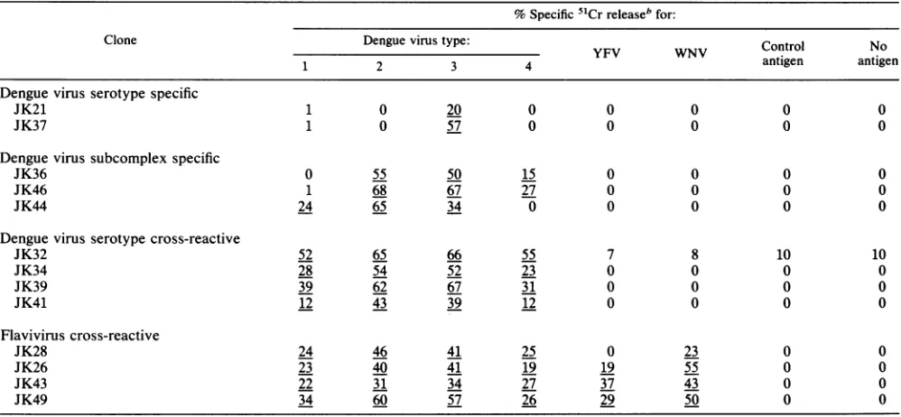
TABLE 1. Virus and dengue virus serotype specificity ofCD4+ CD8- T-cell clones established from donor A infected with dengue virus type 3a
% Specific51Cr releasebfor:
Clone Dengue virus type: Control No
1 2 3 4 antigen antigen
Denguevirus serotype specific
JK21 1 0 20 0 0 0 0 0
JK37 1 0 57 0 0 0 0 0
Dengue virus subcomplex specific
JK36 0 55 50 15 0 0 0 0
JK46 1 68 67 27 0 0 0 0
JK44 24 65 34 0 0 0 0 0
Dengue virus serotype cross-reactive
JK32 52 65 66 55 7 8 10 10
JK34 28 54 52 23 0 0 0 0
JK39 39 62 67 31 0 0 0 0
JK41 12 43 39 12 0 0 0 0
Flaviviruscross-reactive
JK28 24 46 41 25 0 23 0 0
JK26 23 40 41 19 19 55 0 0
JK43 22 31 34 27 37 43 0 0
JK49 34 60 57 26 29 50 0 0
aAtotal of2.5 x 103target cells wasincubatedwith effector cells for 6 h.
bThe percentspecific
51Cr
release was calculatedbyusing the formula described in Materials and Methods. Theeffector/targetratio was 3:1forJK21andJK43;4:1 forJK41; 6:1 forJK36, JK37, JK46, JK44, JK32, JK34, JK39, and JK49; 7:1 for JK26; and 12:1 for JK28. Underlines indicate significant levels of lysis.
Antibody blocking of the lysis of dengue virus antigen-cultured target cells. Monoclonal antibodies B7/21.7, S3/4,
and OKIal recognize HLA-DP, HLA-DQ, and HLA-DR
determinants, respectively.MonoclonalantibodyW6/32
rec-ognizes a framework determinant ofHLA-A, HLA-B, and
HLA-C. B7/21.7 and S3/4 were kindly provided byNancy
Reinsmoen, University ofMinnesota, Minneapolis. OKIal was purchased from Ortho Diagnostic Systems, Inc., Rari-tan, N.J.W6/32was purchased fromAccurateBiochemical
Co., Westbury, N.Y. Atotalof2.5 x 103 51Cr-labeledtarget cells in 0.1 ml were incubatedwith 0.05 ml of1:20diluted
monoclonal antibodies for 30 min. The effector cells were
then added in 0.05 ml, and themixturewasincubated for 6 h. The percent specific
51Cr
release was determined asde-scribed above.
Phenotypic analysis. Anti-Leu-2 (4), anti-Leu-3 (4), and
anti-Leu-4(18) antibodieswere usedas antibodies to CD8,
CD4, and CD3, respectively. Anti-Leu-2, anti-Leu-3, and
anti-Leu-4 antibodieswere purchased from Becton
Dickin-son Co., Mountain View, Calif. Clones were typed with fluorescein isothiocyanate-conjugated monoclonal antibod-ies by direct immunofluorescence methods as described
earlier (12). The percentage of antigen-positive cells was
determined byusingafluorescence-activated cellsorter(no.
440; BectonDickinson Co.).
RESULTS
Virusanddenguevirusserotype specificityofCD4+ T-cell
clones. Denguevirus-specific T-cell clones wereestablished
from lymphocytes of donor A by using limiting-dilution
methodsasdescribed above. Sevencloneswereestablished
by using dengue virus type 3 antigen (15), and six clones
were established by using dengue virus type 2 antigen. Phenotypic analyses with monoclonal antibodies showed that all theclones haveCD3+ CD4+ CD8- phenotypes.
These clones were examinedfor their virus and dengue
virus serotype specificities in cytotoxic activities (Table 1).
JK21and JK37lysedtargetcellscultured withdenguevirus type 3 antigen, but did not lyse target cells cultured with
dengue virus type 1, 2, or 4 or YFV or WNV antigen. Therefore, theyaredenguevirus serotypespecific.JK36and
JK46lysedtargetcellscultured with denguevirus types2,3,
and 4 antigens, but did not lyse target cells cultured with
dengue virus type 1, YFV, or WNV antigen. JK44 lysed
target cells cultured with dengue virus types 1, 2, and 3
antigens, butdid notlysetarget cells cultured with dengue virustype4, YFV,orWNVantigen. Therefore, thesethree
clones aredengue virus subcomplex
specific.
Fourclones,
JK32, JK34,JK39andJK41,lysedtargetcellsculturedwith
dengue virus
antigen
of four serotypes, but did notlyse
targetcellsculturedwith YFVorWNVantigen.
Therefore,
they aredenguevirus serotype cross-reactive.
JK28lysedtargetcells cultured withdenguevirus
antigen
of fourserotypesand WNV
antigen,
butdid notlyse
target cells cultured with YFVantigen. JK26,
JK43, and JK49 lysedtargetcellsculturedwithdengue virusantigen
offour serotypes and YFV and WNVantigens. Therefore,
theseclones areflavivirus
cross-reactive.
These
results indicate thatdengue
virus-specific
CD4+ T cellsare heterogeneous in virus anddengue
virus serotypespecificity and that there are atleast sixpatterns of speci-ficity.
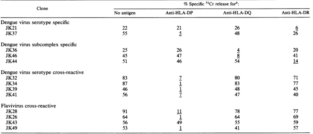
HLArestriction in thelysis of target cellsbyCD4+ T-celi
clones. HLArestrictionof thelysis oftargetcells
by dengue
virus-specific CD4+ T-cell clones were examinedby
using
monoclonalantibodiestoHLAmolecules
(Table 2).
Mono-clonalantibodytoHLA-DPinhibited the
lysis
of target cellsby dengue virus type
3-specific
cloneJK37;
dengue
virusserotype-cross-reactiveclonesJK32,
JK34,
JK39,
andJK41;
andflavivirus-cross-reactive clones
JK26,
JK28,
and JK49.on November 10, 2019 by guest
http://jvi.asm.org/
TABLE 2. HLArestrictionoflysisofdenguevirus type 3antigen-culturedtarget cellsby CD4+T-cellclonesa
% Specific5"Crreleasefor": Clone
Noantigen Anti-HLA-DP Anti-HLA-DQ Anti-HLA-DR
Denguevirus serotype specific
JK21 22 21 26 6
JK37 55 5 48 26
Denguevirussubcomplexspecific
JK36 25 26 4 20
JK46 45 47 8 41
JK44 51 46 54 14
Dengue virusserotypecross-reactive
JK32 83 7 80 71
JK34 87 1 83 77
JK39 46 1 48 45
JK41 56 7 47 40
Flavivirus cross-reactive
JK28 91 11 78 77
JK26 64 1 64 69
JK43 56 49 55 59
JK49 53 1 41 57
aA total of 2.5x 10³targetcellswereincubated witheffector cells for6hinthe presenceof monoclonalantibodiesatfinaldilution of1:80.
bB7/21.7, S3/4, and OKIalwereusedasanti-HLA-DP, anti-HLA-DQ,andanti-HLA-DR, respectively. The percentspecific 51Crreleasewascalculatedby
using the formula described in Materials and Methods.Theeffector/target ratiowas 4:1forJK43; 5:1forJK21,JK39,andJK46;6:1forJK37, JK26, JK32,JK44, and JK49;7:1 for JK36; 11:1 for JK28; 14:1forJK34; and 15:1forJK41.Underlinesindicatesignificant inhibition byeachantibody.
Monoclonal antibody to HLA-DQ inhibited the lysis of
target cells by dengue virus subcomplex-specific clones JK36 and JK46. MonoclonalantibodytoHLA-DRinhibited
thelysisoftargetcells by denguevirustype 3-specific clone JK21 andsubcomplex-specificcloneJK44. Interestingly,the lysis of target cells by flavivirus-cross-reactive clone JK43 was notinhibited by any of the three monoclonal antibodies
toHLA class II orbyanantibodytoHLAclassI.However, the lysis byJK43 was inhibited by a mixture of anti-HLA-DP, HLA-DQ, and HLA-DRantibodiesandbyanantibody
to CD3 (datanotpresented).
These results indicate that dengue virus-specific CD4+
T-cellclonesareHLAclassIIrestrictedand that HLA-DP,
HLA-DQ, and HLA-DR are used asrestrictionelements by
the various clones.
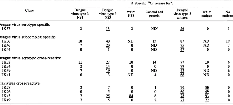
Recognition of NS3 by dengue virus-specific T-cell clones. Wehave previouslyreported thatNS3induceshigh levels of
proliferation responses of donor A PBMC in bulk cultures (17). We next tried to determine whether CD4+ T-cell clones
recognize purified NS3 protein (Table 3). The dengue virus type 3-specific clone JK37, the subcomplex-specific clones JK36 and JK46, and the serotype-cross-reactive clones
JK32, JK34, and JK39lysed target cells cultured withNS3
protein purified from dengue virus type 3-infected cells, but didnot
lyse
target cells cultured with NS1 protein purified fromdengue virus type 3-infected cells or NS3 fromWNV-infectedcells. The flavivirus-cross-reactive clone JK43 lysed target cells cultured with NS3 obtained from dengue virus type3-infected cells or from WNV-infected cells, but did not
lyse
target cells cultured with NS1 obtained from dengue virus type 3-infected cells.Lysis of target cells cultured with the NS3 protein of
dengue virus type 3 by JK34 and JK39 was inhibited by
antibody to HLA-DP (data not presented). JK26, JK28, JK41, JK44, and JK49 did not lyse target cells cultured with NS1orNS3proteins (Table 3). These results suggest that the
NS3 protein contains multiple epitopes recognized by
den-guevirus-specific CD4+ Tcells of variousserotype specific-ities.
DISCUSSION
In this article we have reported that (i) Dengue
virus-specific, human CD4+ CD8- T-cell clones are
heteroge-neous with at least six patterns of virus and serotype
specificities; (ii)HLA restriction ofcytotoxicity by dengue
virus-specific CD4+T-cellclones is alsoheterogeneouswith
HLA-DP, HLA-DQ, and HLA-DR, each being used as
restriction elementsby individual CTL clones;and(iii) 7 of the 12 clones examined recognize epitopes on the NS3
protein. Table 4givesa summaryof these results.
We have previously reported that most of these T-cell clones produce IFN--y after stimulation with dengue virus
antigen (15) and that IFN--y, which up-regulates
FcYR,
augmentsdenguevirus infection ofFc,R-positive monocytic
cells in the presence of
dengue
virus antibodies. Fromtheseobservationswe hypothesized thatCD4+ T cells may
con-tributetothepathogenesisofDHF-DSSby producingIFN--y and by lysing dengue virus-infected monocytes (14, 16). Epidemiologicalstudies haveshownthatDHF-DSS is much
morecommonly observedduringasecondaryinfection with
a different serotype from that which caused the primary infection(3, 7). Althoughthespecificitiesof the clones may
not accurately reflect the specificities of the in vivo,
un-cloned, dengue virus-specific CTL, the identification of
serotype-cross-reactive and flavivirus-cross-reactive CD4+
Tcells supports thepossibilitythatsuch T cellsareactivated
during secondaryinfection withadenguevirus ofa heterol-ogous serotype and that these T cells contribute to the
pathogenesis of DHF-DSS.
Of 13 clones,7 werefoundtorecognize the NS3protein. This resultisconsistentwithourprevious observationthat
purified NS3 induced ahighlevelofproliferation of PBMC from donor A in bulk cultures (17). The clones which
on November 10, 2019 by guest
http://jvi.asm.org/
TABLE 3. Recognition of NS3 protein by dengue virus-specific CD4+ T-cell clonesa
%Specific 51Crreleaseforb:
Clone Dengue Dengue WNV Control cell Dege WNV No
virus type 3 virus type3
protein
virustype 3 antigen antigenDengue virus serotype specific
JK37 2 13 2 NDC 56 0 1
Denguevirus subcomplex specific
JK36 10 40 ND 15 87 ND 19
JK46 7 20 0 ND 75 ND 7
JK44 0 1 0 ND 47 0 0
Dengue virus serotype cross-reactive
JK32 11 27 10 14 77 10 6
JK34 2 14 0 0 79 0 0
JK39 7 19 0 ND 42 ND 6
*JK41 0 3 ND 4 66 ND 0
Flavivirus cross-reactive
JK28 2 7 0 1 70 30 0
JK26 0 5 0 0 60 49 0
JK43 7 25 84 8 78 93 0
JK49 7 5 0 2 77 72 0
aAtotalof2.5 x 103targetcellswereincubated with effector cellsfor 6 h.
bAutologous Epstein-Barr virus-transformed cellswereculturedwithdengue virus type 3 NS1, dengue virus type 3 NS3,WNVNS3, andcontrolcell protein atfinal concentration of20,ug/ml with and dengue virustype 3 and WNV antigens at 1:80 for 24 h prior to addition of effector T cells. Thepercentspecific51Cr
releasewascalculated by usingtheformula described in Materialsand Methods. The effector/target ratio was 4:1 for JK32; 6:1 for JK26, JK37, JK39, and JK44;
7:1for JK46; 8:1 forJK41andJK43; 4:1forJK49; 10:1forJK28andJK34; and 11:1 for JK36.Underlines indicate significantlevels oflysis.
cND, Not done.
recognize NS3 are heterogeneous. They include a dengue virus type 3-specific clone (JK37), subcomplex-specific
clones (JK36 andJK46), dengue virus
serotype-cross-reac-tive clones (JK32, JK34, and JK39) and a flavivirus-cross-reactive clone(JK43). ItwassurprisingtofindthatNS3 is so
immunogenicsinceit is oneofthe sevenflavivirus
nonstruc-turalproteinsandistightly attachedtoendoplasmic reticular
membranes in infected cells (6). NS3 is composed of618
TABLE 4. Summary of denguevirus-specific CD4+ CD8-T-cell clones
Virus andserotype HLA oProtein specificitya Clone restric-
recognzed"
tion NS3 NS1
Serotype specific
D3 JK21 DR NT NT
JK37 DP +
-Subcomplex specific
D2, D3,D4 JK36 DQ +
-JK46 DQ +
-D1,D2,D3 JK44 DR -
-Dengue virusserotypecross-reactive
D1, D2, D3,D4 JK32 DP +
-JK34 DP +
-JK39 DP +
-JK41 DP -
-Flavivirus cross-reactive
D1, D2, D3, D4,WNV JK28 DP -
-Dl, D2,D3, D4,YFV, WNV JK26 DP -
-JK43 ?c +
-JK49 DP -
-aDl,
D2, D3, D4, Dengue virustypes 1, 2, 3,and4,respectively.b+,Recognition; -, norecognition; NT,nottested.
c?, Undetermined.
aminoacids(19) and hasa massof67 kDa. It ishydrophilic
and hasa netpositive charge.Itcontainsconservedepitopes characteristic of helicases(5), andithasalsobeenreported
to have proteinase activity (20). The two largest flavivirus
nonstructuralproteins, NS5(96kDa), which isthoughttobe theviralreplicase,and NS3,arethe mosthighly conserved
amongthe flaviviruses. There is a 75% homology between
theamino acid sequenceofthe NS3of dengue virustype4
(the 814669 strain) and that of dengue virus type 2 (New
GuineaC strain) (10), 62% homology between theNS3s of denguevirus type 4 and WNV,and51% homology between
theNS3s ofdenguevirus type 4 and YFV(19).The presence
of dengue virus subcomplex-specific,
serotype-cross-reac-tive,andflavivirus-cross-reactive T-cellclones isconsistent withthehighlevel of amino acidconservationobservedfor NS3.
The observation that T-cell clones with four different
serotype specificities recognize NS3 strongly suggests that NS3 has atleastfourepitopes recognized by dengue
virus-specific CD4+ Tcells. It will beimportanttolocalizethese
multipleT-cell epitopeson NS3. This will be performed by using recombinant vaccinia viruses
containing
truncated dengue virus NS3 genomeand withsynthetic
peptides.
Itis clear that NS3isnottheonlyprotein
whichcontainsCD4+
T-cell epitopes, since some of the CTL clones do not
recognize either NS3 or NS1. Therefore, identification of otherproteins which contain epitopes
recognized
by these T-cell clones also remainstobe done.We have observed that the viral
proteins
recognized
by
dengue
virus-specific
murine CTL vary with the mousestrains (21).
Therefore,
it islikely
thatdengue
virus-specific
Tcells from other donors who possess different HLA alleles from those ofdonor A will
recognize
differentepitopes
ondenguevirus
proteins.
Togain
acomplete
understanding
ofon November 10, 2019 by guest
http://jvi.asm.org/
[image:5.612.60.301.498.697.2]the T-cell response to dengue viruses in humans, it will be necessary to define the epitopes recognized by dengue
virus-specific T cells from individuals with different major histocompatibility complex antigensaswellasfrom individ-uals infected with various dengueviruses.
ACKNOWLEDGMENTS
We thank Marcia McFadden for technical assistance.
This work wassupported by grants from the U.S. Army Medical Research and Development Command (DAMD 17-86-C-6208) and from the National Institutes of Health(T32-AI07272).
REFERENCES
1. Boyum, A. 1968. Isolation of mononuclear cells and granulo-cytes from human blood. Scand. J. Clin. Lab.Invest.21(Suppl. 97):77-89.
2. Bukowski, J. F.,I. Kurane, C.-J. Lai, M. Bray,B.Falgout, and F.A. Ennis. 1989. Dengue virus-specific cross-reactive CD8+ humancytotoxic Tlymphocytes. J. Virol. 63:5086-5091. 3. Burke,D.S.,A.Nisalak, D. E. Johnson, andR. M.Scott. 1988.
Aprospective study of dengue infections in Bangkok. Am. J. Trop.Med. Hyg. 38:172-180.
4. Engleman,E. G., C. J. Benike, E.Glickman,and R. L. Evans. 1981. Antibodies to membrane structuresthatdistinguish sup-pressor/cytotoxic and helper T lymphocyte subpopulations block the mixed leukocyte reaction in man. J. Exp. Med. 154:193-198.
5. Golbalenya, A.E., A. P.Donchenko,E.V. Koonin,and V. M. Blinov. 1989.N-terminaldomains ofputative helicases of flavi-andpestiviruses may beserine proteases. Nucleic Acids Res. 17:3889-3897.
6. Grun,J. B.,andM. A.Brinton.1987. Dissociation of NS5 from cell fractions containing West Nile virus-specific polymerase activity. J. Virol. 61:3641-3644.
7. Halstead, S. B. 1980. Immunological parameters of togavirus diseasesyndromes, p. 107-173.In R. W.Schlesinger (ed.), The togaviruses: biology, structure, replication. Academic Press, Inc.,NewYork.
8. Halstead, S. B. 1988. Pathogenesis of dengue: challenges to molecularbiology. Science 239:476-481.
9. Innis,B.L.,K.H. Eckels,E. Kraiselbard,D. R.Dubois, G.F. Meadors,D.J. Gubler, D. S. Burke,andW. H.Bancroft. 1988. Virulence ofalive dengue virus vaccine candidate: apossible newmarker of dengue virus attenuation. J. Infect. Dis. 158:876-880.
10. Irie, K., P. M. Mohan, Y. Sasaguri, R. Putnak, and R. Pat-manaphan. 1989.Sequence analysis of cloned dengue virus type 2genome (NewGuinea-C Strain). Gene 75:197-211.
11. Kurane, I., D. Hebblewaite, W. E. Brandt, and F.A. Ennis. 1984. Lysis of dengue virus-infected cells by natural
cell-mediated cytotoxicity and antibody-dependent cell-mediated cytotoxicity. J. Virol.52:223-229.
12. Kurane, I.,D.Hebblewaite,and F. A. Ennis. 1986. Characteri-zationwith monoclonal antibodies of human lymphocytes active in natural killingandantibody-dependent cell-mediated cytotox-icity of dengue virus-infected cells. Immunology 58:429-436. 13. Kurane, I.,B. L.Innis, A. Nisalak, C.Hoke, S. Nimmanitya,A.
Meager, and F.A. Ennis. 1989. Human responses to dengue virus antigens. Proliferative responses and interferon gamma production. J. Clin. Invest.83:506-513.
14. Kurane, I., U. Kontny, A. L. Rothman, and F. A. Ennis. 1989.
Interferon-y production by dengue antigen-specific T lympho-cytes: possible immunopathological role in secondary dengue virus infections, p. 367-370. In R. A. Lerner, H. Ginsberg, R. M. Chanock, and F. Brown (ed.), Vaccines 89: modem approachesto newvaccinesincluding prevention ofAIDS. Cold Spring Harbor Laboratory, ColdSpring Harbor, N.Y. 15. Kurane, I., A. Meager, andF. A. Ennis. 1989. Dengue
virus-specifichuman T cell clones: serotypecross-reactive
prolifera-tion, interferon gamma producprolifera-tion, and cytotoxic activity. J. Exp. Med.170:763-775.
16. Kurane, I., A. L. Rothman, J. F. Bukowski, U. Kontny, J. Janus,B. L.Innis,A.Nisalak, S.Nimmannitya,A.Meager, and F. A. Ennis. 1990.T-lymphocyte responses todengueviruses, p. 301-304. In M. A. Brinton and F. X. Heinz (ed.), New aspects ofpositive-strand RNAviruses. AmericanSocietyfor Microbiology,Washington, D.C.
17. Kurane, I., R. Russell, A. L. Rothman, J. F. Bukowski, C.-J. Lai, M.Bray, B.Falgout, B.Zhao, Y.-M. Zhang, M. Brinton, and F. A. Ennis. 1990. Human T lymphocyte responses to dengue viruses: recognition of dengue virus proteins by T lymphocytes, p. 131-134. In F. Brown,R. M.Chanock, H. S. Ginsberg, and R. A. Lerner (ed.), Vaccines 90: modem ap-proaches to newvaccines including prevention of AIDS. Cold SpringHarborLaboratory,Cold Spring Harbor,N.Y. 18. Ledbetter, J. A., R. L. Evans, M. Lipinski, C.
Cunningham-Rundles,R. A.Good, and L. A. Herzenberg.1981.Evolutionary conservation of surface molecules that distinguish T
lympho-cytehelper/inducerandTcytotoxic/suppressor subpopulations inmouseandman.J. Exp.Med. 153:310-323.
19. Mackow,E.,Y.Makina, B. Zhao,Y.-M.Zhang, L. Markoff, A. Buckler-White, M.Guiler,R.M.Chanock, and C.-J. Lai. 1987. The nucleotide sequence ofdengue type 4 virus: analysis of genescoding for nonstructural proteins. Virology 159:217-228. 20. Preugschat, F., C.-W. Yao, and J. H. Strauss. 1990. In vitro processing of dengue virustype 2nonstructuralproteins NS2A, NS2B, andNS3. J. Virol.64:4364-4374.
21. Rothman, A. L.,I. Kurane, and F. A. Ennis.Unpublished data. 22. Sly,W. S., G. S. Sekhon,R. Kennett, W. F. Bodmer, and J. Bodmer. 1976. Permanent lymphoid lines from genetically marked lymphocytes: success with lymphocytes recovered from frozenstorage.TissueAntigens7:165-172.