0022-538X/88/041364-09$02.00/0
Copyright© 1988, American Society for Microbiology
Location, Transcript Analysis,
and Partial Nucleotide
Sequence
of the
Cytomegalovirus
Gene
Encoding
an
Early
DNA-Binding
Protein with
Similarities
to
ICP8 of
Herpes Simplex
Virus
Type 1
DAVID G. ANDERSt* ANDWADE GIBSON
Virology Laboratories, Department of Pharmac ology andMolecutlar Sciences, the Johns Hopkins University School
of
Medicine,Baltimore,
Maryland
21205Received 2 November 1987/Accepted 20 December 1987
The results presented here locate the gene encoding an early, nonvirion, single-stranded DNA-binding
protein of humanand simian strains ofcytomegalovirus(CMV)[HCMV(Towne) DB140andSCMV(Colburn) DB129, respectively] and provide additional evidence thatthis protein is the CMV homolog of theherpes simplex virus type 1 (HSV-1) major DNA-binding protein (ICP8), as proposed earlier (D. G. Anders, A.
Irmiere,and W.Gibson, J.Virol.58:253-262).The ICP8genewasusedasaprobein Southernanalyses done atmoderate stringencyas an approachtolocatingsimilarsequencesin the CMVgenome. The BamHI Kand EcoRI Vfragments fromthe centerof thelong unique segmentofHCMV(Towne) hybridizedwith theICP8 probeandwereinturnusedtoidentify correspondingsequencesin theEcoRIDfragmentofSCMV(Colburn). RNAprepared fromSCMV(Colburn)-infected cells directed the in vitrosynthesisof DB129. If the RNAwas
firsthybridizedwith the cloned 12.5-kilobaseEcoRIDfragment, in vitrosynthesisof DB129wasspecifically inhibited. Additional hybrid-arrested in vitro translationexperiments withsubclonesspanningtheEcoRI D fragment demonstratedthat the DB129geneislocatedinthe left halfof thatfragment, approximatelybisected byaSalIsite. RNAanalysesidentified3.9-,8.9- ,and 10.0-kilobaseRNAspecies expressedfrom thisregion.
A partial nucleotide sequence of the Colburn region mapping within the boundaries of the 3.9-kilobase transcript, suspected to bethe primary coding species, showed significant sequence similarity to the major DNA-binding proteingenehomologidentified in B95-8Epstein-Barr virus.
Cytomegalovirus (CMV)-infected cells contain an early,
nonvirion, single-stranded DNA-binding protein (DBP) whoseproduction is selectively enhanced by inhibiting viral DNA synthesis,e.g., simianCMV(SCMV) ColburnDB129 andhuman CMV(HCMV) TowneDB140 (2, 2a, 24). Onthe basis of extensive biochemical similarities between this CMVprotein species and the majorDBPofherpes simplex virus (HSV) (i.e., HSV-1 ICP8, HSV-2 ICSP11, and ICSP12), weconcluded that these CMVproteinsare
proba-blehomologs ofthe moreextensively studied HSV DBP (4,
7, 10,25, 26, 31-33, 39, 40, 43, 44). The function of the HSV majorDBPisunknown,butthefindings that it is required for viral DNA replication (5, 7, 36, 37, 42, 47) and may be
involved in regulating viral gene expression (7, 15-17)
un-derscore the importance of tryingtolearnmoreabout it and
itscounterparts in othersystems.
As an approach to establishing whether HCMV DB140 andSCMV DB129arehomologstotheHSV DBP,weused
theclonedHSV-1ICP8genetosearchforcross-hybridizing
sequences in the CMV genome. This general strategy has been useful in many systems (1, 19, 21). The results
pre-sented inthis report show that the cloned HSV ICP8 gene
cross-hybridized with a specific region of the HCMV
ge-nome. Additional experiments confirmed that the
corre-sponding region from SCMV(Colburn) encodes DB129 and identified possible coding transcripts.
*Corresponding author.
t Presentaddress:Virology Laboratories, the Wadsworth Center
for Laboratories and Research, New York State Department of Health, EmpireState Plaza, Albany,NY 12201.
MATERIALS ANDMETHODS
Cells and viruses. Human foreskin fibroblasts were pre-pared, passaged, and infected as previously described (11).
Alsodescribed previously arethe source and conditions of
propagation (11, 13) andthe methodsofvirionisolation(13) and DNA preparation (34) for HCMV(Towne) and SCMV (Colburn).
Plasmids. Plasmid pSG18:SalA (44) containing theSall A subfragment of theHSV-1 KOSfragment EcoRI F (18) was
obtained from D. Knipe. Plasmids pTJ148, containing the SCMV(Colburn) immediate-early region as the Hindlll H
fragment (22), and pSaIG161 containing the Colburn Sall G fragment were provided by G. Hayward. pDGA1 and
pDGA8were madeby excising the HCMV(Towne) BamHI
Kfragment (28)and theSCMV(Colburn)EcoRI Dfragments (23), respectively, froma 1.0% agarosegel and ligatingthe
purifiedfragment intodephosphorylated pUC18 bystandard methods (34). The identities of the resulting clones were
confirmedby restriction analyses andSouthern blotting.The EcoRI Q and V fragments(see Fig. 1)werethen subcloned
from pDGA1 into appropriately prepared pUC18 by the
same technique to yield pDGA2 and pDGA3, respectively. The gel-purified subfragments XbaI B, C, D, and E (see Table 2) of the EcoRI D fragment (i.e., pDGA8) were
subcloned into the XbaI site of pBluescribe M13t
(Strate-gene, San Diego, Calif.), as listed in Table 1. pDGA14,
containing the 4.1-kilobase (kb) EcoRI-to-SalI subfragment ofEcoRI-D, was made by excising from pDGA8 the
frag-ment defined by the unique Sall sites inEcoRI-D (see Fig. 1C)and pUC18 andthenreligating the plasmid.TheSallsite inEcoRI-D liesveryclosetotheleftmost XbaIsite, and thus
1364
on November 10, 2019 by guest
http://jvi.asm.org/
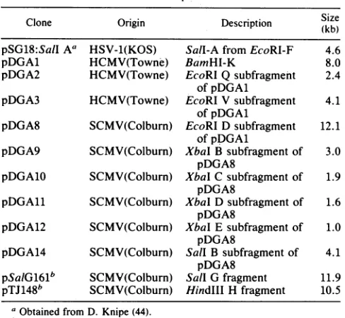
CMV DNA-BINDING PROTEIN GENE 1365 TABLE 1. Description of clones
Clone Origin Description (Skizb)
pSG18:SalI A' HSV-1(KOS) Sall-AfromEcoRl-F 4.6
pDGA1 HCMV(Towne) BamHI-K 8.0
pDGA2 HCMV(Towne) EcoRIQsubfragment 2.4 ofpDGAL
pDGA3 HCMV(Towne) EcoRI Vsubfragment 4.1 ofpDGA1
pDGA8 SCMV(Colburn) EcoRI D subfragment 12.1 ofpDGA1
pDGA9 SCMV(Colburn) XbaI Bsubfragment of 3.0 pDGA8
pDGA10 SCMV(Colburn) XbaI C subfragment of 1.9 pDGA8
pDGA11 SCMV(Colburn) Xbal Dsubfragment of 1.6 pDGA8
pDGA12 SCMV(Colburn) XbaI Esubfragment of 1.0 pDGA8
pDGA14 SCMV(Colburn) Sall B subfragment of 4.1 pDGA8
pSaiG161b SCMV(Colburn) Sall G fragment 11.9 pTJ148b SCMV(Colburn) HindlII H fragment 10.5
aObtained from D. Knipe (44).
bObtained from G. Hayward.
theSall B subclone is equivalenttotheXbaI A fragment in theexperiments described.
Southern blot hybridization. Restriction enzyme-treated DNA was subjected to electrophoresis in 1% agarose gels andtransferred tonitrocellulose(49) with 1OX SSC(1XSSC is 0.15 M NaCl, 0.015 M sodium citrate). Hybridization stringencywascontrolled by varying theformamide
concen-tration. Following4-hprehybridizations in thesamesolution
without probe, filterswereallowedtohybridize overnightat
42°C with about 107 cpm of32P-labeled probe in solutions
containing 1.0 M NaCl,0.01 M Tris (pH 7.4), 0.2% sodium dodecyl sulfate (SDS), 0.01 M EDTA, 100 p.g of salmon
sperm DNA per ml, 5X Denhardt solution (1X Denhardt solution is 0.02% Ficoll-0.02% polyvinylpyrrolidone-0.02% bovine serum albumin), and deionized formamide, at the concentrationgiveninthetextfor eachexperiment. Probes
were 32P-labeled by nick translation (46) or by the random
oligomer-primed synthesismethod(8, 9), asindicated in the text. After hybridization, filters were washed twice for 10 min in 2X SSC-0.1% SDS at room temperature, and then washed twice for 1 h at 62°C in 6X SSC-0.1% SDS (low stringency) or twice for 1 h at 62°C in 0.1X SSC (more stringent). The nitrocellulose filter was air dried, and the boundprobewasvisualized byexposureto KodakX-Omat AR film (Eastman Kodak Co., Rochester, N.Y.) under Cronexintensifying screens(DuPont Co., Wilmington, Del. [30]).
RNA isolation and poly(A)+ selection. Total RNA was
extracted from SCMV(Colburn)-infected cells at 72 h after infection or at 96 h after infection for cultures containing phosphonoformicacid (200 ,ug/ml), by theguanidinium iso-thiocyanatemethod,followedbyultracentrifugation through
a CsCl cushion (6). Polyadenylated RNA was selected by oligo(dT)-cellulose columnchromatography (34).
Invitro translation andhybrid-arrestedin vitro translation. Cell-free translation, directed by total infected-cell RNA, wasperformed by usingarabbit reticulocyte lysate
(Amer-sham Corp., Arlington Heights, Ill.) with [35S]methionine
(translation grade; New England Nuclear Corp., Boston, Mass.), accordingtotheinstructions ofthesupplier.
Hybrid-arrested in vitro translation was done as described by Paterson et al. (38). Hybridization was carried out for 2 h at 42°C in 25 ,ul of a solution containing 80% deionized form-amide, 0.01 M PIPES [piperazine-N,N'-bis(2-ethanesulfonic acid)] (pH 6.4), and 0.4 M NaCl. The reactions were termi-nated by adding 200 ,ul of ice-cold water and 25 ,ug of calf liver tRNA (Boehringer Mannheim Biochemicals,
Indianap-olis, Ind.) and then the mixture was divided into two equal
portions.One of these was heat denatured by incubation for 1 min in a boiling water bath and quick-chilled in a dry ice-isopropanol bath; the other was not heat denatured. Following ethanol precipitation, the reaction products from both the heat-treated and untreated sets were suspended in 1.0
pl
of water and translated as described above. Portionsofthe invitro translation reactions were subjected to
SDS-polyacrylamidegel electrophoresis (SDS-PAGE) and visual-izedby fluorography, as previously described (29), and the
remaining volume was immunoprecipitated.
Immunoprecipitation. The production and specificity of the rabbit monospecific polyclonal anti-DB129 serum used for thesestudies has been described previously (2a).
Immu-noprecipitationwas performedby mixing 10 ,ulofthe
trans-lationreaction with 20
,u1
of 2X RIPA buffer (1X RIPA is 0.05M Tris [pH
7.4]-0.05
M NaCl-0.1% SDS-0.1% NonidetP-40-0.1% sodium deoxycholate-0.001 M
phenylmethylsul-fonyl fluoride) and 10 ,ul of immune serum. Following incubation overnight at 4°C, immune complexes were
col-lected on protein A-Sepharose beads as described before (2a) and subjectedto SDS-PAGE.
Northernanalysis. RNA was resolved in 1% agarose gels (11 by 14 cm) which were prerun for 30 min at 60 V and
containedformaldehyde(34), 0.05M
morpholinepropanesul-fonic acid (MOPS) (pH
7.0),
and 0.001 M EDTA. The electrode buffer was mixed periodically duringelectropho-resisat
100
Vfor 2 h. RNAwastransferred tonitrocellulosein20X SSC,and thenitrocellulose wasbakedat80°C for2 h under vacuum. After prehybridization in the same buffer, hybridizationwasdonefor24hat42°C inabuffercontaining
50% formamide, 5X SSC, 0.02 M sodium phosphate (pH 6.5), 5X Denhardt
solution,
0.05% SDS, and 100p.g
ofsalmon sperm DNA per ml. Approximately 107 cpm of
probe,
32P-labeled
asdescribedin the text,wasusedforeachhybridization. After
hybridization,
thefilterwasrinsed three times for 10 min each at room temperature in 2X SSCcontaining 0.1% SDS and then twicefor 1h eachat
50°C
in0.1X SSC
containing
0.1% SDS and wasfinally
air dried.Bound probewas visualized as describedabove.
DNA sequencing. The 1.0-kb XbaI E subfragment of SCMV(Colburn) EcoRI-D was subcloned into
M13mpl9,
and the sequence of about 300 base
pairs (bp)
from theright-handendwasdetermined
by
usingthedideoxy
method(48) with35S-labeled nucleotides.
RESULTS
Identification of CMV genomic DNA homologous to the HSV-1 majorDBPgene. DNAfrom
HCMV(Towne)
virionswas cleaved separately with
BamHI,
HindIII, and EcoRI;resolvedby
electrophoresis through
a1.0% agarosegel;
andtransferredtonitrocellulose
(49).
The filterwasthenprobed
with 32P-labeled
pSG18:SalIA
insert DNAcontaining
more than90%(i.e.,all but 3' 243bp)
of the HSV-1 ICP8 gene(44;
Table 1), by usingconditions of moderate
stringency
(30%
formamide, Tm
-41°C
[21]),
as described in Materials and Methods. Weak butspecific
hybridization
was seenwithinthe
HindIIl
A,BamHI K,andEcoRI Vfragments
(Fig. 1B),
VOL.62, 1988
on November 10, 2019 by guest
http://jvi.asm.org/
[image:2.612.57.301.81.306.2]A.
HCMV (Towne)
UL
Us
KJ OMU L I J D A H N B 0 C WK2P*TRS F V*
Hindm -
:-iH----i-Hi
lit I LIEZIZI]- i +i4i iiii-ER.0 F G X SN T D KQV EW
ECORI -Il 111l11 1 1 -fi 1
A Y B P I R02U2M* C JZ* t1 1111 1IIII II i i
Z,S, G Y A F E RK C VPWXO 0 M T JU L* B ON BamHI H-1t1I--- H 1_---H11- i--i H l--Hii Ii I i i
B E +" -- B
-pDGAI
B E E B
.... -i
-pDGA2
-pDGA3
C. CMV Cotburn
Y B LW F JSVQM H G UNTO K C A ZP E D R
Sali H 1 H 1 11i-1 11 -11 1- 4-- 1 1 1 H i1 H
K B A SUXPL iL2 D VR F M E H I N G J Q C OTWY
E coRi I
-,,,
tIII, ,.,, .IIl.l t t I II 111EcoRI 1 1 H W 11 I -j Vf -i- , i--i-=J- i !l n H ailH H
k( k pDGA8
D-@
~ o-G
-H
I
Q V
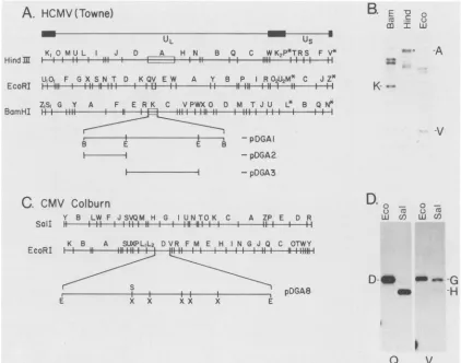
FIG. 1. HCMVsequenceswhichcross-hybridizewith the HSV-1ICP8geneand correspondingsequencesin theSCMV(Colburn)genome.
(A)HindIII,EcoRI, and BamHIrestrictionmapsofHCMV(Towne)(28).Thetoplinedepictsthe general organization of theHCMVgenome,
indicatingthelongand shortunique regions(UL and Us,respectively). Flanking invertedrepeatsareshownassolidboxes. The 8.0-kbBamHI Kfragmentisexpanded,and thepositionsof EcoRI sitesdefiningthesubclonedEcoRIQand EcoRI Vsubfragmentsaremarked.Fragments showingconsistentspecific hybridizationin thisexperimentand others mentioned in thetextareboxed.(B)Southern blotanalysisofHCMV
DNA. Southern blots of HCMVDNAwhichwascleaved withthe indicatedenzymes wereprobed,asdescribed in Materials and Methods,
with the HSV-1 ICP8 gene (i.e., pSG18:SaiA insert DNA) 32P-labeled by the random oligomer-primed synthesis method (8, 9). The
hybridization buffer contained30%formamide, correspondingtoT,,, -41°C (usingaG+Cpercentageof57[45]). Anautoradiogram is shown here. Thefragments whichmost stronglyandreproducibly hybridizedwith the ICP8probe (i.e., BamHI-K, HindIII-A,andEcoRI-V)are
indicated. The circle adjacenttotheHindllllane denotes the Afragment. (C) Sall andEcoRI restrictiOnmapsofSCMV(Colburn) (23).The EcoRI D fragment is expanded to indicate the positions of the XbaI and Sa1l sites (see also Table 2). (D) Southern blot analysis of SCMV(Colburn)DNA.Identical Southern blots ofSCMV(Colburn)DNA, cleaved with theindicatedenzymes,wereprobedasdescribed in Materials and Methods with either nick-translated pDGA2 (Q), or nick-translated pDGA3 (V). The hybridization buffer contained 40%
formamide (T,,, -34°C, calculated as described in the legend to panel B). The positions ofEcoRI-D, Sall-G, and Sall-H areindicated. Abbreviations: E, EcoRI; B, BamnHI; S, Sall;and X,Xbal.
each ofwhich is located around 0.4 onthephysical map of
HCMV(Towne) (Fig. 1A). Although some nonspecific
hy-bridization wasobserved the specificity ofhybridization to the fragments noted above is supported by the following considerations.(i) Hybridizationatthispositionwas
consist-entin all threerestrictionmaps. (ii) Several other bands that
appeared to hybridize well with the ICP8gene probe (e.g.,
those above the BamHI K fragment) contained multiple restriction fragments and thus a greater concentration of nonspecifically hybridized probe. (iii) At higher hybridiza-tion or wash stringencies, nonspecific hybridization was
reduced, relative to specific hybridization with BamHI-K and EcoRI-V; hybridization with these fragments was
ob-servedupto
T",
-34°C (not shown). These results suggested thatICP8-like sequences arepresent in theHCMV(Towne) EcoRIVfragment. TheHCMV(Towne) BamHI K fragment encompassing thecross-hybridizing regionswascloned intothe BarmHI site of pUC18, and subclones containing the left-hand BamnHI-EcoRI fragment (EcoRI Q subfragment) and EcoRI-V were constructed. These clones were
desig-nated pDGA1, pDGA2, andpDGA3, respectively (Fig. 1A; Table 1).
To identify the corresponding region of the SCMV(Col-burn)genome, DNAfromColburn virionswascleavedwith
EcoRI or Sall, sets of both digests were subjected to electrophoresis through a 1.0%agarosegel and transferred to nitrocellulose, and the resulting blotswere probed at Tm -34°C with eitheroftwocontiguous subclones from HCMV (Towne) BamnHI-K (i.e., clones pDGA2orpDGA3; Fig.1D).
Both of these HCMV(Towne) probes hybridized selectively with the SCMV(Colburn) EcoRI D fragment, centered around 0.4 on the physical map (Fig. 1C). They showed different specificities, however, with respect to the Sall fragments comprising the right and left halves of
SCMV(Col-B.
E -0O
1_
a:3 Ir U
m
0-A
-V
D.
,,
V M
L.Li U)
0
-o C
K-w-i
on November 10, 2019 by guest
http://jvi.asm.org/
[image:3.612.93.515.59.391.2]CMV DNA-BINDING PROTEIN GENE 1367 burn) EcoRI-D. Specifically, the rightward EcoRI V
frag-ment of HCMV(Towne) hybridized preferentially with the
rightwardside of SCMV(Colburn) EcoRI-D (i.e., the SalI-G
fragment), and the leftward EcoRI Q fragment of HCMV (Towne) hybridized preferentially with the leftward side of
SCMV(Colburn) EcoRI-D (i.e., Sall-H) (Fig. 1D). This result demonstrates that these regions of the Towne and
Colburngenomes have the same orientation.
Hybrid-arrested in vitro synthesis of DB129 with cloned Colburn EcoRI-D. RNA used in this experiment was
pre-pared from SCMV(Colburn)-infected cells grown in
phos-phonoformate (50) in an effort to selectively increase
syn-thesis of the DB129 message (2). This treatment did not appear to cause any major qualitative changes in the RNA species seen in Northern analyses (see Fig. 3, lane 2, and compare with Fig. 4, Sal-G). The RNA was translated in
vitro in the presence of [35S]methionine, and the products were analyzed by SDS-PAGE, either before (Fig. 2A) or
after (Fig. 2B) immunoprecipitationwith monospecific
poly-clonal anti-DB129 serum (2a). Anti-DB129 serum, but not
preimmune serum (not shown), precipitated a single band which corresponded to DB129 in extracts prepared from
infected cells (Fig. 2B, No Plasmid). In vitro synthesis of
DB129 was nearly eliminated when the infected-cell RNA was hybridized with the entire EcoRI D fragment (i.e.,
pDGA8) before translation (Fig. 2B and D, Eco D). The
location ofsequences which inhibited in vitro synthesis of DB129was better definedby including in this experiment a
A.
B.
series of subclones spanning the Colburn EcoRI D fragment
(Fig. IC and 4; Table 1). Three of the subclones, comprising the left-hand end of the Colburn EcoRI D fragment (i.e., XbaI C, XbaI E, and Sall B), inhibited synthesis of DB129 by six- to eightfold compared with controls (Fig. 2B and D). Clones containing fragments from the right-hand end of Colburn EcoRI-D (i.e., Xba-B and Xba-D) did not affect translation of the DB129 message (Fig. 2B and D). Synthesis of otherproteins was unaffected by hybridization with any of the subclones (Fig. 2A). As a control, cloned DNA from another region of the genome was tested. pTJ148, a plasmid containing the Colburn immediate-early region, was used and found to have no effect on the synthesis of DB129 (TJ148, Fig. 2B and D) but selectively reduced synthesis of the Colburn major immediate-early protein, IE94 (TJ148,
Fig. 2A). Synthesis of other proteins appeared to be unaf-fected. Heating all hybrids before translation restored syn-thesis of the respective proteins (compare Fig. 2B and C;
datafor pTJ148 not shown).
Northern analysis. To identify RNA species that were transcribed from the EcoRI D regionof theSCMV(Colburn) genome, the EcoRI D fragment (i.e., pDGA8) was nick translated and used as a probe in Northern analyses of
polyadenylated RNA prepared from Colburn-infected cells
(Fig. 3). As a control, the blot was probed with pTJ148,
which contains the Colburn major immediate-early region. pTJ148 has been shown to detect a 2.5-kb Colburn transcript in immediate-early RNA from cycloheximide-treated cells
C.
D.
--ao COe U)LIJ
zPFL,icn)xxx
x
,--LY a- "'l oco -, m -,
;5 on: 0-0 -C&2n
2 ZULLCDXXX<X
-0
-~ cccmn
(9)
Z uj c0 - -0 -0 -0zHWLiCO)X>X>X>X
Fm.woquq
6 _ -- MCP-_
X M.
DB129-
IE94-LMA-
I
I
I
DB51-O
FIG. 2. Verification that Colburn EcoRl-D contains DB129-codingsequences. Total SCMV(Colburn) infected-cell RNA used for these experimentswasisolatedasdescribed in Materials and Methodsat96 h after infection from culturescontaining phosphonoformicacid (200
jLg/ml).
Portions ofthe RNA were incubated under hybridization conditions with either noplasmid
or aplasmid
containing the clonedfragmentindicated above eachlane. Afterhybridization, eachreaction mixturewasdivided in half; halfwasheatedat
100°C
for 1 minto denature the hybrids, andthe other halfwas left unheated. After precipitation, allpreparations were translated in vitroas described in Materials and Methods. (A)Asamplefrom eachtranslationreactionwasresolvedbySDS-PAGE. Only the products of the unheated set of reactionsareshown;thepositionof themajor immediate-early protein(IE94;[12, 13])is indicated. Asamplefrom each translation reaction doneusingnondenatured(B)ordenatured(C)RNAwasincubated with rabbitmonospecificanti-DB129serum(2a), and the resulting immune precipitates weresubjectedtoSDS-PAGE.The marker lane contained a[35S]methionine-labeledextract of SCMV(Colburn)-infected cells. Thepositionsofthemajorcapsidprotein(MCP),DB129,the lower matrix protein(LM), andadelayed-earlyDBP, DB51 (14), are indicated (11).(D)Toestimatetherelativeamountof DB129whichwasimmunoprecipitated from each translation reaction, the fluorogram shown in panelBwasanalyzed bydensitometryas previouslydescribed(2). and the resultsare presentedasabargraph.I
i
4
VOL.62, 1988
I .- I ' -,
. : 7 _,,_
:z - '.. ;. ;. -, -1
on November 10, 2019 by guest
http://jvi.asm.org/
[image:4.612.79.549.376.624.2]1368 ANDERS AND GIBSON
1
2
-
8.9
Collburn _co-D
E E
Ix x4 x Xy x
5
~
)) ~12 1
75.0
[image:5.612.318.552.75.421.2]-3.9
ColburnSli-GFIG. 3. RNA species detected by the Colburn EcoRI D
frag-ment. (Lane 1) RNA was isolated from SCMV(Colburn)-infected cellsat72 h afterinfection,andpolyadenylatedRNAwasselected
onoligo(dT)-cellulose. Following electrophoresis througha formal-dehyde-containing1%agarosegeland transfer tonitrocellulose,the RNAwasprobedwithnick-translatedTJ148plasmidDNA(i.e.,the
regionof theColburn IE94gene),the blotwas washed,and bound
probewas visualizedasdescribed in Materialsand Methods. The
positions and estimated sizes of RNA species identified by this control probeare indicatedonthe left. (Lane 2) Theblot, without
removing thepTJ148probe, wasreprobedwith approximatelythe
same number of counts per minute of nick-translated pDGA8 (plasmid containing the cloned Colburn EcoRI D fragment). The
positionsand estimated sizes(in kilobases)of RNAspecies hybrid-izingwith thepDGA8probearemarkedontheright.Atthefarright
arecircles indicatingthepositionsof RNA size markers(Bethesda ResearchLaboratories, Gaithersburg, Md.) whichwere inparallel and visualized by staining.
(22). When used to probe late lytic RNA from a normal
infection, pTJ148identified an RNA of about the same size (estimatedtobe 2.3kb),but also detectedasecond abundant RNA (1.5 kb), as well as two comparatively minor RNAs (5.8 and 5.0kb; Fig. 3, lane 1). When the same blot (i.e.,
pTJ148 probe notremoved) was subsequently probed with
the Colburn EcoRI D fragment (Fig. 3, lane 2), the most intense band observed corresponded to a 2.8-kb RNA.
Additional species of 3.9, 5.0, 8.9, and 10.0 kb were also
detected.
To betterdefinethetranscripts from this region, the XbaI fragments spanning EcoRI-Dwereexcised from pDGA8, gel
purified, 32P-labeled, and used to probe Northern blots of Colburn-infected-cell RNA (Fig. 4). All of the fragments detectedan8.9-kb species. The 3.9- and10.0-kb transcripts were detected by the XbaI A and XbaI E subfragments, whereas the abundant2.8- ,aswellas5.0-kbspecies,andan
additional 2.2-kb species, were detected 'by XbaI-B and
XbaI-D.XbaI-C hybridized with both sets ofRNA species. Theresults of this experiment aresummarized in Table 2.
Partial nucleotide sequence analysis. The results of the
experimentsdescribed aboveindicated that the DB129gene
waslocated inthe left half of ColburnEcoRI-D, spanning the XbaI E fragment. To establish whether this region was
indeed similar to the ICP8 coding region and to aid in defining its organization, a 315-bp sequence fromthe right
endofpDGA12 (i.e., beginningatthe junction ofthe XbaI E andXbaICsubfragments, mapposition0.393, and
proceed-ing leftward; Table 2)wasdetermined by usingthe method of
1-'B 30"
.
bI
r' 5
./,Hi
x4 * g
\
89
3.9-8.9 8--5.
Om _- _
Q
---- ---2.8FIG. 4. Northern analysis with XbaI fragments spanning Col-burnEcoRI-D. Total SCMV(Colburn)-infected-cell RNA was pre-paredasdescribed in thelegendtoFig.2.Equivalent portionswere
subjectedtoelectrophoresis throughaformaldehyde-containing1% agarosegelandtransferredto anitrocellulose sheet whichwasthen cut into six strips. After being baked, each strip was probed as described in Materials and Methods withoneoffive XbaI subfrag-mentswhich span Colburn EcoRI-D (the 0.5-kbXbaI Ffragment was notusedas aprobe)orwith the Sall Gfragment,asindicated above. Positions ofthe respective fragments within EcoRI-D and theirapproximate sizes are indicated at the top. The gel-purified fragmentsused asprobeswere32P-labeledbythe random oligomer-primed synthesis method (8, 9). Bound probe was visualized by autoradiography. Positions ofthe2.8-,3.9- , 5.0- , and8.9-kbRNA speciesareindicated.
TABLE 2. ResultsofNorthernhybridizations
Fragment" Estimated map Fragment RNAspecies coordinates' size(kb) detected(kb)' XbaI-A 0.370-0.388 4.1 3.9, 8.9, 10 XbaI-B 0.411-0.425 3.0 2.2, 2.8, 5.0, 8.9 Xbal-C 0.393-0.415 1.9 2.2, 2.8, 3.9, 5.0, 8.9, 10 XbaI-D 0.404-0.411 1.6 2.2, 2.8, 5.0, 8.9 XbaI-E 0.388-0.393 1.0 3.9, 8.9, 10
pSalG161 0.388-0.442 11.9 2.2, 2.8, 3.9, 5.0, 8.9, 10
"Gel-purified fragments excised from pDGA8 (EcoRI-D) are named in descending order of size.
bOn the basisofa220,000-bp Colburn genomewith theleft-hand sideof the EcoRI Dfragmenttaken tobeatcoordinate0.37.
' Sizes estimatedbycomparisonwiththeBethesda ResearchLaboratories
RNAladder.
5.8
-5.0
--2
8
2.3-1.5
- O-S
J. VIROL.
tl
kb
on November 10, 2019 by guest
http://jvi.asm.org/
[image:5.612.134.227.78.263.2] [image:5.612.317.557.604.686.2]CMV DNA-BINDING PROTEIN GENE 1369 Sanger et al. ([48] Fig. 5). The amino acid sequence
pre-dicted
for each open reading frame of the determinedse-quence was compared with that predicted from the HSV-1
ICP8 gene sequence (43) and with that of the homologous
B95-8 Epstein-Barr virus (EBV) open reading frame BALF2
(3, 43). Clear similaritieswith the sequence of EBV BALF2 were identified by a homology matrix analysis (Fig. 6A).
Withnumbering beginning at the right end of EBV
BamHI-A,the aminoacids of BALF2 stretch leftward from position
617through position1745; sequence similarity was observed roughly between residues 820 and 910. An alignment of the two sequences yielded greater than 30% identity over a
96-amino acid stretch (Fig. 6B). Compared with EBV,
sim-ilarities between this short portion of the DB129 gene sequence and the HSV ICP8 gene were minimal, although some common features were observed. Most notable of
thesefeatureswas the sequenceAla-Leu-Arg at positions 33 to 35 in the Colburnsequence, which also is found in EBV
BALF2.Optimal alignment,which yielded 21% identity over
amino
acids 10 to 105 of the Colburn sequence, requiredintroduction of two single-residue gaps in the DB129
se-quence andone inthe HSV-1 sequence (not shown). DISCUSSION
Thisreportidentifiesthecodingregion for an
SCMV(Col-burn) early DBP DB129 and by extension, its HCMV
(Towne)counterpart DB140 (2, 2a, 11, 13). A clone
contain-ingover90% ofthe HSV-1 ICP8 gene was initially used to
locate cross-hybridizing sequences in the HCMV genome.
Nucdotids #10 20 30 40
TCTAGAC MGCGC AGC TCG
fTC
GTG GM TCTCGG GGATTGTAC GTG CCTL D K R SS F V E S R G L Y V P
Amino Ad #3 10
50 60 79 80 90
GCGGTGAGCGM ACCCTG T TACTATGTTTACACTTCGTGGTGC CAG
A V S E T L F Y Y V Y T S W C
20 30
100 110 1,0 130 140
GCCCTG CGC TTC TCAGAGACCAG GTGCTGAT GM GOGGCCCTGAAG
A L R F S E T K V L I E A A L K
40
150 160 170 180 190
cAG TT0 GTGMCGA6AGC cAG CAG tccGTGMGCTG GCTCCG CA AAG
Q F V ND S S V K L A P H K
50 80
200 210 2,0
2?0
240MGTAC oTC GGGTACAGGAGC CAG GCTG AGCAGTCTGGAG AM GAC
K Y F Q Y Ts Q K L S S L E K D
70 80
250 290 270 290
CACTTAATG CTC AGC GAC GCCGTGATCT60 GAG TG GGGTTCAGc 1Tc
H L M L S D A V I C E L ¢ F S F
90
290 300 310
GCC TCGGTGmTTCTGGAG TCGGWC TA
A S V F L D S A Y
100
FIG. 5. Partial nucleotide sequence of theXbaI Esubfragment from Colburn EcoRI-D. Nucleotide sequence was determined as
described in Materials and Methods. Nucleotides are numbered
fromtherightend of XbaI-Easdrawn inFig.1 and4,with thefirst
base assignedas 1. Amino acidsare indicatedbystandard single-letterabbreviations. The readingframegiving thededucedamino
acid sequence shown was selected for its amino acid sequence
similaritytoEBVBALF2(seeFig.6).
A.
N
LL
m
>
Colburn
XbaI E
10 20 30 40 50 60 70 80 90 l00 110
U3-I--
-850-
--860---
--
a---
-870- - - -
va-
--88
0--890
I_-
-----_
- --900---- - - -
I--~Io-- - -I-
-B.
10 30 50
* * *
SRGLYVPAVSETLFYYVYTSWCOALRFSETKVLIEAALKQFVNDSQQSVKLAPHKKYFGY ** * ** * *N*M * * ** * * M**
lRqfYnsdlSrcmheo 1YTgloQALRvrrvgkLvElleKOslqDqakvoKvAPIKofpas 817
70 90 105
* * *
TSOKLSSLEKDHLbMLSDAVICELGFSFASVFLDSAY
* * ** * *9**** *
Ti...SphdsgaLMlvDsaoCELavSyApomLeash 909
COLBURN XboI-E
EBVBALF-2
FIG. 6. (A) Homology matrix comparison of deduced amino acid sequencesfrom the ColburnXbaI Esubfragment of EcoRI-D and B95-8 EBV BALF2 (3). Comparison was done by using Pustell programs(InternationalBiotechnologies, Inc., New Haven, Conn. [41]).TheColburn sequenceis plotted on the horizontal axis, and the EBV BALF2 sequenceis plotted on the vertical axis.Arangeof 30amino acidswasused,and apoint was plotted only when25%or moreof the residues inagiven window matched. Other parameters were set to thefollowing numbers: hash level, 1; scale factor, 0.95; step,1;andjump,1.Thefigureshown is redrawn from theoriginal plot. (B)Analignmentof theColburnand EBVBALF2 sequences. Athree-amino-acid gapwasintroduced into the EBV sequenceto maximize matches.Equivalent residuesaremarkedbyanasterisk. Thenumbering system isexplained in thetext.
An HCMV restriction fragment containing the identified region was then used to locate similar sequences in the
SCMV(Colburn) genome to take advantage of
previously
characterized antisera that had been raised
against
the Colburn DB129protein
(2a).Hybrid-arrested
translation experiments andNorthernanalyses were then used to cor-relate the protein with its coding sequence and to identify possible coding transcripts.Finally,
apartial
nucleotidesequenceobtainedfromtheColburnDB129geneestablished
its similarity to the gene forthe EBV DBP that is
homolo-gous toHSV
ICP8.
The first series of
experiments
demonstrated that the cloned HSV ICP8 genecross-hybridized
with DNA se-quences around 0.4 on thephysical
map of the HCMVgenome. Hybridization was detected
only
underconditions ofmoderatestringency
(equivalent
to Tm-340C
orlower),
indicating limited nucleotide sequence
similarity.
A clonecontaining
the identified HCMV sequence was constructedandusedin
experiments
whichestablishedthatSCMV(Col-burn) contains a
related,
colinear(i.e.,
around 0.4 on itsphysical map)sequencewhich ispresentin thesamerelative
-To%f
I I I I I I I I I I 1 920
VOL.62, 1988
on November 10, 2019 by guest
http://jvi.asm.org/
[image:6.612.325.553.75.380.2] [image:6.612.64.298.389.660.2]orientation. These CMV sequencesarethemselves
approx-imately
colinear with theposition
of the ICP8 gene in theHSV-1 genome
(43)
andcorrespond
to a region of theHCMV(AD169)
genome with sequencesimilarity
to theHSV ICP8 gene
(T.
Kouzarides,
A.Bankier,
K.Weston,
S. C.
Satchwell,
S.Beck,
andB. G.Barrell,
Abstr. 11thInt.Herpesvirus Workshop, 1986,
p.60;
B.Barrell, personal
communication).
Hybrid-arrested
in vitro translationexperiments
whichwere done by
using
subclones of the cross-hybridizingColburn
region
established that DB129 is coded for within theleft half of the EcoRI Dfragment.
Positive andnegative
controlsdemonstrated the
specificity
of thehybrid-arrested
translationassay. This
approach
wasused instead ofhybrid
selection fortwo reasons.
(i)
Itismorespecific
forsequenceswithin the
coding body
of thetranscript (35).
(ii)
Theanticipated large
size and lowabundance of DB129 mRNAdiminished the chances for its
successful
selection. Theavailability
of amonospecific
antiserum(2a)
provided
ameansto
identify
thesmallamountsof translationproduct
in theseexperiments.
Northern
analyses
identified six RNAspecies
that wererecognized by
theset ofsubclones used forhybrid-arrested
translation
experiments
(Fig.
4and7).
Ofthese,
the 3.9- and 10.0-kb RNAs are the mostlikely
to betranscripts
forDB129,
sincethey
hybridized
withonly
those EcoRIsub-fragments
thathybrid
arrested DB129synthesis (XbaI-A,
XbaI-E,
and XbaI-C). The 2.8- and5.0-kbspecies
arise fromsequences located to the
right
(as shown inFig.
7) of the DB129 gene, and thepredicted
maximum size ofaprotein
encoded
by
theabundant 2.8-kbRNA(i.e.,
100kilodaltons[kDa])
would be somewhat smallerthan the size estimatedby
SDS-PAGE forDB129(i.e.,
129kDa).
Anarrangementofthe RNA
species
detected with EcoRI-D thatis consistentwith the results presented in this report is shownin Fig. 7.
Results of the
sequencing
experiments indicated that theDB129geneis transcribed fromrighttoleft,asdrawnin this
figure.
Since the 3.9-kbtranscript is large enoughtoencodeDB129,
it issuspected
tobetheprimary coding species.
Wenote that the HSV ICP8 gene is transcribed in the same
direction as is the DB129 gene and
gives
rise to twoapparently 5' coterminal RNAs of4.5 and 10.2 kb (20, 44)
that areclose in size totheColburn3.9- and 10.0-kb RNAs and may be counterparts. Thereportby Kemble et al. (24)
that the mRNAfor the homologous HCMV(Towne)geneis
10to 12kb in sizesuggests thatprocessing ofthe transcript
fortheHCMVgene maydiffer from that ofthe
SCMV(Col-burn)
and HSV-1 genes.The partial nucleotide sequenceobtained forthe Colburn
DB129gene, and its similarity to the BALF2 open reading
frame ofEBV, aided in positioning the DB129 gene more
precisely,
as follows. Given that the DB129 and EBVBALF2proteinsareabout the samesize(i.e., observed, 129
kDa,
and predicted, 123 kDa, respectively) and assumingthatnomajorrearrangements existbetween these CMV and EBVgenes, the DB129aminoacid sequence predicted here
begins
about200residues downstream from its aminotermi-nus. Therefore, the 5' end of the DB129 coding region is
predicted to begin about 0.6 kb to the right of the XbaI
E-XbaI Cjunctionand end about 2 kb to the left of the
Sall
site inthe EcoRI Dfragment.This position for the 3' end is
consistent with the finding that cDNA clones to DB129 extendleftward fromtheXbaI E subfragment about 2 kb into the XbaI A fragment (L. Robson, D. Anders, and W.
Gibson,
unpublished results).We have previously identified the HCMV counterpart to
Colburn
Eco-D
E
12.1 s
E
ColburnSal-G
11.9
Clones
X-'E' X-'
1.0 0.5
X-.C*
X-'D'4.1 E H
t-1.9 1.6
K H B Sm
x x x x x
10.0
8.9 1
-5.0
3' 3 5'
B E
Physical Map
1
RNA
Species
i -2.8
J
Fragments
whichhybridarrestDB129translation
FIG. 7. Summary relating hybrid-arrested in vitro translation and Northern blot results. Theapproximate positionsoftranscripts are shown, and the fragments which hybrid arrested in vitro synthesisof DB129areindicated. Thetranscriptionalorientationof the3.9-kbRNA,suspectedtoencodeDB129,ispredictedfrom the nucleotide sequence data. Restriction sites are as follows: Sm, SmaI; H,HindIll;K,KpnI;othersareasindicatedin thelegendto Fig. 1.Sizesgivenareinkilobases.
DB129
(2, 13), DB140,
andhaveshownittobeimmunolog-ically
reactive withmonospecific
antiseraprepared against
purified
DB129(2a).
Evidencepresented
here indicates thatthegenefor DB140 is encoded within the BamHIK
fragment
of
HCMV(Towne),
around 0.4on thephysical
map.Cross-hybridization
experiments
showed that theSCMV(Colburn)
region containing
the DB129 genecorresponds
to HCMV(Towne)sequences
spanning
theEcoRIQ-EcoRIVjunction(Fig.
1). Taken together with the Colburn nucleotidese-quence
data,
these resultspredict
that the DB140 geneextends leftward from Towne EcoRI-V into the
adjacent
EcoRI
Q fragment.
This locationcorrespondstothe identi-fied ICP8-like sequences ofHCMV(AD169)
(unpublisheddata;
Kouzaridesetal.,
Abstr. 11th Int.Herpesvirus
Work-shop, 1986; B. Barrell, personal communication) and the
HCMV
region
which was detected by expression cloninganddeterminedtoencodeanICP8-likeproteinwith proper-ties of DB140 (24). Furthermore, the partial nucleotide sequence that we obtained from the Colburn DB129 gene showed
striking
(i.e., 86%) similarity with theHCMV(AD169)
ICP8-like gene identified in nucleotidese-quence
analysis
of theHCMV(AD169)genome (D. Anders,W.
Gibson,
and B. Barrell, unpublished results). Theobser-vation thatthe ICP8 gene probe did not hybridize as well withtheHCMV(Towne)EcoRIQfragmentas it did with the EcoRI V
fragment
suggests that the 5' end of the gene is more conservedbetween HSV and CMV than the 3' end or that the 5' end may contain a small conserved region.on November 10, 2019 by guest
http://jvi.asm.org/
[image:7.612.320.556.75.369.2]CMV DNA-BINDING PROTEIN GENE 1371
Three lines ofevidence support the conclusion thatCMV
proteins DB129 and DB140 are homologs of HSV ICP8, as suggested previously(2). First, they exhibit similar biochem-ical properties, including an early time of synthesis and comparatively low abundance, a nuclear location, absence from thematurevirion,a size of approximately 130 kDa,and the capacity to bind to DNA (2, 2a, 24). Second, the genes for these CMV proteins cross-hybridize with the ICP8 gene (Fig. 1B), are approximately colinear on the physical map (Fig. 1A and C), are transcribed in the same relative direc-tion (Fig. 7), and in the case of the DB129 gene, give riseto aputativecoding transcript which is similar in size to thatfor ICP8 (Fig. 3, 4, and 7). And third, a partial nucleotide sequence of the gene that encodes DB129 shows clear similaritytothe EBV open reading frame BALF2 (Fig. 6),a putative homolog of the HSV ICP8 gene (43). Similarities suchas these in both individualgene sequences, and inthe relative arrangements of blocks of genes (27), add to evi-dence for an evolutionary link among the herpes group viruses. It is noteworthy that the EBV BALF2 sequence from around amino acids 840 through 900 (as numbered here), like the corresponding Colburn sequence (Fig. 6), shows only weak similarity to the corresponding ICP8 se-quence, althoughflanking sequences show a clearsimilarity (43). In this regard, the greater similarity ofthe CMV and EBV genes in this region, bycomparisonwith HSV-1,may be of functional or evolutionary significance.
In summary, we have identified the coding region for DB129 andprovided evidence byhybridization experiments and sequence comparison that it and HCMV DB140 repre-sent CMV homologs ofthis herpesvirus group ofcommon DBP species. The identification and cloning of these CMV genes will facilitate their biochemical and genetic analysis and help establishtheirrole during infection.
ACKNOWLEDGMENTS
We thank David Knipefor clone pSG18:SalA,Gary Haywardfor clones pSalG161 and pTJ148, Lori Robsonfor theoligo(dT)-selected RNA, and MarieHardwick for providing the use of her computerfor sequence comparisons and Ivan Auger for performing some addi-tional comparisons by using GENALIGNon the BIONET computer resource and FASTP. We also thank Bart Barrell for helpful information regardingtheHCMV(AD169) nucleotide sequence and Bob LaFeminaand Gary Hayward andmembersof his laboratory for helpful discussions. Weappreciatetheexcellent technical assis-tance of Donna Woodsand Jennifer Kidd.
This workwas aided by Public Health Service grantsAl13718,Al 22711, and Al 19373 from the National Institute of Allergy and Infectious Diseases and by American Cancer Society Institutional grant IN-llX to W.G. D.G.A. was supported during the initial period of this work by Public Health Service Individual National Research ServiceAwardAl 07293-01from theNational Institute of Allergy and Infectious Diseases.
LITERATURECITED
1. Alestrom, P., A. Stenlund, P. Li, A.Bellett, andU. Pettersson. 1982. Sequence homology betweenavian and human adenovi-ruses. J. Virol. 42:306-310.
2. Anders, D. G., A. Irmiere, and W. Gibson. 1986. Identification and characterization of a major early cytomegalovirus DNA-binding protein. J.Virol. 58:253-262.
2a.Anders,D. G.,J. R. Kidd, and W. Gibson. 1987.Immunological characterizationofan earlycytomegalovirussingle-strand DNA binding with similarities to the HSV major DNA-binding pro-tein. Virology 161:579-588.
3. Baer, R., A.T. Bankier, M. D. Biggin, P. L. Deininger, P. J. Farrell,T. J.Gibson, G.Hatfull, G. S. Hudson, S. C.Satchwell, C. Seguin, P. S. Tuffnell, and B. G. Barrell. 1984. DNA
sequence andexpression of the B95-8 Epstein-Barr virus ge-nome.Nature(London) 310:207-211.
4. Bayliss, G. J.,H.S.Marsden, and J. Hay. 1975.Herpessimplex virusproteins:DNA-binding proteins ininfected cells and in the virusstructure. Virology68:124-134.
5. Chiou,H.C.,S. K.Weller,and D. M.Coen. 1985. Mutations in the herpes simplex virus major DNA-binding protein gene leading to altered sensitivity to DNA polymerase inhibitors. Virology 145:213-226.
6. Chirgwin, J. M., A. E. Przybyla, R.J. MacDonald,and W.J. Rutter. 1979. Isolation of biologically active ribonucleic acid from sources enriched in ribonuclease. Biochemistry 18:5294-5299.
7. Conley, A.J.,D. M.Knipe,P.C.Jones,and B.Roizman. 1981. Moleculargenetics of herpessimplex virus. VII. Characteriza-tion of a temperature-sensitive mutant produced by in vitro mutagenesis and defective in DNAsynthesisand accumulation of -ypolypeptides. J. Virol. 37:191-206.
8. Feinberg, A. P., and B. Vogelstein. 1983. A technique for radiolabeling DNA restriction endonuclease fragmentsto high
specific activity. Anal. Biochem. 132:6-13.
9. Feinberg, A. P.,and B. Vogelstein. 1984. Addendum. A tech-nique forradiolabelingfragmentstohighspecificactivity.Anal. Biochem. 137:266-267.
10. Fenwick,M.L.,M.J.Walker,andJ.M.Petkevich.1978. On the association of virus proteins with the nuclei of cells infected with herpessimplexvirus. J. Gen. Virol. 39:519-529.
11. Gibson,W. 1981.Structural and nonstructuralproteinsof strain Colburncytomegalovirus. Virology 111:516-537.
12. Gibson,W.1981. Immediate-earlyproteins of human cytomeg-alovirus strainsAD169,Davis,and Towne differ in
electropho-reticmobility. Virology 112:350-354.
13. Gibson, W. 1983. Protein counterparts ofhuman and simian cytomegaloviruses. Virology 128:391-406.
14. Gibson, W.,T.Murphy, andC. Roby. 1981.
Cytomegalovirus-infected cells containaDNA-binding protein.Virology 111:251-262.
15. Godowski,P.J.,and D. M.Knipe. 1983.Mutations in the major DNA-bindingproteingeneofherpessimplexvirustype1result in increased levels of viral gene expression. J. Virol. 47:478-486.
16. Godowski, P. J., and D. M. Knipe. 1985. Identification of a
herpes simplexvirus function that represses late gene expres-sion fromparental viral genomes. J. Virol. 55:357-365. 17. Godowski,P.J.,and D. M.Knipe. 1986.
Transcriptional
controlof herpesvirus gene expression: gene functions
required
for positive and negative regulations. Proc. Natl. Acad. Sci. USA 83:256-260.18. Goldin, A. L., R. M. Sandri-Goldin, M. Levine, and J. C. Glorioso. 1981. Cloning of herpes
simplex
virus type 1se-quences representingthewhole genome. J. Virol. 38:50-58. 19. Heilbronn, R., G. Jahn, A. Burkle, U.-K. Freese, B.
Flecken-stein,and H.zurHausen.1987. Genomiclocalization, sequence analysis, and transcription ofthe
putative
human cytomegalo-virus DNApolymerase gene.J. Virol. 61:119-124.20. Holland, L. E., R. M. Sandri-Goldin, A.L. Goldin, J. C. Glorioso, and M. Levine. 1984.
Transcriptional
andgenetic
analysesof theherpessimplexvirustype1genome: coordinates 0.29to 0.45. J.Virol. 49:947-959.21. Howley, P. M., M. A. Israel, M.-F. Law, and M. A. Martin. 1979. A rapid method for
detecting
andmapping homology
betweenheterologous DNAs.J. Biol. Chem. 254:4876-4883. 22. Jeang, K.-T., M.-S. Cho,and G. S.
Hayward.
1984. Abundantconstitutiveexpressionof theimmediate-early94K
protein
from cytomegalovirus (Colburn) in a DNA-transfected mouse cellline. Mol.Cell. Biol. 4:2214-2223.
23. Jeang, K.-T., G. S. Hayward. 1983. A
cytomegalovirus
DNA sequencecontaining tracts oftandemly
repeated
CA dinucleo-tideshybridizestohighlyrepetitive dispersed
elementsinmam-malian cellgenomes. Mol. Cell. Biol. 3:1389-1402.
24. Kemble, G.W., A. L. McCormick, L. Pereira, and E. S. Mo-carski. 1987. A
cytomegalovirus
protein
withproperties
of herpes simplex virus ICP8:partial
purification
ofthepolypep-VOL.62, 1988
on November 10, 2019 by guest
http://jvi.asm.org/
tide andmapposition ofthegene.J. Virol.61:3143-3151. 25. Knipe, D. M., M. P. Quinlan, and A. E. Spang. 1982.
Charac-terization of two conformational forms of the major DNA-bindingprotein encoded by herpessimplex virus1.J. Virol. 44: 736-741.
26. Knipe, D. M.,and A. E.Spang. 1982. Definition ofaseries of stagesin the association oftwoherpesviralproteinswiththecell nucleus. J. Virol. 43:314-324.
27. Kouzarides, T.,A. T. Bankier, S. C.Satchwell, K.Weston, P. Tomlinson, andB.G.Barrell. 1987. Large scale rearrangement of homologous regions in the genomes of HCMV and EBV. Virology 157:397-413.
28. LaFemina, R. L., and G. S. Hayward. 1980. Structural organi-zation of theDNAmolecules from humancytomegalovirus, p. 39-55. In B. N.Fields,R.Jaenisch, and C.F.Fox(ed.), Animal virus genetics, vol. 28. Academic Press, Inc., New York. 29. Laskey, R. A.,and A. D.Mills.1975.Quantitative film detection
of 3H and "4C inpolyacrylamidegels byfluorography. Eur. J. Biochem. 56:335-341.
30. Laskey, R. A., and A. D. Mills. 1977. Enhanced autoradio-graphic detection of32P and1251usingintensifying screensand hypersensitized film. FEBS Lett. 82:314-316.
31. Lee, C. K., and D. M. Knipe. 1983. Thermolabile in vivo DNA-binding activity associated with a protein encoded by mutantsof herpessimplex virus type 1.J.Virol. 46:909-919. 32. Leinbach, S. S., J. F. Casto, and T. K. Pickett. 1984.
Deoxyri-bonucleoprotein complexes and DNAsynthesis of herpes sim-plexvirus type1. Virology 137:287-296.
33. Littler, E., D. Purifoy, A. Minson, and K. L. Powell. 1983. Herpes simplex virus non-structural proteins. III. Function of themajor DNA-binding protein. J. Gen. Virol. 64:983-995. 34. Maniatis, T., E. F. Fritsch, and J. Sambrook. 1982. Molecular
cloning: alaboratory manual. Cold Spring Harbor Laboratory, Cold Spring Harbor, N.Y.
35. Miller, J. S., B. M. Paterson, R. P. Ricciardi, L. Cohen, and B. E.Roberts. 1983. Methodsutilizing cell-free protein-synthe-sizing systems for the identification of recombinant DNA mol-ecules. MethodsEnzymol. 101:650-674.
36. O'Donnell, M. E., P. Elias, B. E. Funnell, and I. R. Lehman. 1987. Interaction between the DNA polymerase and single-strandedDNA-binding protein (infected cell protein 8) of herpes simplex virus 1.J. Biol. Chem.262:4260-4266.
37. O'Donnell,M.E., P. Elias,andI.R.Lehman. 1987. Processive replication of single-stranded DNA templates by the herpes simplex virus-induced DNA polymerase. J. Biol. Chem. 262:
4252-4259.
38. Paterson,B.M., B. E. Roberts, and E. L. Kuff. 1977. Structural gene identification and mapping by DNA * mRNA hybrid-ar-rested cell-free translation. Proc. Natl. Acad. Sci. USA 74: 4370-4374.
39. Powell,K.L.,E.Littler, and D.J. M. Purifoy. 1981. Nonstruc-tural proteins of herpes simplex virus. II. Majorvirus-specific DNA-binding protein. J. Virol. 39:894-902.
40. Purifoy,D.J. M., andK.L.Powell. 1976. DNA-bindingproteins inducedby herpessimplex virus type 2 in HEp-2 cells. J. Virol. 19:717-731.
41. Pustell, J., andF. C. Kafatos.1984.Aconvenient andadaptable package of computer programs for DNA and protein sequence management, analysis, and homology determination. Nucleic Acids Res. 12:643-655.
42. Quinlan, M. P., L. B. Chen, and D. M. Knipe. 1984. The intranuclear location ofa herpes simplex virus DNA-binding protein is determined by the status of viral DNA replication. Cell 36:857-868.
43. Quinn, J. P.,and D.J.McGeoch. 1985.DNAsequence of the region inthe genome of herpes simplex virus type1containing the genes for DNA polymerase and the major DNA-binding protein. Nucleic Acids Res. 13:8143-8163.
44. Rafield, L. F.,and D. M.Knipe. 1984. Characterization of the majormRNAs transcribed from the genes forglycoprotein B andDNA-binding protein ICP8 of herpes simplex virus type 1. J. Virol.49:960-969.
45. Rapp,F.1983. The biology ofcytomegaloviruses, p. 1-53. InB. Roizman (ed.), The herpesvirus, vol. 2. Plenum Publishing Corp., New York.
46. Rigby, P. W. J., M. Dieckman, G. Rhodes, and P. Berg. 1977. Labeling deoxyribonucleic acidtohighspecific activity invitro by nick translation with DNA polymerase I. J. Mol. Biol. 113: 237-251.
47. Ruyechan,W.T., and A. C. Weir. 1984.Interaction with nucleic acids and stimulation of the viral DNA polymerase by the herpes simplex virus type 1 major DNA-binding protein. J. Virol. 52:727-733.
48. Sanger, R.,S.Nicklen,and A. R. Coulson. 1977. DNA sequenc-ing with chain-terminating inhibitors. Proc. Natl. Acad. Sci. USA 74:5463-5467.
49. Southern, E. M. 1975. Detection of specific sequences among DNAfragments separated by gelelectrophoresis.J. Mol. Biol. 98:503-517.
50. Wahren, B.,and B.Oberg. 1979. Inhibition of cytomegalovirus lateantigens by phosphonoformate. lntervirology 12:335-339.


![FIG. 6.were30sequencesprogramsthemoreTheB95-8Aplot.step,maximize[41]). three-amino-acid amino (A) Homology matrix comparison of deduced amino acid from the Colburn XbaI E subfragment of EcoRI-D and EBV BALF2 (3)](https://thumb-us.123doks.com/thumbv2/123dok_us/1343121.88023/6.612.64.298.389.660/sequencesprogramsthemoretheb-aplot-maximize-homology-comparison-deduced-colburn-subfragment.webp)

