Structure-based Molecular Design:
Identification of Modulators of the NCS1:D2
Interaction
Thesis submitted in accordance with the requirements of the
University of Liverpool
For the degree of
Doctor in Philosophy
by
Victoria Elizabeth Pedder
i
Abstract
The interactions between specific proteins (PPIs) is known to be critical for numerous
biological processes, implicating them in many pathological conditions, thus
modulation of PPIs has substantial therapeutic potential. The complexity, topography
and, in some cases, the hydrophobic nature of the PPIs presents a considerable
challenge. One important PPI of therapeutic interest, that has been implicated in the
treatment of bi-polar and schizophrenia disorders, occurs between neuronal calcium
sensor 1 (NCS1) and the dopamine receptor 2 (D2).
The research detailed in this thesis describes the application of structure-based drug
design (SBDD) to select small molecule compounds for synthesis and biophysical
assessment against the NCS1 D2 target. The use of a structure-based drug design
method, has been seen in previous PPI studies and uses a combination of
techniques, including computational modelling used in conjunction with “hit
identification” and ”hit to lead” optimisation processes in a drug discovery pipeline.
The biophysical analyses of the first generation synthesised were hampered by
problems associated with limited aqueous solubility, restricting the determination of
accurate affinity values. Thus a second generation of ligands were developed with
addition of solubilising groups to the scaffold, based on that of the compound
1-
benzyl-N-((2-methoxy-4,6-dimethylpyridin-3-yl)methyl)-3,5-dimethyl-1H-pyrazole-4-carboxamide (Inhibitor 2). The second generation of compounds displayed
improved aqueous solubility, in particular;
1-(4-chlorobenzyl)-3,5-dimethyl-N-((5-(morpholine-4-carbonyl)pyridin-3-yl)methyl)-1H-pyrazole-4-carboxamide (Inhibitor
5), presented the most promising hit.
A fragment based approach was also investigated, adapting the SBDD approach by
developing a computational pipeline to select 28 compounds from a library of 1137
for biophysical screening. A two-step biophysical screening protocol was developed;
employing high throughput NMR techniques, five fragments were identified alongside
a hit fragment candidate 5-methyl-3-phenyl-1H-pyrazole (4.21).
This research presents two applications of an in silico screening protocol able to
identify ligands targeting PPIs. Through verification via biophysical techniques, a
number of compounds were determined as hits however, no affinity for the target was
determined. This project highlights that despite some successes, many challenges
ii
Table of Contents
1.1.1 NCS1 and the dopamine receptor D2 ... 2
1.1.2 Frq1 and pik1 ... 7
1.3.1 IL-2 ... 11
1.3.2 Interactions involving the human papilloma virus HPV ... 12
1.3.3 ZipA and FtsZa ... 13
1.3.4 TNF ... 13
1.3.5 Case Study 1: Developing small molecule modulators of P53 and MDM2 over the decades… ... 14
1.3.6 Case Study 2: Targeting the interaction of Bcl-XL and BaK ... 20
1.4.1 Pharmacophore Modelling ... 26
1.4.2 Pharmacophore-based virtual screening ... 28
1.4.3 Molecular Docking ... 29
1.4.4 Physicochemical parameters ... 30
1.4.5 Pareto Ranking ... 32
1.5.1 The peptide-based approach ... 34
1.5.2 Natural Product inspired ligand design ... 37
1.5.3 Chemistry inspired through Diversity Orientated Synthesis (DOS) .... 38
1.6.1 Protein Nuclear Magnetic Resonance (NMR) Spectroscopy ... 41
1.6.2 Isothermal titration Calorimetry ... 45
1.6.3 Fluorescence Spectrophotometry ... 48
iii
General experimental procedures ... 103
2.3.4.1 Results and Discussion ... 104
2.3.6.1 Inhibitor 1 ... 126
2.3.6.2 Inhibitor 2 ... 127
2.3.6.3 Comparison of binding affinity determination between inhibitors 1 and 2 with NCS1 ... 127
2.3.8.1 Inhibitor binding assay ... 133
3.1.1 Introduction ... 139
3.1.2 Adapting the modelling pipeline: Computational optimisation techniques ... 140
iv
3.2.2 Synthesis of Inhibitors 4 and 5 ... 148
3.2.3 Summary ... 155
3.3.1 NMR Spectroscopy ... 156
3.3.2 Binding affinity determination by ITC ... 181
3.3.3 Binding affinity determination using Fluorescence Spectroscopy .... 185
Summary ... 188
4.1.1 NMR method for ligand screening ... 194
4.1.2 Examples of Fragment-based drug development ... 196
4.1.2 Aims of Chapter ... 200
4.3.1 Primary Screen ... 212
4.3.1. Secondary Screen ... 215
4.3.2. Summary ... 227
5.1.1 Structure-based drug design ... 231
5.1.2 The fragment-based approach ... 233
5.1.3 Concluding remarks ... 234
6.1.1 Pharmacophore Selection Protocol 1 ... 239
6.1.2 Online Screening Database Protocol 2 ... 239
6.1.3 Molecular Docking Methods ... 239
6.1.4 Ligand-based Screening and Selection ... 240
6.1.5 Second Generation Inhibitors ... 242
6.1.6 Fragment Library ... 243
6.2.1 Analysis Techniques and Reagents ... 250
6.2.2 1-(3,5-dimethylphenyl)-3-methyl-1H-pyrazole-5-amine[245] ... 252
6.2.3 1,3,6-trimethyl-1H-pyrazolo-3,4-pyridine-ethylester[246] ... 253
6.2.4 1,3,6-trimethyl-1H-pyrazolo-3,4-pyridine-carboxylic acid [246] ... 254
v
6.2.6 2-methoxy-4,6-dimethylnicotino-3-nitrile [251] ... 257
6.2.7 N-Benzyl-3,5-dimethylpyrazole [324] ... 258
6.2.8 N-Benzyl-3,5-dimethyl-1-H-pyrazole-4-carbonylchloride [253] ... 259
6.2.9 Inhibitor 2 [325] ... 260
6.2.10 (1,2,4)-triazolo (4,3-a) pyridine-3 (2H)-thione [326] ... 261
6.2.11 2-chloro-N-(3,4,5- trimethoxyphenyl)acetamide [327] ... 262
6.2.12 Inhibitor 3... 263
6.2.13 tert-Butyl-5-bromonicotinate[328]... 264
6.2.14 tert-Butyl-5-cyanonicotinate [329]... 265
6.2.15 tert-Butyl-5-(aminomethyl)nicotinate [287] ... 266
6.2.16 tert-Butyl 5-((2,2,2-trifluoroacetamido)methyl)nicotinate [330] ... 267
6.2.17 5-((2,2,2-trifluoroacetamideo)methyl)nicotinic acid [331] ... 268
6.2.18 2,2,2-trifluoro-N-((5-(morpholino)pyridin-3-ylmethanone [288,332] ... 269
6.2.19 (5-(aminomethyl)pyridin-3-yl)(morpholino)methanone [333] ... 270
6.2.20 (E)-ethyl 2-acetyl-3-hydroxybut-2-enoate [290] ... 271
6.2.21 Ethyl N-benzyl-3,5-dimethyl-1H-pyrazole-4-carboxylate [290] ... 272
6.2.22 N-benzyl-3,5-dimethyl-1H-pyrazole-4-carboxylic acid [290] ... 273
6.2.23 Inhibitor 4 [288,332] ... 274
6.2.24 Ethyl 3,5-dimethyl-1H-pyrazole-4-carboxylate [290] ... 275
6.2.25 Ethyl N-(4-chlorobenzyl)-3,5-dimethyl-1H-pyrazole-4-carboxylate [252] .. ... 276
6.2.26 N-(4-chlorobenzyl)-3,5-dimethyl-1H-pyrazole-4-carboxylic acid [334] 277 6.2.27 Inhibitor 5 [288,332] ... 278
6.2.28 tert-Butyl-5-bromo-2-oxo-1,2-dihydropyridine-3-carboxylate[328] ... 279
6.2.29 tert-Butyl-5-(9H-fluorenylmethoxycarbonyl)-amino methyl nicotinate[335] ... 280
6.2.30 3H-[1,2,3]triazolo[4,5-b]pyridin-3-yl N -benzyl-3,5-dimethyl-1H-pyrazole-4-carboxylate [288,332] ... 281
6.2.31 N,1-dibenzyl-3,5-dimethyl-1H-pyrazole-4-carboxamide [288,332] ... 282
6.3.1 Materials ... 283
6.3.2 Methods ... 286
A.2.1 One Set of Sites fitting functions [212] ... 329
vi
vii
Acknowledgements
There have been many people throughout the course of my PhD that I would like to
thank, firstly my supervisors Dr Andrew Carnell, Professor Lu-Yun Lian and Dr Neil
Berry. Your guidance, support, encouragement and patience with me has been
invaluable to my experience over the past four years.
I have also been very privileged to work in two excellent labs within the Department
of Chemistry and the Institute of Integrative Biology; Firstly to the members of Lab
1.84 passed and present, to Katie and Lee, thank you for your constant ridicule over
our well scheduled tea breaks, I believe they call it character building?! Lab C and
the NMR centre, there’s far too many of you to name, thank you for welcoming me to
biology, for being so understanding and putting up with this clumsy chemist.
To the costa crew; Dr Hannah Davies, Dr Jill Madine and Dr Marie Phelan without our
weekly meetings over tasty beverages I do not know how I would have coped with
the scientific and moral dilemmas of the past four years. Marie has been especially
helpful not just whilst working within the NMR centre, but even when I was working in
chemistry. For having the patience of a saint with my continuous questioning and for
reading the entirety of this thesis I will be forever grateful.
The lady who inspired me to study Chemistry, who never gave up on me during my
time at school and sixth form Mrs Shan, you were always so kind, understanding and
believed in my abilities when perhaps others were not so sure. I was lucky enough
to be one of the many pupils who over the course of your career you took under your
wing and helped achieve their goals. Thank you for all that you have done for me
and many more, there is not enough Guinness in the whole of Ireland that could repay
your kindness to us all.
Finally, perhaps the most important thankyou is to my family especially my mum and
dad, you have always had faith in me even when I have not had faith in myself. It
can’t be easy having a PhD student for a daughter, especially one that lives 222 miles
away who likes to talk to you about her day in work every day and still gets lifts home
for the holidays!! I can honestly say that without your love and support through
university I would not have been able to get this far and because of that I dedicate
viii
List of Abbreviations and Definitions
Å Ångström
AMPA α-amino-3-hydroxy-5-methyl-4-isoxazolepropionic acid receptor
APS Ammonium persulfate
ASP Astex statistical potential
Bcl-xL B-cell lymphoma-extra large
Bak BH3 Pro apoptotic peptide with a BH3 domain
D2R Dopamine receptor 2
d Doublet
dd Doublet of doublets
DCC N,N-dicyclohexylcarbodiimide
DCM Dichloromethane
DIPEA N, N-Diisopropylethylamine
DMAP 4-Dimethylaminopyridine
DPPF 1, 1’-Bis (diphenylphosphino) ferrocene
EDCI 1-Ethyl-3-(3-dimethylaminopropyl) carbodiimide
EF Hand Helix loop helix motif
ELISA Enzyme linked immunosorbent assay
ES Electrospray
Fmoc-Cl Fluorenylmethyloxycarbonyl chloride
FP Fluorescence polarisation
FRET Fluorescence resonance energy transfer
frq1 Frequenin
FtsZa Filamenting temperature-sensitive mutant Z A
ix
HATU 1-Bis(dimethylamino)methylene-1H,1,2,3-triazolo(4,5b)pyridinium3-oxihexafluorophosphate
HDM2 Human protein double minute
HOBt Hydroxybenzotriazole
HPV Human papilloma virus
HTS High throughput screening
HRMS High resolution mass spectroscopy
HSQC Heteronuclear single quantum coherence
IC50 Half maximal inhibitory concentration
IL-2 Interleukin-2
IR Infrared spectroscopy
ITC Isothermal titration calorimetry
KD Disassociation constant
m Multiplet
MDM2 Mouse double protein minute
MeCN Acetonitrile
MeI Iodomethane
MeOD Deuterated methanol
MeOH Methanol
m/v Mass per volume
mw Molecular weight
N Normal (concentration units)
NCS Neuronal calcium sensor
NCS1 Neuronal calcium sensor 1
NEt3 Triethylamine
x
OMe Methoxy
PDB Protein data bank
Pd2[dba]3 Tris(tribenzylideneacetone)dipalladium(0)
Pd[dba]2 Bis(dibenzylideneacetone)palladium(0)
PGDS Prostaglandin D synthase
pik1 Phosphatidylinositol 4-kinase
PLP Piecewise linear potential
PPI Protein-protein interactions
ppm Parts per million
P53 Tumour suppressor protein
q Quartet
RMSD Root mean squared deviation
s Singlet
SAR Structure activity relationship
SBDD Structure based drug design
SDF Standard delay format
SDS-PAGE Sodium dodecyl sulfate- polyacrylamide gel electrophoresis
SPR Surface plasmon resonance
TEV Tobacco etch virus
THF Tetrahydrofuran
TLC Thin layer chromatography
TMED Tetramethylethylenediamine
TNF Tumour necrosis factor
VDW Van der Waals
TOCSY Total correlation spectroscopy
xi
WT Wild type
w/v Weight per volume
ZipA Z interacting protein A
Alpha
β Beta
1-D One dimensional
2-D Two dimensional
Chapter 1
1
1 Introduction
Neuronal Calcium
Sensor Proteins
This thesis is concerned with a class of Ca2+ binding proteins that has been implicated
in a number of neurological disorders. Such proteins can be subdivided into two
classes (a) buffers and (b) sensors, depending on their affinity for Ca2+, binding off
rates and functionality.[1] Members of the Ca2+buffer class include parvalbumin,
calbindin and calretinin however, they shall not be discussed further as it is members
of the sensors that are of interest to this research.[2]
The neuronal Ca2+ sensors (NCS) are a family of EF-hand containing Ca2+ binding
proteins.[1] The NCS proteins can be subdivided into five groups (A - E), depending
upon their appearance in evolution, or their sequence similarity and have been
identified in numerous organisms from yeast to man.[1,3] The members of the five
sub-classes include: class A (NCS proteins), class B (neurocalcin and hippocalcin),
class C (recoverin), class D (guanylyl cyclase activating proteins, GCAPs) and finally
class E (potassium channel inactivating proteins, KChIPs).
Members of the NCS family are able to bind Ca2+ with varying affinity and as such
have different purposes, NCS proteins regulate signal transduction in the brain and
retina.[4] Recoverin is only expressed in the retina, where it has been found to be a
sensor in rod and cone cells whereby it controls the desensitisation of rhodopsin.[4]
Many other NCS proteins are expressed in the brain and have diverse functions for
example, neurocalcins and visinin-like proteins are implicated in synaptic plasticity
through their regulation of guanylate cyclase and nicotinamide acetylcholine
receptors.[5]
Other proteins such as KChIP, hippocalcin and NCS1 are involved in the control of
neuronal excitability through their interactions with ion channels.[6–8] Within the NCS
proteins, there is a large degree of sequence identity which would suggest that they
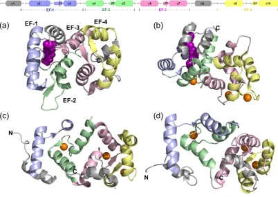
would have conserved three dimensional structures.[3,4] This is observed in both the
X-ray crystallography or NMR structures of Ca2+ bound un-myristoylated NCS
proteins (Figure 1.1).[4]
The evolutionary conservation of the NCS proteins can be observed, all are
approximately 200 amino acid residues in length and possess four EF hands (four
helix loop helix motif’s).[3,9] Of those four, only three are able to bind Ca2+ (although
recoverin and KChIP1 can only bind two).[3,9] Furthermore it is the first EF hand in
2
the 14 mammalian NCS proteins 11 are N-terminally myristoylated which enables
them to anchor to cell membranes, albeit using different mechanisms.[3,10]
Despite the similarities, the ability of the different NCS proteins to recognise different
sets of physiological ligands suggests that they must have some distinguishing
structural difference, which therefore makes the NCS family potential druggable
candidates.[4]
No PPI inhibitors have as yet been identified for NCS1 a member of this important
class of proteins.
Figure 1.1- A comparison of some of the different NCS protein structures- (Top) a generalised representation of the secondary structure of a member of the NCS proteins colour in accordance to the four EF hands, light blue (EF-1), light green (EF-2), light pink (EF-3), and pale yellow (EF-4). (a) Myristoylated mammalian recoverin (PDB 1iKU [11]) the myristoyl group coloured in purple, the four EF hands can be seen in light blue (EF-1), light green (EF-2), light pink (EF-3), and pale yellow (EF-4) (b) Myristoylated mammalian GCAP1 (PDB 2r2I [12]) the myristoyl group coloured in purple, with the Ca2+ as orange spheres, again the four EF hands can be seen in light blue, light green, light pink, and pale yellow. (c) Rat NCS1 (PDB 5AEQ [13]) the four EF hands are coloured in accordance with the previous structures and the Ca2+ as orange spheres. (d) S.cerevisae frq1 (PDB 1FPW [14]) the four EF hands are coloured in accordance with the previous structures and the Ca2+ as orange spheres.
1.1.1 NCS1 and the dopamine receptor D2
Mammalian NCS1 also known as human frequenin (Figure 1.1 c) has been implicated
in numerous physiological functions including the regulation of neuro transmitter
release, synaptic plasticity, learning and memory function.[15–18] An investigation
into the interacting partners, indicate that NCS1 is involved in many protein-protein
[image:18.595.76.479.248.533.2]3
interacting proteins identified are phosphatidylinositol-4-kinase (PI4K) and its yeast
orthologue pik1, the dopamine receptors D2R and D3R, ARF1, the interleukin
receptor accessory protein like-1 IL1RAPL1, TRPC5 channels and InsP(3)
receptors.[19–27] Of these wide ranging interacting partners, one of considerable
interest and importance is the dopamine receptor D2 due to its role within the central
nervous system.
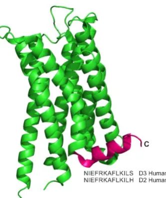
The dopamine receptor D2R is a member of the G-protein coupled receptors
(GPCRs) of which there are five (D1-D5) that can be subdivided into two families; the
D1 like family which include D1 and D5 and the D2 like family including D2, D3 and
D4.[28] Comparisons between the dopamine receptor D3 and D2 lead to the
prediction that D2 is composed of seven transmembrane α helices, which are
connected through three intracellular and three extracellular loops.[29] Alongside an
amphipathic α helical C-terminal domain as seen with D3, the amino acid sequence
in the C-terminus of D3 differs from that of D2 by only one residue (Serine D3 for
[image:19.595.236.403.370.569.2]Histidine D2, Figure 1.2).[29]
Figure 1.2- The structure of the dopamine D2/D3 receptor (PDB 3PBL) with the C-terminal helix coloured magenta and the amino acid sequence annotated. The D2R peptide is predicted as being α helical, the C-terminal region of D3 which only differs from the sequence of D2R peptide by one residue is also known to form an amphipathic α helix as shown here.[27,29]
The biological implications of the dopamine receptor D2 include dopaminergic
signalling within the central nervous system, addictive behavioural disorders, bi-polar
disorder and schizophrenia.[30,31] Interestingly, the expression levels of NCS1, are
increased in patients with bi-polar disorder and schizophrenia when under an
4 Figure 1.3- The chemical structures of the antipsychotic drugs haloperidol 1.1 and clozapine 1.2.
The mechanism for the interaction of NCS1 with D2 is both direct and indirect in
nature; the Ca2+ sensitive interaction between NCS1 and D2 causes the
desensitisation of the D2 receptor towards dopamine to be reduced (the direct effect
Figure 1.4).[32] However, the interaction between NCS1 and the glycoprotein
coupled receptor kinase 2 (GRK2) blocks the GRK2 mediated phosphorylation of the
D2 receptor (this is the indirect effect Figure 1.4).[32] The Ca2+dependent nature of
the NCS1 interactions are regulated through changes in the Ca2+ concentration, this
occurs through the release of Ca2+ from intracellular stores and Ca2+ channels (Figure
1.4).[32]
Figure 1.4- A representative view of the interaction between NCS1 and dopamine receptor D2 (D2R) at the synapses (adapted from [32]). The Ca2+ sensitive interaction between NCS1 and D2R causes attenuation of the D2 receptor desensitisation. The interaction of NCS1 with the glycoprotein coupled receptor kinase 2 (GRK2) is also Ca2+ dependent and it blocks the GRK2 mediated phosphorylation of the D2 receptor. The Ca2+ dependent nature of NCS1 is regulated through changes in the Ca2+ concentration through the release from intracellular stores and Ca2+ channels.
The physiological relevance of the NCS1-D2 interaction has heralded it as a
promising target for molecular intervention. No PPI inhibitors have as yet been
[image:20.595.139.414.367.614.2]5
With the structures of numerous members of the NCS proteins already determined
using X-ray crystallography or NMR spectroscopy, it was suggested that the
NCS1-D2R interaction could occur in a similar manner.[33] It was observed in the previous
cases, that the interaction of a ligand or peptide occurred in a large hydrophobic cleft
that was formed by a number a hydrophobic residues.[4,34–37] The residues in
question were known to be conserved throughout the NCS family, suggesting that
this mode of binding could be adopted across the NCS proteins.[38] However, the
specificity of each interaction would occur through differences in the size of the cleft
most likely in relation to the size of the binding partner.[38] In the case of NCS1 it
was found that the presence of Ca2+ caused a large hydrophobic cleft to become
exposed as with previous NCS proteins, this furthered the assumption that this could
be the site for the interaction of a protein or ligand.[33]
More recently an extensive study into the different crystal structures of NCS1
complexes and truncated Ca2+ bound proteins by Lian et al. elucidated a number of
interesting and significant features (Figure 1.5).[13] Utilising a number of techniques
including X-ray crystallography, it was confirmed that two D2R peptide molecules
bound to NCS1, as had been postulated by previous computational and NMR
spectroscopic based studies.[13,38]
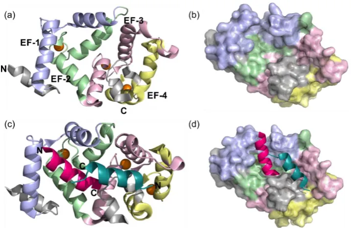
Figure 1.5- Structure of rat Ca2+ NCS1 complexed with dopamine receptor D2R peptide (PDB
5AER). (a) Cartoon representation of the NCS1 structure when in complex with the dopamine receptor D2R peptide omitting the D2 peptide. The EF hands are coloured accordingly EF-1 pale blue, EF-2 pale green, EF-3 pale pink and EF-4 cream the three Ca2+ orange spheres. (b) The molecular surface of NCS1 coloured as before, with the hydrophobic ligand binding site observed running down the centre. (c) Cartoon representation of the NCS1 D2R peptide complex, NCS1 coloured as before with the two C
[image:21.595.139.498.427.660.2]6
A number of key hydrophobic residues within the conserved hydrophobic groove were
implicated in the binding interaction between the protein and the peptide (Figure
1.6).[13] The binding of NCS1 to the two peptides can be split into two specific
regions of the protein, the N-lobe and C-lobe, with one peptide molecule binding to
each region separately. The two D2R peptides were found to bind to NCS1 with their
C-termini orientated towards the centre of NCS1 (Figure 1.5). Those implicit residues
of NCS1 found to undergo an interaction within the N-lobe include Trp30, Phe34,
Phe48, Ile51, Tyr52, Phe55, Phe85 and Leu89.[13] Interestingly when compared to
other NCS protein and peptide complexes, the evolutionary conservation of these
residues indicate their importance for binding within the complexes.[13] In
comparison, the C-lobe was found to make fewer intermolecular contacts which
include Phe54, Ile128, Tyr129, Met131, Val132, Val136, Leu138, Ala182, Tyr186
(Figure 1.6 b and Figure 1.7). However, despite having fewer contacts to the D2R
peptide, it appeared to undergo significant structural changes upon binding. These
changes include the induced-fit structural stabilisation of one side of the hydrophobic
crevice between residues Val132 - Leu138. In the uncomplexed protein (Figure 1.1
c) this region was found to be unstructured, however in the presence of the D2R
peptide (Figure 1.5 b) a helical conformation is adopted.[13] Interestingly it is this C
-lobe made up of the EF-3 and EF-4 regions that are the least conserved within the
NCS family members. It could be suggested that the residues within this region that
are stabilised upon D2R peptide binding could give rise to target specificity and as
[image:22.595.73.480.497.659.2]such could be key for therapeutic purposes.
Figure 1.6- Structure of rat Ca2+ NCS1 in complex with D2R peptide-the key hydrophobic residues
7 Figure 1.7- The Intermolecular interactions of rat Ca2+ NCS1 with the D2R peptide. (a) A detailed
zoom of the hydrophobic interactions of the sidechain residues from the N-lobe of NCS1 (coloured as Figure 1.5) with the C-terminal helix of D2R peptide 1 (magenta). (b) A detailed magnification of the hydrophobic interactions of the sidechain residues from the C-lobe of NCS1 (coloured as before) with the C-terminal helix of D2R peptide 2 (teal).
1.1.2 Frq1 and pik1
As well as mammalian NCS1, it has been found that the yeast Saccharomyces
cerevisiae and Schizosaccharomyces pombe genomesencode a protein that shares a sequence homology of greater than 60% with mammalian NCS1.[4] These
homologs have different roles in their respective genomes. For example in the fission
yeast S. pombe the homologue known as Ncs1 is responsible for the regulation of
sporulation and Ca2+ tolerance.[39,40] In the budding yeast S. cerevisiae, the
orthologue is known as frq1 and it is essential for cellular growth.[26] A key
protein-protein interaction has been found to occur between frq1 and phosphatidylinositol
4-kinase (pik1) (Figure 1.8). The pik1 protein is fundamental for vesicular trafficking in
the late secretory pathway, nuclear functions and cytokinesis, frq1 is necessary for
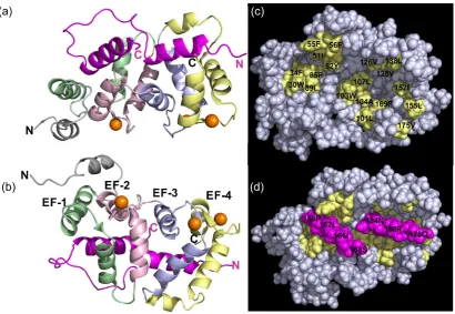
8 Figure 1.8- The frq1 pik1 complex. (a) View from the binding interface and (b) the binding face rotated 180°, both cartoon representations of NMR structure of the interaction between yeast frq1 and pik1. The four EF hands of frq1 can be seen in light green, light pink, light blue and cream, with the two alpha helices of pik1 in magenta. (c) and (d) Space fill representations from the binding interface (c) of frq1 alone with its key hydrophobic residues in yellow (d) frq1 and pik1 with the key residues of pik1 annotated (adapted from [34]).
The pik1 peptide is composed of two α-helices, the N-terminal helix from residues
127 - 135 and the C-terminal helix 156 - 167, the two helices are connected by a
twenty residue loop.[34] The stoichiometry of binding deduced from NMR studies
was defined as 1:1, in contrast to that of the 2:1 stoichiometry determined for the D2R
peptide NCS1 interaction .[34] As with the NCS1 D2R peptide interaction the
protein-protein interaction between frq1 and pik1 involves two lobe regions of frq1 interacting
independently with two α helices of pik1. The hydrophobic residues of the N-terminal
helix of pik1 interacts with the C-terminal residues of frq1.[34] Similarly, the
hydrophobic residues on the C-terminal helix of pik1 contact the N-terminal
hydrophobic groove of frq1 (Figure 1.8).[34]
It was discovered through the results of NMR studies by Strahl et al. that in the
complex of frq1 and pik1 the terminal residues of frq1 at both the N and C terminus
are solvent exposed and disordered. This lead to the possibility that the N-terminal
myristoyl group of uncomplexed frq1 may actually be involved in recruiting pik1 to the
membranes.[34]
Work by Lim et al. found that frq1 underwent a large conformational change induced
9
hydrophobic residues in the C-terminal to become solvent-exposed enabling it to
interact with pik1.[4] Furthermore through their NMR analysis, the interaction
between the C-terminal helix of pik1 and frq1 was found to be more stable in
comparison to the corresponding interaction of the N-helix of pik1.[34] A number of
hydrophobic residues on the C-terminal helix of pik1 were found to make extensive
contacts with the aromatic and other hydrophobic residues on frq1 (Figure 1.9 a).
Figure 1.9- (a) Close up view of the C-terminal helix of pik1 (magenta) with the sidechains of the N -terminal hydrophobic groove of frq1 (pale yellow). (b) Close up view of the N-terminal helix of pik1 (magenta) with the sidechains of the C-terminal hydrophobic groove of frq1 (pale yellow) (adapted from [34]).
The network of strong and intricate intermolecular hydrophobic interactions explain
the high affinity of the interaction between the two proteins.[34] It suggested that the
interaction of the C-terminus of pik1 was the anchoring point between the peptide and
the protein, facilitating further interaction between the second N-helix of pik1 and
frq1.[34]
Frequenin has been found to have a high degree of evolutionary conservation with a
similar secondary structure to the mammalian NCS1, with the EF hand motifs EF-2,
EF-3 and EF-4 bound to Ca2+ ions.[41] Another similarity drawn between the two
homologues and other NCS proteins is that they have a hydrophobic pocket, found
at the surface between two of the EF-hand-containing lobes as well as the solvent
exposed N and C termini.[41]
The structural similarities and the fact that the two orthologues NCS1 and frq1, both
interact with pik1, suggest that NCS1 may be responsible for the regulation of the
activity of pik1 in mammalian cells.[34] The knowledge that the hydrophobic residues
within the C-terminal helix of pik1 make considerable contact with aromatic and other
hydrophobic sidechains of frq1, could be useful when designing inhibitors.[34] Such
10
Met165.[34] This small cluster of hotspot residues could be used to generate a
starting template or three dimensional representation, from which a computational
structure-based drug design (sbdd) method could be applied. The sbdd method
would allow for the selection of novel compounds, able to replicate the area of 3-D
space that the hotspot residues represent for a pharmacophore based search. The
compounds would subsequently be synthesised and tested using a variety of
biophysical methods.[34]
There are a variety of different computational applications used in structure-based
drug design process (Section1.4), and a number of protein-protein interactions (PPIs)
have been targeted therapeutically, using computational applications in development
of drug-like compounds.
Protein-Protein Interactions (PPIs) as Drug Targets
PPIs are involved in nearly all biological processes and cellular function is therefore
reliant on their regulation.[42] Due to their ubiquitous nature and central role, there
has been an increasing interest in targeting the interface between two interacting
proteins as a potential therapeutic target.[42,43] Inhibition of protein-protein
interactions has seen significant recent advances, although it does present a number
of challenges because of the nature of protein-protein interaction surface.[43] The
surfaces involved in PPIs are typically large, often flat with complicated topography,
and a large portion of the surface area of the interfaces are buried.[44,45] Despite
these large interfaces there may only be a small number of influential residues
essential for a high affinity binding interaction [46–48], thereby, reducing the actual
challenges posed when developing small molecule inhibitors and making the
targeting of protein-protein interactions a realistic and viable drug development
strategy.
A typical passively absorbed drug profile follows Lipinski’s ‘Rule-of-five’ which
includes a MW < 500 Da. Due to the nature of protein-protein interfaces molecules
which are targeted at them tend to have a high molecular weights.[43,49] They also
tend to have an increased hydrophobicity, a higher number of hydrogen bond donors,
hydrogen bond acceptors and a higher ring complexity in comparison to other
drugs.[50]
At present excluding peptidomimetics there is no well-established structure-based
approach for the design of PPI modulators using defined computational pipelines.
There is also a general lack of small molecule starting points for drug design as there
11
interfaces.[44,50] It can be difficult to distinguish between true binding from
artefactual binding and current small molecule compound libraries tend to be biased
towards certain protein classes, making them more specific to certain targets and less
general.[44,50]
Despite the difficulties PPI targets present, there have been a number of notable
success stories with several drugs are already in clinical use and some others are
being evaluated in clinical trials.[51]
The inhibition of PPIs past and present
Research into the design and synthesis of small molecule inhibitors has been
conducted for a number of key protein-protein interactions that are believed to play a
vital role in many biological processes. Due to the central role of PPIs the number of
systems studied is increasing very rapidly, with some of the most interesting being,
studies on binding to IL-2, HPVE2, ZipA, TNf, HDM2 and Bcl-XL.[43] Here we shall
briefly discuss some of the approaches developed to target these interactions, in
particular those of HDM2 and Bcl-XL (Sections 1.3.5 and 1.3.6).
1.3.1 IL-2
IL-2 (Interleukin-2) is a cytokine that is of particular interest due to its role in the
activation of killer T-cells in an immune response and has been linked to the rejection
of tissue grafts.[52] The design of a series of small molecules able to bind to the
receptor of IL-2, was conducted by Sunesis Pharmaceuticals, without prior knowledge
of the structure of the IL-2 bound to its receptor (IL-2R).[53] The initial hit to lead
process started with a small peptidomimetic designed to mimic the structure of IL-2
binding region; however it was found to bind to IL-2 itself rather than the IL-2 receptor
and so was not taken any further.[54]
Interestingly, the subsequent discovery process was carried out utilising a
combination of techniques and using the original unsuccessful compound as a
template starting point. The compound was divided into its four constituent
fragments, from which the subsequent structure activity relationship studies (SAR)
elucidated one key fragment. Optimisation of this fragment was carried out through
a number of steps including further compound library screening processes to evolve
the target compounds.[43,54]
To ensure a greater success rate at each separate stage the lead compounds were
characterised using a number of assays including enzyme-linked immunosorbent
12
The most successful small molecule designed was SP4206 (1.3 Figure 1.10) which
interestingly, does not resemble the IL-2R.[53] In fact it is believed that if the crystal
structure of the IL-2 receptor complex had been used in the design process then
SP4206 would not have been discovered, highlighting the occasional unpredictability
and sometimes serendipitous nature of drug discovery.[43]
1.3.2 Interactions involving the human papilloma virus HPV
The protein-protein interaction between the viral transcription factor E2 and the viral
helicase E1 is important in the life cycle of the human papilloma virus (HPV).[55] HPV
is associated with cervical cancer and warts, and small molecule treatment regimens
are available with limited success.[56] Therefore the development of such drugs to
target HPVE2 and prevent its interaction with E1 is a key target for pharmaceutical
companies.[56] A research group at Boehringer Ingelheim was able to utilise High
Throughput Screening (HTS) to identify a class of compounds known as
indadiones.[56] These compounds were found to disrupt moderately the
protein-protein interaction between E2 and E1; however, further target optimisation and
medicinal chemistry efforts led to the development of the 1.4 (Figure 1.10). This
compound exhibited a lowered IC50 value of 6 nM, with a higher ligand efficiency than
the original binding partner target E1.[55,57,58] Thought to be due to the ability of
the compound to bury its hydrophobic surface area, rather than spanning across the
[image:28.595.165.391.468.696.2]vast protein-protein interface.[55,58]
13
1.3.3 ZipA and FtsZa
In another study the interaction between ZipA and FtsZa was examined, ZipA is a
membrane anchored protein that interacts with a homologue of eukaryotic tubulin,
FtsZa through terminal carboxy domains.[59] These proteins were found to be the
main constituents of a structure known as a septal ring found in gram negative
bacteria, and is responsible for their separation and replication.[59] The interaction
between the proteins was studied using X-ray crystallography, NMR spectroscopy
was employed to screen a diverse set of 825 compounds.[60] The NMR study found
seven hits that bound to ZipA at the same site as FtsZa and is an example of early
drug design processes.[60] However, despite this early indication that ZipA may be
“druggable” further medicinal chemistry and SAR were unable to generate any small
molecules able to penetrate deep within the surface of ZipA which is in stark contrast
to the findings of studies on IL-2, HPVE2, HDM2 and Bcl-XL.[43,60,61] This is an
excellent example of the challenges that are involved in targeting complex
interactions associated with two interacting proteins.
1.3.4 TNF
Tumour Necrosis Factor (TNF) is also a cytokine and is an important biological target
due to its involvement in the inflammatory response.[62] It is the interaction between
TNF and its receptors, TNFR1 and TNFR2, that the small molecule inhibitors are
designed to disrupt, these targeted therapeutics have been approved for the
treatment of arthritis.[62] Fragment screening lead to the development of a group of
small molecules, that were found to interfere with TNF binding, through the
displacement of one of the three monomers that constitute the TNF-α homotrimer.[63]
However due to their moderate affinities, these compounds are not considered as
potential drug candidates.[63]
14
1.3.5 Case Study 1: Developing small molecule modulators of P53 and
MDM2 over the decades…
An extensively investigated protein-protein interaction is that between the tumour
suppressor gene p53 and HDM2.
HDM2, also known as the human protein double minute, was found to be an excellent
target in the treatment of cancer.[64] The initial research into MDM2, the mouse
orthologue, found that it bound to the tumour suppressor protein p53, increasing its
degradation and blocking its transcriptional activity which results in tumour
suppression.[65] The interaction of MDM2 with p53 also results in the nuclear
movement of p53 into the cytoplasm and away from its site of action.[66]
The design and synthesis of compounds to modulate this interaction has been of
substantial interest over a number of decades. However despite keen research
interest, the development of small molecule modulators was not successful for a long
period of time, indicative of the complexity involved in the design of modulators for
PPI targets.[67]
The interaction between the two proteins MDM2 and p53 was found to involve of a
small segment of the N-terminus transcription activation domain of p53 (residues
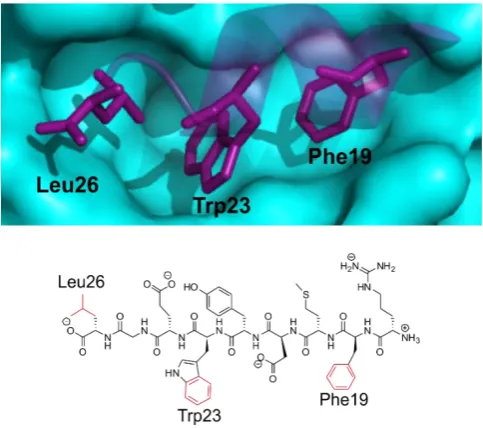
13-19) and a hydrophobic pocket within MDM2.[68] Furthermore X-ray crystallography,
indicated the primary involvement of three sidechains of the hydrophobic residues;
Phe19, Trp23 and Leu26, which form a short α helix on p53 located within a deep
pocket in MDM2 (Figure 1.13).[67,69]
One of the earliest success stories of inhibiting the two interacting proteins was
reported by Vassilev et al. who developed a series of cis-imidazoline compounds,
more commonly known as Nutlins (Figure 1.12).[70] High-throughput NMR
spectroscopy screening of a diverse library of compounds, followed by extensive
optimisation strategies from the initial leads resulted in three racemic hit compounds
Nutlin-1 and Nutlin-2. However interestingly, of the two enantiomers of Nutlin-3 only
one is active and this is known as Nutlin-3a.[70] The three Nutlin compounds were
found to disrupt the p53-MDM2 interaction with IC50 values of 260 nM, 140 nM and
90 nM, respectively.[70] Determination of the crystal structure of the complex
between MDM2 and Nutlin-2, indicated that the compound was able to mimic the
sidechains of Phe19, Trp23 and Leu26 on the p53 helix, through investigations of the
corresponding interaction sites on MDM2. Since its initial publication Nutlin-3a has
15 Figure 1.12 The chemical structure of Roche’s cis-imidazoline compounds the Nutlins, designed to inhibit the p53 MDM2 interaction. The two racemic compounds Nutlin-1 and Nutlin-2 and the active enantiomer Nutlin-3a.
Cummings et al. at Johnson and Johnson used the proposed three key residues from
the interacting sequence of a p53 9mer segment (Figure 1.13), as a template in the
design of their benzodiazepine derived inhibitors.[67] When screened against their
biological target, these benzodiazepine derivatives presented half maximal
concentration (IC50)values ranging from 125 μM - 0.4 μM.[67] A crystal structure of
one of the benzodiazepine inhibitors with MDM2 (PDB 1T4F), indicated it was able to
successfully mimic the three sidechain residues, this structural data further supported
a competition assay with the native peptide.[67,71]
Figure 1.13- The three hydrophobic sidechain residues of the p53 helix (purple) highlighted in the deep pocket of MDM2 (cyan) (PDB 1T4F), against the corresponding 9mer segment of p53 with the sidechains highlighted below (adapted from [67]).
Ding et al. elucidated a key hydrogen bonding interaction between the NH of the
indole ring of sidechain from Trp23 to the MDM2 backbone using x-ray
crystallography.[68] Therefore the design strategy of small molecule inhibitors
developed by Ding et al. involved searching for chemical moieties that could mimic
[image:31.595.197.439.414.628.2]16
mimic the hydrogen bonding and hydrophobic contributions of the Trp23 sidechain
and this was therefore used as a core structure for the starting point of the
structure-based strategy.[68] From here a substructure search was conducted to identify
natural products that contained the oxindole ring, among those identified were
spirotryprostatin which contained a spiro-oxindole core.[68]
Molecular modelling studies using computational docking, found these compounds
fitted poorly into the MDM2 cleft due to steric clashes, but the spiropyrrolidone ring
presented a rigid scaffold from which the residues of Phe19 and Leu26 could be
mimicked through the addition of hydrophobic sidechains.[68] Candidate compounds
were then designed through the variation of the sidechains, their most potent inhibitor
1.10 (Figure 1.14) was found to have a KD value of 86 nM with MDM2.[68] It was an
effective inhibitor of cellular growth in cancer cells with WT p53 and exhibited
selectivity over cancer cells with deleted p53, with minimal toxicity to healthy cells and
presented a promising lead compound for optimisation.[68]
Figure 1.14- The chemical structures of three non peptidic inhibitors of p53 and MDM2, a product of the structure-based design and synthesis process approach developed by Ding et al. (adapted from [68,69]).
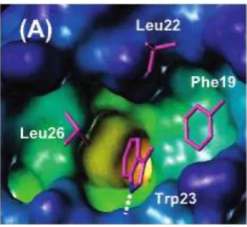
The three residue mimic was also used to design other inhibitors; however through
the use of fluorescence polarisation assays it was discovered that despite the high
binding affinities of these molecules they were significantly less potent.[69] This
indicated the presence of other interactions, confirmed with the discovery of a fourth
residue Leu22 involved in the binding interface (Figure 1.15).[69] Molecular modelling
techniques such as computational docking using the crystal structure was then used
to optimise 1.10 bound to MDM2.[69] The findings suggested that a chain extension
at the carbonyl end could interact with the desired fourth residue.[69]
The computational docking programme GOLD was the next step in the process,
predicting binding modes of several different chain extensions. The fact that the
Leu22 residue was not buried within a hydrophobic pocket, but rather solvent exposed
meant that the group could experiment with different polar moieties that could also
17
Through fluorescence polarisation (FP) assays, the initial target 1.11 (Figure 1.14)
displayed an inhibition constant (Ki) of 13 nM and an increased potency, six times
greater than 1.10.[69] The synthetic route to obtain 1.11 utilised an asymmetric 1,
3-dipolar cycloaddition reaction as the main step in the stereospecific synthesis and is
shown in Scheme 1.1.
Figure 1.15- X-ray structure of p53-MDM2 complex (PDB 1YCR). In addition to the three residues Phe19, Trp23 and Leu26, Leu22 was also found to play a role in the interaction. MDM2 cavity is coloured according to its depth, the deep buried region coloured in yellow with the external solvent exposed regions in blue and the sidechains of the key residues in magenta (adapted from [69]).
Further medicinal chemistry driven optimisation of 1.11, involved attempts to improve
the metabolic stability of the compounds through the incorporation of fluorine groups
on the benzene lead to a reduction in their potency. However, development of
another ligand 1.12 from the scaffold of 1.11 with the incorporation of a fluorine group,
resulted in a Ki of 3 nM. When Ding et al. used their fluorescence polarisation assay
to compare their newly designed inhibitor to that of Hoffman la Roche’s most
[image:33.595.228.407.173.337.2]18 Scheme 1.1- The three step stereospecific synthetic route to spiro-oxindole derived p53 MDM2 modulators 1.11 and 1.12 developed by Ding et al. (adapted from [69]). (a) 4 Å molecular sieves, toluene; (b) 2-morpholin-4-yl-ethylamine/THF or 1(2-aminoethyl)-piperadine/THF, RT; (c) Pb(OAc)4, CHCl2-MeOH (1:1), 0 ˚C.
This is an excellent example by Ding et al. of the use of rational drug design and
guided by computational modelling used alongside chemical synthesis and structural
biology to develop and optimise small molecule inhibitors of PPIs.
More recently an extensive investigation into the structure-based rational design
behind the discovery of a novel piperidinone series of p53 MDM2 inhibitors was
reported by Rew et al. (Figure 1.16).[72] Their investigative studies were based on a
rigid core that was capable of holding two aromatic rings, to again target the key
residues of p53. Initial results led to the development of 1.19 a 1,3,5,6-tetra
substituted piperidinone which had an IC50 of 2.42 μM.[72] By simply changing the
configuration of the acetic acid component and converting the diaryl from cis to trans
followed by isolation of the active enantiomer 1.20, increased the potency up to 70
fold.
This new optimised inhibitor was found to be capable of forming electrostatic
interactions between the acetic acid component and an imidazole sidechain of
MDM2.[72] They subsequently reported that optimisation processes of the N-alkyl
substituent on 1.20 resulted in compounds that were designed to occupy the Phe19
pocket, improved solubility through increasing the polarity of the compound and
creating van der Waals contacts to MDM2.[72] Furthermore the discovery of a third
hit compound 1.21 came from optimizing the piperidinone scaffold so that the trans
-diaryl conformation was stabilised with substitution of a methyl moiety on the same
19
compound 1.22 was obtained through optimisation of 1.21, it demonstrated an
excellent efficacy with a binding constant (KD) of 0.4 nM and was selective for its
target with an improved oral bioavailability and pharmacological studies predicted it
to have a long half-life in humans.[72]
Figure 1.16- The five piperidinone inhibitors developed by Rew et al. The development process involved the initial hit compound 1.19, optimisation processes led to 1.20, which in turn was optimised to compound 1.21 and ultimately led to the most potent compound 1.22 which had a KD of 0.4 nM. The synthetic procedures employed in this vast hit to lead optimisation project
followed multi step syntheses which are summarised in Scheme 1.2. From the simple
starting compound 2-(3-chlorophenyl) acetic acid 1.23 a six step synthetic procedure
(a) involving such processes as conjugate addition, NaBH4 reduction and the
Staudinger reaction lead to 1.24 in its cis-diaryl form.
A further three steps converted the cis-diaryl 1.24 into the first hit compound the cis
-diaryl 1,3,5,6-tetrasubstituted piperidinone 1.19. The second hit compound 1.20 was
generated from the enantiomer of trans-diaryl 1.24, the reactions include N-alkylation,
followed by allylation at the C3 position and finally oxidative cleavage of the alkene at
C3 using catalytic amounts of ruthenium tetroxide. The six synthetic procedures to
obtain 1.21 included some of the following protection of the amide nitrogen of the
trans-aryl piperidinone 1.24, methylation at C3, deprotection followed by N-alkylation and oxidative cleavage of the alkene as before.
A further three successive reduction and oxidation reactions using the Des-Martin
periodiane from an analogue of 1.21, resulted in the final and most successful lead
compound 1.22 in a yield of 75%. This is an excellent example of a scaffold based
synthesis that is able to utilise a wide variety of synthetic organic chemistry
techniques to achieve optimisation of hit to lead. Although the processes may be
lengthy and not high throughput, the synthesis which revolves around further
functionalisation of a core scaffold, works in sequential manner that is efficient at
20 Scheme 1.2- The general synthetic approach to obtain the hit compounds 1.19, 1.20, 1.21 and 1.22. The processes involved multistep syntheses originating from the parent starting material 2-(3-chlorophenyl) acetic acid 1.23 which is converted to the piperidinone core 1.24 in a six step synthesis 87 – 88% yield (a). The piperidinone core is presented as a general scaffold without any stereochemistry, however to achieve the desired stereochemistry of 1.19, 1.20, 1.21 and 1.22 the reagents and conditions of (a) were altered as necessary. The cis-diaryl 1.20 is converted to the first hit compound the cis-diaryl 1,3,5,6-tetra substituted piperidinone 1.19 in a four step synthesis 79% yield (b), the second developed compound the trans-diaryl carboxylic acid 1.20 was synthesised from trans -diaryl 1.22 in a three step synthetic procedure 89% (c). A six step synthetic procedure 47 – 80% (d) was used to transform the trans-diaryl scaffold of 1.24 to the carboxylic acid 1.21 and finally a nine step procedure (e) was employed to achieve the most successful hit compound 1.22 80% yield.[72]
Further to these examples of the numerous compounds developed as MDM2
inhibitors, at least two compounds have more recently reached clinical development.
MDM2 inhibitors include an oral treatment for advanced stage solid tumours (Johnson
and Johnson, Phase 1), and R7112 (Hoffmann-La Roche, Phase 1) an oral treatment
for hematologic neoplasms and advanced solid tumours.[73]
1.3.6 Case Study 2: Targeting the interaction of Bcl-XL and BaK
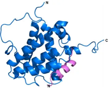
The second case study to be discussed is that of Bcl-XL and BaK, within the Bcl2
family, Bcl-XL inhibits programmed cell death and BaK promotes apoptosis.[74] The
interactions between the two proteins controls the sensitivity of the cell towards
apoptosis through the antagonism of the two different functions.[74] Bcl-XL contains
two centralhydrophobic α-helices which are in turn surrounded by five amphipathic
helices, it binds tightly to a 16 amino acid region (BH3) of BaK (residues 72-87)
21 Figure 1.17- The Bcl-XL (blue) / BaK (Violet) complex derived from NMR studies by Sattler et al. (PDB ID 1BXL, adapted from [74]).
Considerable research has been conducted by Hamilton et al. into targeting the
interface with small molecule antagonists to promote apoptosis in unhealthy cells.[75]
Several approaches were used to identify small molecule inhibitors of Bcl-XL, including
oligomeric aryl-aryl and aryl-carboxamide components, in these instances the
substituent’s would mimic the distance and angular projection of the sidechains from the α helix of its binding partner BaK3.[75] Hamilton utilised intramolecular hydrogen
bonds along with the steric interactions to help position the peripheral functionalities
and instil a rigidity to the structure to aid pre-organisation at a molecular level.[75]
Through the use of chemical synthesis alongside structural biological techniques
such as, X-ray crystallography, fluorescence polarisation (FP), isothermal titration
calorimetry (ITC) and NMR spectroscopy studies, Hamilton was able to develop
enaminone 1.25, benzoylurea 1.26, terphenyl 1.27 and terepthalamide 1.28
scaffolds.[75–77] These four novel scaffolds were able to successfully mimic the residues of an α-helix (Figure 1.18).[75–77]
Figure 1.18- The α-helix small molecule mimetics developed by Hamilton and co-workers. enaminone 1.25, benzoylurea 1.26, terphenyl 1.27 and terepthalamide 1.28 scaffolds where R1, R2 and R3 are the different positions of functionalisation.[75–77]
The BaK3 helix has been shown through crystallographic evidence to contain four
[image:37.595.232.409.70.212.2]22
role in its interaction with Bcl-XL.[77] The original terphenyl derivatives 1.27 were the
first compounds synthesised to mimic projection of these sidechains, they adopted a
staggered conformation with substituents that projected their groups at the i, i+4 and
i+7 positions as with the α helix of BaK3.[76] The synthesis was developed using O
-alkylated phenols and sequential Suzuki aryl-aryl cross coupling reactions.[76] The
synthesis was lengthy and the compounds were found to be extremely hydrophobic
with a predicted logP = 9.25 and low aqueous solubility logS = -10.76.[77]
The terephthalamides 1.28 were developed as a second generation of small
molecules and were simplified in terms of structure; they were also designed to have
a better solubility profile. [77] The two terphenyl rings of 1.27 were replaced by two
functionalised carboxamides, which were able to replicate the planar nature of the
phenyl rings through the rigidity and restricted rotation about the amide bond.[77] The
synthesis of these compounds was simplified when compared to that of the
terphenyls, involving O-alkylation and amide couplings and was found to be much
more efficient.[77] In comparison to the terphenyls, terephthalamides predicted logP
= 4.42 and a solubility constant logS= -5.35 were much more desirable.[77] The
enamine scaffolds 1.25 were synthesised using an even faster and efficient synthesis
(Scheme 1.3). This involved initial Claisen condensation reaction to give the
di-ketone 1.30.[78] Subsequent reaction with BF3OEt2 resulted in the
1,3-diketonatoboron difluoride complex which in the final reaction was reacted with m
-toluidine to produce the desired enamine 1.25.[78]
Scheme 1.3- The synthetic approach to the enamine scaffold 1.25. The synthetic approach involves a Claisen condensation reaction of 1.29 using (a) NaH, EtOAc, EtOH and ether to give the diketone 1.30. (b) The subsequent reaction of the diketone with BF3OEt2 to give the di-fluoride compound 1.31 in 84% yield, (c) which is finally reacted with m-toluidine to give the target compound the enamine 1.25 in 70% yield.
Finally, the benzoylurea compounds 1.26 were designed based on the enamine
scaffold, the synthetic approach involved the synthesis of two fragments; an amide
and an isocyanate which were coupled to form 1.26 (Scheme 1.4).[75] The two
substituent fragments were synthesised using similar synthetic approaches involving
23 Scheme 1.4- Coupling of an amide and isocyanate substituents to give the benzoylurea scaffold 1.26 in 43% yield (adapted from [75]).
These examples of the extensive investigations conducted by Hamilton et al. not only
produced efficient small molecule inhibitors of the Bcl-XL protein. They were also able
to provide a detailed understanding of the small molecule bound Bcl-XL structure
through informed computational docking programmes and numerous biophysical
techniques.[75–77]
With these case studies of different PPIs and the alternate approaches employed to
target them, it is clear that the process involves the combination of many techniques
and iterative of design, synthesis and testing.
The successful compounds were developed using computational modelling to predict
aspects such as binding poses and solubility. A variety of medicinal chemistry
techniques were used alongside diverse synthetic organic chemistry, to realise a
range of scaffolds that were able to either mimic the positions of amino acid
sidechains, or target certain hotspot residues. When used in conjunction with
structural knowledge of the biological target and a number of biophysical techniques,
it lead to improving the initial hit compounds towards lead molecules and in some
cases, ultimately to the clinic.
This interdisciplinary approach was successful in targeting protein-protein
interactions implicated in numerous biologically relevant viruses, diseases and
24
Computational applications of structure-based drug design
In designing inhibitors for protein-protein interactions, the complex nature of the
systems involved means that conventional high throughput screening methods may
not be an effective way to generate hit compounds. This may be observed in a high
number of false positives that occur due to the hydrophobic nature of many binding
[image:40.595.79.482.218.470.2]sights. Indeed it is clear that a new pre-screening strategy is necessary.[79]
Figure 1.19- The computational workflow applied in this thesis, following a general structure-based drug design process. Pharmacophore generation is used to screen a library of compounds to replicate an area of three dimensional space, the compounds are then docked into the target and their ADMET properties subsequently calculated. The information is then combined and a balanced selection protocol is undertaken to select the best possible candidate for synthesis. At each stage the number of potential hits is reduced.
As discussed previously, computational applications have found use in the
development of drug-like compounds to target protein-protein interactions. As seen
with the development of inhibitors to target the Bcl-XL and BaK3 interaction (Section
1.3.6), Hamilton et al. designed four novel scaffolds which are able to mimic the amino
acid sidechains at positions i, i+4 and i+7 of the BaK3 helix. Once two scaffold
structures had been designed, the computational programme QikProp was used to
calculate certain drug-like parameters, such as the solubility profile of each ligand to
compare and contrast the two and determine if the scaffolds were suitable.[77] The
conformations of the compounds in solution were then investigated using the
molecular modelling facility MacroModel, wherein they employed energy minimisation
25
MacroModel were then superimposed onto the native BaK3 α-helix, to compare the
positions of the functionalised substituents, to the i, i+4 and i+7 amino acid sidechain
positions on the BaK3 α-helix.[77]
Ding et al. also used the computational docking programme Gold, to calculate the
binding pose of their initial hit spiro-oxindole 1.10 (1.3.5) and from that develop a
number of analogues including their lead compound 1.12.[69] These simple
computational processes aided in the selection of the compounds to synthesise and
is an example of how computational techniques can aid success when used in the
process of targeting PPIs.
The use of a structure-based drug design method, as seen in previous PPI studies
(1.2) uses a combination of techniques. The methodology involves computational
modelling, used in conjunction with “hit identification” and ”hit to lead” optimisation
processes in a drug discovery pipeline (Scheme 1.5).[80,81] Furthermore, to improve
this computational screening process, it is more favourable to start with the best
selection of ligands to be computationally analysed and selected for synthesis.
26 Scheme 1.5- The structure-based drug design pipeline. The flow process applied in the SBDD protocol follows two phases, the “hit identification” phase and the “hit to lead” phase. A number of processes occur in the hit identification phase and these include (a) Target identification and preparation of the binding-site. (b) Preparation of the compound library used to screen hits for the target. (c) Molecular docking methods, analysis of the compounds predicted binding interactions with the target. This is followed by the hit to lead phase, selection of a lead compound (adapted from [82]).
1.4.1 Pharmacophore Modelling
The term pharmacophore was defined by Ehrlich as ‘a molecular framework that
carries the essential features responsible for a drug’s biological activity.[83] The
composition of the pharmacophore encompasses the steric and electronic
characteristics necessary to ensure optimal interactions between a molecule and
27
features is important for the development of a successful pharmacophore model.[84]
There are two types of pharmacophore model, ligand-based and structure-based.
The first works in the absence of a 3-D structure of the macromolecular target; it takes
a group of active molecules and compares their common 3-D features.[83] A
structure-based pharmacophore model works directly with the 3-D structure of a
macromolecular target or macromolecule-ligand complex.[83]
1.4.1.1 Ligand-based pharmacophore modelling
The ligand-based method was not used for the purposes of this thesis and so shall
not be discussed in great detail. However, briefly the method involves two main steps.
Firstly the conformational flexibility of each ligand has to be represented by creating
conformers, and secondly the ligands then have to be aligned and the common
features extracted to generate the pharmacophore.[83] This approach does not come
without a number of challenges such as the ability to model the ligands flexibility and
selecting the best method to align the molecules.[85,86] Perhaps a more challenging
question is finding a way to select the best group of compounds to generate the
pharmacophore, ensuring that the selection is diverse and not biased.[86]
1.4.1.2 Structure-based pharmacophore modelling
In the work presented here structure-based modelling was used. This involves
analysis of the complementary chemical features of the active site and the spatial and
chemical relationships, allowing a pharmacophore model with selected features to be
built.[83] It can be further subdivided into two categories: (i)
macromolecule-ligand-based whereby the complex structure is available to determine the hotspot residues
between the two binding partners.[83], and (ii) macromolecule- based which is simply
the macromolecule target without the natural ligand.
There are a number of modelling programmes which are able to determine the key
residues, either in the complex or within the macromolecule itself and these can be
used to generate a pharmacophore.[83] As with the ligand-based model, this
approach does have a number of limitations, such as the need for the structure of a
macromolecule target or macromolecule-ligand complex. Even if the complex or
structures are available there are often a vast number of chemical features that have
to be rationalised before a pharmacophore can be generated computationally.[83] To
overcome these problems a number of algorithms have been developed with the
ability to generate hotspot pharmacophores from the structure of the macromolecule
itself. It does so by identifying cavities within the structure and comparing these


![Figure 1.10- Structures of two developed small molecule inhibitors (1.3) SP4206 an inhibitor developed for targeting IL-2 (1.3) the so called “Compound 23” developed by Boehringer Ingelheim for the HPV: E2 interaction (structures adapted from [53,55,58] )](https://thumb-us.123doks.com/thumbv2/123dok_us/8044287.221911/28.595.165.391.468.696/structures-developed-inhibitors-inhibitor-boehringer-ingelheim-interaction-structures.webp)