A COMPARATIVE STUDY TO FIND THE
EFFECTIVENESS OF PRESSURE BIOFEEDBACK
VERSUS ISOMETRIC EXERCISES OF DEEP
NECK FLEXORS IN REDUCING CHRONIC NECK
PAIN USING NECK DISABILITY INDEX
By
(Reg. No . 27101805)
PADMAVATH COLLEGE OF PHYSIOTHERAPY
A COMPARATIVE STUDY TO FIND THE
EFFECTIVENESS OF PRESSURE BIOFEEDBACK
VERSUS ISOMETRIC EXERCISES OF DEEP
NECK FLEXORS IN REDUCING CHRONIC NECK
PAIN USING NECK DISABILITY INDEX
By
(Reg. No . 27101805)
Under the guidance of
Mr. K. KUMAR , M.P.T. , MIAP.,
Associate Professor,
Padmavathi College of Physiotherapy
Submitted in Partial fulfillment of the requirements for the Degree of Master of Physiotherapy
From
The Tamilnadu Dr. M.G.R. Medical University, Chennai
PADMAVATH COLLEGE OF PHYSIOTHERAPY
CERTIFICATE
This is to certify that the project entitled
“A COMPARATIVE
STUDY TO FIND THE EFFECTIVENESS OF PRESSURE
BIOFEEDBACK VERSUS ISOMETRIC EXERCISES OF
DEEP NECK FLEXORS IN REDUCING CHRONIC NECK
PAIN USING NECK DISABILITY INDEX”
Submitted by
the candidate
(Reg. No . 27101805)
is a bonafide work done in partial fulfillment of the requirements for the Degree of Master of Physiotherapy from
The Tamilnadu Dr. M.G.R. Medical University,
Chennai
Guide Principal
Viva-voce Examination held on ________________
DECLARATION
I hereby declare and present my dissertation entitled
entitled
“A
COMPARATIVE STUDY TO FIND THE EFFECTIVENESS
OF PRESSURE BIOFEEDBACK VERSUS ISOMETRIC
EXERCISES OF DEEP NECK FLEXORS IN REDUCING
CHRONIC NECK PAIN USING NECK DISABILITY
INDEX”
the outcome of the original research work undertaken
and carried out be me , under the guidance of
Mr. K. KUMAR , M.P.T. , MIAP.,Associate Professor , Padmavathi College of
Physiotherapy, Periyanahalli, Dharmapuri , Tamilnadu.
I also declare that the material of this dissertation had not
formed in any basis for the award of any other Degree previously
from the Tamilnadu Dr. M.G.R. Medical University, Chennai.
ACKNOWLEDGEMENT
First and foremost I thank
LORD ALMIGHTY
for
showering the blessings who always been my source of strength
and guided me in all endeavors leading to the completion of this
project.
My heartful gratitude to the Honorable Chairman
Mr.M.G.SEKAR,B.A.B.L.
Padmavathi College of Physiotherapy,
Periyanahalli, for providing me the valuable opportunity for doing
my Bachelor Degree in Physiotherapy.
My sincere and devoted thanks to my project guide
Mr. K. KUMAR, M.P.T. , MIAP.,
Associate Professor for
Padmavathi College of Physiotherapy , for his inspiration and
guidance throughout this thesis.
I wish to express my sincere thanks to
Mr. K.KUMAR,
M.P.T.,M.I.A.P.,
Principal, Padmavathi College of Physiotherapy,
for his valuable advice , suggestions and encouragements in
making this project a successful one.
I express my special thanks to all of my
FRIENDS
for
sharing their knowledge and support each and every step of this
thesis work.
I take this golden opportunity to thank each and every patient
who took part in this study, for his or her kind cooperation and
needed information
DEDICATED TO MY BELOVED
PARENTS , STAFFS
AND
TABLE OF CONTENTS
CHAPTERS Page No
I. INTRODUCTION
1. Introduction 1
II. REVIEW OF LITERATURE
1. Review of Literature 28
III. MATERIALS & METHODOLOGY
1. Research Design 34
IV. RESULTS
1. Research Design 39
V. DISCUSSION
1. Discussion 53
VI. CONCLUSION
1. Conclusion 59
REFERENCES 61
APPENDICES
ANNEXURE - I 68
ANNEXURE - II 73
INTRODUCTION
Neck pain is a major problem in the society, with an increasing sedentary population especially with reliance on computer technology in the workplace.
Pain is defined as a sensation characterized by a group of unpleasant perceptual and emotional experiences that triggers autonomic, psychologic and somatomotor response associated with actual (or) potential damage to the tissues1. Mechanical neck pain may be defined as pain secondary to overuse of a normal anatomic structure (or) pain secondary to injury (or) deformity of an anatomic structure2.
Neck pain is considered to be chronic if it last for more than 3 months of duration and pain that continues after the stimulus is removed (or) the tissue damage heals. Chronic neck pain is becoming increasingly prevalent in society estimations indicated that 67% of individuals will suffer neck pain at some stage throughout life. The current research incidence of chronic neck pain in Bangalore has been estimated as 35% and the median age as 27 years and its ranges between 18 to 52 years.
the surrounding neck musculature. The ligaments role in stabilization occurs mainly at end of range postures3. While muscles supply dynamic support in activities around the neutral and mid range postures, which are commonly adopted during functional daily tasks. In the presence of injury (or) pathology, the role of the muscular system becomes even greater which highlights the need to address the muscle system during both the assessment and rehabilitation of patient with neck pain.
stabilization role with resulting altered mechanics and an altered load distribution on articular and soft tissues the cervical tissues are subjected to adverse stress and chronic strain with resultant pain.
Further more it was demonstrated that these stresses could be relieved if the supporting muscles principally the upper and deep cervical flexors were functioning at a level where they could repeatedly hold a low load inner range contraction without postural function. There is clinical evidence that the upper and deep cervical flexor that are the important muscles for cervical segmental and postural control lose their endurance capacity in patients with neck pain. The increased understanding of the tonic supporting role of the deep neck flexors and their functional differentiation from the superficial flexors realizes the need to develop a test that would target these muscles in relative isolation from their superficial counterparts.
spine. It is based on the anatomical interrelated action of the deep muscles to support and stabilize the cervical spine as well as produce a flattening of the normal cervical spine lordosis4. This test is conduced in supine lying with the head neck region supported in neutral position. The action is reasoned to recruit all the deep neck flexors to hold the head and cervical region in a static position. As the muscles are deep and unable to be palpated directly, an indirect quantification of their ability to hold the cervical spine position is gained by monitoring the steady position of he neck with an inflatable air filled pressure sensor (stabilizer Chattanooga south pacific) that is positioned sub occipitally behind the neck. Work is currently proceeding to establish the validity of this test although initial clinical data suggest that it can depict a deficit in function in patients with neck pain. This dysfunction improves with retraining and parallels a reduction in symptoms.
Panjabi introduced an innovative model of the spinal stabilization system which serves as an appropriate model for understanding the entity of spinal stability and instability and fits the clinical paradigm for the assessment and treatment in the neck pain patients.
The model incorporates
¾ Passive subsystem - osseous and articular structures and the spinal ligaments.
¾ Active subsystem - force generating capacity of the muscles.
¾ Neural control subsystem – control of these muscles.
It appears the local stability system dysfunction only develops after the onset of pain and pathology. Patient education may be an important component in the non surgical treatment of patients with segmental instability. . Patients also should be made aware of the importance of maintaining muscle strength and endurance, particularly in the muscles of the cervical spine. Fatigue can adversely affect the ability of the spinal muscles to respond to imposed loads, and general strengthening programs have been shown to be effective in patients with chronic Neck pain5.
Anecdotal evidence suggests that cervical flexor muscles become dysfunctional in the presence of neck pain, further simple clinical mechanical measures have demonstrated a reduction in the strength and endurance capabilities of the deep cervical flexor muscles in neck pain patients.
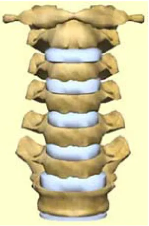
ANATOMY
FIGURE 1
The Cervical spine is made up of first seven vertebrae in the spine; it starts just below the skull and ends at the top of thoracic spine. The Cervical spine has a backward “C” shape lordotic curve and is much more mobile than either of thoracic or lumbar regions of the spine. The Cervical spine has special openings in each vertebra for the arteries that carry blood to the brain.
The first two vertebral bodies in the cervical spine are called Atlas and Axis. Atlas is named after a mythical Greek god who supported the weight of the world on his shoulders because this is the vertebral body that supports the weight of the head. The Atlas and Axis vertebra in the cervical spine differ from all other vertebrae because they are designed primarily for rotation.
The Atlas has a thick anterior arch and thin posterior arch with two prominent masses. The Axis sits underneath the Atlas and has a bony knob called the Odontoid Process that articulates up with the Atlas. It is this mechanism that allows the head to turn side to side.
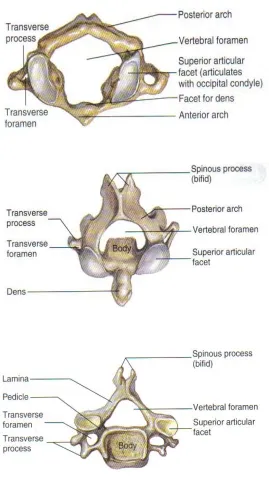
VERTEBRAE
FIGURE 2
The entire Spinal column is joined together by ligaments that allow the Spine to bend and twist carrying the weight of the human body with just the right balance of strength and flexibility. Special joints between each of the vertebral bodies called Facet joints allow the individual bones of the spine to move and rotate with respect to each other .These joints are important because they can be source of pain if they become arthritic.
Each vertebra is shaped in a special way so that when they are stacked together, Spinal cord is protected from damage or injury by the bones of the entire spinal column. Vertebrae support the majority of the weight imposed on the spine. The body of each vertebra is attached to a bony ring that consists of several parts.
INTERVERTEBRAL DISC
It is located in between each vertebrae and functions as a shock absorber and as joints; they are designed to absorb the stresses carried by the Spine while allowing the vertebral bodies to move with respect to each other. They are made up of a strong outer ring of fibers called the Annulus Fibrosis and a soft centre called the Nucleus Pulposus. The outer layer Annulus Fibrosis helps to keep the inner layer Nucleus Pulposus intact. The Annulus is made up of very strong fibers that connect each vertebra together. The Nucleus of the disc has a very high water content making it very moist.
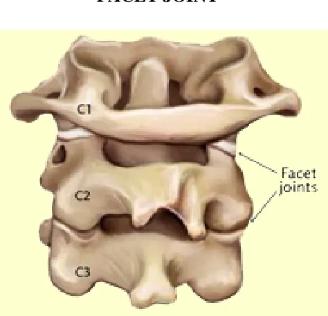
[image:19.612.209.446.408.636.2]FACET JOINT
FIGURE 3
The Facets connect the bony arches of each of the vertebral bodies. There are two Facet joints between each pair of vertebrae, one on each side. Facet joints connect each vertebra with the next vertebrae above and below. They are primarily designed to allow the vertebral bodies to rotate with respect to each other.
NEURAL FORAMEN
The Neural foramen is the opening where the nerve roots exit the spine and travel to the rest of the body. There are two Neural foramen located between each pair of vertebrae, one on each side. The foramen creates a protective passage way for the nerves that carry signals between the Spinal cord and rest of the body.
SPINALCORD AND NERVE ROOTS
The Spinal cord is surrounded by spinal fluid inside this sac. The nerves in each area of the Spinal cord connect to specific parts of the body. The nerves of the cervical spine go to the upper chest and arms, the nerves also carry electrical signals back to the brain creating sensations. Damage to the nerves, nerve roots or spinal cord can lead to symptoms such as pain, tingling, numbness and weakness.
LIGAMENTS OF CERVICAL VERTEBRAL COLUMN
occipital protuberance and it is probably a major stabilizer of head and neck.
MUSCLES AND FASCIA OF THE CERVICAL SPINE
The muscles of the neck can be defined by anatomic limits, innervations or function. Because the cervical spine is the most mobile section of the spine, it contains the most elaborate a specialized muscle system of the spine. Many muscle groups that move the trunk and limbs also attach to the spinal column.
The muscles that closely surround the bones of the spine are important for maintaining posture and help the spine to carry the loads created during normal activities, work and play. The four pre vertebral muscles of the neck are the longus colli (cervicis), the longus capitis, rectus capitus anterior and rectus capitis lateralis. These are weak flexors of the head and neck. They extend from the base of the skull to superior mediastinum. They partially cover the anterior aspect of the vertebral column. They are covered anteriorly by the thick pre vertebral fascia. These fascia forms planes and compartments in which deeper structures of the neck are organized.
subcutaneous fat, the platysma muscle, the external jugular vein and cutaneous sensory nerves. The superficial layer surrounds all the deeper structures of neck. Next to the anterior to the cervical spine are oesophagus, trachea and thyroid gland. These structures are covered by intermediate fascial layer separate from the prevertebral fascia. The middle layer of the deep cervical fascia encloses the strap muscles and extends laterally to the scapula. The deepest layer of the deep fascia is the prevertebral fascia, which covers the scalenus muscles, longus colli muscles and the anterior longitudinal ligament. A number of important structures are located between these fascial layers.
BLOODSUPPLY OF THE CERVICAL SPINE
The vertebral artery is the major source of blood supply for the cervical spine and the cervical portion of the spinal cord. The vertebral arteries are usually the first and largest branch of the subclavian artery on each side.
BIOMECHANICS OF CERVICAL SPINE
the cervical spine is necessary for making a complete assessment of the neck of patients who have cervical problems. The normal biomechanics and pathomechanics of the cervical spine have been learned from static mechanical testing of cadaveric specimens in the laboratory. It is well established that forces and stresses can be applied to the spine in any combination of flexion, extension, rotation and shear. These stresses affect the entire Motion segment including the intervertebral disc, zygapophyseal ligaments, un co vertebral joints and the other ligamentous structures. The muscles and fascial attachments interact with the cervical spine to accommodate load, alter forces and direct motion.
Damage or irritation of the spinal cord and nerve roots in addition, to prevent incapacitating deformity (or) pain due to structural changes, while the issue of clinical instability is particularly germane to traumatic injuries of cervical spine, the subject also applies to inflammatory disorders such as rheumatoid arthritis and degenerative spondylolisthesis. Range of motion in the sub axial cervical spine can be helpful in making decisions about instability. The maximum anteroposterior translation on a lateral radiograph under physiologic loads has been measured. A difference in angulations greater than 110 between two cervical segments on a lateral radiograph also suggests abnormal motion.
Kinematics is the examination of the motion of bodies without consideration of the influencing forces. The two factors that determine the kinematics of vertebral motion are the geometry of the articulating surfaces and the mechanical properties of the connecting structures. The function of cervical spine may be divided into two sections, that of the upper segment above C3 and that of the lower segment from C3 to C7. Most of the axial rotation in upper cervical spine occurs at the atlantoaxial joint.
which represents about 50% of the axial rotation in the neck, with the lower cervical spine contributing the other 50% of rotation. There are also about 40 of axial rotation at the occipito cervical junction. The rotation of C2 on C3 is physically limited by the anatomic locking of the anterior tip of the articular process of C3 on the lateral process of the axis. The lower cervical segment includes C3 through C7 with foraminal openings for the spinal nerve roots that supply the upper extremities. Motion in the lower cervical spine includes flexion, extension, lateral flexion, and rotation.
In forward flexion, the anterior disc space undergoes compression with widening posteriorly simultaneous separation and shear of the posterior elements occur. An anterior shearing force is placed on the disc with elongation of annular fibrosis. Forward gliding of superior vertebrae occurs on the inferior vertebrae with widening of facet joint. In extension of the cervical spine, the posterior aspect of the disc compresses and the anterior portion elongates. The facet joint glides posteriorly positions of the cervical spine affect intra discal pressure. In supine it is least and in extension it is greatest.
direct contact of the vertebral laminae, zygapophyseal joints and the posterosuperior Spinous process.
As the neck flexes the spinal canal lengthens with the posterior wall elongating to a greater degree than the anterior wall. Conversely when the neck extends, the canal shortens, with the anterior wall. The spinal cord ascends and descends in the spinal canal as the neck is flexed and extended posture is a neuromuscular reaction to proprioceptive impulses from the periphery and the feeling that posture is appropriate is a learned process. The posture will be considered normal by the nervous system even if the places the musculoskeletal system at a mechanical disadvantage. In later life these positions may result in fatigue and neck pain.
NECK DISABILITY INDEX
recreation. Total score can range from 0 to 50 where 0 is considered as highest level of function and 50 has lowest level of function.
MUSCLE HOLDING CAPACITY
Muscle holding capacity is used to measure the holding capacity of low load, inner range isometric contraction of the muscle.
CRITERIA FOR MECHANICAL NECK PAIN 7
¾ Pain is usually cyclic and episodic.
¾ Morning stiffness or pain is common.
¾ There is pain on forward flexion and often also on returning to erect position.
¾ Pain is often produced or aggravated by extension, lateral flexion, rotation and exercises.
¾ Pain usually becomes worse over the course of the day.
¾ Pain is relieved by change of position especially when lying down or in flexed Posture.
NEED FOR THE STUDY
“Pleasure is often a visitant but pain cruelly clanged on us” kart M.
Pain is defined as the sensory, emotional experience associated with actual (or) potential damage to the tissues, it is predicted that prevalence rate of neck pain will continue to rise as the computers have made a sweeping and drastic change in our working environment. These complaints are often grouped together as occupational overuse syndromes (or) work related neck disorders.
Mechanical neck pain remains an almost universal condition. Mechanical Neck
pain is a descriptive term commonly used for a mechanically originating, non- discogenic
Neck pain, which is provoked by physical activity and relieved by rest. This is a chronic
dull aching pain of varying intensity affecting the spine.
control. Deficits in muscle coordination which could result in poor support and potential overload on cervical structures.
Mayoux et al (1994) highlighted the importance of longus colli for postural control of the cervical curve. Beeton and Jull in 1994 found that there is a evidence that the upper and deep cervical flexors tend to lose their endurance capacity in patients with neck pain.
Effective management of this condition is vital not only for the relief of symptoms but perhaps more importantly, for the prevention of recurrent episodes of cervical pain personal suffering and lost work productivity. A number of studies have demonstrated a reduction in the strength and endurance capabilities of both deep and superficial cervical flexes muscles in patients with neck pain.
Exercise interventions are important for effective management of patients with neck pain. However there is a consensus on optimal exercise prescription.
hypothesis a low load craniocervical flexion test has been developed by Jull et al to investigate the anatomical action of the deep cervical flexors, specifically of the longus colli in synergy with the longus capitis.
Various methods are used to treat patients with neck pain. These include exercise therapy, massage, ergonomic advice, electrotherapy, short-wave diathermy and spinal manipulative therapy. Manipulative or manual therapy is one of the fundamental treatment methods used by physical therapists, osteopaths, chiropractors and manual medicine practitioners in the management of neck pain.
There is evidence that manipulative therapy can be effective for the relief of pain and restoration of motion in the short term, but this therapy has not met the challenge of lessening persistent and recurrent episodes of neck pain. This was also our clinical experience and, in addition, general neck exercises appeared to have equal limitations for the goal of controlling pain and preventing recurrent or persistent episodes of pain.
could provide cervical segmental stabilization that insight was gained into the type of therapeutic exercise that may be beneficial for supporting the spinal joints, controlling pain and preventing recurrent bouts of neck pain.
Based on the available evidence on the spinal joint stabilization, I intended to study stabilization programme using the muscle system to protect the spinal joint structures from further repetitive micro trauma, recurrent pain and degenerative changes. Pressure biofeedback is a device designed to teach and measure various muscle functions. So using pressure biofeedback which may be more beneficial in re-educating deep neck flexors, which are the major small muscles directly attached to the cervical vertebrae, and more prone to weakness in neck pain patients.
OBJECTIVES
¾ To study the efficacy of stabilizer pressure biofeedback of Deep
Neck flexors helps in reducing chronic neck pain using Neck
Disability Index.
¾ To study the efficacy of isometric exercises of Deep Neck flexors
helps in reducing chronic neck pain using Neck Disability Index.
¾ To compare the results obtained by stabilizer pressure biofeedback
and isometric exercises of Deep Neck flexors in reducing chronic
HYPOTHESIS
NULL HYPOTHESIS
There is no significant difference between the effectiveness of stabilizer pressure biofeedback and isometric neck exercises of Deep Neck flexors in reducing chronic neck pain using Neck Disability Index.
RESEARCH HYPOTHESIS
REVIEW OF LITERATURE
¾ Falla D (2004)8 has analyzed deficits in the motor control of deep and superficial cervical flexor muscles in people with chronic neck pain.
¾ Grand R J, Jull G A (1997)9 found that an exercise programme that focuses on specific load training of key supporting muscles of the neck and shoulder girdle has potential beneficial effects to upper quadrant musculoskeletal system.
¾ Ylinen J, Tkala (2003)10 Concluded in his study that strength and endurance training with a 12 day institutional programme followed by advice to exercise regularly at home were effective methods for decreasing pain and disability in women with chronic neck pain.
¾ Gustawa Stendig-Lindberg (2004)11 stated that daily application of isometric exercise for 6 seconds only by using two thirds of maximal contractile force, results in a optimal increase of muscle strength.
perform the CCFT may be clinical evidence of an impairment that characterizes neck pain, regardless of origin.
¾ Dr. Deepak sharan (2001-2006)13 found that the age between 18 to 52 are usually affected by neck pain and the median age is 27 years.
¾ Peter D Aker (1996)14 concluded that there is a little clinical data available from clinical trails to support many of treatments for mechanical neck pain and in general conservative interventions have not been studied enough in detail to assess the efficacy (or) effectiveness adequately.
¾ Vernon (1991)15 demonstrated that the neck disability index has achieved a high degree of reliability and internal consistency.
¾ Panjabi (1998)16 stated that the osteoligamentous system contributes 20% to the mechanical stability of the cervical spine while 80% is provided by the surrounding neck musculature.
¾ Janda (1994)17 suggests that the cervical flexors muscles become dysfunctional in the presence of neck pain and further demonstrated a reduction in the strength and endurance capabilities of cervical flexor muscles in neck pain patients.
¾ Merskey H (1986)19 stated that pain occurs (or) is associated with actual (or) potential damage to the tissues.
¾ Taimola (2000)20 stated that if the neck pain lasts for more than 3 months of duration is said to be chronic.
¾ Chiu TT (2002)21have demonstrated a reduction in the strength and endurance capabilities of both deep and superficial cervical flexor muscles in patients with neck pain.
¾ Mayoux (1994)22 highlighted the importance of longus colli for postural control of cervical curve.
¾ Beeton and Jull (1994)23 concluded that there is a evidence that the upper and deep cervical flexor lose their endurance capacity in patients with neck pain
¾ Falla D, Jull G (2006)24 concluded that an endurance - strength exercise regime for the cervical flexors muscles is effective in reducing myoelectric manifestations of superficial cervical flexor muscle fatigue as well as increasing cervical flexion strength in group of patients with chronic non severe neck pain.
limit the amplitude and velocity of trunk excursion caused by arm movement.
¾ Panjabi MM (2003) 26found that the spinal muscles provide significant stability to the spine as shown by both in vitro experiments and mathematical models concerning the role of neuromuscular control system, increased body sway has been found in patients with low back pain, indicating a less efficient muscle control system with decreased ability to provide the needed spinal stability.
¾ Peter white (2004)27 concluded that short core neck pain questionnaire has been found to be valid as a brief neck disability index questionnaire for use of patients with mechanical neck pain.
¾ Michael S, Conley (1995)28 concluded from the results of his study, has proved that few selected muscles which have been examined in human electromyographic studies neck muscle function and morphology can be studied at a detailed level using exercise induced shifts in magnetic resonance images.
¾ Pierre cot e (1998) 30in his cross sectional study shows that neck pain is highly prevalent in Saskatchewan and that it significantly disables 4.6% of the adult population.
¾ Hodges and Richardson (1996-1997)31 suggested that specific muscles with a muscle group have been found to be dysfunctional they are the deep muscles h direct vertebral attachments that span the vertebrae and have more influence on joint control rather than torque production.
¾ Watson and Trott (1993)32 suggested there is a clinical evidence that the upper and deep cervical flexors that are important muscles for cervical segmental and postural control lose their endurance capacity in patients with neck pain.
¾ Winters and Peles (1990)33 on studying the interaction of several neck muscles by computer modeling, noted that if only the large muscles of the neck were simulated to produce movement, this resulted in regions of local segmental instability particularly in near upright or neutral postures deep muscle activists was required to stiffen (or) stabilize the segments in functional mid ranges.
¾ Jull (1994)35 suggested that the holding capacity of the upper and deep cervical flexors were determined by their ability to sustain an inner range upper cervical flexion position in supine lying.
¾ Jull G, Barrett C (1999)36 in his clinical use of the test suggests that an ideal controlled performance of the deep cervical flexors can increase the pressure to 30mmHg and hold this pressure for 10 seconds.
MATERIALS AND METHODOLOGY
POPULATION
¾ Chronic Neck pain subjects between 25-50 yrs of both genders
SOURCE OF DATA
¾ Government General Hospital, Tamilnadu.
¾ Padmavathi College of Physiotherapy, OPD, Dharmapuri.
¾ Clinics in and around Dharmapuri.
SAMPLE SIZE
¾ Sample size is 30
SAMPLING DESIGN
¾ Simple Random Sampling
RESEARCH DESIGN
¾ Experimental evaluation comparative study.
INCLUSION CRITERIA
¾ Subject with Chronic neck pain
¾ Subjects between the age of 25-50 years
¾ Subject with Mechanical neck pain
EXCLUSION CRITERIA
¾ Acute cervical disc prolapse
¾ Recent cervical vertebral Fracture
¾ Recent ligament and muscle injuries in neck region
¾ Subject with cervical spinal deformities
¾ Subject with radiating pain along the upper limb and head
¾ Subject with neurological problem.
¾ Open wounds around neck.
¾ Tumors of cervical origin
¾ Vertebro Basilar Insufficiency
¾ Recent dental fracture and conditions like mandibular fracture
MATERIALS
¾ Stabilizer Pressure Biofeedback
¾ Towel
FIGURE 4
TOOLS
¾ Neck Disability Index
¾ Muscle Holding Capacity using stabilizer pressure biofeedback
PROCEDURE
[image:44.612.128.460.128.373.2]assessment includes Neck Disability Index and Muscle Holding Capacity was taken once in every week for four weeks, with treatment sessions carried out thrice every week.
Subjects are divided into two groups by Random sampling method.
Group A- Stabilizer pressure biofeedback
Group B- Isometric Exercise
Group A was treated with SPB in supine lying with Chin tuck in, to strengthen the deep neck flexors for 10 sec hold for 10 repetitions for 4 weeks duration
Training of Deep Neck Flexors using SPB
Patient lies supine with the head and cervical spine in neutral position. A folded towel may be placed beneath the patient head to obtain neutral position if necessary. The patient is instructed to place the tip of the tongue on the roof of the mouth and keep the jaw relaxed this prevents the patient from fixing the jaw and substituting the hyoid muscles.
pressure. Position the 3 folded pressure cell under the neck so that it abuts against the occiput and reading should be checked to maintain it in 20 mm Hg. The movement patient has to perform is a gentle nodding of the head, as if they are saying ‘Yes’. Instruct the patient to gently nod and just one mark on the pressure dial and see if the patient can hold the position steadily. If successful relax and repeat at each target position up to 30 mm Hg, Hold for 10 sec breathe normally. Perform 10 repetitions each, twice daily, three days per week for four weeks38 39.
Group B was treated with Isometric exercises in supine lying with Chin tuck in, to strengthen the deep neck flexors for 6 sec hold for 10 repetitions for 4 weeks duration
Training of Deep Neck Flexors using Isometric Neck Exercise
The patient is asked to lie in supine position and head placed in neutral position, then by placing the rolled towel behind the neck and instruct the subject to perform slight chin tuck and then press the towel placed behind the neck and hold the contraction for 6 sec without any movement. Perform 10 repetitions, twice daily, three days per week for four weeks40, 41, 42.
RESULTS
RESEARCH DESIGN
[image:47.612.135.514.413.723.2]A Experimental evaluation comparative study consisting of 30 patients with Neck pain randomized in to two groups; 15 subjects in Group A (Stabilizer Pressure Biofeedback) and 15 subjects in Group B (Isometric Exercise) is undertaken to study and compare the effects of treatment in reducing the neck pain.
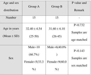
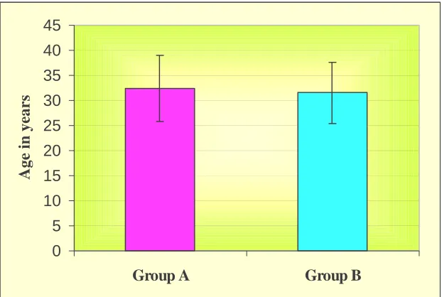
TABLE 1
AGE AND SEX DISTRIBUTION
Age and sex distribution
Group A Group B
P value and Remark
Number 15 15 -
Age in years (Mean ± SD)
32.40 ± 6.54 (25-50)
FIGURE 7
0 5 10 15 20 25 30 35 40 45
Group A Group B
Age in years
FIGURE 8
M ale 67.7%
Female 33.3%
Group A
M ale 40.0% Female
60.0%
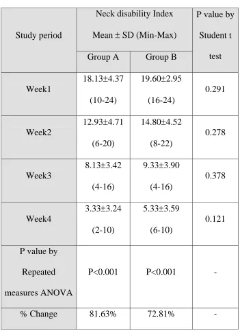
TABLE 2
NECK DISABILITY INDEX
Study period
Neck disability Index Mean ± SD (Min-Max)
P value by Student t
test Group A Group B
Week1
18.13±4.37 (10-24)
19.60±2.95 (16-24)
0.291
Week2
12.93±4.71 (6-20)
14.80±4.52 (8-22)
0.278
Week3
8.13±3.42 (4-16)
9.33±3.90 (4-16)
0.378
Week4
3.33±3.24 (2-10)
5.33±3.59 (6-10)
0.121
P value by Repeated measures ANOVA
P<0.001 P<0.001 -
% Change 81.63% 72.81% -
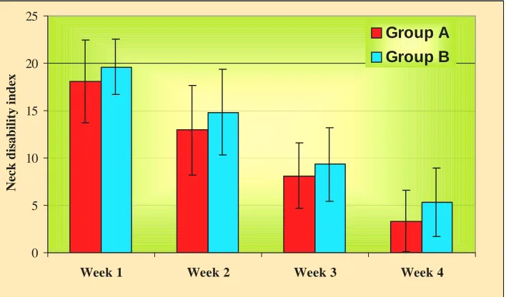
FIGURE 9
0 5 10 15 20 25
Week 1 Week 2 Week 3 Week 4
N
eck disa
bility
inde
x
[image:50.612.151.495.410.635.2]Group A Group B
FIGURE 10
0 5 10 15 20 25
Week 1 Week 2 Week 3 Week 4
Neck disability inde
x
Group A
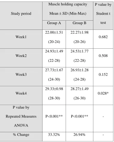
TABLE 3
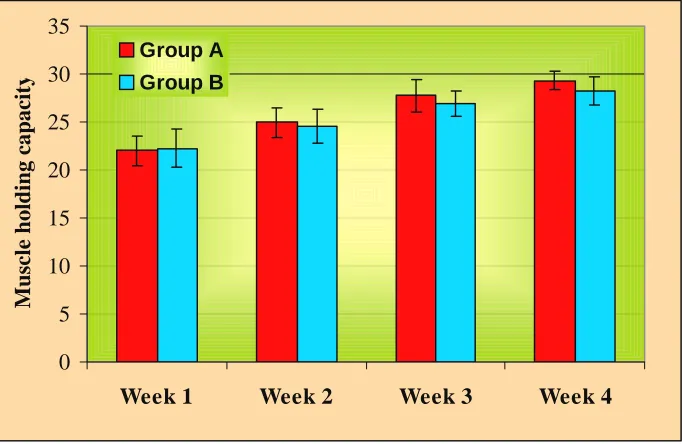
MUSCLE HOLDING CAPACITY
Study period
Muscle holding capacity Mean ± SD (Min-Max)
P value by Student t
test Group A Group B
Week1
22.00±1.51 (20-24)
22.27±1.98 (20-26)
0.682
Week2
24.93±1.49 (22-28)
24.53±1.77 (22-28)
0.508
Week3
27.73±1.67 (24-30)
26.93±1.28 (24-28)
0.152
Week4
29.33±0.98 (28-30)
28.27±1.49 (26-30)
0.028*
P value by Repeated Measures
ANOVA
P<0.001** P<0.001** -
% Change 33.32% 26.94% -
# Analysis of Covariance has been used to find the significance MHC at Week 4 taking into account of variations at Week1, week2 and Week3.
FIGURE 11
0 5 10 15 20 25 30 35
Week 1 Week 2 Week 3 Week 4
Muscle holding capacit
y
Group A
[image:52.612.150.497.413.637.2]Group B
FIGURE 12
0 5 10 15 20 25 30 35
Week 1 Week 2 Week 3 Week 4
Muscle holding capacit
y
Group A
TABLE 4
COMPARISON OF NDI BETWEEN TWO GROUPS
Study
Period
Group
Neck Disability Index
Normal Mild
Disability Moderate disability Severe disability Complete disability Week 1 Group A (n=15)
- 2(13.3%) 13(86.7%) - -
Group B
(n=15)
- - 15(100.0%) - -
Week 2
Group A
(n=15)
- 10(66.7%) 5(33.3%) - -
Group B
(n=15)
- 9(60.0%) 6(40.0%) - -
Week 3
Group A
(n=15)
3(20.0%) 11(73.3%) 1(6.7%) - -
Group B
(n=15)
1(6.7%) 11(73.3%) 3(20.0%) - -
Week 4
Group A
(n=15)
12(80.0%) 3(20.0%) - - -
Group B
(n=15)
FIGURE 13
0 10 20 30 40 50 60 70 80 90 100
Pe
rc
e
n
ta
g
e
Group A Group B Group A Group B Group A Group B Group A Group B Normal Mild disability Mod.disability
TABLE 5
COMPARISON OF NDI INDIVIDUAL TASKS BETWEEN TWO GROUPS
Tasks Group
Study period
Week1 Week2 Week3 Week4 Mean ± SD
(Median)
Pain intensity
A 2.60±0.74 (2)
2.00±0.66 (2)
1.53±0.64 (1)
0.73±0.59 (1) B 2.81±0.74
(3)
2.13±0.92 (2)
1.33±0.49 (1)
1.00±0.54 (1)
Personal care
A 2.67±0.72 (3)
1.93±0.79 (2)
1.40±0.63 (1)
0.27±0.46 (0) B 2.93±0.70
(3)
2.13±0.92 (2)
1.07±0.59 (1)
0.53±0.52 (1)
Lifting
A 2.47±0.83 (3)
1.87±0.64 (2)
1.13±0.64 (1)
0.53±0.52 (1) B 2.47±0.92
(2)
2.09±0.79 (2)
1.20±0.56 (1)
0.60±0.63 (1)
Reading
A 2.13±0.64 (2)
1.67±0.90 (2)
0.73±0.70 (1)
0.20±0.41 (0) B 2.53±0.74
(2)
2.07±0.70 (2)
1.07±0.70 (1)
0.40±0.63 (0)
Headache A 0 0 0 0
B 0 0 0 0
Concentration A 2.60±0.51 (3)
1.40±0.74 (1)
0.80±0.78 (1)
B 2.33±0.49 (2)
1.80±0.68 (2)
1.20±0.56 (1)
0.60±0.74 (0)
Work
A 2.20±0.68 (2)
1.80±0.68 (2)
1.27±0.46 (1)
0.80±0.68 (1) B 2.13±0.35
(2)
1.73±0.70 (2)
1.20±0.41 (1)
0.87±0.52 (1)
Driving
A 1.20±1.20 (2)
0.60±0.91 (0)
0.40±0.63 (0)
0.20±0.41 (0) B 2.07±0.26
(2)
1.53±0.74 (2)
1.00±0.66 (1)
0.40±0.51 (0)
Sleeping A 0 0 0 0
B 0 0 0 0
Recreation
A 2.27±0.70 (2)
1.67±0.72 (2)
0.87±0.64 (1)
0.33±0.62 (0) B 2.27±0.46
(2)
1.73±0.59 (2)
1.27±0.46 (1)
STATISTICAL METHODS
Chi-square / Fisher Exact test has been used to find the significance of Neck disability Index between Group A and Group B during the study period. Student t test (Two tailed) has been used to find the significance of Neck disability Index and Muscle holding capacity between Group A and Group B. Repeated Measures ANOVA has been used to find the significance of Neck disability Index and Muscle holding capacity during the study period for each group separately.
1. Chi-Square Test
Ei Ei Oi
∑
−=
2
2 ( )
χ , Where Oi is observed frequency and Ei is Expected
frequency
2. Fisher Exact Test
TABLE 6
Class1 Class2 Total
Sample1 a b a + b
Sample2 c d c + d
Fisher Exact Test statistic=
∑
∑
= + + + + ! ! ! ! 1 ! )! ( )! ( )! ( )! ( d c b a n d b c a d c b a p3. “t” –test for two population means ( variance unknown but equal)
Objective: To investigate the significance between the means of two populations ) 2 / 1 1 / 1 ( ) ( ) ( 2 2 1 2 1 n n s x x t + − − −
= μ μ
Where 2 2 1 ) 2 2 ( ) 1 2 ( ) 1 1 ( ) 1 1 ( 2 1 2 2 1 1 2 − + − − + − − =
∑
=∑
− n n x x n x x n s n i n i4. ANACOVA: Analysis of Covariance has been used to find the significance of difference of post treatment between groups keeping the Pre treatment scores as covariates.
Procedure is as follows SPT=
∑∑
−N TxTy
xy , y is post treatment scores and x is Pre treatment
SPB =
N TxTy ni
TxiTyi −
∑
SPW = SPT-SPB
SS`YT = SSyt -
xt T
SS SP2
: SS`YW = SSYw -
XW W
SS SP2
: SS`YB = SS`YT - SS`YW
ANACOVA TABLE
[image:59.612.119.569.116.499.2]TABLE 7
Source of
variation df SSx SP SSy SS`y MSS`y F ratio Between
groups
(k-1) SSXB SPB SSYB SS`YB MSS`YB F
Within groups
(N-k-1) SSXW SPW SSYW SS`YW MSS`YW -
Total (N-2) SSXT SPT SSYT SS`YT - -
There is no significant difference between age and sex distribution between the Group A and Group B
Comparison of decrease of NDI between Group A and Group B
There is a significant decrease of NDI to 3.33 in Group A which is much lower when compared to Group B 5.33, with p = 0.169#. Hence research or alternate hypothesis is accepted.
Comparison of NDI individual tasks between 2 groups
There is significant difference in all individual tasks expect in headache and sleeping components.
Comparison of increase of MHC between Group A and Group B
MHC is significantly increased in Group A which when compared to Group B with p = 0.006.
STATISTICAL SOFTWARE
DISCUSSION
This study is an attempt to assess the efficacy of stabilizer pressure biofeedback and isometric exercise to reduce chronic neck pain by activating the deep neck flexor muscles in chronic neck pain subjects.
Patients with chronic neck pain who were treated with stabilizer pressure biofeedback (Group A) have shown statistically better improvements in reduction of pain and muscle holding capacity than the isometric exercise (Group B). The design of the study (which include random assignment to study group)
The effect of the treatment were achieved in four weeks of duration that most probably due to the effective activation of deep neck flexors. Most of previous studies suggested that in neck pain patients, deep neck flexors activation is decreased and also the muscle holding capacity is reduced.
Jull G stated that the anatomical interrelated action of the deep neck muscles are to support and stabilize the cervical.
Watson and Trott stated that the upper and deep cervical flexors contraction is important to stabilize the spine by creating a tension over the cervical fascia. In turn this stabilizes the cervical spine and forms stable base for the movement and functional activities. The current study was focused on generalized neck pain, mechanical in origin.
Beeton and Jull stated that deep neck flexors are the key muscle for the stabilization of the cervical spine. There is a significant dysfunction of this muscle has also been implicated in neck pain patients.
Hence the deep neck flexors help in improving the strength and there by relieve chronic neck pain. SPB was proved as a preventive measure, retraining the stabilization capacity of the deep neck flexors might reduce the effect of cervical structures from stress. Chi Square Test / Fisher Exact Test have been used to find the significance of NDI between Group A and Group B the study period. Student t test has been used to find the significance of difference of pre and post set groups keeping the NDI and MHC during the study period for each group separately.
Group B (5.33) with p=0.169 by ANACOVA test. Improvement in MHC is significantly increased in Group A which when compared to Group B with the p=0.006** by ANACOVA test.
The percentage reduction of NDI also significantly reduced in Group A (80%) compared with Group B (60%). test. When individual components are compared between Group A and Group B, the Pain intensity, personal care, reading, concentration, driving and recreation components shows marked improvement in reduction of NDI value at the end of 4th week and there is more difference is seen in between the values of Group A and Group B. when comparing components of lifting and work there is marked reduction in the NDI value but the difference between the groups is less, and components of Headache and sleeping has equal score of Zero from the initial procedure, Since the mechanical neck pain relieves during rest and headache is excluded from the study.
The design of this study precludes determination of which aspect of the treatment program produced the changes in neck pain.
Both the Groups showed significant improvement in first two weeks and third and fourth week Group B has shown slight improvement it may be due to isolation of specific muscle group contraction, motor control and relearning due to visual feedback. Increase in isometric group may be due to involvement of other global muscles. when comparing NDI and MHC of both the groups , Group A showed significantly better improvement due to visual biofeedback.
This study also coincides with Grant Jull who states that the stabilizer pressure biofeedback is more significant in isolating the Deep neck flexors muscles specifically and there by relieve neck pain.
This study may show marked significant changes in statistical and theoretical aspect when carried out for a longer duration.
Isometric exercises also help in reducing neck pain, but the effect is little bit slow compared to SPB, Since Stabilizer pressure biofeedback has proven to be effective treatment measure in reducing chronic neck pain.
LIMITATIONS
¾ The outcome measure was only neck disability index and muscle holding capacity.
¾ Study population is selected only from Bangalore.
¾ Confounding variables like Range of motion, postural adjustment are not used.
¾ Manipulation technique was not considered.
RECOMMENDATIONS AND SUGGESTIONS
¾ This study revealed only with deep flexors muscles, other
muscle groups also can be considered in further studies in
reducing neck pain.
¾ The same study may be explored for specific neck pain
conditions
¾ This study can also be done for radiating pain conditions.
¾ The beneficial treatment effect can be followed for the
persistence of recovery
¾ The present study may be done in larger population for better
outcomes
¾ The study was focused in supine lying position only. This can
also be done in other functional positions
¾ The study can be carried out for longer duration to show better
results.
¾ This study can be further carried out in combination of both
CONCLUSION
This study concluded that the SPB have better improvement in
reduction of pain by giving correct feedback for the isolation of deep
neck flexors contraction in chronic neck pain patients with the help of
NDI score. From this study it is also noted that the basic
characteristics of age and sex are not having a direct impact on chronic
neck pain subjects.
SUMMARY
The study is done to find out the comparison of stabilizer pressure biofeedback and isometric exercise for reduction of pain by contracting the deep neck flexors in chronic neck pain subjects with the help of NDI value.
REFERENCES
1. Seeley Stephen, Anatomy and physiology, 5th edition, Tate 2000.
2. Bernstein Wiesel boden, Neck pain medical drug and comprehension
management 2001.
3. Harms Ringdahl K, Ekholm J Schuldtk, Nemoth G, Arborelius, Load
movements and myoelectric activity when the cervical spine is held in
full flexion and extension. Ergonomics;1986:29(12): 1539-1552
4. Jull G. Deep cervical flexor muscle dysfunction in whiplash
musculoskeletal pain; 2000:8:143 -154.
5. Comer fords MJ, Mott ram SL, Functional stability retraining:
Principles and strategies for managing mechanical dysfunction.
Manual Therapy 2001;6(1):3-14
6. Carr J and Shepherd R B ,A motor relearning programme for stroke
London :William Heinemann;1987: pp29-33
7. David J, Magee, Orthopedic physical assessment, published by W B
8. Falla D. Unraveling the complexity of muscle impairment in chronic
neck pain. Manual therapy; 2004:9:125-133.
9. Grant RJ , Jull GA and Spencer TJ :Active stabilization training for
screen based key board operators a single case study , Australia
Journal of physiotherapy;1997: 43: 235-242
10.Ylinen J, Tkala. Active neck muscle training in the treatment of the
chronic neck pain in women, a randomized controlled trial. JAMA
;2003:No289: pp2509-2516
11.Gustawa Stendig. Therapeutic exercises, E medicine :2004
12.Thomas Tai Wing. Performance of the craniocervical flexion test in
subjects with and without chronic neck pain: Orthop sports phys ther;
2005:vol 35: pp 567-571
13.Dr. Deepak Sharan, Repetitive strain injuries, incidence of mechanical
neck pain in Bangalore 2001-2006
14.Peter D Aker, Conservative management of mechanical neck pain;
systemic overview and meta analysis BMJ ;1996: Vol 313;No
15.Vernon H, The Neck disability index :A study of reliability and
validity: journal of manipulative physiol ther;1991: Vol 14:No
7:pp409-415
16.Panjabi M M, Chole wicki J, Nibu K, Critical load of the human
cervical spine: an in vitro experimental study. Clinical biomechanics
;1998:13:11-17
17.Janda V Muscles and motor control in cervicogenic disorders:
assessment and management .In grant R ,editor .physical therapy of
the cervical and thoracic spine.newyork:Churchill Livingstone
;1994:195-216
18.Cote P, Cassidy J D, Carroll L, The Saskatchewan health and back
pain survey, the prevalence of the neck pain and relative disability in
Saskatchewan adults. Spine 1998:23(15):1689-1698.
19.Merskey H “Classification of Chronic pain: Description of chronic
20.Taimola S, Takala E P, Asklof T, Active treatment of chronic neck
pain: A prospective randomized intervention. Spine 2000;
25:1021-1027
21.Chiu T T, Losk. Evaluation of Cervical range of Motion and Isometric
neck muscle strength: reliability and validity .Clin Rehabil; 2002:
16:851-858.
22.Mayoux-Benhamou MA, Reval M et al. Longus colli has a postural
function of cervical curvature. Surgical and radio logic anatomy;
1994:16:367-371.
23.Beet on and Jull, Effectiveness of manipulative physiotherapy in the
management of cervicogenic headache single case study
physiotherapy; 1994:80:417-423.
24.Falla D. Unraveling the complexity of muscle impairment in chronic
neck pain. Manual therapy; 2004:125-133
25.Moseley GL and Hodges PW. Are the changes in postural control
associated with pain caused by pain inference? Clin J pain; 2005:
26.Panjabi MM, Clinical spinal instability and low back pain .J
Electromyography and kinesiol.2003; 13(4):71-79.
27.Peter white. The core outcomes for neck pain: Validation of a new
outcome measure. Spine 2004: Vol 29:No17;1923-1930
28.Conley B et al, non invasive analysis of human neck muscle function.
Spine1995: 20:2505-2512
29.Deborah L Falla. Patients with Neck pain demonstrate reduced electro
myographic activity of deep cervical Flexor muscles during
performance of the craniocervical flexion test. Spine 2004: Vol 29; No
29: pp 2108-2114.
30.Pierre Cote DC,MSC, J .David Cassidy.The saskatchewen health and
back pain survey:The prevalence of neck pain and related disability in
Saskatchewan adults.Spine1998:Vol23;No15:pp1689-1698.
31.Hodges PW, Richardson CA. Delayed postural contraction of
transversus Abdominis in low back pain associated with movement
32. Natural head posture and upper cervical flexor muscle performance.
Cephalgia; 1993:13: pp 272-284.
33.Winters J M, Peles J D. Neck muscle activity and 3D head kinematics
during quasistatic and dynamic tracking movements. In: winters J M,
Woo S L editors. Multiple muscle systems: Biomechanics and
movement organization. New York: Springer; 1990: pp 461-480.
34.Cholewicki J and McGill S, mechanical stability of the in vivo lumbar
spine: Implication for injury and chronic low back pain. Clinical
biomechanics; 1996: 1-15.
35.Jull G .Physiotherapy management of the cervical spine .In Giles L,
Singer K. clinical anatomy and management in cervical spine pain,
UK: Butterworth Heinemann; 1998:168-197.
36.Jull G Barrett C, Magee R, HO P. Further clinical classification of the
muscle dysfunction in cervical headache. Cephalgia 1999:19:179-185.
37.Falla D, Jul G, An electro myographic analysis of the deep cervical
flexor muscles during craniocervical flexion. Physical therapy;
38.H. Duane Saunders and robin Saunders Ryan, Evaluation, treatment
and prevention of musculoskeletal disorders, Spine; 2004, vol 1,
39.Ruth grant, Clinics in physical therapy of the cervical and thoracic
spine, published by Churchill Livingstone, New York; 1997.
40.Carrie M Hall, Lori Thein Brady, Therapeutic exercises moving
towards to function; 1998: pp 532.
41.M D Robert, H Shmerling, what to do about Neck pain, Harvard
medical school 2000
42.Carolyn Kishner, Lynn Allen Colby. Therapeutic exercises foundation
and techniques 4th edition published by Jaypee brothers, New Delhi;
2002: pp 80-83
43.Bernard Rosner, Fundamentals of Biostatistics, 5th Edition, Duxbury
2000.
44. M. Venkataswamy Reddy, Statistics for Mental Health Care Research
NIMHANS Publication, India 2002.
45. Basmajain J. Control and training of individuals motor units, science;
APPENDICES
ANNEXURE I
INFORMED CONSENT FORM
TITLE
Comparison of Stabilizer pressure biofeedback and Isometric Neck
exercises in reducing Chronic Neck pain using NDI.
PURPOSE OF THE STUDY
I ………have been informed by Mr. M.
Sitharthan that this study is done to find out stabilizer pressure
biofeedback/Isometric Neck exercise will reduce pain, this study has role
to play in reducing Chronic Neck pain and improve my function by
reducing the disability.
PROCEDURE
I understand that I will be randomized and put into one of the
exercise protocol, either stabilizer pressure biofeedback for 10 sec hold
for 10 repetitions or Isometric exercises for 6 sec hold and 10 repetitions.
I will be explained about the intensity at which I have to perform the
supervision and guidance of Mr. M. Sitharthan and follow the instructions
given by her.
RISK AND DISCOMFORT
I understand that there is no potential risk associated with the
treatment programme, and I will not experience any discomfort during
the exercises.
I understand that Mr. M. Sitharthan will accompany me during the
Treatment
BENEFITS
The Stabilizer pressure biofeedback and Isometric Neck exercises
will help in reducing pain
ALTERNATIVES
Other treatment alternatives are explained to me with their benefits
and limitations.
CONFIDENTIALITY
I understand that the information produced by this study will be
confidential. If the data are used for publication in the medical literature
as photographs and audio or video tapes will be used only with
REQUEST FOR MORE INFORMATION
I understand that I may ask any question about the study at any
time to Mr. M. Sitharthan and she is available to answer my question.
Copy of this concern form will be given to me to keep for my careful
reading
REFUSAL OR WITHDRAWAL OF PARTICIPATION
I understand that my participation is voluntary and I may refuse to
withdraw consent and discontinue participation at any time. I also
understand that she may terminate my participation in the study at any
time after she has explained the reasons for doing so
INJURY STATEMENT
I understand that the exercises which I am going to perform are
most unlikely to cause any injury or further deteriorate my condition if
performed under the guidance of Mr. M. Sitharthan. In such case medical
attention will be provided, but no further compensation will be provided.
I understand my agreement to participate in this study and I am not
waiving any of my legal rights.
I confirm that Mr. M. Sitharthan has explained me about
the purpose of the study, the study procedure and the possible risk and
benefits that I may experience. I have read and I have understood this
………
………
SUBJECT
DATE
………..
………
WITNESS TO SIGNATURE
DATE
I have explained to sri /smt………..the
purpose of the research, the procedure required and the possible risks and
benefits, to the best of my ability.
……….
………
INVESTIGATOR
ANNEXURE II
ASSESSMENT PROFORMA
o Patient Name : o Age :
o Sex :
o Occupation : o Address :
o Study setup/ Source : o Presenting Complaint : o Past History :
o Personal History : o Occupational History :
o Subjects with Age group 25-50 : Yes / No
o Subjects with Chronic Mechanical Neck pain : Yes / No o Subjects with cervical vertebral fractures : Yes / No o Subjects with cervical dislocations : Yes / No
o Subjects with Radiating pain to upper limb and head : Yes / No o Subjects with TMJ dysfunction: Yes / No
o Subjects with Tumor of cervical origin : Yes / No
o Subjects with Migraine : Yes / No o On Observation :
o On palpation : o On Examination : Neck Disability Index
Week 1 2 3 4
Stabilizer Pressure Biofeedback Isometric Exercise
Muscle Holding Capacity
Week 1 2 3 4
Stabilizer Pressure Biofeedback Isometric Exercise
o Spurling / Compression Test : Positive / Negative o VBI Test : Positive / Negative
ANNEXURE III
NECK DISABILITY INDEX
1.PAIN INTENSITY
o I’ve no pain at the moment o The pain is very mild at the
moment
o The pain is moderate at the moment
o The pain is fairly severe at the moment
o The pain is very severe at the moment
o The pain is the worst imaginable at the moment
2.PERSONAL CARE(Washing, Dressing) etc
o I can look after myself normally without causing extra pain
o I can look after myself normally, but it causes extra pain
o It is painful to look after myself, I am slow and careful
o I need some help but manage most of my personal care
o I need help every day in most aspects of self care
o I don’t get dressed, wash with difficulty and stay in bed
3.LIFTING
o I can lift heavy weights without extra pain
o I can lift heavy weights but it gives me extra pain
o Pain prevents me from lifting heavy weights off the floor, but I can manage if they are conveniently positioned, for example on a table
o Pain prevents me from lifting heavy weights but I can manage light to medium weights if they are conveniently positioned o I can lift very light weights o I cannot lift or carry anything
at all.
4.READING
o I can read as much as I want to with no pain in my neck
o I can read as much as I want to with slight pain in my neck
o I can read as much as I want with moderate neck pain
o I can’t read as much as I want because of moderate neck pain o I can hardly read at all because of
5.HEADACHES
o I have no headaches at all o I have slight headaches which
come in frequently
o I have moderate headaches which come infrequently
o I have moderate headaches which comes frequently
o I have severe headaches which comes frequently
o I have headaches almost all the time
6.CONCENTRATION
o I can concentrate fully when I want to with no difficulty
o I can concentrate fully when I want to with slight difficulty
o I have fair degree of difficulty in concentrating when I want to o I have a lot of difficulty in
concentrating when I want to o I have a great deal of difficulty in
concentrating when I want to o I cannot concentrate at all
7.WORK
o I can do as much work as I want to do
o I can only do my usual work, but no more
o I can do my usual work, but no more
o I cannot do my usual work o I can hardly do any work at all o I can’t do any work at all
8.DRIVING
o I can drive my car without any neck pain
o I can drive my car as long as I want with slight pain in my neck
o I can drive my car as long as I want with moderate pain in my neck o I can’t drive my car as long as I
want because of moderate pain in my neck
o I can hardly drive at all because of severe pain in my neck
9.SLEEPING
o I have no trouble sleeping
o My sleep is slightly disturbed(less than 1 hr. sleepless)
o My sleep is mildly disturbed(1-2 hrs. sleepless)
o My sleep is moderately disturbed (2-3 hrs. sleepless)
o My sleep is greatly disturbed(3-5 hrs. sleepless)
o My sleep is completely disturbed (5-7 hrs. sleepless)
10. RECREATION
o I am able to engage in all my recreation activities with no neck pain at all
o I am able to engage in all my
recreation activities. With some pain in my neck
o I am able to engage in most, but not all of my usual recreation activities because of pain in my neck
o I am able to engage in a few of my usual recreation activities because of pain in my neck
o I can hardly do any recreation activities because of pain in my neck
o I can’t do any recreation activities at all
SCORES (OUT OF 50)
0-4 = NO DISABILITY 5-14 = MILD DISABILITY]
15-24 = MODERATE DISABILITY 25-34 = SEVERE DISABILTY
MASTER CHART
STABILIZER PRESSURE BIOFEEDBACK
SL
NO AGE SEX WEEK1 WEEK2 WEEK3 WEEK4
NDI MHC
NDI MHC
NDI
MHC NDI MHC
1 40 F 16 22 10 26 8 30 4 30 2 25 F 10 24 6 26 4 28 0 30 3 30 M 22 22 18 24 12 24 8 28 4 34 F 24 20 20 22 10 26 4 28 5 32 M 18 22 10 26 8 30 2 30 6 35 M 16 22 8 24 4 28 0 30 7 29 F 22 20 18 24 10 28 4 30 8 50 M 24 20 20 24 16 28 10 28 9 32 M 16 24 12 26 6 28 0 30
10 35 M 10 24 8 28 4 30 0 30
11 26 F 18 24 10 26 8 26 3 28 12 25 M 16 22 12 24 6 28 0 30 13 26 M 22 20 18 24 12 26 8 28 14 32 M 20 22 14 24 8 28 4 30 15 35 M 18 22 10 26 6 28 3 30
ISOMETRIC EXERCISE
SL
No AGE SEX WEEK 1 WEEK 2 WEEK 3
WEEK 4
NDI MHC NDI
MHC NDI MHC
NDI MHC
1 36 F 24 20 20 24 16 28 8 28
2 30 F 22 20 18 22 12 26 12 26
3 28 M 18 22 14 24 8 26 4 28
4 42 M 24 20 22 24 16 28 10 28
5 28 M 18 24 14 26 6 28 4 30
6 27 F 16 24 12 26 6 28 2 30
7 38 M 22 20 18 22 10 26 6 26
8 30 F 16 24 10 26 8 28 4 28
9 26 M 18 24 8 26 4 28 0 30
10 26 F 20 22 14 24 8 26 4 28
11 28 F 18 26 8 28 6 28 4 30
12 227 F 24 20 22 22 16 24 10 26
13 28 F 20 22 16 24 8 26 8 28
14 45 F 18 22 14 24 8 26 4 28