JOURNAL OFVIROLOGY, Mar. 1994, p. 1475-1485 Vol.68, No.3 0022-538X/94/$04.00+()
Copyright © 1994,American Society for Microbiology
Subcellular Distribution of Human Immunodeficiency Virus
Type
1
Rev and
Colocalization of Rev with RNA Splicing
Factors
in a
Speckled Pattern in the Nucleoplasm
KARL-H. KALLAND,I* ANNE MARIE SZILVAY,' ERIK LANGHOFF?, AND GUNNAR HAUKENES' The NationalCentreforResearch in Virology, DepartmentofMicrobiology andImmunology, The Gade Institute,
UniversityofBergen, BergenHigh TechnologyCentre, N-5020 Bergen, Norway,I and DanaFarber Cancer Institute, Boston, Massachusetts 021152
Received 15 October 1993/Accepted29November 1993
The human immunodeficiency virus type 1 (HIV-1) Rev (regulator of virion protein expression) protein
exemplifies a new type of posttranscriptional regulation. One main function of Rev is to increase the cytoplasmic expression of unspliced and incompletely spliced retroviral mRNAs from which viral structural proteins aremade. In that way, Rev is essential in order to complete the retroviral life cycle. The biology of Rev inthe host cell hasremained elusive. In this study, a complex distribution of Rev in single cells was found. Rev wasfound in the cytoplasm, in a perinuclear zone, in thenucleoplasm,and in the nucleoli. In thenucleoplasm,
Revcolocalized in a speckled pattern with host cell factors known to assemble on nascenttranscripts. Those factors are involved in theprocessing ofheterogeneous RNA to spliced mRNA in the nucleoplasm of all cells. Thedistribution of Rev wasdependentonly on Rev andhost cellinteractions,since neither the Rev target RNA norother HIV proteins were expressed in the cells. Rev was found in the same subcellular compartments of cells treated for extendedperiods with cycloheximide, an inhibitor of protein synthesis. This finding implies thatRev shuttles continuouslybetweencytoplasmic andnucleoplasmic compartments. The results suggest a potential role for Rev both in the RNA-splicing process and in the nucleocytoplasmic transport of Rev-dependent HIVmRNA.
The regulators of virion protein expression (abbreviatedRev
and Rex in lentiviruses and oncoviruses, respectively) repre-sent anovel classofregulatory proteins first discovered inthe
retroviruses humanimmunodeficiency virus (HIV) (Rev) (25, 70)and human T-cellleukemia virus (HTLV) (Rex) (40).The
expression of unspliced and incompletely spliced HIV-1 mRNArequiresRev at anunknownstep(s)between transcrip-tionand translation. The retroviral structuralcomponents are made from the Rev-dependent unspliced and incompletely spliced mRNAs. Therefore, maturevirions are not formed in the absence of Rev (65,73).The Rev-dependent mRNAs are
characterized bytwotypesofcis-actingsequences, asingleRev responseelement(RRE) (20, 50)and severalcis-acting
repres-sive sequences (CRSs) (64). These elements are removed in
thecompletely splicedHIVmRNAswhichdonotrequireRev
for cytoplasmic appearance and translation. The RRE is
present in the HIV envregion between the two exonsofrev and folds into acomplex stem-loop RNAsecondarystructure
(50) that binds Rev with high affinity (18, 33, 52, 56, 78) in
vitro. MutationsthatcompromiseRREstructureabrogateRev
binding and Rev function (21, 36, 41, 52, 56). The CRSs are less well defined and mapped (13, 22, 46, 64, 68) but by definition prevent expression of the HIV-1 mRNAs in which
they are present unless counteracted by Rev activity. One
hypothesis is that CRSs bind inhibitory host cell structures which are displaced by Rev following its binding tothe RRE
(15, 32).
Site-directed mutagenesis of the 116-amino-acid (aa) Rev
protein has delineated severalfunctional domains(34, 47,51,
*Corresponding author. Mailing address: Centre for Virology,
Bergen High Technology Centre. N-5020 Bergen, Norway. Phone: 47-55-544510. Fax:47-55-544512. Electronic mail address: kalland@a
bio.uib.no.
55). Mutations in a basic domain (aa 35 to 50) compromise
nuclear and nucleolar accumulation. The basic domain addi-tionally binds specifically to the RRE RNA (14, 47). Other mutations affect Rev multimerization (51, 55). The activation domain(aa 73to90),which containscharacteristically spaced
leucine residues, may generate transdominant negative mu-tants that repress Rev wild-type function in cotransfection experiments(47, 51, 74). Ithas beenreported thatthe activa-tion domainends with critical acidicresidues(53).
Different experiments have suggested that the splicing ap-paratus(10,45),the RNA transport system(22, 26, 31,50),and
thecytoplasmicpolysomes (1,17) arepotentialtargetsfor Rev
activity. Morphological studies have shown that Rev is
im-ported to the nucleus and accumulates in the nucleolus (14, 16). TostudyRev in the contextofthe single cell, apanel of monoclonal antibodies (MAbs) was raised against
recombi-nant Rev expressed in Escherichia coli and selected for
reac-tivity in indirectimmunofluorescence in Rev-expressing cells. Rev wasdistributedtothecytoplasm,to aperinuclearzone,to the nucleoli, and tothe nucleoplasmicspeckles believed tobe sites of processing of RNA polymerase II pre-mRNA. The
continuous shuttling of Rev between thedifferent subcellular
compartments supports a model in which the role of Rev is
both to interfere with splicing of HIV mRNA and to be
involvedin HIV mRNA transport and incytoplasmiceventsof
HIV mRNAexpression.
MATERIALS ANDMETHODS
Plasmid constructs. Plasmid pSVCMV rev contains the
HIV-1 IIIB revcDNA (BamHI-EcoRI fragment derivedfrom
pH3art[64])downstream of thesimianvirus 40originand the
cytomegaloviruspromoter and upstream of the simianvirus40
poly(A) signal ina pGEMbackbone. In prev 81 (74),the rev 1475
on November 9, 2019 by guest
http://jvi.asm.org/
1476 KALLAND ET AL.
cDNA mRNA transcription is driven by the cytomegalovirus
immediate-early promoter. pSV3gal was from Promega
Bio-tec.
Cell lines andtransfection. COS-1 cellswere transfectedby
using standard DEAE-dextran and calcium phosphate meth-ods, withthesameresults.Oneday following transfection,cells
were trypsinized, plated on 12-mm-diameter Assistant
cover-slips, fixed inmethanol between36 and72 hposttransfection, andexamined.HeLa/crev8 cells and HeLa/cnef cells (gifts from Bryan Cullen, Howard Hughes Medical Institute, Durham, N.C.) were made essentially as described for HeLa/ctat cells
(48). In HeLa/crev8 cells,revisexpressed from the full-length
revcDNA present in pcrev (49). HeLa/crev8 cells were grown
in Iscove's medium (GIBCO) supplemented with fetal calf
serum and 5 x 10⁻⁷ Mmethotrexate.
Autoimmune seraand MAbs. The human autoimmune sera
usedrecognize nucleoplasmicspeckles (Sigma and Chemicon), nucleoli(ANA-N; Sigma), the mitotic spindle(Chemicon),and lamin A/C (a gift from Frank McKeon, Harvard Medical School, Boston, Mass.). The sera were characterized by
fluo-rescent double labeling with MAbs of known specificity. The autoimmune antilamin serum colocalized with an anti-lamin
A/C MAb (Frank McKeon). MAb anti-SC35 (28) detects an
essentialnon-small nuclear ribonucleoprotein(snRNP) RNA-splicing factorlocalized in nucleoplasmic speckles. MAb Y12 (44) recognizesthesmantigencommontoU1, U2, U4/U6,and U5 snRNPs which all are present in speckles. MAb LalB5 detects the La antigen (RNA polymerase III termination factor and a nucleocytoplasmic shuttle protein [4]). MAb
anti-B" (30) detects the U2 snRNP-specific B" protein in the speckles. H20 (6, 61) detects the specific modification of snRNAs, 2,2,7-trimethylguanosine (m3G caps) present in the speckles. Antibodies against heterogeneous nuclear ribo-nucleoproteins (hnRNP) Al (4B10) and C (4F4) (12) were
used. Other control MAbs were anti-B23 (54) and anti-C23
(27), directed against major nucleolar antigens involved in nucleocytoplasmic shuttling, and MAbs against nuclear pore
complexes (RL-1 [69] and MAb350 [19]) and an anti-rRNA MAb(44). Anti-cyclin A and anti-cyclin Bi werefrom
Pharm-ingen.An anti-C-terminal Revpolyclonal rabbit serum was a
gift from M.-L. Hammarskjold and David Rekosh (State Universityof NewYork at Buffalo, Buffalo, N.Y.).
Immunofluorescence analysis. Coverslips with cells were
washed twice inphosphate-buffered saline (PBS) and fixed in ice-coldmethanol foratleast10min. Followingtwowashes in PBS, the coverslips were incubated in 0.5% bovine serum
albumin (BSA) in PBS for 15 min. The first antibody at appropriatedilution inPBS-and0.5%BSAwasadded for1h.
PBSwashesweredonebeforeandafterincubationwith Texas red-or fluoresceinisothiocyanate (FITC)-conjugated
second-aryantibodies for0.5h.FITCanti-human (Boehringer), FITC anti-mouse (Boehringer), and Texas red anti-mouse (Amer-sham) conjugateswere diluted 1:100in 0.5% BSA in PBS.
Generation and characterization of anti-Rev hybridomas. Three BALB/c mice were immunized with 50 Fg of purified
recombinant HIV-1 Rev expressed in E. coli (American Bio-technology, Boston, Mass.) in a volume of 500 pl with 50%
Freund'scomplete adjuvant. Three boosts of 50 p.gofRev in
PBS and Freund'sincomplete adjuvant and afourth boost of 50 ,ug ofRev inPBS were given at monthly intervals. Three days followingthe lastboost, thespleenwasremoved and the
splenocytes were fused with the myeloma cell line NSO by
standard methodsusing polyethylene glycol 4000. The initial screening of antibody producing hybridomas was done by enzyme-linked immunosorbentassay,using 100ngofRevper
well.IndirectimmunofluorescenceofRev-expressing cellswas
used for the selection of hybridomas that generated strong
fluorescent signals. The selected hybridomas were cloned by
fixed dilutions until monoclonal cultures were obtained. Four different hybridomas,9G2, 8E7,IG10,and IG7, workedwellin
indirect immunofluorescence and also detected Rev in West-ern immunoblot assays. The MAbs were mapped by using a panel of14overlapping synthetic peptides spanning the whole length of theRevprotein(ABT, USA). IG10and1G7 reacted specifically withasynthetic peptide correspondingtoRevaa96 to 105 (GVGSPQILVE), and 8E7 and 9G2 reacted with a peptide corresponding to Rev aa 70 to 84 (PVPLQLPPLER LTLD).
A kit (Zymed) was used for theisotyping ofantibodies. 9G2
and 8E7 both belonged to the immunoglobulin G2a kappa
subtype, andIG10 and 1G7 belongtotheimmunoglobulin G2b kappa subtype.
Synchronization and arrest of the cell cycle of HeLa/crev
cells. The procedure used was essentially that described pre-viously (11). HeLa/crev cells were grown in Iscove's medium supplemented with 1% fetal calfserum for2 days.Ge,-arrested
cells were exposed to aphidicolin at 10 p.g/ml in complete
medium for 12 to 15 h. Cells arrested by aphidicolin in the G1/S
stage of the cell cycle were released by two washes and
incubation in complete growth medium. The cells were
moni-tored by the number and appearance of mitoticspindles, using
a human anti-mitotic spindle serum (Chemicon).
RESULTS
Selection of hybridomas producing anti-Rev MAbs strongly reactive in indirect immunofluorescence. A panel of hybrido-mas secreting anti-Rev MAbs was generated. Four of twelve
hybridomas that specifically detected Rev in a Western blot analysis reacted strongly in immunofluorescence with HeLa/
crev cells and showed no background staining of HeLa/cnef
cells. These antibodies (9G2, 8E7, IG10, and 1G7) generated
the same fluorescent patterns, although they mapped to at least two distinct epitopes of Rev. The eight other antibodies either did not generate detectable fluorescence in HeLa/crev cells or generated nonspecific labeling by staining of cells that did not express Rev.
Specificity of the anti-Rev MAbs. The different anti-Rev MAbs specifically detected Rev in cell lysates of
HeLalcrev
cells, cells transiently transfected with pSVCMVrev, and
HIV-1 IIIB-infected C8166 cells, using both a radioimmuno-precipitation assay and Western blotting. The fluorescent patterns of Rev detected with the anti-Rev antibodies 8E7, 9G2,IG10, and 1G7 were present only in Rev-expressing cells like HeLa/crev cells (Fig. 1) and COS-1 cells transfected with the rev expressor plasmids pSVCMVrev (Fig. 2) and prev 81 (74) (not shown). HeLa/cnef cells and mock-transfected COS-1 cells did not stain with the anti-Rev antibodies when used in parallel experiments (not shown). Cells transfected with pSV3gal (Promega Biotec) expressed 3-galactosidase in
5-bro-mo-4-chloro-3-indolyl-f3-D-galactopyranoside (X-Gal) color substrate assay (62) but did not stain with the anti-Rev antibodies. The various FITC, rhodamine, and Texas red conjugates used in the absence of the anti-Rev antibodies or other primary antibodies or sera used in this study generated only a faint background fluorescence, in contrast to the parallel experiments including the primary antibodies.
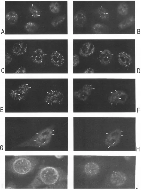
Patterns of Rev accumulation revealed by the anti-Rev MAbs. The anti-Rev MAbs visualized several new subcellular localizations of Rev (Fig. 1 and 2). In addition to the previously recognized nucleolar (Fig. 1C,E, and G and 2A) and diffuse nucleoplasmic (Fig. 1G) accumulation, Rev was detected as J. VIROL.
on November 9, 2019 by guest
http://jvi.asm.org/
HIV-1 rev AND SUBCELLULAR DISTRIBUTION 1477
A
-C
D
E
F
G
--H
FIG. 1. Indirect immunofluorescent doublelabelingofHeLa/crev cellsshowingRev in thesnRNP-richnucleoplasmicspeckles(small
arrows),
in the nucleoli(arrowheads),inaperinuclearzone,andin thecytoplasm(curvedarrow).Left-right pairsofpanelsrepresentthesamemicroscopic
field (magnification, x400). (A, C, E,and G) Anti-Rev MAb 9G2 anda Texas red anti-mouse conjugate; (B andF) human autoimmune sm-positiveserum;(D)humanautoimmuneserumagainstnucleoli, ANA-N; (H)human autoimmuneserumagainstlamins. The human antisera inpanels B, D, F, andHwerevisualizedbyusinganFITC anti-humanconjugate.
VOL.68, 1994
on November 9, 2019 by guest
http://jvi.asm.org/
[image:3.612.104.506.63.670.2]A
C
E
0
3
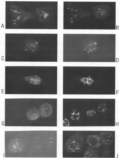
FIG. 2. Indirectimmunofluorescentdoublelabeling of COS- I cellstransfcctedwithpSVCMVrev,andfixed64 hlater. Left-right pairs of panels represent the samemicroscopicfield (magnification, x600). (A, C, E, and G)Anti-Rev MAb 9G2aind a Texasrcd
aniti-mousc
conjugate; (I) anti-SC35 MAb and a Texas red anti-mouse conjugate; (B, D, F, H, andJ) human sm-positive serum (speckled) and an FITC anti-human conjugate. Shownare the nucleoli (arrowheads), perinuclear zone (split arrow), cytoplasm (curved arrow), and localization ofnucleoplasmic snRNPs(small arrows).1478
on November 9, 2019 by guest
http://jvi.asm.org/
[image:4.612.71.545.48.679.2]HIV-1 rev AND SUBCELLULAR DISTRIBUTION 1479 TABLE 1. Colocalizations between Rev and host cell factors,
determined byindirect immunofluorescentdoublelabeling
Colocalization'
MAb orserum Nucleoplasmic Perinuclear zone (antigen recognized) speckles
sm Rabbit sm Rabbit
serum anti-Rev serum anti-Rev MAb
8E7(HIV-1 Rev) + + +
9G2(HIV-1 Rev) + + +
4B10 (hnRNPAl) + +
4F4(hnRNP C) + +
H20(m3Gcaps) + +
Y12(sm antigen) + +
Anti-B" (U2snRNPB") + +
Anti-SC35(SC35) +/- +1-
-Antilamin (lamin A/C) MAb350 (nuclear pore) RL-1 (nuclear pore)
LalB5(La antigen) -/+ -/+ - -/+
Anti-B23 (B23) - - - -/+
Anti-C23(nucleolin) - - - -/+
Anti-rRNA (rRNA) - - - -/+
Serum
Smpositive(smantigen) + +
ANA-N(nucleolus) - - - +
Antilamin(lamin)
a+,both antibodies of thecorresponding coordinateslabel thenucleoplasmic speckles ortheperinuclear zone; -,only oneof the twoantibodieslabels the speckles or the perinuclear zone;+/-,considerable doublelabeling;-/+,some doublelabeling.
distinct speckles within thenucleoplasm(Fig. 1A, C,and E and
2E) and additionally inaperinuclearzone (Fig. 1C, E,andG
and2C) from which Rev-containingrays sometimes extended into the cytoplasm (Fig.2C). Rev wasalsodiffusely distributed inthe cytoplasm ofmanycells(Fig.1C and G and2C).In some
cells, mainly thenucleoli stained (Fig. 1E and2A); inothers,
mainly the nucleoplasmic speckles (Fig. 1A and 2E), the perinuclear zone (Fig. 2C),or thecytoplasm (Fig. 1G, upper
right) stained. Any combination of those basic accumulations
wasfound. In repeated experiments ofnonsynchronized
cul-tures, theprevalence of the basic subcellular distributions of
Rev wascomparable.
Characterizationof humanautoimmuneseraby fluorescent
double labeling.Sm-positive seragenerateaspeckled nucleo-plasmic immunofluorescent staining, indicating that mRNA
splicingcomponents arenonrandomlydistributed in the
nucle-oplasm (44). MAbs against the sm antigen ofsnRNPs (44),
RNA-splicing factors like U2AF(77)and SC35(28),andsome hnRNPs(12, 23, 59)generate asimilarspecklednucleoplasmic immunofluorescent staining. Additionally, poly(A) RNA (9), m3G cap RNA (6, 61), Ul, U2, and U5 snRNAs (8), and specific mRNA transcripts (35, 75) have been localized to similar nucleoplasmic speckles. Whether distinct subsets qf
speckles exist is still unclear. The functional relationships
between speckles and a number of morphological distinct,
subnucleardomains including focior coiled bodies(8, 43, 60, 79), hnRNP L domains (8, 59), nuclear dots (3), andPIKAs
(66) await elucidation.
Thisstudywilldeal withspecklesasthefluorescentpattern
generatedby the sm-positive autoimmunehuman serum.The
speckled immunofluorescent colocalizations between defined subcellular components are shown in Fig. 3 and Table 1. In
methanol-fixed cells, antibody Y12, directed against the sm
antigen, completely colocalized with the speckles generated by the sm-positive human serum. In addition, both stained the nucleoplasmdiffusely (Fig. 3A and B). Extensive colocalization between the speckles of the sm-positive serum, the m3G caps, theU2snRNP-specific protein B", hnRNP Al (Fig. 3C and D), and hnRNP C(Fig. 3Eand F) was found. In some prepara-tions, a partial colocalization between the sm-positive serum andthe LalB5 antibody (4), directed against the La antigen, was found(Fig. 3G and H). The SC35 staining was sometimes different from the sm staining, consisting of larger, irregular structuresenriched around the nucleolus(Fig. 4H). Theextent
of colocalization between the SC35 staining and the sm-positive staining surprisingly appeared variable in methanol-fixed cells in repeated experiments.
HIV-1 Rev associated with splicing factors in the plasm. The autoimmune sera reacting with the same nucleo-plasmic speckles as MAbs to spliceosomal components were
used in combination with the anti-Rev MAbs. Colocalization between Rev and thenucleoplasmicspecklesis evident in Fig. 1A, B, E, and F and Fig. 2E and F. Almost all cells stained positive with autoimmune sera or MAbs against subcellular structures.Incomparison,aminority of thesamecells stained strongly with the Rev antibodies (Fig. 1, 2, and 4). Only the autoimmune serum with the nucleoplasmicspeckledreactivity colocalized with the nucleoplasmic Rev speckles. Neither the human antinucleolar(Fig. 1C andD) nor the human antilamin (Fig. 1G andH) serum double stained thenucleoplasmic Rev
spots.
In double-labeling experiments using 9G2 or 8E7 and a
polyvalent rabbit antiserum directed against theRev C-termi-nal part, the same HeLa/crev cells stained with overlapping fluorescent patterns (Fig. 4A and B). The results of the fluorescent double labeling using thepolyclonal anti-Rev
se-rum and MAbs against defined subcellular components are
displayed in Table 1 and Fig. 4. Extensive colocalization in nucleoplasmic speckles between Rev and B" (Fig. 4C and D), the smantigen(Fig.4EandF), and hnRNP Al, hnRNPC, and m3G caps (Table 1) was evident. The colocalization between Rev and SC35 appeared sensitive to slight variations of the immunofluorescence procedure and varied(Fig.4G andH).In
less than 10% ofRev-positive cells, significant speckled colo-calization between Rev and LalB5 (Fig. 41 andJ) existed.
Rev accumulates in a perinuclear zone enriched in other host cell antigens that are transported across the nuclear
membrane. A large proportion of Rev-expressing HeLa and COS cells exhibitedapronouncedperinuclear accumulationof
Rev (Fig. 1G and 2C). In some cases, Rev-containing rays extended beyond the zone and into the cytoplasm (Fig. 2C). The zone wasoutside the areas thatwere stainedby both the antispeckled serum (Fig. 2C and D) and the antilamin serum
(Fig. IG and H). Double labeling between anti-nuclear pore MAbs (RL-1 and MAb350) and the rabbit anti-Rev (not shown) did not clearly resolve the extent of colocalization between the nuclear membrane and the perinuclear zone. From FITC and Texas red overlays (not shown) and conven-tional fluorescence microscopy, it seemed that the Rev zone didnotoverlap with the nuclear part of the nuclearmembrane
butoverlappedor wascontiguoustothecytoplasmicside of the
nuclear membrane. Several characterized antibodies stained
theRev perinuclear zone, including the LalB5 antibody (Fig.
41andJ) and MAbsagainstthemajornucleolarproteins, B23
and C23 (not shown). Notably, all of these proteins belongto a growing class of proteins known to shuttle between the
nucleus and the cytoplasm.
ThedifferentRevstainingpatternsdidnotvary insubclones of the HeLa/crev cell cultureorwith the abundance of Rev in VOL.68, 1994
on November 9, 2019 by guest
http://jvi.asm.org/
c
G
B
D
[image:6.612.72.545.43.679.2]IH
FIG. 3. Characterization of the humansm-positiveserum(Chemicon)inpanelsB, D, F, H,andJ,usingindirectimmunofluorescent double labelingofHeLa/crev cells with MAbs Y12(A), 4B10(C),4F4(E), La1B5(G),andMAb350(I).Thesmantibodieswerevisualized with rhodamine anti-human conjugates, and the MAbs were visualized with FITC anti-mouse conjugates. See Table 1 for results and more extensive characterizationperformedwith additional antibodies.Some of thedouble-labeledspeckles areindicated(bars).
1480
on November 9, 2019 by guest
http://jvi.asm.org/
A
Cs
E
3
D
[image:7.612.68.554.48.689.2]G
FIG. 4. Indirectimmunofluorescent doublelabelingofHeLa/crevcells. All cells inpanels A,C, E, G,and Iwerelabeled withapolyclonalrabbit anti-RevserumandaTexas redanti-rabbitconjugate.Inpanelsontheright,thefollowingMAbs andanFITC anti-mouseconjugatewereused: 8E7anti-Rev(B),anti-B"(D),Y12(F), anti-SC35(H),andLa1B5 (J).Some of thedouble-labelednucleoplasmicspecklesareindicated(bars).
1481
on November 9, 2019 by guest
http://jvi.asm.org/
1482 KALLAND ET AL.
the cells. Toexamine the possibility that aseries of subpopu-lations ofRev-expressing cells accounted for distinct subsets of unique Rev distributions, many new subclones were grown from the originalHeLa/crev8 cells. In all of the subclones that generated positive Rev fluorescence, all of the original Rev distribution patterns reappeared (not shown). All of the dif-ferent HeLa/crev clones showed a pronounced cell-to-cell variation in the amount of Rev expression. Rev did not preferentially accumulate in any of the particular subcellular compartments when the amount of Rev protein present in each cell varied from high to barely detectable. Low amounts of Rev were visible in the cytoplasm only (Fig. 1C and G), in the nucleoli (Fig. 1C), and in the speckles only (Fig. 1A).
Inhibition of new protein synthesis by using cycloheximide didnotchange the subcellulardistribution patternsofRev.To
examine whether afinaldestination of Rev accumulation could be defined, the HeLa/crev cells were incubated with cyclohex-imide at 50jig/ml toinhibit newsynthesis of protein. As long asthecellsappearedreasonably healthy, for up to 8 to 10 h in the presenceof cycloheximide, the distribution of Rev between the cytoplasm, the perinuclear zone, the nucleoli, and the nucleoplasmic speckles continued.
The different subcellular localizations of Rev were present
during interphase of HeLa/crev cells. Two approaches were used to determine the relationship between the stage of the cell cycle and the Rev distribution patterns. Cells were syn-chronizedby serum starvation and next treated with aphidico-lin for arrest in the
G1/S
stageof the cell cycle. Time points weredone for cells released from aphidicolin arrest. Cells were double stained by anti-Rev MAbs and a human anti-mitotic spindle serum. Rev was detected in the cytoplasm, in the perinuclear zone, in the nucleoli, and in the nucleoplasmic speckles in G1, S, and G2 stages (not shown).Inthe second approach, immunofluorescent double labeling was done with rabbit Rev and mouse monoclonal anti-cyclinAandanti-cyclin
Bi.
Fromthe cellcycle-specific expres-sionandsubcellularlocalization of cyclin A and cyclinBi(57), it was concluded that Rev was expressed and distributed with comparable patterns throughout the interphase (not shown). Rev was also detected in mitotic cells.Rev-induced change of speckled morphology in vivo. Inthe Rev-expressing COS-1 cells, two types of changes of the speckled staining pattern were observed. In 60 to 80% of Rev-expressing cells with Rev present in the nucleoplasm, the humanantispeckled serum stained the nucleoplasm in a retic-ularpattern. Revclosely colocalized with the reticles (Fig. 2E andF). Inthe other Rev-expressing cells, the diffuse
compo-nent of the staining was often subdued, and the speckles changed to a globular appearance (Fig. 2B, H, and J). In
contrasttothereticles, the globules usually did not colocalize with Rev. Instead, Rev was present as crescents in the nucle-oplasm between the globules (Fig. 2G and H), or Rev was not detected in the nucleoplasm but was detected in the other compartments (Fig. 2A and B). Double labeling showed that anti-SC35 (Fig. 21/J) and the Y12 MAb (not shown) stained
theglobules. In Fig. 2I and J, the extreme size that the changed splicing structures sometimes evidenced is shown. A similar
change of the staining of the sm antigen has been reported
previously and has been associated with inhibition of mRNA transcription (5). Globular speckles such as those in Fig. 21 or the intense reticular Rev staining such as that in the Rev-expressing COS cells in Fig. 2F were not found in HeLa/crev cells and mock-transfected COS cells.
DISCUSSION
Wefound that Rev colocalizes withmRNA-splicing factors in a speckled pattern in the nucleoplasm of cells.Accumulating evidence shows that the nucleoplasmic speckles represent active sites of both transcription and mRNA processing (37, 38, 76). A long-standing unsolved issue is whether Rev regu-lation of HIV mRNA expression is at the level of RNA splicing, RNA transport, or otherposttranscriptional steps.In
nuclear extracts in vitro, splicing factors assemblestepwiseon
the pre-mRNA into a multimolecular structure termed the spliceosome,from which correctly spliced mRNAs are eventu-allyreleased (reviewed by Green[29]). The elements required for RNA splicing in vitro are located in speckles in the nucleoplasm that include snRNP proteins (61, 71, 72), the m3G caps of snRNAs (61), specific snRNAs (8), the RNA-splicing factor SC35 (28), poly(A) RNA (9), and specific pre-mRNAs
(35,75).
Recently,by using a combination of in situ hybridization and immunofluorescence (76), it was demonstrated that introns and exons ofspecific mRNA transcripts emerged from indi-vidualSC35-containing speckles. The introns and exons sepa-rated along a track emanating from thosespeckles. In HeLa cells infected with adenovirus, RNA polymerase II, cellular splicing factors, and hnRNPs redistributed to the sites of adenovirus mRNAtranscription (38).Aseries of observations nowsuggeststhattranscription and RNA-splicing events occur in or close to thenucleoplasmic speckles that Rev colocalizes with. Splicing of retroviral mRNAs is a rate-limited event, probably due to splice sites that are suboptimal as recognized bythe splicingmachinery (10). A complex differential splicing
(67)of the approximately 9-kb mRNA that is transcribed from the HIV-1 genome is one of the main strategies utilized by HIV-1 to expressitscondensed genetic information. The viral structural products that are encoded by the full-length and singly spliced mRNAs are Rev dependent, while proteins translatedfrom fully spliced mRNAs such as Rev itself and the other essential HIV-1 regulatory protein, Tat, are not Rev dependent. Therefore, it should come as no surprise that the splicing process may be a target for retroviral regulation. Chang and Sharp(10)have shown thatif splicing is made more efficient by substituting
P-globin
splice sites for the suboptimalHIV splice sites, Rev cannot rescue the intron from being removed by splicing although the RRE is present in cis. A role ofUl snRNAin the formation of Rev-dependent env mRNA hasbeen demonstrated (45). In a cell-free splicing system, a small Rev-dependent inhibition of splicing of RRE-containing
RNAhas been demonstrated. A synthetic peptide correspond-ing to the basicdomain of Rev significantly inhibited splicing apparently by blocking entry of U4/U6 and U5 snRNPs into the spliceosome (42). The present demonstration of Rev colocalization with the splicing factors in the nucleoplasm corroborates the view that one strategic site of Rev activity is within the splicing machinery of the cell.
Itmight be argued that overexpression of Rev can lead to oversaturation of the host cell transport apparatus or to nonspecific binding of Rev to host cell RNA. It was evident, however, that barely detectable amounts of Rev in the cells werespecifically distributed to the cytoplasm (Fig. 1C and G), the nucleoli (Fig. 1C), and the speckles (Fig. 1C), arguing against the possibility that overexpression is a confounding parameter leading to artificial Rev distributions in this study. The pronounced variation of Rev expression levels within single cells of the same culture (Fig. 1 and 4) could not be related to different stages of the cell cycle and was apparent in newly established subclones of the HeLa/crev culture. Most J. VIROL.
on November 9, 2019 by guest
http://jvi.asm.org/
HIV-1 rev AND SUBCELLULAR DISTRIBUTION 1483
likely, thevaryingRevexpressioncanbeattributedtounstable control mechanisms for the artificially introduced rev, gene.
The wide range of abundance levelsofRevin individual single cells show thatevenwhen theamountofRevwithin the cellis very low, Revis specifically transported into both the nucleoli and the speckles. Notably, the RRE is made neither in the HeLa/crev cells nor in the Rev-expressing COS-1 cells
exam-ined, nor is any other HIV-1 protein expressed. It can
there-fore be concluded thatthe initial distribution ofRevwithinthe host cell is based entirelyupon Rev and host cell interactions. Rev must therefore contain the information not only for nuclear and nucleolar import but also for the RRE-indepen-dent localization to nucleoplasmic speckles.
It has previouslybeen shown thatRevpromotes the nuclear-to-cytoplasmic distribution of RRE RNA (22, 26, 31, 50). In one study of the analogousHTLV-I Rex protein, itwasshown
that Rex affects not only the nuclear-to-cytoplasmic distribu-tion of Rex-dependent RNA but also the intranuclear distri-bution of Rex-dependent RNA (39). The demonstration of Rev-dependent polysomal loading of RRE RNA is consistent with the possibilitythat Rev has both nuclear and cytoplasmic functions (1, 2, 17). In the present study, it was found that
when protein synthesis was inhibited by cycloheximide for up
to 10 h, Revcontinuedtoaccumulate unabatedinthedifferent subcellular compartments. It has previously been shown that nucleocytoplasmic protein transport isnot inhibited by cyclo-heximide undersimilar conditions (5, 7,58). The results show that there is no preferred final destination of Rev accumula-tion. Instead, a continuous redistribution of Rev between the subnuclearand cytoplasmic compartments occurs.
Several host cell proteins that shuttle between the nucleus and the cytoplasm(B23, C23, and La) colocalized with Rev in the perinuclearzone.Asimilar perinuclearzonehaspreviously
been identified as a translationally active ribosome-rich
com-partment (5);a similar zone hasalso been associated with the passive attachmentstage of the active protein import through the nuclearpore complex (63).
The role of the nucleolus remains enigmatic, since this structure hasno known role inprocessingortransportof RNA polymerase TI-transcribed mRNA, the presumed target for Rev when the RRE is present in those mRNAs as well. One
possibility isthat Revexploitsthe high protein import capacity of the nucleolus to penetrate the nucleus. It has been found that Rev binds specifically to a major nucleolar protein, B23
(24), that shuttlesbetween the cytoplasm andthe nucleus (7). In this study, colocalization between Rev and B23was found
both in the nucleolus and in the perinuclear zone.
This study has identified new subcellular compartments of Rev distribution. The RRE-independent and continuous dis-tribution ofRevbetween the cytoplasm, the nucleoli, and the nucleoplasmic speckles supports the following model of the Rev mechanism of function. Inthe speckles, Rev may
dissoci-ate assembly of splicing factors on nascent HIV mRNA transcripts. By dual binding to the RRE of Rev-dependent mRNAs and to host cell factors involved innucleocytoplasmic transport, Rev may mediate transport of such mRNAs to
cytoplasmatic translation.
ACKNOWLEDGMENTS
WethankB.Cullen for helpfuldiscussionsand the HeLa/crev8and
Hela/cnefcelllines.We thank W. A. Haseltinefor stimulating
discus-sions and support. We thank M.-L. Hammarskj6ld, D. Rekosh, G.
Blobel,X.-D. Fu, T. Maniatis, L.Gerace, J. A. Steitz, F. McKeon, R.
Luhrmann, A.I. Lamond, M. Bachmann, G. Dreyfuss, and S. Pifiol-Romaforantibodies,L. K.VenkateshandG.Chinnaduraiforprev 81,
and Y.-C. SungforpSVCMVrev. WethankD.Hellandfor adviceand
help. Beth Johannesen and Sissel Aadland are acknowledged for
excellent technical assistance.
This work was supported by the Norwegian Cancer Society. The Norwegian Research Council for Science and Humanities, The Nor-wegian Odd Fellow Foundation, and Familien Blix' Fond.
REFERENCES
1. Arrigo, S. J., and
I.
S. Y. Chen. 1991. Rev is necessary for translationbut not cytoplasmic accumulation of HIV-1vif, vpr and env/vpul 2 RNAs. Genes Dev. 5:808-819.2. Arrigo, S. J., S. Heaphy, and J. K. Haines. 1992. In vivo bindingof wild-type and mutant human immunodeficiency virus type 1 Rev proteins: implications for function. J. Virol. 66:5569-5575. 3. Ascoli, C. A., and G. G. Maul. 1991. Identification of a novel
nuclear domain. J. CellBiol. 112:785-795.
4. Bachmann, M., W. J. Mayet, H. C.
Schroder,
K. Pfeifer, K.-H. Meyer zumBiischenfelde, and W. E. G.Muller. 1986.Association of La and Ro antigens with intracellular structures in HEp-2 carcinoma cells. Proc. Natl. Acad. Sci. USA 83:7770-7774. 5. Bachmann, M., H. C. Schroder, D. Falke, and W. E. G. Muller.1988. Alteration of the intracellular localization of the Laprotein compared with the localization of U snRNPs, p. 101-125. hI R. Peters (ed.), Nucleocytoplasmic transport. Academic Press, San Diego, Calif.
6. Bochnig, P., R. Reuter, P. Bringmann, and R.Luhrmann. 1987. A monoclonal antibody against 2,2,7-trimethylguanosine that reacts with intact class U, small nuclear ribonucleoproteins as well as with
7-methylguanosine-capped
RNAs. Eur. J. Biochem. 168:461-467.7. Borer, R. A., C. F. Lehner, H. M. Eppenberger, and E. A. Nigg. 1989. Major nucleolar proteins shuttle between nucleus and cytoplasm. Cell 56:379-390.
8. Carmo-Fonseca, M., R. Pepperkok, B. S. Sproat, W. Ansorge, M. S. Swanson, and A.
I.
Lamond. 1991. hI v,ivo detection of snRNP-richorganelles in thenuclei of mammalian cells. EMBOJ. 10:1863-1873.9. Carter, K. C., K. L. Taneja, and J. B. Lawrence. 1991. Discrete nuclear domains of poly(A)RNA and their relationship to the functional organization of the nucleus.
J.
Cell Biol.115:1191-12t)2. 10. Chang, D. D., and P. A. Sharp. 1989. Regulation by HIV Revdepends upon recognition of splice sites. Cell 59:789-795. 11. Chida, K., andP. K. Vogt. 1992.Nuclear translocation of viralJun
but not of cellular Jun is cell cycle dependent. Proc. Natl. Acad. Sci. USA89:4290-4294.
12. Choi, Y. D., and G. Dreyfuss. 1984. Monoclonal antibody charac-terization of the C proteins of heterogeneous nuclear ribonucleo-protein complexes in vertebrate cells. J. Cell Biol. 99:1997-20)0)4. 13. Cochrane,A.W.,K.S.Jones, S. Beidas, P. J.Dillon,A. M.Skalka,
and C. A. Rosen. 1991. Identification and characterization of intragenic sequences which repress human immunodeficiency vi-rus structural geneexpression. J. Virol. 65:5305-5313.
14. Cochrane, A. W.,A.Perkins, andC. A. Rosen. 1990. Identification of sequences
important
in the nucleolar localization of human immunodeficiency virus Rev: relevance of nucleolar localization to function. J. Virol. 64:881-885.15. Cullen, B. R. 1992. Mechanism of action of regulatory proteins encoded by complex retroviruses. Microbiol. Rev.56:375-394. 16. Cullen, B. R., J. Hauber, K. Campbell, J. G. Sodroski, W. A.
Haseltine, and C. A. Rosen. 1988. Subcellular localization of the human immunodeficiency virus trans-acting art gene product. J. Virol. 62:2498-2501.
17. D'Agostino, D. M., B. K. Felber, J. E. Harrison, and G. N.
Pavlakis.
1992. The Rev protein of human immunodeficiency virus type1 promotes polysomal association and translation ofgag/poland vpu/env, mRNAs. Mol. Cell. Biol. 12:1375-1386.
18. Daly, T. J., K.S. Cook, G. S. Gray, T. E. Maione, and J. R. Rusche. 1989. Specific binding of HIV-1 recombinant Rev protein tothe Rev-responsive element in vitro. Nature (London)342:816-819. 19. Davis, L., andG. Blobel. 1986.Identification and characterization
of a nuclearpore complex protein. Cell 45:699-709.
20. Dayton, A. I., E. F. Terwilliger, J. Potz, M. Kowalski, J. G.
Sodroski, and W. A. Haseltine. 1988. Cis-acting sequences respon-sive to the rev gene product of the human immunodeficiency virus.
VOL. 68? 1994
on November 9, 2019 by guest
http://jvi.asm.org/
1484 KALLAND ET AL.
J.AcquiredImmuneDefic.Syndr. 1:441-452.
21. Dayton, E. T., M. Douglas, D. Powell, and A. Dayton. 1989. Functional analysis of CAR, the target sequence for the rev
proteinof HIV-1. Science 246:1625-1629.
22. Emerman, M., R. Vazeux, and K. Peden. 1989. The rev gene product of the human immunodeficiency virus affects
envelope-specificRNAlocalization. Cell 57:1155-1165.
23. Fakan, S., G. Leser, and T. E. Martin. 1984. Ultrastructural distributionof nuclearribonucleoproteins asvisualizedby immu-nocytochemistry onthin sections. J. Cell Biol. 98:358-363. 24. Fankhauser, C.,E.Izaurralde,Y.Adachi,P.Wingfield,and U.K.
Laemmli. 1991. Specific complex of human immunodeficiency virus type 1 Rev andnucleolar B23proteins: dissociation by the Rev responseelement. Mol. Cell. Biol. 11:2567-2575.
25. Feinberg, M. B., R. F. Jarret, A. Aldovini, R.C. Gallo, and F. Wong-Staal. 1986. HTLV-III expression andproduction involve
complex regulationatthelevels ofsplicingandtranslation of viral RNA.Cell 46:807-817.
26. Felber,B. K.,M.Hadzopoulou-Cladaras, C.Cladaras,T. Copel-and,andG. N.Pavlakis. 1989. Revprotein of human
immunod-eficiencyvirustype 1affects thestabilityand transportofthe viral
mRNA.Proc. Natl. Acad. Sci. USA 86:1495-1499.
27. Freeman, J. W.,A.Chatterjee,B.E.Ross,andH.J. Busch. 1985. Epitope distribution and immunochemical characterization of nucleolar phosphoprotein C23using ten monoclonal antibodies. Mol. Cell. Biochem. 68:87-96.
28. Fu,X.-D.,andT.Maniatis. 1990. Factorrequiredfor mammalian
spliceosome assembly is localized to discrete regions in the
nucleus. Nature (London)343:437-441.
29. Green,M.R. 1991. Biochemical mechanisms ofconstitutiveand regulated pre-mRNA splicing.Annu. Rev. CellBiol.7:559-599. 30. Habets,W.J.,M. H.Hoet,B. A. W. DeJong,A. VanderKemp,and
W.J. vanVenrooij. 1989. Mapping of B cell epitopeson small nuclearribonucleoproteinsthatreactwithhumanautoantibodies
aswell as with experimentally inducedmouse monoclonal anti-bodies.J. Immunol. 143:2560-2566.
31. Hammarskjold,M.-L., J. Heimer, B.Hammarskjold,I.Sangwan, L.Albert,and D.Rekosh.1989. Regulationof human immunod-eficiencyvirusenv expression by therev geneproduct. J. Virol. 63:1959-1966.
32. Haseltine,W. A.1991.Regulationof HIV-1replication,p.1-42. In W. A. Haseltine and F.Wong-Staal(ed.), Geneticstructureand regulationof HIV-1. Raven PressLtd,NewYork.
33. Heaphy, S.,C.Dingwall,I.Ernberg,M.J.Gait, S. M.Green,J. Karn, A. D. Lowe, M. Singh, and M. A. Skinner. 1990. HIV-1 regulator of virion expression (rev) protein binds to an RNA stem-loop structure located within the rev response element region.Cell 60:685-693.
34. Hope,T.J.,D.McDonald,X. Huang, J. Low,and T. G.Parslow. 1990. Mutational analysis ofthe human immunodeficiencyvirus type 1 rev trans activator: essential residues near the amino terminus. J. Virol. 64:5360-5366.
35. Huang, S., and D. L. Spector. 1991. Nascent pre-mRNA
tran-scripts are associated with nuclear regions enriched in splicing
factors. GenesDev.5:2288-2302.
36. Huang, X.,T.J.Hope,B.L.Bond,D.McDonald,K.Grahl,and T. G. Parslow. 1991. Minimal rev-response element for type 1 humanimmunodeficiencyvirus.J.Virol.65:2131-2134.
37. Jackson,D.A.,A. B. Hassan, R.J. Errington, and P. R. Cook. 1993. Visualizationof focal sites oftranscription within human nuclei. EMBO J. 12:1059-1065.
38. Jimenez-Garcia,L.F.,and D. L.Spector. 1993. In vivo evidence that transcription and splicing are coordinated by a recruiting mechanism. Cell 73:47-59.
39. Kalland,K.H.,E. Langhoff,H.J. Bos, H.Gottlinger, and W.A. Haseltine. 1991. Rex-dependent nucleolar accumulation of HTLV-I mRNA. New Biol.3:389-397.
40. Kiyokawa, T.,M.Seiki,S.Iwashita,K.Imagawa,F.Shimizu, and M.Yoshida.1985.
p27x-1I
andp21x-1,,
proteinsencodedbythe pX sequenceof human T-cell leukemiavirustype I.Proc.Natl.Acad. Sci. USA 82:8359-8363.41. Kjems, J., M. Brown, D. D. Chang, and P. A. Sharp. 1991. Structuralanalysis of theinteractionbetween the human
immu-nodeficiency virus rev protein and the rev responsive element. Proc. Natl. Acad. Sci. USA 88:683-687.
42. Kjems,J., and P. A. Sharp. 1993. The basic domain ofrevfrom humanimmunodeficiencyvirus type 1specificallyblocks the entry of U4/U6-U5 small nuclear ribonucleoprotein in spliceosome
assembly. J.Virol.67:4769-4776.
43. Lamond, A.I., andM. Carmo-Fonseca.Thecoiledbody. Trends Cell Biol., in press.
44. Lerner,E.A.,M.R. Lerner,C. A. Janeway, and J.A.Steitz.1981. Monoclonal antibodies to nucleicacid-containingcellular constit-uents: probes for molecular biology and autoimmune disease. Proc. Natl. Acad. Sci. USA 78:2737-2741.
45. Lu, X., J.Heimer,D.Rekosh, and M.-L.Hammarskjold.1990.Ul small nuclear RNA plays a direct role in the formation of rev-regulated human immunodeficiency virus env mRNA that remainsunspliced. Proc. Natl. Acad. Sci. USA87:7598-7602. 46. Maldarelli, F.,M.A.Martin, and K. Strebel. 1991. Identification
of posttranscriptionally active inhibitory sequences in human immunodeficiency virus type 1 RNA: novel level of gene regula-tion. J.Virol.65:5732-5743.
47. Malim, M. H., S. Bohnlein, J. Hauber, and B. R. Cullen. 1989. Functional dissection of the HIV-1 revtrans-activator-derivation ofatrans-dominant repressor ofrevfunction. Cell 58:205-214. 48. Malim, M.H., R. Fenrick, D. W. Ballard, J. Hauber, E.Bohnlein,
andB.R.Cullen. 1989.Functionalcharacterizationofacomplex protein-DNA-binding domain locatedwithin thehuman immuno-deficiency virus type 1 long terminal repeat leaderregion. J. Virol. 63:3213-3219.
49. Malim, M. H., J. Hauber, R. Fenrick, and B. R. Cullen. 1988. Immunodeficiency virus rev trans-activator modulates the expres-sionof the viralregulatory genes. Nature(London)335:181-183. 50. Malim, M. H., J. Hauber, S.-Y. Le, J. V. Maizel, and B. R. Cullen. 1989. The HIV-1 rev transactivator acts through a structured target sequence to activate nuclear export of unspliced viral mRNA. Nature (London) 338:254-257.
51. Malim, M. H., D. F.McCarn, L. S. Tiley, and B. R. Cullen.1991. Mutational definitionofthe human immunodeficiency virus type 1 rev activationdomain. J. Virol. 65:4248-4254.
52. Malim, M. H., L. S. Tiley, D. F. McCarn, J. R. Rusche, J.Hauber, and B. R. Cullen.1990. HIV-1 structural gene expressionrequires binding of therevtrans-activatortoitsRNA target sequence.Cell 60:675-683.
53. Mermer, B., B. K. Felber, M. Campbell, and G. N. Pavlakis. 1990. Identification of trans-dominant HIV-1 rev protein mutants by direct transfer ofbacterially produced proteins into humancells. NucleicAcids Res. 18:2037-2044.
54. Ochs, R., M.Lischwe, P.O'Leary,and H. Busch. 1983. Localiza-tion of nucleolar phosphoproteins B23 and C23 during mitosis. Exp.Cell Res. 146:139-149.
55. Olsen, H. S., A. W. Cochrane, P. J. Dillon, C. M. Nalin, and C. A. Rosen. 1990. Interaction of the human immunodeficiency virus type 1 rev protein with a structured region in env mRNA is dependent on multimer formation mediated through a basic stretch of amino acids. Genes Dev. 4:1357-1364.
56. Olsen, H. S., P. Nelbock, A. W. Cochrane, and C. A. Rosen. 1990. Secondary structure is the major determinant for interactionof HIVrevprotein with RNA. Science 247:845-848.
57. Pines, J., and T. Hunter. 1991. Human cell division: the involve-ment of cyclins A andBi, and multiple cdc2s, p. 449-463. In The cellcycle LVI. Cold Spring Harbor Laboratory Press, Cold Spring Harbor, N.Y.
58. Pinol-Roma, S., and G. Dreyfuss. 1992. Shuttling of pre-mRNA binding proteins between nucleus and cytoplasm. Nature (Lon-don) 355:730-732.
59. Pifiol-Roma, S., M. S. Swanson, J. G. Gall, and G. Dreyfuss. 1989. A novel heterogeneous nuclear RNP protein with a unique distribution on nascent transcripts. J. Cell Biol. 109:2575-2587. 60. Raska, I., L. E. C. Andrade, R. L. Ochs, E. K. L. Chan, C.-M.
Chang, G. Roos, and E. M. Tan. 1991. Immunological and ultrastructural studies on the nuclear coiled body with autoim-mune antibodies. Exp. Cell Res. 195:27-37.
61. Reuter, R., B. Appel, P. Bringmann, J. Rinke, and R. Luhrmann. 1984. 5'-terminal caps ofsnRNAs are reactive with antibodies J. VIROL.
on November 9, 2019 by guest
http://jvi.asm.org/
HIV-1 rev AND SUBCELLULAR DISTRIBUTION 1485 specific for 2,2,7-trimethylguanosine in whole cells and nuclear
matrices. Exp. Cell Res. 154:548-560.
62. Riabowol, K. T., J. S. Fink, M. Z. Gilman, D. A. Walsh, R. H. Goodman, and J. R. Feramisco. 1988. The catalytic subunit of cAMP-dependent protein kinase induces expression of genes containing cAMP-responsive enhancer elements. Nature (Lon-don)336:83-86.
63. Richardson, W. D., A. D. Mill, S. M. Dilworth, R. A. Laskey,and C.Dingwall. 1988. Nuclear protein migration involves twosteps: rapid binding at the nuclear envelope followed by slower translo-cationthrough nuclear pores. Cell 52:655-664.
64. Rosen, C. A., E. Terwilliger, A. Dayton, J. Sodroski, and W.A. Haseltine. 1988. Intragenic cis-acting art-gene-responsive se-quencesof the human immunodeficiency virus. Proc. Natl. Acad. Sci. USA 85:2071-2075.
65. Sadaie, M. R., T. Benter, and F. Wong-Staal. 1988. Site-directed mutagenesis of two trans-regulatory genes (tat-III,trs) of HIV-1. Science239:910-913.
66. Saunders, W. S., C. A. Cooke, and W. C. Earnshaw. 1991. Compartmentalization within the nucleus: discovery of a novel subnuclearregion.J.CellBiol.115:919-931.
67. Schwartz, S., B.K.Felber, D. M. Benko, E. M.Fenyo,and G. N. Pavlakis.1990.Cloning and functional analysis of multiplyspliced mRNAspecies of human immunodeficiency virus type 1. J. Virol. 64:2519-2529.
68. Schwartz, S., B.K.Felber, and G. N. Pavlakis.1992. Distinct RNA sequences inthe gag region of humanimmunodeficiencyvirus type 1decrease RNAstability and inhibit expression in the absence of Revprotein. J. Virol.66:150-159.
69. Snow, C. M., A. Senior, and L. Gerace.1987. Monoclonal antibod-iesidentifyagroupof nuclear pore complexglycoproteins.J.Cell Biol. 104:1143-1156.
70. Sodroski, J., W. C. Goh, C. Rosen, A. Dayton, E. Terwilliger, and W. A. Haseltine. 1986. A second post-transcriptional activator gene required for HTLV-III replication. Nature (London) 321: 412-417.
71. Spector, D. L. 1990. Higher order nuclear organization: three-dimensional distribution of small nuclear ribonucleoprotein par-ticles. Proc. Natl. Acad. Sci. USA87:147-151.
72. Spector, D. L., W. H. Schrier, and H. Busch. 1983. Immunoelec-tronmicroscopiclocalization of snRNPs. Biol. Cell 49:1-10. 73. Terwilliger, E., R. Burghof, R. Sia, J. Sodroski, W. A. Haseltine,
and C.Rosen.1988. The art gene product of human immunode-ficiency virus is required for replication. J. Virol. 62:655-658. 74. Venkatesh, L. K., and G. Chinnadurai. 1990. Mutants in a
conservedregionnearthecarboxy-terminusof HIV-1 Revidentify functionally important residues andexhibita dominantnegative phenotype. Virology 178:327-330.
75. Wang, J., L.-G. Cao, Y.-L. Wang, and T. Pederson. 1991. Local-ization of pre-messenger RNA at discrete nuclear sites. Proc. Natl. Acad.Sci. USA 88:7391-7395.
76. Xing, Y., C. V. Johnson, P. R. Dobner, and J. B. Lawrence. 1993. Higher level organization of individual gene transcription and RNAsplicing.Science 259:1326-1330.
77. Zamore, P. D., andM. R.Green. 1991. Biochemical characteriza-tion of a U2snRNP auxiliary factor: an essential pre-mRNA splicing factor with a novel intranuclear localization. EMBO J. 10:207-214.
78. Zapp, M. L., and M. R. Green. 1989. Sequence-specific RNA binding by the HIV-1 Revprotein.Nature(London)342:714-716. 79. Zhang, M., P. D. Zamore,M.Carmo-Fonseca,A.I.Lamond,and M. R.Green.1992.Cloningandintracellular localizationof the U2 small nuclear ribonucleoprotein auxiliary factor small subunit. Proc.Natl.Acad. Sci. USA 89:8769-8773.
VOL.68, 1994