0022-538X/96/$04.0010
Copyrightq1996, American Society for Microbiology
Identification of Cell Surface Molecules That Interact
with Pseudorabies Virus
AXEL KARGER
ANDTHOMAS C. METTENLEITER*
Institute of Molecular and Cellular Virology, Friedrich Loeffler Institutes, Federal Research
Centre for Virus Diseases of Animals, D-17498 Insel Riems, Germany
Received 3 October 1995/Accepted 21 December 1995
The alphaherpesvirus pseudorabies virus (PrV) has been shown to attach to cells by interaction between the
viral glycoprotein gC and cell membrane proteoglycans carrying heparan sulfate chains (HSPGs). A secondary
binding step requires gD and presumably another, hitherto unidentified cellular receptor. By use of a virus
overlay protein binding assay (VOPBA), cosedimentation analyses, and affinity chromatography, we identified
three species of cell membrane constituents that bind PrV. By treatment with EDTA, peripheral HSPGs of very
high apparent molecular mass (>200 kDa) could be extracted from Madin-Darby bovine kidney cells. Binding
of PrV to these HSPGs in the VOPBA was sensitive to enzymatic digestion with heparinase or papain.
Cosedimentation analyses indicated that binding between PrV and high-molecular-weight HSPG depended on
the presence of gC in the virion. In addition, adsorption of radiolabeled PrV virions to cells could be inhibited
by the addition of purified high-molecular-weight HSPG. By using urea extraction buffer, a second species of
HSPG of approximately 140 kDa could be solubilized. Binding of PrV to this HSPG in the VOPBA was also
dependent on the presence of heparan sulfate, since reactivity was abolished after suppression of
glycosami-noglycan biosynthesis with NaClO
3and after heparinase treatment. In addition to HSPG, in cellular
mem-brane extracts obtained by treatment with mild detergent, a 85-kDa memmem-brane protein was demonstrated to
bind to PrV in the VOPBA and affinity chromatography. In summary, we identified three species of cell
membrane constituents that bind PrV: a peripheral HSPG of high molecular weight, an integral HSPG of
approximately 140 kDa, and an integral membrane protein of 85 kDa. It is tempting to speculate that
interaction between PrV and the two species of HSPG mediates primary attachment of PrV and that the 85-kDa
protein is involved in a subsequent attachment step.
Several alphaherpesviruses, such as herpes simplex virus
types 1 and 2 (HSV-1 and HSV-2) (52), varicella-zoster virus
(54), pseudorabies virus (PrV) (36, 42), and bovine herpesvirus
1 (BHV-1) (39), as well as the betaherpesvirus human
cyto-megalovirus (7) and the gammaherpesvirus BHV-4 (51) attach
to target cells by interaction of the virion with heparan
sulfate-carrying proteoglycans (HSPGs) of the cell surface. In HSV-1,
PrV, and BHV-1, the viral glycoprotein gC is primarily
respon-sible for this interaction (10, 15, 28, 36, 39). Attachment of
wild-type virus is significantly reduced by addition of
compet-ing heparin durcompet-ing the attachment process, by digestion of the
target cell membrane with heparinase, or by the absence of gC
in the virion. In PrV, gC
2mutants have been shown to be
resistant to heparin inhibition (36). In addition, mutated
mu-rine L cells with defects in heparan sulfate biosynthesis can be
infected with gC
2PrV with the same efficiency as wild-type
cells, indicating that in PrV gC represents the only viral
glyco-protein productively interacting with cell surface heparan
sul-fate during the attachment process (23). Interaction with other
glycosaminoglycans (GAGs), e.g., chondroitin sulfate, appears
to play a minor role, if any. Therefore, gC
2PrV initiates
infection by a pathway which is independent of cell surface
proteoglycans. In contrast, gC
2HSV-1 mutants remain
sensi-tive to heparin competition and lack of cell surface GAGs
because of an interaction between virion gB and cellular
pro-teoglycans (1, 14).
Attachment of PrV to Madin-Darby bovine kidney (MDBK)
cells is a biphasic process (55) requiring gC for an initial
bind-ing step which is sensitive to competition by exogenous
hepa-rin. For a second, heparin-resistant attachment, gD is necessary
(22), presumably by interaction with a hitherto unidentified
cellular receptor. A similar finding has been reported for
BHV-1 (22). In HSV-1, cumulative evidence also indicates that
gD is a receptor-binding protein (30). HSV-1 gD is required
for stable virus attachment to the cell surface (11). A truncated
water-soluble form of gD inhibits penetration but not
adsorp-tion of HSV-1 and binds to a limited number of sites on Vero
cell surfaces (18). HSV-1 gD has been shown to bind to the
mannose 6-phosphate receptor (4, 5), although the significance
of this interaction for viral entry is unclear. The basic fibroblast
growth factor (bFGF) receptor has also been described to act
as an HSV-1 receptor (19). However, the results could not be
reproduced, and the role of this protein in HSV-1 infection is
questionable (37, 38, 44). Recently, a receptor-binding
func-tion has also been proposed for the BHV-1 gB, although a
corresponding cellular receptor has not been identified (27).
Therefore, aside from cellular HSPG, no specific receptor
for any alphaherpesvirus has been unequivocally identified.
This could at least partially be due to the fact that attachment
of alphaherpesviruses to cells is a complex process in which
several components of the viral membrane interact with
dif-ferent cell membrane constituents. Examples of multiple
in-teractions in ligand-receptor binding are known. For example,
two distinct binding sites for the bFGF receptor in addition to
a heparin-binding domain are present on bFGF protein.
Inter-action of all three receptor-binding domains with their cellular
counterparts is required for mitogenic activity of bFGF (47). In
human immunodeficiency virus type 1, the formation of a
ter-nary complex between the viral glycoprotein gp120 and two
proteins of the cell surface, i.e., CD4 and an unidentified
HSPG, has been shown to occur (41).
* Corresponding author. Phone: 49-38351-7102. Fax: 49-38351-7151.
2138
on November 9, 2019 by guest
http://jvi.asm.org/
and radiolabeled on pig kidney (PSEK) cells. For titration, adsorption assays and preparation of cell extracts, MDBK cells were used.
Adsorption assay.Adsorption assays were performed as described earlier (22). PrV virions were labeled with [3H]thymidine by the procedure of Kaplan and
Ben-Porat (20) and purified in a sucrose step gradient as described previously (22). To test inhibition of attachment by anion-exchange chromatography frac-tions I, IIA, IIB, and IIB-H, [3H]thymidine-labeled PrV (70,000 cpm) was mixed
with the indicated amount of protein and diluted to a final volume of 60ml in phosphate-buffered saline (PBS) containing 1% bovine serum albumin (BSA) (PBS-A). After 1 h of incubation on ice, 50ml of the mixture was added to 200
ml of PBS-A and used for the adsorption assay. [3H]thymidine-labeled PrV
preincubated in PBS-A served as a control. Only total binding was determined (22).
Anion-exchange chromatography.For anion-exchange chromatography (mod-ified as described in references 13 and 53), a column of 5 ml of DEAE-Sephacel (Pharmacia, Freiburg, Germany) was washed with urea extraction buffer {7.2 M urea, 0.1% 3-[(3-cholamidopropyl)-dimethyl-ammonio]-1-propane sulfonate (CHAPS), 50 mM sodium acetate-acetic acid (pH 5.8), 5 mM benzamidine, 0.1 mM N-ethylmaleimide, 0.2 mM phenylmethylsulfonyl fluoride} and subsequently with urea extraction buffer supplemented by 1 M NaCl and equilibrated in urea extraction buffer. Urea extracts from labeled (106
cpm) or unlabeled (5 to 10 mg of protein) MDBK cells were applied to the column at a flow rate of 0.5 ml/min. Unbound material was removed with a minimum of 10 column volumes of urea extraction buffer. Bound protein was eluted with a linear gradient from 0 to 1,000 mM NaCl in urea extraction buffer at a flow rate of 0.5 ml/min. Elution of proteins was monitored at 280 nm, and fractions of 0.5 ml were collected. When radiolabeled urea extracts were fractionated, aliquots of 50ml were taken from each fraction and radioactivity was determined by liquid scintillation counting. Single fractions of the unlabeled material were dialyzed against deionized water, freeze-dried and analyzed by VOPBA.
Anion-exchange high-pressure liquid chromatography (HPLC) of EDTA-sol-uble membrane proteins of MDBK cells.The anion-exchange column (LKB 2133-500 Glas Pac column TSK DEAE 5 PW 8x75) was washed with 5 column volumes each of 50 mM Tris-HCl (pH 7.4), TN1000 (50 mM Tris-HCl [pH 7.4], 1 M NaCl), AC50 (50 mM sodium acetate-acetic acid [pH 4.5]), and again 50 mM Tris-HCl (pH 7.4) at a flow rate of 1.0 ml/min. For sample application and separation, a flow rate of 0.5 ml/min was used. EDTA extracts were applied to the column, and the column was washed with TN150 (50 mM Tris-HCl [pH 7.4], 150 mM NaCl) until baseline levels were monitored at 280 nm and then washed with AC50 until baseline was again reached. After equilibration with 5 column volumes of TN150, proteins were eluted by a linear NaCl gradient. Fractions of 0.5 ml were collected, concentrated, and desalted in Centricon-10 microconcen-trators (Amicon, Beverly, Mass.). For analysis in the VOPBA and in cosedimen-tation and adsorption inhibition assays, buffer was changed to PBS and pools were concentrated to a protein content of approximately 1 mg/ml in Centricon-10 microconcentrators. For chemical degradation of GAGs with trifluoromethane-sulfonic acid (9), aliquots of fraction IIB were diafiltrated with deionized water in Centricon-10 microconcentrators and freeze-dried.
Biotinylation of PrV virions.Purified PrV (22) was pelleted and resuspended in biotinylation buffer (50 mM Na2CO3-NaHCO3[pH 8.5], 150 mM NaCl;
modified as described in reference 17) at a protein concentration of 1 mg/ml. After brief sonication, 20mg of NHS-LC-Biotin (Pierce, Rockford, Ill.) was added per mg of PrV protein from a 10-mg/ml stock solution in dimethyl sulfox-ide. The mixture was shaken for 1 h on ice, and the reaction was terminated by addition of Tris-HCl (pH 8.5) to a final concentration of 50 mM. Biotinylated virus was collected by ultracentrifugation (TLA45 rotor, 45,000 rpm, 48C), washed once in PBS, and resuspended in PBS at a protein concentration of 2 mg/ml. After biotinylation, the titer of PrV was reduced by 50% at maximum as measured by plaque assay. Biotinylated virions were stored at2708C.
Cosedimentation analysis.Na2 35
SO4-labeled fraction IIB from the
anion-exchange HPLC was diluted to 100ml in PBS and clarified (TLA45 rotor, 45,000 rpm, 45 min, 48C) to remove any precipitates. Thereafter, 100mg of purified wild-type PrV or PrV-gC2virions was resuspended in the clarified solution. After 1 h of incubation on ice, the mixture was layered onto a 1-ml cushion of 40% sucrose in 10 mM Tris-HCl (pH 7.4)–1 mM EDTA in a test tube for a TLS55 rotor. After centrifugation (40,000 rpm, 1 h, 48C), the supernatant was carefully removed, the pellet was resuspended in 50ml of sample buffer, and
gation (50Ti rotor, 1 h, 40,000 rpm, 48C) and concentrated, and the buffer was changed to TN50/150 in Centricon-30 microconcentrators, yielding the EDTA extract.
After treatment with EDTA (see above), remaining cell surface proteins were biotinylated as described previously (34), with the following modifications. Cells were resuspended in biotinylation buffer (50 mM Na2CO3-NaHCO3[pH 8.5],
150 mM NaCl) at 23107cells per ml and kept on ice for 15 min; 25mg of
NHS-LC-Biotin (stock solution of 10 mg/ml in DMSO; Pierce) was added per 107
cells. After 30 min of incubation on ice with occasional shaking, biotinylation was terminated by addition of NH4Cl from a 1 M stock solution to a final
concen-tration of 10 mM. After 10 min, cells were pelleted by low-speed centrifugation and washed three times with Tris-buffered saline (50 mM Tris-HCl [pH 7.4], 150 mM NaCl). Biotinylated surface proteins were extracted by resuspending 107
biotinylated cells in 1 ml of lysis buffer (1% CHAPS–1% Nonidet P-40–0.1% sodium deoxycholate–0.2 mM phenylmethylsulfonyl fluoride–10 mM N-ethylma-leimide in PBS) and incubating the mixture for 1 h on ice. Cells and cellular debris were removed by ultracentrifugation (TLA45 rotor, 45 min, 48C, 45,000 rpm). The extract was concentrated to approximately 5 mg of protein per ml in Centricon microconcentrators.
For extraction of total membrane proteins, 107
MDBK cells were washed with PBS and then incubated at room temperature for 1 h with 10 ml of urea extraction buffer. Cells and cellular debris were removed by low-speed centri-fugation, and the supernatant was cleared by ultracentrifugation (50Ti rotor, 45,000 rpm, 1 h, 48C), resulting in the urea extract. Extraction with urea extrac-tion buffer yielded approximately 5 mg of protein per 107
cells.
Enzyme reactions.For heparinase treatment, monolayers of MDBK cells grown in 24-well tissue culture dishes were washed with PBS-A, overlayed with 250ml of PBS-A containing 2.5 U of heparinase, and incubated at 308C for 2 h. Monolayers were then washed with PBS-A and used for an adsorption assay as described above. Aliquots of fractions from anion-exchange chromatography and anion-exchange HPLC containing 10mg of protein were digested with 10 U of heparinase and 1 U of heparitinase per ml in a volume of 50ml of PBS for 2 h at 308C. For chondroitinase treatment, 1 U/ml was used. For papain digestion, 10
mg of protein of fraction IIB was diluted to 50ml in papain buffer (100 mM sodium acetate-acetic acid [pH 6.2], 50 mM EDTA), a grain of cysteine-HCl was added, and the reaction was performed with 1 U papain at 568C for 24 h. After 8 h, an additional unit of papain was added. All enzymes were purchased from Sigma, Deisenhofen, Germany.
Inhibition of GAG biosynthesis with NaClO3.MDBK cells were passaged twice in SO4-deficient minimum essential medium (MEM) supplemented with
10% dialyzed fetal calf serum and 30 mM NaClO3. Cells were then incubated in
SO4-deficient MEM supplemented with 100 mCi of Na235SO4
(Amersham-Buchler, Braunschweig, Germany) per ml for 72 h. For chromatography or anion-exchange HPLC, labeled cells were extracted with urea extraction buffer or with PBS-EDTA as described for unlabeled cells. Control cells were passaged and grown in MEM containing 0.811 mM NaSO4and additional 30 mM NaCl.
VOPBA.Cell extracts were separated by SDS-PAGE and blotted to nitrocel-lulose. Unspecific binding was blocked by incubation with PBS-A for 1 h at room temperature followed by incubation in 5% dry milk powder in PBS for 1 h. Nitrocellulose sheets were washed in binding buffer (125 mM Tris-HCl [pH 7.0], 50 mM NaCl, 10 mM MgCl2, 1% BSA) and overlaid with biotinylated PrV at a
concentration of 10mg/ml. After 16 h incubation at 48C, unbound virus was removed by three brief washes with precooled binding buffer. Nitrocellulose sheets were then treated with streptavidin-biotinylated horseradish peroxidase complex (diluted 1:2,500 in binding buffer; Amersham) for 20 min at 48C. After three washes with binding buffer, bound PrV was detected by using an Amer-sham ECL kit. Usually, exposure times of 1 min were sufficient.
Affinity chromatography of biotinylated integral membrane proteins on a PrV affinity matrix.For the preparation of a PrV affinity matrix, 10 mg of gradient-purified wild-type PrV virions were extracted with 10 ml of lysis buffer for 1 h on ice. Insoluble material was removed by ultracentrifugation (50Ti rotor, 49,000 rpm, 1 h, 48C). The supernatant buffer was changed to coupling buffer (100 mM NaHCO3, 1% Nonidet P-40, 500 mM NaCl; adjusted to pH 8.3 with HCl) by
diafiltration in Centricon-30 microconcentrators. If precipitates were observed, they were removed by ultracentrifugation (see above). The clarified supernatant usually contained approximately 3 mg of protein. Coupling of the extracted PrV envelope proteins to CNBr-activated Sepharose 4B (Pharmacia) was performed
on November 9, 2019 by guest
http://jvi.asm.org/
as recommended by the manufacturer, with the modification that all buffers were supplemented with 1% Nonidet P-40 to solubilize integral membrane proteins. For one column, 3 mg of protein was coupled to 300 mg of CNBr-activated Sepharose 4B (1-ml gel bed volume). PrV envelope protein extracts were tested by immunoblotting in comparison with purified whole virions to ensure correct presence of viral glycoproteins gB, gC, gD, gE, and gH, for which specific immunological reagents were available (data not shown).
Biotinylated integral membrane proteins were fractionated by affinity chroma-tography immediately after extraction without prior freezing, which was found to lead to precipitation and loss of material. The buffer for samples containing 1 mg of biotinylated integral membrane proteins was changed to TC (50 mM Tris-HCl [pH 7.4], 0.1% CHAPS). Precipitates were removed by ultracentrifugation (TLA45 rotor, 45,000 rpm, 45 min, 48C), and the volume was adjusted to 1 ml. After equilibration of the affinity matrix with TC, 1 mg of biotinylated integral membrane proteins in 1 ml of TC was applied, and the mixture was shaken in an overhead tumbler for 2 h at room temperature. Unbound material was removed with TC (five times with 5 ml each time) and TC150 (150 mM NaCl in TC; five times with 5 ml each time), and bound proteins were eluted by three sequential washes with 1 ml of TC150-Hep (50mg of heparin per ml in TC150), TC500 (500 mM NaCl in TC), TC1000 (1,000 mM NaCl in TC), and 4 M urea. Eluates were concentrated, and buffer was changed to TC in Centricon-30 microconcentrators. Concentrated samples were subjected to SDS-PAGE and blotted to nitrocellu-lose, and unspecific binding was blocked by 0.1% Tween-20 in PBS. After addition of streptavidin-peroxidase, biotinylated proteins were detected by chemiluminescence.
RESULTS
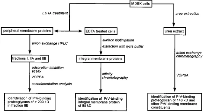
Experimental design.
The experimental approach is
de-picted in Fig. 1. For the isolation and analysis of all cell
mem-brane constituents, urea extraction followed by
anion-ex-change chromatography was performed. To separate integral
and peripheral membrane proteins, cells were treated with
EDTA, which released cell surface structures bound by
diva-lent cations. These molecules were further separated by
anion-exchange HPLC and characterized by adsorption inhibition
assay, cosedimentation analysis, and VOPBA. EDTA-treated
cells were surface biotinylated and extracted in
detergent-con-taining lysis buffer to solubilize integral membrane proteins,
which were further analyzed by affinity chromatography on
immobilized PrV envelope proteins.
Identification of PrV-binding proteoglycans in urea extracts
of MDBK cells.
MDBK cells were extracted in urea extraction
buffer, a buffer especially suitable for the solubilization of
proteoglycans (53), and extracts were fractionated by
anion-exchange chromatography on DEAE-Sepharose in a linear
NaCl gradient. The elution profile is shown in Fig. 2A. When
cellular GAGs had been labeled with Na
235SO
4prior to
ex-traction, radioactively labeled material appeared in fractions
30 to 40, indicating that these fractions contained
proteogly-cans (Fig. 2A). Every second fraction was tested for
PrV-binding proteins in the VOPBA (Fig. 2B). Biotinylated Pr
virions bound to several protein species in fractions eluting at
different salt concentrations. In particular, in fractions 22 to 28
eluting at medium salt concentrations, several PrV-binding
proteins, including proteins of 85 and 90 kDa, were present. In
fractions 30 to 40, which also contained the material which
could be metabolically labeled with Na
235
SO
4
(Fig. 2A), a
protein of approximately 105 kDa was detected, as well as a
smear of material ranging from 116 to 170 kDa with a
maxi-mum intensity at approximately 140 kDa. In addition, material
with a molecular mass of
.
200 kDa was also detected in
fractions 28 to 34. A similar profile was obtained when African
green monkey (Vero) cells were analyzed under identical
con-ditions (data not shown).
To identify proteoglycans among the PrV-binding cell
mem-brane constituents, cells were incubated in medium containing
NaClO
3, which inhibits biosynthesis of cellular GAGs, and
extracted in parallel with the untreated cells analyzed as
de-scribed above. As shown in Fig. 2C, inhibition of GAG
bio-synthesis did not affect or only marginally affected detection of
proteins in fractions 22 to 28 as well as reactivity of the
105-kDa protein in fractions 30 to 32. In contrast, reactivity with
the material resulting in the broad bands in Fig. 2B, as well as
reactivity with the
.
200-kDa species, was completely lost. This
[image:3.612.138.479.74.260.2]result shows that in the VOPBA, biotinylated PrV is able to
interact with cell surface GAGs and that the reactivity is lost
when biosynthesis of GAGs is inhibited. This correlates with a
decrease in attachment of radiolabeled Pr virions on
chlorate-treated cells by 90% compared with unchlorate-treated controls (data
not shown). Reactivity in the VOPBA could also be abolished
when corresponding fractions were digested with
heparinase-heparitinase (data not shown).
FIG. 1. Experimental design. Total membrane proteins of MDBK cells were extracted with urea extraction buffer, separated by anion-exchange chromatography, and analyzed by VOPBA, leading to identification of an approximately 140-kDa PrV-binding proteoglycan and additional PrV-binding membrane constituents. Peripheral membrane proteins were released by treatment of cells with EDTA and separated by anion-exchange HPLC. Analysis by adsorption inhibition assay, VOPBA, and cocentrifugation with purified virions identified.200-kDa PrV-binding HSPG(s). Integral membrane proteins were obtained from EDTA-extracted cells after surface biotinylation and extraction with detergent-containing lysis buffer. They were further analyzed by affinity chromatography on immobilized PrV envelope proteins, resulting in the demonstration of an 85-kDa PrV-binding protein.
on November 9, 2019 by guest
http://jvi.asm.org/
Identification of water-soluble peripheral PrV-binding
pro-teins of MDBK cells.
Treatment of MDBK cells with EDTA
released radioactively labeled material when cells had been
incubated in medium supplemented with Na
235
SO
4
, indicating
the presence of proteoglycans in the EDTA extract.
Fraction-ation by anion-exchange HPLC yielded three major fractions
designated I, IIA, and IIB (Fig. 3A), of which fraction IIB
contained all material that could be labeled by Na
235
SO
4. To
assay for biological activity, abilities of the different fractions to
inhibit attachment of PrV to target cells were assayed. As
shown in Fig. 3B, when different amounts of proteins of
frac-tions I, IIA, and IIB were mixed with radiolabeled Pr virions
and virus attachment was compared with that of an untreated
control, only fraction IIB was able to inhibit attachment of PrV
to MDBK cells in a dose-dependent manner, whereas fractions
I and IIA showed no effect (Fig. 3B). After digestion of
frac-tion IIB with heparinase and an addifrac-tional chromatography
(IIB-H), the inhibitory activity of fraction IIB was eliminated,
indicating that the proteoglycans present in fraction IIB which
are responsible for the inhibition of the attachment of PrV to
MDBK cells carried heparan sulfate GAGs. Again, this result
correlates with attachment studies on intact cells in which
treatment of cell surfaces with heparinase reduced attachment
by approximately two-thirds (36).
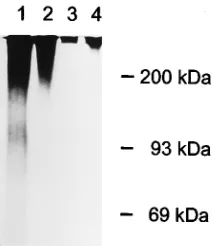
found after degradation with papain (Fig. 4B, lane 5), or
tri-fluoromethanesulfonic acid (Fig. 4B, lane 6). In summary,
these data show that proteoglycans with apparent molecular
masses of
.
200 kDa, predominantly those carrying heparan
sulfate, are able to specifically bind Pr virions in the VOPBA.
Since EDTA-detachable high-molecular-weight HSPGs are
solubilized in the absence of detergent, it was possible to
an-alyze their binding to intact PrV virions in suspension. After
incubation with radiolabeled fraction IIB, wild-type PrV and
PrV-gC
2were sedimented through a 40% sucrose cushion,
and radiolabeled PrV-binding proteins were identified by
flu-FIG. 2. Anion-exchange chromatography of urea extracts on DEAE-Sepha-rose. (A) Elution profile of proteins monitored at 280 nm (——) in a linear NaCl gradient (– – –). · · · , material which could be metabolically labeled by Na2
35
SO4. (B) VOPBA of single fractions (every second fraction) from the
[image:4.612.58.300.71.402.2]chromatography with biotinylated PrV virions after separation in SDS-PAGE. Fraction numbers are given. Positions of molecular weight marker proteins in SDS-PAGE are indicated. (C) VOPBA as in panel B except that cells were grown in medium supplemented by NaClO3to inhibit GAG biosynthesis.
FIG. 3. Anion-exchange HPLC of EDTA extracts from MDBK cells. MDBK cells were extracted with PBS-EDTA, and the extracts were fractionated by anion-exchange HPLC. (A) Elution profile of proteins by the indicated NaCl gradient (– – –) was monitored at 280 nm (——). Three major fractions were obtained and designated I, IIA, and IIB. · · · , material which could be meta-bolically labeled with Na2
35
SO4. The fractions were desalted and further
char-acterized in an adsorption inhibition assay (B) using different amounts of pro-tein. Virus adsorption is indicated in relation to the attachment of untreated control samples. IIB-H indicates fraction IIB after heparinase treatment.
on November 9, 2019 by guest
http://jvi.asm.org/
[image:4.612.317.547.330.655.2]orography after SDS-PAGE of the obtained pellet. As shown
in Fig. 5, lane 2, the high-molecular-weight HSPG did indeed
bind to wild-type Pr virions in suspension. In the presence of 50
m
g of heparin per ml, reactivity of HSPG with Pr virions was
abolished (Fig. 5, lane 4), which again correlates with data
from attachment assays (22). In the absence of gC (Fig. 5, lane
3), HSPGs from fraction IIB were unable to bind to virions,
which is in accordance with data from infectivity experiments
using gC-negative PrV mutants and cell lines deficient in GAG
biosynthesis (23). Labeled material present on top of the gel in
lanes 3 and 4 most likely constitutes insoluble aggregates which
pass through the sucrose cushion. Together, these data identify
HSPG with a high apparent molecular mass of
.
200 kDa
exhibiting characteristics predicted from biological assays.
Identification of an 85-kDa PrV-binding integral membrane
protein.
When urea extracts of MDBK cells were fractionated
by anion-exchange chromatography and single fractions were
analyzed by VOPBA, several proteins were found to be able to
bind biotinylated PrV virions (Fig. 2B and C). For further
characterization of integral PrV-binding membrane proteins,
EDTA-extracted cells (see above) were surface biotinylated
and extracted with detergent-containing lysis buffer.
Solubi-lized integral membrane proteins (Fig. 6, lane 1) were then
reacted by affinity chromatography with immobilized PrV
en-velope (glyco)proteins and sequentially eluted with buffer
con-taining 50
m
g of heparin per ml and 150 mM NaCl (Fig. 6, lane
2), 500 mM NaCl (Fig. 6, lane 3), 1 M NaCl (Fig. 6, lane 4), or
4 M urea (Fig. 6, lane 5). Eluates were analyzed for the
pres-ence of biotinylated proteins after SDS-PAGE and protein
blotting. Under the conditions applied, a single protein species
of 85 kDa was partially eluted by 50
m
g of heparin per ml and
150 mM NaCl (Fig. 6, lane 2). Most of the remaining 85-kDa
protein could be recovered by elution with 500 mM NaCl (Fig.
6, lane 3). Harsher elution conditions did not result in an
increase in the 85-kDa protein, nor did they lead to recovery of
significant amounts of other proteins. Only a weak protein
band of 56 kDa was additionally observed in the 1 M NaCl
eluate (Fig. 6, lane 4). Therefore, from the biotinylated cell
surface proteins remaining after extraction of peripheral
mem-brane constituents with EDTA, an 85-kDa protein which
bound to immobilized PrV envelope proteins was identified.
DISCUSSION
Initiation of infection by alphaherpesviruses is thought to
require a cascade of interactions between different viral and
cellular membrane components (11, 35, 46). In HSV-1, PrV,
and BHV-1, gC mediates primary attachment by interacting
with cell surface components which consist of or contain GAG
(15, 36, 39), in particular heparan sulfate (23, 45, 52). Whereas
gC appears to be the only PrV virion envelope protein capable
of interacting productively with heparan sulfate and thus
lead-ing to infection, in HSV-1 and BHV-1, gB is also able to bind
cell surface GAG (6, 14). Since gC proteins are nonessential
for replication of HSV-1, PrV, and BHV-1, gC
2PrV has to
initiate infection by a gC- and GAG-independent mechanism,
whereas gC
2HSV-1 and BHV-1 may still use a
GAG-depen-dent attachment mechanism.
[image:5.612.94.267.68.201.2]Although the analysis of cell lines of either hamster (CHO)
or murine (L) origin deficient in GAG biosynthesis
demon-strated the importance of these carbohydrates in
alphaherpes-virus adsorption (12, 45), biochemical analyses have so far
been limited to characterization of the requirements of the
GAG for inhibition of virus attachment (31). In this respect,
HSV-1 and PrV have been shown to differ (50), presumably as
[image:5.612.374.500.72.196.2]FIG. 4. Characterization of PrV-binding proteoglycans in fraction IIB by VOPBA. (A) Ten-microgram aliquots of protein of fractions I (lane 1), IIA (lane 2), and IIB (lane 3) were separated by SDS-PAGE and tested in the VOPBA. (B) Ten micrograms of fraction IIB (lane 1) was digested with heparinase-hepariti-nase (lane 2), chondroitiheparinase-hepariti-nase (lane 3), hepariheparinase-hepariti-nase-heparitiheparinase-hepariti-nase and chondroiti-nase (lane 4), papain (lane 5), and trifluoromethanesulfonic acid (lane 6), sep-arated by SDS-PAGE, and tested in the VOPBA.
FIG. 5. Cosedimentation of PrV with radiolabeled peripheral proteoglycans extracted from MDBK cells. Fraction IIB of the EDTA extract was metabolically labeled with Na2
35
SO4(lane 1). After incubation with wild-type PrV (lane 2) or
PrV-gC2(lane 3) virions for 1 h on ice, virions were sedimented through a 40% sucrose cushion. In lane 4, incubation of wild-type PrV with radiolabeled fraction IIB was performed in the presence of 50mg of heparin per ml. Proteins that had bound to the virions were analyzed by SDS-PAGE and autoradiography.
FIG. 6. Affinity chromatography of biotinylated integral membrane proteins from MDBK cells on a PrV affinity matrix. Membrane extracts from gradient-purified wild-type PrV virions were immobilized on Sepharose, and biotinylated integral membrane proteins (lane 1) from MDBK cells were applied. Unbound material was removed by repeated washes with TC and TC150. Bound proteins were sequentially eluted with TC150 supplemented with 50mg of heparin per ml (lane 2), TC500 (lane 3), TC1000 (lane 4), and 4 M urea (lane 5). Eluates were collected, concentrated by ultracentrifugation, and separated by SDS-PAGE. After blotting to nitrocellulose, biotinylated proteins were detected after incu-bation with streptavidin-peroxidase by chemiluminescence.
on November 9, 2019 by guest
http://jvi.asm.org/
[image:5.612.127.234.542.665.2]competing heparin than that of wild-type PrV. A functional
attachment domain has been mapped to the amino-terminal
one-third of PrV gC (amino acids 25 to 157 [10]), indicating
that both domains within gC mediate distinct interactions. In
addition, glycoprotein D of PrV, HSV-1, and BHV-1 has
sub-sequently been shown to be required for secondary stable
at-tachment (22, 33). For several years gD has been proposed to
interact with a cell surface protein on the basis of the finding
that soluble gD can inhibit penetration of HSV-1 by binding to
a limited number of sites on the cell surface (18). Recently,
soluble truncated HSV-1 gD has been shown to bind to the
mannose 6-phosphate receptor in a ligand blotting assay (5).
Binding to this protein has also been suggested for
varicella-zoster virus (54). However, the significance of this interaction
is unclear. In a photoaffinity labeling study, purified BHV-1
and an anti-idiotypic antibody which mimics a BHV-1 gD
epitope have been used to screen cell surfaces for BHV-1- or
BHV-1 gD-binding proteins. A 60-kDa protein could thus be
found only in bovine cell lines which are permissive for BHV-1
infection (48).
Since these studies indicated the interaction of several virion
proteins with different cell surface molecules, we used two
techniques, i.e., VOPBA (2, 8, 32, 40) and cosedimentation,
which allow us to study the attachment of intact PrV virions to
isolated cell membrane components, a prerequisite to detect
virus-cell interactions involving more than one viral envelope
protein. Standard procedures like affinity chromatography or
precipitation of receptor-ligand complexes might fail under
these circumstances, since experimental conditions (presence
of detergents, low concentration of ligands, absence of tertiary
structure, absence of lipid envelope) might not support such a
complex process. Differential extraction led to the isolation of
total, peripheral, and integral cell membrane components.
In-fectivity assays proved that biotinylation under our conditions
did not decrease infectivity of the virus preparation by more
than twofold, indicating that the topology of the viral envelope
was not overtly disturbed. Our studies identified two species of
HSPG which differ in molecular weight and membrane
distri-bution. A
.
200-kDa HSPG can be released from the cell
surface with EDTA, indicating that it represents a peripheral
membrane constituent attached to the cell via divalent cations.
Several proteoglycans with this property have been described
(16). In addition, a presumably integral HSPG of ca. 140 kDa
with the capability to bind PrV virions in the VOPBA has been
identified.
Binding to HSPG was sensitive to heparinase or proteinase
treatment, to competition by exogenous heparin, and to growth
of cells in chlorate medium. In cosedimentation, PrV gC
2was
deficient in binding to high-molecular-weight HSPG compared
with wild-type PrV. Therefore, inhibition of reactivity in
VOPBA and lack of cosedimentation with HSPG correlated
with inhibition of attachment of PrV to intact cells, indicating
that the conditions in the VOPBA reflect the situation with
respect to attachment of PrV to cultured cells, at least
regard-anchored HSPG also play a role in PrV attachment and
whether the 140-kDa HSPG is indeed GPI anchored is
cur-rently under investigation.
Since interaction of virion gC with cellular HSPG probably
represents only the first step in the infection of cells by
wild-type PrV, cellular extracts were also analyzed for PrV-binding
components other than HSPG. Among several proteins in urea
extracts of cell membranes, a 85-kDa polypeptide was found to
be capable of binding to biotinylated PrV virions in VOPBA. A
protein of identical molecular mass could also be identified by
affinity chromatography of cell membrane extracts obtained by
mild detergents on immobilized virus envelope proteins. Part
of the bound protein eluted in the presence of 50
m
g of heparin
per ml at physiological conditions (150 mM NaCl); part of the
protein required 500 mM NaCl, which indicates an
electro-static interaction between virion protein(s) and the 85-kDa
protein, which might thus act as an additional PrV receptor.
We are currently analyzing whether this 85-kDa protein
cor-relates with the common saturable receptor for PrV and
HSV-1 found on Vero cells which is different from heparan
sulfate (26).
The process of initiation of infection by alphaherpesviruses
is still poorly understood. Glycoproteins gB, gC, and gD have
been implicated in receptor binding (29, 35, 46), and various
cellular virus-binding proteins have been described (4, 19, 52).
On the basis of the currently available data, we hypothesize
that the following events may occur during attachment and
infectious entry of PrV. Interaction between virion gC and
peripheral high-molecular-weight HSPG (capture receptor)
leads to a primary binding of virion to target cells. This binding
is stabilized through interaction of gC with the 140-kDa
inte-gral membrane HSPG, a process which might be defective in
an internal PrV-gC
2mutant lacking amino acids 157 to 290
(55). Interaction of virion gD with another cell membrane
component, perhaps related to the 85-kDa protein described
here, then further strengthens binding of virus to target cells
and initiates fusion between viral and cellular membranes. It
needs to be emphasized that except for the initial conversion of
heparin-sensitive to heparin-resistant attachment (22), it is
un-clear whether these postulated interactions occur sequentially
or simultaneously.
It appears conceivable that even more viral protein-cellular
receptor interactions might take place, and in the VOPBA
several other protein species besides the
.
200- and 140-kDa
HSPGs and the 85-kDa protein bound to intact biotinylated
PrV virions, although the significance of these interactions
remains unknown. In the VOPBA, the original conformation
of the virion envelope is retained to a large degree, as shown by
only a very modest reduction in viral infectivity after
biotiny-lation. In contrast, fractionation of cell membranes leads to
disruption of higher-order structure between cell membrane
components. However, the availability of virus mutants devoid
of either essential or nonessential virion envelope
glycopro-teins allows one to assay the contribution of single viral surface
on November 9, 2019 by guest
http://jvi.asm.org/
proteins to the first steps of virus infection, and a combination
of genetical approaches (1, 12) with the biochemical
tech-niques described here will help to characterize the respective
cellular reaction partners.
ACKNOWLEDGMENTS
This study was supported by grant Me 854/2-3 from the Deutsche Forschungsgemeinschaft and grant ERB-CHRX-CT92-0029 from the EEC.
REFERENCES
1. Banfield, B., Y. Leduc, L. Esford, K. Schubert, and F. Tufaro. 1995. Sequen-tial isolation of proteoglycan synthesis mutants by using herpes simplex virus as a selective agent: evidence for a proteoglycan-independent virus entry pathway. J. Virol. 69:3482–3489.
2. Boyle, J., D. Weismiller, and K. Holmes. 1987. Genetic resistance to mouse hepatitis virus correlates with absence of virus-binding activity on target tissues. J. Virol. 61:185–189.
3. Brown, D., and J. K. Rose. 1992. Sorting of GPI-anchored proteins to glycolipid-enriched membrane subdomains during transport to the apical cell surface. Cell 68:533–544.
4. Brunetti, C. R., R. L. Burke, B. Hoflack, T. Ludwig, K. Dingwell, and D.
Johnson.1995. Role of mannose 6-phosphate receptors in herpes simplex virus entry into cells and cell-to-cell transmission. J. Virol. 69:3517–3528. 5. Brunetti, C. R., R. L. Burke, S. Kornfeld, W. Gregory, F. R. Masiarz, K. S.
Dingwell, and D. C. Johnson.1994. Herpes simplex virus glycoprotein D acquires mannose 6-phosphate residues and binds to mannose 6-phosphate receptors. J. Biol. Chem. 269:17067–17074.
6. Byrne, K. M., D. Horohov, and K. Kousoulas. 1995. Glycoprotein B of bovine herpesvirus-1 binds heparin. Virology 209:230–235.
7. Compton, T., D. Nowlin, and N. R. Cooper. 1993. Initiation of human cyto-megalovirus infection requires initial interaction with cell surface heparan sulfate. Virology 193:834–841.
8. Crane, S., J. Buzy, and J. Clements. 1991. Identification of cell membrane proteins that bind visna virus. J. Virol. 65:6137–6143.
9. Edge, A. S., C. R. Faltynek, L. Hof, L. E. Reichert, and P. Weber. 1981. Deglycosylation of glycoproteins by trifluoromethanesulfonic acid. Anal. Biochem. 118:131–137.
10. Flynn, S., B. Burgett, D. Stein, K. Williamson, and P. Ryan. 1993. The amino-terminal one-third of pseudorabies virus glycoprotein gIII contains a functional attachment domain, but this domain is not required for the effi-cient penetration of Vero cells. J. Virol. 67:2646–2654.
11. Fuller, A. O., and W.-C. Lee. 1992. Herpes simplex virus type 1 entry through a cascade of virus-cell interactions requires different roles of gD and gH in penetration. J. Virol. 66:5002–5012.
12. Gruenheid, S., L. Gatzke, H. Meadows, and F. Tufaro. 1993. Herpes simplex virus infection and propagation in a mouse L cell mutant lacking heparan sulfate proteoglycans. J. Virol. 67:93–100.
13. Hascall, V. C., and J. H. Kimura. 1982. Proteoglycans: isolation and char-acterisation. Methods Enzymol. 82:769–800.
14. Herold, B. C., R. Visalli, N. Susmarski, C. Brandt, and P. G. Spear. 1994. Glycoprotein C-independent binding of herpes simplex virus to cells requires cell surface heparan sulphate and glycoprotein B. J. Gen. Virol. 75:1211– 1222.
15. Herold, B. C., D. WuDunn, N. Soltys, and P. G. Spear. 1991. Glycoprotein C of herpes simplex virus type 1 plays a principal role in the adsorption of virus to cells and in infectivity. J. Virol. 65:1090–1098.
16. Ho¨o¨k, M.1984. Cell-surface glycosaminoglycans. Annu. Rev. Biochem. 53: 847–869.
17. Inghirami, G., M. Nakamura, J. Balow, A. Notkins, and P. Casali. 1988. Model for studying virus attachment: identification and quantitation of Ep-stein-Barr virus-binding cells by using biotinylated virus in flow cytometry. J. Virol. 62:2453–2463.
18. Johnson, D., R. L. Burke, and T. Gregory. 1990. Soluble forms of herpes simplex virus glycoprotein D bind to a limited number of cell surface recep-tors and inhibit virus entry into cells. J. Virol. 64:2569–2576.
19. Kaner, R. J., A. Baird, A. Mansukhani, C. Basilico, B. Summers, R.
Flork-iewicz, and D. Hajjar.1990. Fibroblast growth factor receptor is a portal of cellular entry for herpes simplex virus type 1. Science 248:1410–1413. 20. Kaplan, A. S., and T. Ben-Porat. 1961. The action of 5-fluorouracil on the
nucleic acid metabolism of pseudorabies virus-infected and non-infected rabbit kidney cells. Virology 13:78–92.
21. Kaplan, A. S., and A. Vatter. 1959. A comparison of herpes simplex and pseudorabies viruses. Virology 7:394–407.
22. Karger, A., and T. C. Mettenleiter. 1993. Glycoproteins gIII and gp50 play dominant roles in the biphasic attachment of pseudorabies virus. Virology
194:654–664.
23. Karger, A., A. Saalmu¨ller, F. Tufaro, B. W. Banfield, and T. C. Metten-leiter. 1995. Cell surface proteoglycans are not essential for infection by
pseudorabies virus. J. Virol. 69:3482–3489.
24. Laemmli, U. K. 1970. Cleavage of structural proteins during the assembly of the head of bacteriophage T4. Nature (London) 227:680–685.
25. Langeland, N., and L. J. Moore. 1990. Reduction of HSV-1 binding to BHK cells after treatment with phosphatidylinositol-specific phospholipase C. FEBS Lett. 277:253–256.
26. Lee, W.-C., and A. O. Fuller. 1993. Herpes simplex virus type 1 and pseu-dorabies virus bind to a common saturable receptor on Vero cells that is not heparan sulfate. J. Virol. 67:5088–5097.
27. Li, Y., S. van Drunen Littel-van den Hurk, L. Babiuk, and X. Liang. 1995. Characterization of cell binding properties of bovine herpesvirus 1 glycop-roteins B, C, and D: identification of a dual cell-binding function of gB. J. Virol. 69:4758–4768.
28. Liang, X., L. Babiuk, and T. Zamb. 1993. Mapping of heparin-binding structures on bovine herpesvirus 1 and pseudorabies virus gIII glycoproteins. Virology 194:233–243.
29. Liang, X., L. A. Babiuk, S. van Drunen Littel-van den Hurk, D. R.
Fitz-patrick, and T. J. Zamb.1991. Bovine herpesvirus 1 attachment to permis-sive cells is mediated by its major glycoproteins gI, gIII, and gIV. J. Virol. 65: 1124–1132.
30. Ligas, M. W., and D. C. Johnson. 1988. A herpes simplex virus mutant in which glycoprotein D sequences are replaced byb-galactosidase sequences binds to but is inable to penetrate into cells. J. Virol. 62:1486–1494. 31. Lycke, E., M. Johansson, B. Svennerholm, and U. Lindahl. 1991. Binding of
herpes simplex virus to cellular heparan sulphate, an initial step in the adsorption process. J. Gen. Virol. 72:1131–1137.
32. Maisner, A., J. Schneider-Schaulies, K. Liszewski, J. Atkinson, and G.
Her-rler.1994. Binding of measles virus to membrane cofactor protein (CD46): importance of disulfide bonds and N-glycans for the receptor function. J. Virol. 68:6299–6304.
33. McClain, D., and A. O. Fuller. 1994. Cell-specific kinetics and efficiency of herpes simplex virus type 1 entry are determined by two distinct phases of attachment. Virology 198:690–702.
34. Meier, T., S. Arni, S. Malarkannan, M. Poincelet, and D. Hoessli. 1992. Immunodetection of biotinylated lymphocyte-surface proteins by enhanced chemiluminescense: a nonradioactive method for cell-surface protein anal-ysis. Anal. Biochem. 204:220–226.
35. Mettenleiter, T. C. 1994. Initiation and spread ofa-herpesvirus infections. Trends Microbiol. 2:2–4.
36. Mettenleiter, T. C., L. Zsak, F. Zuckermann, N. Sugg, H. Kern, and T.
Ben-Porat.1990. Interaction of glycoprotein gIII with a cellular heparinlike substance mediates adsorption of pseudorabies virus. J. Virol. 64:278–286. 37. Mirda, D. P., D. Navarro, P. Paz, P. L. Lee, L. Pereira, and L. T. Williams. 1992. The fibroblast growth factor receptor is not required for herpes sim-plex virus type 1 infection. J. Virol. 66:448–457.
38. Muggeridge, M., G. Cohen, and R. Eisenberg. 1992. Herpes simplex virus infection can occur without involvement of the fibroblast growth factor receptor. J. Virol. 66:824–830.
39. Okazaki, K., T. Matsuzaki, Y. Sugahara, J. Okada, M. Hasebe, Y. Iwamura,
M. Ohnishi, T. Kanno, M. Shimizu, E. Honda, and Y. Kono.1991. BHV-1 adsorption is mediated by the interaction of glycoprotein gIII with heparin-like moiety on the cell surface. Virology 181:666–670.
40. Ravindranath, R., and M. Graves. 1990. Attenuated murine cytomegalovirus binds to N-acetylglucosamine, and shift to virulence may involve recognition of sialic acids. J. Virol. 64:5430–5440.
41. Roderiquez, G., T. Oravecz, M. Yanagishita, D. Chequer Bou-Habib, H.
Mostowski, and M. A. Norcross.1995. Mediation of human immunodefi-ciency virus type 1 binding by interaction of cell surface heparan sulfate proteoglycans with the V3 region of envelope gp120-gp41. J. Virol. 69: 2233–2239.
42. Sawitzky, D., H. Hampl, and K.-O. Habermehl. 1990. Comparison of hepa-rin-sensitive attachment of pseudorabies virus (PRV) and herpes simplex virus type 1 and identification of heparin-binding PRV glycoproteins. J. Gen. Virol. 71:1221–1225.
43. Schreurs, C., T. C. Mettenleiter, F. Zuckermann, N. Sugg, and T. Ben-Porat. 1988. Glycoprotein gIII of pseudorabies virus is multifunctional. J. Virol. 62: 2251–2257.
44. Shieh, M.-T., and P. G. Spear. 1991. Fibroblast growth factor receptor: does it have a role in the binding of herpes simplex virus? Science 253:208–209. 45. Shieh, M.-T., D. WuDunn, R. Montgomery, J. Esko, and P. G. Spear. 1992. Cell surface receptors for herpes simplex virus are heparan sulfate proteo-glycans. J. Cell Biol. 116:1273–1281.
46. Spear, P. G. 1993. Entry of alphaherpesviruses into cells. Semin. Virol. 4: 167–180.
47. Springer, B. A., M. W. Pantoliano, F. A. Barbera, P. L. Gunyuzlu, L. D.
Thompson, W. F. Herblin, S. A. Rosenfeld, and G. W. Book.1994. Identifi-cation and concerted function of two receptor binding surfaces on basic fibroblast growth factor required for mitogenesis. J. Biol. Chem. 269:26879– 26884.
48. Thaker, S. R., D. L. Stine, T. J. Zamb, and S. Srikumaran. 1994. Identifi-cation of a putative cellular receptor for bovine herpesvirus 1. J. Gen. Virol.
75:2303–2309.