`“CHRONIC RECURRENT ABDOMINAL PAIN AND
EVALUATION OF DISEASE BY VARIOUS
DIAGNOSTIC MODALITIES”
Dissertation submitted to
The Tamil Nadu M.G.R Medical University
Chennai- 600032
In partial fulfillment of the Regulations of the award of degree of
M.S. General Surgery
DEPARTMENT OF GENERAL SURGERY
Coimbatore Medical College Hospital
Coimbatore – 641018
CERTIFICATE
This is to certify that this dissertation titled “CHRONIC RECURRENT ABDOMINAL PAIN AND EVALUATION OF
DISEASE BY VARIOUS DIAGNOSTIC MODALITIES” submitted
to the Tamil Nadu Dr. M.G.R. Medical University, Chennai in partial fulfillment of the requirement for the award of M.S Degree Branch - I (General Surgery) is a bonafide work done by Dr. BALA MURUGAN.A
post graduate student in General Surgery under my direct supervision and guidance during the period of September 2011 to November 2012.
Dr.Vimala, M.D.
Dean,
Coimbatore Medical College Hospital Prof.S.Natarajan, M.S.
Associate Professor
Dept. of General Surgery
Coimbatore Medical College Hospital
Prof. P.V. Vasantha Kumar, M.S.
Professor and Head of the Department
Dept. of general Surgery
DECLARATION
I hereby declare that the dissertation entitled “CHRONIC RECURRENT ABDOMINAL PAIN AND EVALUATION OF
DISEASE BY VARIOUS DIAGNOSTIC MODALITIES” was done
by me at Coimbatore Medical College Hospital, Coimbatore – 641018 during the period of my post graduate study for M.S. Degree Branch-1 (General Surgery) from 2010 to 2013.
This dissertation is submitted to the Tamil Nadu Dr. M.G.R. Medical University in partial fulfillment of the University regulations for award of M.S., Degree in General Surgery.
Dr. BALA MURUGAN. A
Post Graduate Student M.S. General Surgery
ACKNOWLEDGEMENT
It gives me immense pleasure to express my deep sense of gratitude to my Unit Chief Prof. Dr. S. NATARAJAN, M.S. General Surgery, Department of General Surgery, Coimbatore Medical College Hospital, for his excellent guidance and valuable suggestions during the course of study and in preparation of this dissertation.
I am grateful to Prof. Dr. P.V.VASANTHA KUMAR, M.S.,
Professor and Head of the Department of General Surgery, Coimbatore Medical College Hospital , for his guidance throughout this study. I also express my heartfelt thanks for the associate professors of the college,
Dr.Elango, Dr. Swaminathan, Dr. Ranganathan, Dr.Ravindran and
Dr. Saradha for their suggestions at the apt time that has helped me in the completion of this work.
I am grateful to my Assistant Professors, Dr.S. DURAIRAJ, M.S.,
Dr. MEENA, M.S., for their help and guidance throughout this study.
I express my gratitude to Dr.VIMALA, Dean, Coimbatore Medical College Hospital for permitting me to use the clinical material for the study.
I express my thanks to my friends and all others who have helped me in the preparation of this dissertation.
LIST OF ABBREVIATIONS
CSF - Cerebro Spinal Fluid
CCK - Cholecystokinin
CT - Computerised Tomography
D Cells - Delta Cells
ELISA - Enzyme linked Immuno Sorbent Assay GI - Gastro Intestinal
H2 Receptors - Histaminic
H Pylori - Helicobacter Pylori
Ig - Immunoglobulin
L Appendix - Laparoscopic Appendicetomy LA - Local Anaesthesia
Los - Lower Oesophageal Sphinter PPI - Proton Pump Inhibitors LFT - Liver Function Tests
NSAIDS - Non Steroidal Anti Inflammatory Drugs OGD Scopy - Oesophago gastro Duodenoscopy
SAAG - Serum Asctic Albumin Gradient
ABSTRACT
`“CHRONIC RECURRENT ABDOMINAL PAIN AND
EVALUATION OF DISEASE BY VARIOUS DIAGNOSTIC
MODALITIES”
OBJECTIVES :
This study is done to make a definitive diagnosis by using various
diagnostic modalities available at Coimbatore Medical College Hospital and to
identify the Common etiologies for Chronic Recurrent Pain.
METHODS
This study was conducted at Coimbatore Medical College Hospital. A
sample size of 50 patients with complaints of Chronic Recurrent pain abdomen
were taken into this study. The patients with Acute Abdomen, patients
undergoing previous surgeries and urological and Gynaecological causes were
excluded from this Study.
RESULTS
Of the total 50 patients studied, it was found that the incidence was more
commoner in males, and patients between 31-40 years were commonly
affected.16 of the 50 patients were Alcoholics for more than 10 years and
Chronic smokers and vegetarians were less commonly affected than people
taking mixed diet. The patients with low Socio Economic status were
commonly affected in this study. This study also showed that OGD scopy
revealed the diagnosis in patients with peptic ulcer disease, GORD and
Gastritis, CT was confirmatory in case of Chronic pancreatitis and colonoscopy
Neoplastic Growth. Of the total 50 patients 43 patients were treated
conservatively of which 17 got relieved of their symptoms and 7 patients were
subjected to surgical treatment of which 4 patients got relieved of their
symptoms.
CONCLUSION
The common etiology for chronic recurrent pain abdomen in adults at
Coimbatore Medical College Hospital was peptic ulcer, chronic pancreatitis,
Gastritis, GORD, TB abdomen followed by malignancy. For patients with upper
abdominal pathology, endoscopy proves to be a confirmatory diagnosis and for
patients with lower abdominal pathology CT abdomen and diagnostic
Laparoscopy gives better information in arriving at a diagnosis. For 5 out of
total 50 patients, definitive diagnosis could not be established inspite of all
investigations. These patients were classified as chronic intractable abdominal
pain, which is found to be more common in females. They were given placebo
and referred to psychiatrists for further management.
KEY WORDS
Chronic Recurrent Abdominal Pain, Peptic Ulcer, Chronic Pancreatitis,
GORD, Gastritis, TB Abdomen, Chronic Appendicitis, USG Abdomen, CT
CONTENTS
S.NO TOPIC PAGE NO.
1 INTRODUCTION 1
2 AIMS & OBJECTIVES 2
3 REVIEW OF LITERATURE 3
4 MATERIALS & METHODS 67
5 OBSERVATIONS 69
6 DISCUSSION 85
7 SUMMARY 88
8 CONCLUSION 90
9 BIBLIOGRAPHY
10 PROFORMA
INTRODUCTION
Chronic abdominal pain is defined as constant or intermittent pain persisting for more than 3 months.
Intermittent pain may be referred to as recurrent abdominal pain with intervals of asymptomatic periods.
It presents a diagnostic and treatment challenge to the general surgeons and it remains a common surgical problem.
Specific diagnosis could not be arrived despite all necessary investigations.
The cost of health care is high, due to need for sophisticated investigations and the treatment cost.
AIMS AND OBJECTIVES
To make a definitive diagnosis by using various diagnostic modalities available at Coimbatore Medical College Hospital.
To identify the common etiologies for chronic recurrent pain.
REVIEW OF LITERATURE
About 70 years ago Hutchinson stated that in the process of treating chronic pain abdomen the important thing is to diagnose the patient at an earlier time. If she has set her feet on the slippery slope which leads to successive operations she is undone.
VARIOUS CAUSES OF CHRONIC RECURRENT PAIN ABDOMEN
PAIN ARISING FROM VISCERA
Peptic ulcer
Gastro esophageal reflux disease
Gastritis due to Helicobacter pylori
Ulcer due to intake of Non-steroidal anti-inflammatory drugs
Inflammatory bowel disease
Neoplasia and psychiatric cause
Chronic pancreatitis
Chronic appendicitis
PAIN ARISING FROM ABDOMINAL WALL
• Iatrogenic peripheral nerve injuries
• Hernias
• Pain abdomen of spinal origin
• Spontaneous rectus sheath hematoma
PSYCHIATRIC CAUSES
UROLOGICAL CAUSES
GYNAECOLOGICAL CAUSES
CLINICAL EXAMINATION
With patient on supine position abdomen palpated in the usual manner
Over the tender spot with the palpating fingers of the surgeon
The patient is asked to raise his head from the bed,there by contracting the muscles of abdomen
Once the muscles get tensed, the surgeon reapplies his fingers and asked the patient if the pain has changed
The patient will have decreased pain or tenderness, because the tensed muscle will shield the intra-abdominal viscera, there by the pain will be
decreased.
Commonest chronic abdominal pain encountered clinically are recurrent peptic ulcer diseases, chronic pancreatitis, GERD, gastritis, TB abdomen and malignancy.
CHRONIC INTRACTABLE ABDOMINAL PAIN
Chronic intractable abdominal pain is defined as abdominal pain that persists for 4 to 6 months when there is no definite pathological diagnosis made after proper medical history and investigation.
PEPTIC ULCER
ANATOMY
1. The stomach is an elegant organ described by Wallace P.Ritchie Jr.
2. Stomach develops during 5th week of gestation as a dilatation of embryonic foregut.
3. The stomach occupies the left upper quadrant of the abdomen. The parts of stomach are lesser curvature, greater curvature, fundus,incisura angularis, body, pyloric portion.
BLOOD SUPPLY OF STOMACH
ARTERIES
2. The Right gastric artery-A branch of common hepatic artery, supplies stomach with numerous branches along the lesser curvature and anastomosis with left gastric artery.
3. The Right gastro epiploic artery, a branch of gastro duodenal artery supplies the greater curvature of stomach and anastomosis with left gastro epiploic artery. It is the source of bleeding in duodenal ulcer.
4. The left gastro epiploic artery-A branch of splenic artery supplies greater curvature, numerous short branches (5to7) also supplies the fundus.
LYMPHATIC DRAINAGE OF STOMACH
Lymph nodes draining the stomach are 1. Hepatic group.
2. Sub pyloric nodes. 3. Gastric group
a. superior group b. Inferior group. 4. Pancreaticolineal group.
COURSE OF VAGUS AND INNERVATION OF STOMACH
THE ANTERIOR VAGUS NERVE
In the abdomen, the anterior vagus gives a branch to pyloric antrum, hepatic flexure, fundal branches, in the lesser omentum as Nerve of Laterjet and supplies acid secreting parts of stomach.
CROW’S FOOT
From 5-7cms proximal to pylorus, anterior vagus nerve divides into branches, this division has been called as CROW'S FOOT, the pyloric antrum being supplied by these the most.
THE POSTERIOR VAGUS NERVE
It gives a branch to coeliac ganglion and supplies antrum.
PHYSIOLOGY
GASTRIC PEPTIDES
GASTRIN
It is produced by G cells in the gastric antrum. Release of gastrin is facilitated by food in contact with stomach. Gastrin secretion is inhibited by Somatostatin and luminal acid. It plays a major role in gastric phase of acid secretion and gastric mucosal defense system, preventing gastric injury from luminal irritants.
SOMATOSTATIN
It is produced by D cells, stimulus is antral acidification, inhibitory being acetyl choline from vagus. It inhibits parietal cell acid secretion.
Other gastric peptides are histamine, ghrelin, gastrin releasing peptide.
STIMULATED ACID SECRETION
CEPHALIC PHASE
GASTRIC PHASE
It is stimulated by mechanical distension of stomach, there by activating stretch receptors to elicit vasovagal reflex. Food interacts with antral G cells there by stimulating gastrin release. It is responsible for 60 to70% of acid output.
INTESTINAL PHASE
It accounts for 10% of the acid secreted in response to meal and is mediated by entry of chyme into small bowel.
GASTRIC BARRIER FUNCTION
The major determining factor in maintaining gastric mucosal defense is the amount of blood flow. When there is 50% reduction in gastric mucosal blood flow, the effects are minimal on the gastric mucosa, however marked mucosal injury occurs when blood flow is decreased to 75%.Restitution is a process by which damaged mucus cells are replaced by surface mucus cells migration along the basement membrane.
ulcers are caused by decreased defensive factors, increase aggressive factors or decrease in both.
EPIDEMIOLOGY
Peptic ulcer disease is more common in females due to increased smoking habits and increased ingestion of NSAIDS and decrease among males the reason being unknown. The incidence of gastric ulcer remains increased during these years, but incidence of duodenal ulcer seems to be decreasing. Patients seek admission for complication of gastric ulcer such as bleeding, perforation,this may be due to increased intake of non-steroidal anti-inflammatory drugs.
PATHOGENESIS
HELICOBACTER PYLORI INFECTION
receptors, for recognition of H pylori, thus it can survive only in gastric epithelium.
Mechanism by which H pylori initiates gastric injury are
1. By toxic mediators.
2. Immune response inducted locally.
3. Increased acid secretion due to increased gastrin level.
TOXIC MEDIATORS
Toxic mediators are ammonia (from urease activity), mucinase-degrades mucus, cytotoxins, epithelial cells damage induced by phospholipases, and mucosal injury induced by platelet activating factor. In addition gastro intestinal injury is also caused by mucosal immune response caused by H pylori.
Duodenal ulcer (90%) , Gastric ulcer (60-90%), Acute and Chronic Gastritis , Gastric Cancer. For all These Diseases H. pylori Plays a Major Etiological Factor. Duodenum is commonly affected in H.pylori Infection.
NON STEROIDAL ANTI- INFLAMMATORY DRUGS:
complications. These patients often gives History of Associated Use Of Anticoagulants, Steroids and a poor GI Event .
NSAIDS Induces Both Acute and Chronic Gastro duodenal Injuries Where Acute Lesions Appear Within Two Weeks of NSAIDS Ingestions Resulting in Gastric Erosions and Hyperemic Mucosa , Whereas Chronic Injury Appears one Month After Ingestion ,manifesting as ulcerations or erosions in The Gastric Antrum and Duodenum. Stomach is Commonly Affected by NSAIDS.
PATHOPHYSIOLOGY OF DUODENAL ULCER:
Duodenal Ulcer is caused by multiple etiologies. Altered secretory mechanisms play a major role in causation of duodenal ulcer, which is due to increased duodenal acid load, increased day time and nocturnal acid secretion and decreased secretion of bicarbonates. Duodenal ulcer is caused due to interaction of H pylori and NSAIDS and pepsin and acid secretion.
GASTRIC ULCER PATHO PHYSIOLOGY
2. And type 11 gastric ulcers constitute 15% located in the body of stomach associated with duodenal ulcers, caused due to increased acid secretion.
3. Pre pyloric ulcers are type III gastric ulcers constituting 20% of the lesions, caused due to increased acid secretion.
4. Type IV gastric ulcers occurs high near the esophago gastric secretion along the lesser curvature.
People between the ages of 55to65 years are commonly affected and people in low socio economic status are commonly affected. The exact cause of gastric ulcer is still unknown. Predisposing conditions are females, age more than 40, NSAIDS or aspirin ingestion, gastritis, delayed gastric emptying causing gastric stasis, infection with H pylori, chronic alcohol and smoking, long term infections and steroid therapy.
DUODENAL ULCER
CLINICAL FEATURES
1. Pain Abdomen
2. Perforation
The incidence of duodenal ulcer perforation is 5%, where the ulcer of duodenum penetrates into the peritoneal cavity causing peritonitis, creating a surgical emergency where immediate surgical intervention is needed.
3. Bleeding
Bleeding from duodenal ulcer constitutes 25% of all GI bleeding, and it is the most common cause of death in patients with peptic ulcer disease. The source of bleeding is from gastro duodenal artery, where duodenal ulcer has eroded it.
4. Obstruction
Gastric outlet obstruction is manifested due to acute inflammation of duodenum, manifested by vomiting and a delayed emptying of stomach .The prolonged vomiting may leads to hypokalemic hypocholeremic metabolic alkalosis due to loss of potassium, chloride, hydrogen from gastric juice loss.
GASTRIC ULCER
1. Type II and III gastric ulcer manifests as bleeding, where as massive bleeding may be a presentation of type IV gastric ulcer. 2. Type II OR III gastric ulcer can presents with gastric outlet
obstruction.
3. Only 8to20% patients require surgical interventions from complications of gastric ulcer.
DIAGNOSIS
All routine investigations should be done such as LFT, Creatinine, complete blood count, calcium levels. A serum gastrin level should be done. X ray chest in erect view should be done to rule out perforation.
1. Upper GI radiography and OGD scopy are the best investigations for diagnosing peptic ulcer disease.
2. HELICO BACTER PYLORI TESTING
It may be invasive and non-invasive. Rapid urease test, culture and histology are the invasive tests where endoscopy is required. Serology and carbon labeled urea breath test are non-invasive tests.
3. SEROLOGY
eradication after treatment. Rapid office based immune assays and ELISA are available serological tests.
4. UREA BREATH TEST
It is a non-invasive test having 95% sensitivity to detect H pylori, and best test to document eradication. It samples the entire stomach and is cheap. Principle of this test is ability of H pylori to hydrolyse urea. The patient is asked to take carbon isotope labeled urea using 13c or 14c .After taking carbon isotope, urea will be broken to ammonia and labeled bicarbonate is excreted in the patients breath as carbon di oxide, quatified by mass spectrometry.
5. RAPID UREASE ASSAY
This method has a specificity of 98% and sensitivity of 90% and within few hours results can be arrived. The principle of this test is the ability of H pylori to hydrolyse urea. Endoscopy is required for this test.
6. HISTOLOGY
H Pylori IN GASTRIC MUCOSA
7. CULTURE:
By mean of endoscopy culture can be done after obtaining a bit of Gastric mucosa. However it is expensive and requires expertise. The sensitivity and specificity are 80% and 100% respectively.
8. UPPER GASTROINTESTINAL RADIOGRAPHY
This is the picture of benign appearing gastric ulcer protruding
medially from lesser curvature of stomach.
9.FIBER OPTIC ENDOSCOPY
The best investigation for diagnosing gastric ulcer is endoscopy. The sensitivity approaches 90% if multiply biopsies are performed for cytology. Benign ulcer have regular, edges rounded, flat and ulcer base is smooth, whereas malignancy appears as mass protruding into the lumen, on the folds may be nodular, Fused clubbed surrounding the ulcer crater.
ENDOSCOPIC VIEW OF GASTRIC ULCER
TREATMENT
MEDICAL MANAGEMENT
1. It involves lifestyle modifications, stoppage of cigarette and Alcohol, discontinuing of aspirin or NSAIDS, Neutralization of Acid secretion, H.pylori eradication.
ANTACIDS
H2 RECEPTOR ANTAGONISTS
These are similar to Histamine. The potent drug is famotidine. H2 Receptor Antagonists administered by continues infusion produces more uniform acid inhibition. Studies done indicate that H2 Receptor Antagonists after 4 weeks of therapy has 70 to 80% healing rates whereas after 8 weeks of therapy, 80 to 90% healing rates observed.
PROTON PUMP INHIBITORS
Proton pump inhibitors are the potent anti-secretory agents classified under benzimidazoles. They are more potent than H2 receptor antagonists by binding to catalytic alpha sub unit of proton pump. Studies have showed that after 4 weeks PPIS produces 85% healing rates and after 8 weeks it produces 96% healing rates. Concurrent use of proton pump inhibitors, antacids and H2 Receptor Antagonists is contraindicated.
TREATMENT OF H PYLORI INFECTION
Consideration should be given to the management of below three, in the treatment of H.pylori infection
a. Symptoms b. Ulcer
The symptoms and ulcer healing can be achieved by use of Anti Secretory agents. With stoppage of NSAIDS, Recurrence can be prevented. Patients have 72% Healing Rates after H.pylori therapy for duodenal ulcers.
Various triple Regimens used are
1. Proton pump inhibitor in combination with amoxicillin or clarithromycin, and metronidazole.
Duration of therapy is 2 weeks, twice a day. These are commercially available as Helidac. After 2 weeks, 90% eradications rate is achieved. For failure of the above treatment quadruple therapy with bismuth is recommended.
CHRONIC PANCREATITIS
DEFINITION
ANATOMY OF PANCREAS
It is a retro peritoneal organ weighing 80grams.Pain (all) and kreas (flesh).It is divided into head, body and tail constituting 30% and 70% respectively. Pancreas has exocrine and endocrine part. Exocrine part constitutes 80 to 90% of pancreas. The pancreatic duct after branching into inter lobular and intra lobular duct ends in acini.Numerous endocrine cells called islets of Langerhans are distributed throughout the pancreas.
Pancreas develops from ventral and dorsal pancreatic duct from day 26 of gestation. Ampulla of vater also called as Main duodenal papilla, its anatomy is variable.
SPHINTER OF ODDI is a complex of 1. Inferior choledochal sphincter. 2. Superior choledochal sphincter. 3. Ampullary sphincter
4. Pancreatic sphincter.
PHYSIOLOGY
Pancreas also synthesizes proteins at a good rate (per gram of tissue.)90% of protein functions as digestive enzymes. During the process of protein synthesis proteolytic enzymes are secreted in an inactive form which is important in preventing pancreatitis.
CAUSES
1. Alcohol
2. Pancreatic duct obstruction after trauma or stricture
3. after acute pancreatitis
4. Pancreatic cancer causing duct obstruction.
5. Rare causes like annular pancreas and pancreas divisum.
6. Cystic fibrosis.
7. Hereditary pancreatitis.
8. Idiopathic.
PATHOGENESIS
1. There is still no proper explanation of alcohol causing chronic inflammation is still unknown. May be genetic and metabolic factors may be the cause.
3. In auto immune pancreatitis auto antibodies and immunoglobulin IgG4 concentration increased.
4. Young adults living in warm climates of Kerala have a higher incidence of pancreatitis, have increased incidence of diabetius.These come under idiopathic.
CLINICAL FEATURES
1. Main symptom is pain .Patient will have epigastric pain if the disease is localized to head of pancreas; on the other hand left subcostal pain and back pain are common if the disease is confined to pancreas. Pain may be dull aching and gnawing, and pain may be radiating to the left shoulder.
2. Frequent analgesic use, weight loss is common.
3. Due to loss of exocrine function patient will have steatorrhea, due to loss of endocrine function patient will have diabetes.
INVESTIGATIONS
1. Serum Amylase
It will be increased only in the early stage of the disease.
2. X ray abdomen
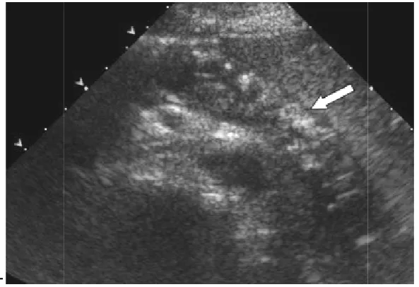
Ultra sound abdomen can diagnose chronic pancreatitis with a sensitivity and specificity of
duct dilatation, enlargement or atrophy of
contours with increased echogenicity can be appreciated in ultrasound abdomen. It is useful for follow up of complications of pancreatitis such as pseudocysts and fluid collection.
USG ABDOMEN
-Figure showing
4. ENDOSCOPIC
It has got 93% sensitivity in diagnosing chronic pancreatitis. By doing endoscopic ultrasound and
pancreatitis with a sensitivity and specificity of 98% and 100% respectively. Advantages
Ultra sound abdomen can diagnose chronic pancreatitis with a specificity of 60-70% and 80-90% respectively. Pancreatic duct dilatation, enlargement or atrophy of pancreas, irregular
contours with increased echogenicity can be appreciated in ultrasound is useful for follow up of complications of pancreatitis such as pseudocysts and fluid collection.
USG ABDOMEN –CHRONIC PANCREATITIS
showing Hypo echoic gland with multiple punctuate
calcifications (arrow).
4. ENDOSCOPIC ULTRASOUND
It has got 93% sensitivity in diagnosing chronic pancreatitis. By doing endoscopic ultrasound and MRCP, we can diagnose chronic pancreatitis with a sensitivity and specificity of 98% and 100% respectively. Advantages being it is less invasive than ERCP and can be Ultra sound abdomen can diagnose chronic pancreatitis with a respectively. Pancreatic pancreas, irregular pancreatic contours with increased echogenicity can be appreciated in ultrasound is useful for follow up of complications of pancreatitis such
CHRONIC PANCREATITIS
c gland with multiple punctuate
[image:37.595.170.467.304.507.2]used for coeliac plexus block and drainage of be sedated, and its limi
5. COMPUTED TOMOGRAPHY
Moderate to severe pancreatitis can be diagnosed with a sensitivity and specificity of 74
dilatation of ducts,
This is the CT picture showing
stones in patient with hereditary pancreatitis.
eliac plexus block and drainage of pseudocysts. Patient and its limited availability is its disadvantages.
TOMOGRAPHY
Moderate to severe pancreatitis can be diagnosed with a sensitivity and specificity of 74-90% and 85% respectively. On CT we can find
calcification and parenchymal atrophy.
This is the CT picture showing multiple calcified
stones in patient with hereditary pancreatitis.
pseudocysts. Patient should its disadvantages.
Moderate to severe pancreatitis can be diagnosed with a sensitivity CT we can find calcification and parenchymal atrophy.
multiple calcified intra ductal
6. MAGNETIC RESONANCE
PANCREATICOGRAPHY
The early and mild form of chronic pancreatitis can be It has got sensitivity and specificity of 65% and90%
noninvasive. Anomalies of pancreatic duct can be
disadvantage is that no therapeutic procedures can be done in MRCP.
7. ENDOSCOPIC RETROGRADE CHOLANGIO PANCREAT
GRAPHY (ERCP)
It is the gold standard imaging procedure for and diagnosing chronic
procedure can be performed but it requires common bile duct or pancreatic duct cannulation. The
hemorrhage are its disadvantages.
This is ERCP
irregularly
MAGNETIC RESONANCE CHOLANGIO
PANCREATICOGRAPHY (MRCP)
The early and mild form of chronic pancreatitis can be
has got sensitivity and specificity of 65% and90% respectively. It Anomalies of pancreatic duct can be
disadvantage is that no therapeutic procedures can be done in MRCP.
NDOSCOPIC RETROGRADE CHOLANGIO PANCREAT
(ERCP)
It is the gold standard imaging procedure for planning,
and diagnosing chronic pancreatitis. Main advantage is therapeutic procedure can be performed but it requires common bile duct or
cannulation. The complications such a are its disadvantages.
is ERCP picture showing pancreatic and its branches
irregularly dilated in chronic pancreatitis.
The early and mild form of chronic pancreatitis can be diagnosed. respectively. It is Anomalies of pancreatic duct can be detected. The disadvantage is that no therapeutic procedures can be done in MRCP.
NDOSCOPIC RETROGRADE CHOLANGIO PANCREATICO
planning, treatment advantage is therapeutic procedure can be performed but it requires common bile duct or complications such as bile leakage,
its branches
TREATMENT
The cause of pain in chronic pancreatitis is due to increased peri neural inflammation, increased pressure in large and small ducts
MEDICAL TREATMENT
The patients with chronic pain abdomen should be initially treated with acetaminophen and non-steroidal anti-inflammatory drugs, if refractory, tramadol or propoxyphene may be tried. Narcotic analgesics should not be used as it may precipitate gastroparasis.The patient should be advised to avoid alcohol and smoking, and he should be referred to a pain management specialist.
NEURO ABLATIVE PROCEDURES
CT or Endoscopic ultrasound guided coeliac block may be tried for intractable pain.
PANCREATIC ENZYMES
ANTI OXIDANTS
Anti-oxidants such as vitamins C,E,selenium,Beta carotene, L – methionine can significantly decrease pain if taken for a long time.
USE OF ENDOSCOPY IN TREATING CHRONIC PANCREATITIS
1. The use of endoscopy in treating chronic pancreatitis is, with elevated sphincter of oddi pressures- pancreatic sphinterotomy can be done endoscopically.
2. Pancreatic duct stones can be broken endoscopically.
3. Pancreatic duct stricture can be treated endoscopically by dilatation of stricture.
MANAGEMENT OF MALABSORPTION
Patient develops steatorrhea when his lipase level decreases to 10% of normal. Hence the main process of treatment is through is through exogenous replacement of lipase by means of pancreatic enzymes.
SURGICAL INTERVENTION OF CHRONIC PANCREATITIS
The indications for surgery in chronic pancreatitis are
1. Pain
PAIN
If patients lifestyle is limited by pain or the pain continues to be present even after abstinence from alcohol completely and intake of analgesics.Proper investigations should be done to define pancreatic and ductal anatomy, and also the merits and demerits of the surgery should be properly explained to the patient, because even after surgery the patient may have persistent pain and loss of exocrine and endocrine function can occur.
FOR SMALL DUCTS –DRAINAGE PROCEDURES
Trans duodenal sphincteroplasty of the common bile duct with pancreatic septotomy i.e. division of septum between pancreatic duct and bile duct can be done for small pancreatic ducts (4-6mms).Patients having multiple duct strictures cannot be managed by these procedures.
FOR DILATED DUCTS –DRAINAGE PROCEDURES
The pancreatic duct should be dilated more than 1 cms for drainage procedure. The ideal procedure would be to create an anastamotic connection between the intestinal lumen and dilated duct. The various procedures are the PUESTOW and ROCHELLE modification and HO and FREY procedure.
PUESTOW PROCEDURE
By Puestow procedure the entire pancreatic duct is opened longitudinally,and the opened pancreas is invaginated to a Roux en Y loop of jejunum. Complete decompression of duct can be achieved but it requires splenectomy.
PARLINGTON AND ROCHELLE MODIFICATION
Parlington and Rochelle modified the Puestow procedure by creating a side to side anastomosis between the opened pancreatic duct and jejunal loop, thus splenectomy is avoided in this procedure.
HO and FREY PROCEDURE
Ho and Frey said a new procedure whereby head of pancreas is removed, Marsupialization of duct is achieved. In this procedure complete duct decompression can be achieved and longer pancreatico jejunostomy can be performed. This procedure can be done when the dilatation of duct is moderate.
RESECTIONAL PROCEDURES
Resective procedures are Distal pancreatectomy ( body and tail of pancreas resected), Whipples procedure (head and uncinate process resected),Subtotal pancreatectomy where by a small rim of pancreas retained along the inner curvature of duodenum, and total pancreatectomy. After total pancreatectomy brittle diabetes can occur. In chronic pancreatitis inflammatory process in head of pancreas decides the symptoms and further progression of the disease in rest of the gland. Hence after resection of pancreatic head, pain relief was achieved in more than 70to80%of patients.
intestinal function. In Beger's procedure duodenum and distal bile duct are preserved, and coring of head of pancreas is done.
Distal pancreatectomy is done for patients with chronic pancreatitis, where the disease is confined to the tail of the pancreas .This procedure is not done if the disease is extending to the entire gland, because recurrence in head is common, if re-surgery is done the patient will be let out without any functioning endocrine tissue. Distal pancreatectomy is usually combined with splenectomy, but spleen can be preserved if vascular supply is adequate.
TOTAL PANCREATECTOMY
1. Total or near total pancreatectomy is done for patients for whom drainage procedures have failed
2. Small ducts and patients who have undergone distal pancreatectomy with persisting pain.
TUBERCULOSIS ABDOMEN
PATHOGENESIS
Two pathological entities are described namely 1. Ulcerative
2. Hyperplastic
ULCERATIVE TYPE
The ulcerative type is the severest of the two in which the virulence of the organism is more than the host resistance. This type is caused by swallowing of infected sputum. The organism reaches the terminal ileum producing transverse ulcers and undermined edges.
HYPERPLASTIC TYPE
CLINICAL FEATURES
1. Patient presents with signs of tuberculosis such as weight loss, malaise, chronic cough, sweating and evening raise of temperature, altered bowel habits, and intermittent vague abdominal pain with abdominal distension.
2. Due to perforation of tuberculous ulcer in the small bowel, patient may present with features of peritonitis.
3. Patient may present with multiple fistula in ano.
4. In hyperplastic type a mass may be palpable in the right iliac fossa.
5. The patient is usually chronically ill and the abdomen on palpation had a doughy feel.
6. The patient may also present in the terminal stage with signs of intestinal obstruction with abdominal pain, faeculent vomiting and distension for which urgent laparotomy is required.
INVESTIGATIONS
LABAROTORY TESTS
1. The common abnormality is elevated ESR in more than 90% of patients.
3. Positive tuberculin test has no value in endemic countries, because those who receive bacillus calmette-gurein vaccine have a high rate of positivity even in healthy individuals. It may be used as a screening test in non-endemic countries.
4. Ascitic Fluid Analysis
a. The ascetic fluid analysis has a high WBC count.
b. The sensitivity approaches 81% if the total white cell count is more than 500/mm3.
c. The total white cell count is within the normal range if the patient has associated with cirrhosis and AIDS.
d. Tuberculous ascitic fluid has high total protein > 2.5g/dl. serum ascetic fluid albumin gradient (SAAG) is less than1.1.
e. The value of lactate dehydrogenase is elevated to more than 90units/l, low ph, and an ascitic fluid; blood glucose ratio less than 0.96.
5. Adenosine deaminase activity is a useful diagnostic test for tuberculosis ascites with a sensitivity of 95%.But if the patient has associated HIV infection, ADA activity is less useful.
By means of Abram’s needle or cope’s needle, blind biopsy of the peritoneum can be done in the presence of ascites. These procedures have low complication rates but mortality has been reported. Bowel perforation is the main risk of blind biopsy.
Under LA, open biopsy of parietal peritoneum is safer.
LAPAROSCOPIC PICTURE-TB ABDOMEN
Straw coloured ascites with scattered multiple whitish nodules all
over peritoneum with omental thickening.
8. By means of ultrasonography or CT guidance, targeted biopsy specimens can be obtained from diseased areas such as lymph nodes which are enlarged.
Miliary tubercles are the characteristic finding, where the peritoneal lining loses its smooth glistening surface, appearing rough and irregular.
10. Histological examination –caseating granuloma can be seen.
11. Imaging studies
a. PLAIN RADIO GRAPH
Calcification of the mesenteric lymph nodes, in the liver, spleen and pancreas calcified granulomas can be seen. Air fluid level can be seen in patients with intestinal obstruction.
b.USG
In USG peritoneum appears thickened, irregular ,poor echoic, nodular or sheet like appearance. It is sensitive for detection of small quantities of fluid.
CLUB SAND WICH APPEARANCE
The presence of alternating echogenic and echo free layers produced by the bowel wall, serosa and the adjacent bowel loop causes the characteristic CLUB SAND WICH appearance.
C.CT Abdomen
In CT the nodularity and thickening of the peritoneum and mesentery can be easily identified.
D.BARIUM STUDIES
Irregularity and thickening of the mucosal folds are the earliest abnormalities. In barium swallow compression of oesophagus with mediastinal nodes can be seen.
In advanced disease mucosal ulcers, bowel lumen deformity, and stricture can be seen in advanced disease.
FLEISCHNER SIGN-Narrowing of terminal ileum with thickening of ileo caecal valve.
STERLIN SIGN-Fiber optic terminal ileum opening into a contracted caecum is suggestive of intestinal tuberculosis.
DOUBLE CONTRAST BARIUM ENEMA –ILEO CAECAL TB
Marked retraction of ileo caecal area with incompetent ileo
ENTEROLYSIS (small bowel enema)
Best method of assessing the small bowel. It has got low sensitivity rate. Crohn’s disease and lymphoma are confused with TB on barium studies.
e. ENDOSCOPIC BIOPSY
By means of colonoscopy ileo caecal region can be asessed. Proximal small bowel loops assessed by enteroscopes,
Findings are mucosal ulcerations, nodularity, deformity, and narrowing and bowel stricture. Ulcers in TB are transversely arranged and have sharply defined margins with the surrounding mucosa. By means of endoscopy carcinoma and lymphoma can be excluded.
f.SEROLOGICAL STUDIES
ELISA has got sensitivity and specificity with accuracy of 80% .It can detect mycobacterium infection, active disease and patients with previous BCG inoculation.
g. POLYMERASE CHAIN REACTION
TREATMENT
The treatment for intestinal tuberculosis is similar to the management of Crohn’s disease. After completion of medical treatment, intestine should be looked through laparoscopy for strictures. If the patient has features of sub-acute intermittent intestinal obstruction resection and anastomosis can be done. Limited ileo colic resection can be done with anastomosis between terminal ileum and ascending colon. Alternative procedures are Right hemicolectomy and stricturoplasty.
Treating patients in the emergency department present a challenge to the surgeon. Since most of the patients are from low socio economic status they presents at the extremity with acute small bowel obstruction. The patient may be sick from malnutrition, anaemia,dehydration and other systemic evidence of active pulmonary tuberculosis.
On laparotomy if the patient has terminal ileal stricture, side to side ileo transverse anastomosis should be done. Before laparotomy the patient should be resuscitated properly.
FOLLOW UP
tuberculous drugs the patient should be reviewed for the disease and also the byepass procedure done should be checked.
On the basis if the patient has negative sputum smears, weight gain and normal inflammatory markers Elective Right hemicolectomy can be performed. After right hemicolectomy it is supplemented by stricturoplasty.
If perforation is the complication of TB abdomen, affected segment should be resected. If there is no any gross peritoneal contamination resection and anastomosis can be done at the first stage, on the other hand if there is any gross peritoneal contamination resection and exteriorization can be done and once after the patient got stabilized resection and anastomosis done as second procedure.
CHRONIC APPENDICITIS
ANATOMY
The base of appendix is located where all the three taenia converge at inferior aspect of caecum. The tip of appendix has many locations, common being Retro caecal, pelvic 30% and Retro peritoneal 7%.
PHYSIOLOGY
The appendix has no function in the adults.
HISTORY
1. Reginald Fitz of Baston in 1886 told that the cause of right lower quadrant pain is appendix. The word appendicitis was coined by him and he advised early intervention for this disease.
2. The migration of pain in appendicitis was first described by Chester Mcburney.
3. Kurt semm in 1982, a gynecologist was the first to do laparoscopic appendicectomy and after her, it gained popularity all over the world.
PATHOGENESIS
2. Pin worms an internal parasite is also responsible for luminal obstruction.
3. Chronic appendicitis is seen in cystic fibrosis where lumen of the appendix is occluded by mucoid material.
CLINICAL FEATURES
1. Patient may present with recurrent episodes of acute appendicitis and at each time, spontaneous resolution occurs with use of antibiotics, analgesics.
2. Clinical signs of chronic appendicitis similar to acute appendicitis but symptoms have prolonged course.
CRITERIA FOR CHRONIC APPENDICITIS
1. Presence of symptoms for more than 2 weeks.
2. Relief of symptoms after appendicectomy.
3. On pathological examination confirmation of chronic appendiceal inflammation.
DIFFERENTIAL DIAGNOSIS
1. Crohn’s disease.
Stump Appendicitis
After appendicectomy if the appendicular stump is not properly buried this appendiceal stump can act as small appendix leading to frequent obstruction and inflammation causing recurrent pain.
CT – Stump appendix can be seen as-Focal thickening of caecal apex with pericaecal fat stranding.
Recurrent appendicitis and chronic appendicitis are difficult to distinguish from acute appendicitis on CT.
Diagnosis
1. Done mainly by Clinical assessment.
WBC count – leucocytosis .
Urine Analysis – Renal causes to be excluded.
Pregnancy Test – To rule out ectopic pregnancy.
2. Ultra Sound
Useful for excluding other diseases that mimic chronic appendicitis
3. CT Findings for chronic Appendicitis
a. Appendix is enlarged more than 6 mms.
c. Periappendiceal inflammatory mass can be seen in the advanced stage and disease.
d. Ceacum and ileum may be seen thickened (Arrow Head Sign) in appendicoliths.
Laparoscopy
Therapeutic and Diagnostic Laparoscopy can be done.
Complications
Appendiceal perforation can lead to peritonitis and appendicular mass.
Treatment
Treated by Open Surgery or Laparoscopy
Open Appendicectomy
Various incisions used for appendicectomy are Mcburney (oblique), modified Mcburney (curvilinear) over the Langer line also called lanz incisions, Rockey – Davis (Transverse Incision) muscle splitting incision and Rutherford Morrison s incision.
Laparoscopic Appendicectomy
Carcinoma Caecum
The caecum, Appendix, Ascending colon, transverse colon, descending colon, sigmoid colon, rectum and Anal canal forms part of large bowel and it extend from ileo caecal valve to the Anus. The large bowel has fixed and mobile parts. The caecum, transverse, sigmoid colons are mobile whereas the ascending colon and descending colon are fixed to the posterior abdominal wall.
Caecum
1. The caecum forms the saccular commencement of colon.
2. Caecum occupies the right iliac fossa and it lies over the iliacus muscle.
3. It may cross the pelvic brim and may occupy the position of true pelvis
4. caecum is an intraperitoneal structure, but it has no mesentery possessing considerable range of mobility.
Relations of Caecum
ILEO CAECAL VALVE
At the point ileum enters caecum, there are 2 flaps containing circular muscle fibers derived both from caecal and ileal musculature. It functions as functional sphincters but its function is still doubtful.
At the posterior-medial border of caecum the appendix taken origin and it can be traced by following the anterior taenia to its junction with the other two taenia. The length of appendix is 8 to 10 cm in length, diameter 5 to 10 mms.
Surgical Resection for Cancer
The whole of the gut supplied by right colic and ileo colic artery and their branches, its related peritoneum should be removed, so that all lymph territory that converges on these vessels is removed.
Superior mesenteric plexus supplies caecum by sympathetic and parasympathetic nerves.
Blood Vessels to the area of Resection
Structures removed in Right Hemicolectomy
Caecum, ascending colon, hepatic flexure, proximal one third of transverse colon, terminal 15 cms of ileum.
Physiology
It takes place in the process of fluid and electrolyte reabsorption. The "Sac like" morphology of caecum and its distensible nature, it can adapt to storage of large volumes of semi liquid chyme entering through ileo caecal valve from small bowel.
Normal colon function a) Water absorption
Colon can absorb 5 to 6 Liters of fluid per day, majority of colonic absorption take place in right colon.
b) Electrolyte Transport
By active processes, in exchange of potassium and bicarbonate sodium and chloride absorption occurs.
Nutrition
Colonic gas
By fermentation of colonic bacteria about 2000ml of colonic gas produced is composed of swallowed nitrogen and oxygen, and carbon di-oxide and methane produced on fermentation.
Clinical Features
1. Patient may present with severe anaemia.
2. Mass in the right iliac fossa may be the presentation.
3. It may be the apex of intussusception, with symptoms of sub-acute intestinal obstruction.
4. Presentation may be of metastatic disease to liver, lungs, brains, skin, and bone.
INVESTIGATIONS
Flexible Sigmoidoscopy
Colonoscopy
This is the choice of investigation for colorectal cancer and this is the confirmatory test for colonic cancer.
It not only detect primary lesion but also detect secondary deposits.
It gives a definitive histological diagnosis before operating the patient.
Bowel should be prepared properly before the produce.
Disadvantage is small risk of perforation.
Inability to get caecum in 10% of cases, by experienced endoscopist.
This is now the diagnostic confirmatory test for colonic malignancy.
Colonoscopy Technique
Preparation of bowel by giving clear fluids 48 hours before procedure.
Enema before beginning a procedure.
Poly ethylene glycol in hypertonic lavage preparation used to purge the colon for 4 hours.
Proper digital rectum examination should be done for dilating the rectum, and then Colonoscope should be introduced.
After the positioning the colonoscope in the rectal vault the colonoscope should be introduced, ahead only if the lumen of bowel is clearly visible.
Perforation may be caused if blind insertion of colonoscope done.
The colonoscope would reach the caecum by 100 cm or less, splenic flexure can be reached 50 to 55 cm.
Mercedes Sign
On colonoscopy caecum is demonstrated as an arch where the three taenia converge. This is called Mercedes sign. Therapeutic applications of colonoscopy are polypectomy and biopsy.
Radiology
It colonoscopy is contra indicated double contrast – barium enema can be used.
Carcinoma is seen as constant irregular filling defect. False positive 1 - 2%,
False Negative 7 – 9%
This is the barium enema picture showing polypoid
carcinoma arising from caecum.
USG Abdomen
To detect liver metastasis
CT Abdomen
To asses local invasion and pelvic invasion
Virtual Colonoscopy or CT Colonography
It may replace colonoscopy as the gold standard investigation in future where it can detect polyps of size 6 mm.
Preparation of colon is essential Preparation consists of 3 elements
DIET
Clean fluid diet 24 – 48 hrs, before the examination.
Purgation
Cleaning the bowel can be achieved by dry and wet preparations. Wet preparation can be done by using PEG (Poly ethylene glycol), followed by 2 sachets of sodium picosulphate 24 hours prior to the procedure.
Colonic Distension
For a good quality CTC examination, adequate colonic distention is essential. Distension can be achieved by the use of spasmolytics by method of insufflation.
Reading CTC examination
After the above procedure, About 1000 to 1200 images will be yield in a complete two position examination.
Preoperative Preparation
1. 48 hours before surgery, the patient should be advised to take only fluids.
2. On the day of operation sodium Pico sulphate used to purge the colon.
3. Stoma site is selected preoperatively after consulting with stoma care specialist.
4. Before surgery the patient should be started on subcutaneous Heparin and prophylactic antibiotics should be administrated.
Test of Operability
1. Presence of secondary deposits in liver.
2. Peritoneal seedings.
3. Lymph nodes draining the involvement segment should be assessed.
4. Growth should be examined for mobility and operability.
GORD (GASTRO OESOPHAGEAL REFLUX DISEASE)
Clinical features
1. Retrosternal burning pain, epigastric pain and regurgitation constitutes classical triad of GORD.
2. Symptoms are provoked by fat & spicy foods, stopping or exercises, hot beverages, citrus drinks, alcohol, nocturnal reflux.
3. Due to stricture dysphagia occur and it’s a late sign.
4. Since GORD is such a common disorder, it should be anticipated when patient presents with oesophageal symptoms that are unusual or defy diagnosis after series of investigation.
Diagnosis
1. Patient may present with less typical symptoms such as chest pain laryngeal or pulmonary symptoms.
2. In most cases, diagnosis is assumed rather than proven and treatment is empirical.
3. Investigation in only required when diagnosis is in doubt, when patient does not respond to Proton Pump Inhibitor or if dysphagia is present.
5. There could be reflux esophagitis – peptic stricture or Barrett’s esophagus
(Due to increase in use of Proton Pump Inhibitor leading to rapid healing of early mucosal lesions).
6. There is strong correlation between worsening endoscopic appearances and oesophageal acidification on PH testing.
7. If symptoms persist despite treatment, oesophageal manometry and 24 hour PH recording guides in diagnosis and management.
Diagnostic Measurement in GORD
1. 24 hrs pH recording is the 'gold standard' for diagnosis of GORD.
2. Most important manometric findings are TLOSR's.
3. Length and pressure of Los is also important.
4. Achalasia is differentiated by slow undulations in pH trace and complete absence of peristalsis whereas in GORD there are rapid bursts of reflux in pH trace ,but peristalsis is not totally absent.
PH Monitoring
This study is done after placing thin catheter containing 1 or 2 solid catheter in the oesophagus. The electrode connected to data recorder, and the electrodes are placed 5 to 10 cm apart, in the pH 2.7. They are capable of sensing electrons.
Inferences from the study are
1. Longest episode to reflux.
2. Total number of reflux episodes .
3. Number of episodes lasting more than 5 minutes. 4. Extent of reflex in upright and supine position.
5. The total score is obtained using all of the above parameters. The score obtained is Demeester Score and it should be less than 14.7.
Esophagogram
This test is performed for patients with symptoms of GERD who are to undergo surgery and whose symptoms do not respond properly after treatment.
Oesophageal lengthening procedure has to be done, if during the study if mediastinal gastro oesophageal junction does not invaginate into the peritoneal cavity. Diverticula, para esophageal hernias can be discovered on Esophagogram.
Endoscopy
1. It can exclude other diseases.
2. In can detect the presence of peptic esophageal injury.
3. Severity of Injury can be measured using a scoring system called savary-miller interpretation.
a. Erythema
b. Linear Ulceration c. confluent ulceration d. Stricture
4. Barrett’s esophagus is extremity of mucosal injury.
New and evolving endoscopic techniques
GORD treatment has been attempted by new techniques such as suturing devices, Radio frequency energy and injector polymers.
NOTES
Various surgeries that can be done using NOTES are 1. Cholecystectomy
2. Appendicectomy 3. Peritonoscopy
In Humans both pure and hybrid procedures have been performed.
In this procedure, after advancing endoscope into the rectum, mouth, surgery is done after puncturing the viscus.
Endoscopic staplers are useful for performing resection of Gastric carcinomas.
Endoscopic suturing has been used for narrowing of stomach after gastric bypass surgery.
Capsule endoscopy is one of the latest imaging techniques of the bowel.
OTHER INVESTIGATIONS
1. Scintigraphy study can be useful to evaluate can reflux and esophageal clearance.
2. It can detect motility disorder and gastro esophageal reflux.
4. Patients having laryngeal symptoms of Gastro esophageal reflux can be screened by laryngoscopy and stroboscopic examination.
5. Findings in this procedure are Laryngeal mucosal inflammation, mucus tension abnormalities and subglottic stenosis.
Therapeutic Interventions of Upper GI Endoscopy
1. Monopolar (or) Bipolar probes can be used to coagulate bleeding points.
2. Rubber banding (or) Injection sclerotherapy are used to treat Esophageal varices.
3. Pyloric, esophageal strictures can be treated by means of dilatation by using hydrostatic balloons and obstructive esophageal tumors . Stomach vascular lesions are treated by laser energy through the scope.
4. Barrett’s esophagus, dysplasia and early carcinoma can be treated by endoscopic mucosal resection.
5. Suck-and-ligate and suck-and-cut are the EMR techniques employed.
MANAGEMENT OF UNCOMPLICATED GORD: MEDICAL
MANAGEMENT
1. Simple measures like weight loss, avoiding smoking, avoiding consumption of alcohol, tea or coffee, avoidance of large late night meals and modest head-up tilt of bed (similar effect to taking the receptor antagonist).
2. 'Step Down' PPI treatment for 8 weeks are the most effective treatment for GERD.
SURGERY
Endoscopic Treatment
They include endoscopic suturing to plicate gastric mucosa just below the cardia to accentuate angle of His, Radio frequency ablation at sphincteric level and injection of submucosa polymers into lower esophagus.
They prove temporary symptomatic improvement.
Failure rates at 1 years are > 50%.
GASTRITIS
Types of Gastritis
1. Hypertrophic Gastritis (Menetrier’s disease). 2. Antral Gastritis
Hypertrophic Gastritis
1. Also called as Menetrier’s disease or Hypoproteinemic Hypertrophic Gastropathy is a pre malignant condition characterized by gastric folds in the fundus to stomach, giving cobble stone appearance,
2. This condition in associated with excessive mucus production, hypochlorhydria and protein loss from stomach.
3. The cause is unknown.
4. It may be due to over expression of TGF – Alfa.
Antral Gastritis
1. Antral gastritis is associated with peptic ulcer.
2. The cause due to H-pylori Infection.
3. Patient with peptic ulcer had antral gastritis proven histologically
4. The most infection is confined to antrum.
Stress Gastritis
Clinical Features
1. Stress gastritis occurs after physical trauma, shock, sepsis, respiratory failure and it can lead to gastric bleeding which may be life threatening.
2. Stress gastritis is characterized by superficial (Non ulcerating) erosions. That usually starts in the Acid secreting portion of stomach and progressing distally.
3. Cushing's Ulcer – Occur in central nervous system disease.
4. Curling' Ulcer – It occurs as a Result of Thermal Burn injury. involving more than 30% of the body surface area.
5. Stress gastritis changes with time duration. It is considered to be early if they appear with in the first 24 hours after injury. The lesions are multiple and shallow, with isolated area of erythema along the focal hemorrhage.
6. Frank bleeding may occurs if the lesion erodes into the Submucosa where blood vessels are located.
8. In stress gartitis, the ulcers usually seen in the fundus of the stomach, they are seen rarely on the distal stomach.
9. Late acute gastritis is usually seen 24 to 72 hours after injury and there is tissue reaction around a clot.
10. Late lesions appears similar in appearance to regenerating mucosa around the area of gastric ulcer healing site .
Investigation
- Endoscopy for confirmatory diagnosis.
- It is used to differentiate stress gastritis from other causes of GI Haemorrhage.
Therapy
- Patient with upper GI bleeding requires the correction of coagulation abnormalities. Fluid resuscitation should be done properly.
- Fresh frozen plasma and plates should be given to the patient if the patient has clotting anomalies (or) platelet deficiencies.
MATERIALS AND METHODS
Time period of study : - September 2011 to November 2012
AGE OF PATIENTS –older than 13years
GENDER OF PATIENTS –male and female
STUDY AREA –Coimbatore medical college hospital
STUDY POPULATION-patients presenting to the surgical department with features of chronic recurrent pain abdomen
SELECTION CRITERIA
INCLUSION CRITERIA
Chronic recurrent pain abdomen
Age more than 13years
EXCLUSION CRITERIA
Acute pain abdomen
Patients treated by psychiatrists
Coagulopathy, severe cardiopulmonary disease
Previous surgeries
STUDY DESIGN
Prospective observational study was conducted on 50 patients admitted with chronic recurrent pain abdomen
PARAMETERS STUDIED
Age
Sex
Dietary pattern
Smoking pattern
Scio economic status
Habit of beeten nut chewing
Type of pain
Location of pain
Etiology in males
Etiology in females
Diagnostic investigation which was confirmatory
Treatment given –conservative and surgical
OBSERVATIONS MADE
OBSERVATIONS
SEX WISE DISTRIBUTION
The incidence is more common in males than females in a ratio of 2:1. Males comprised 66% of our study compared to 33% in females. However the incidence in females is catching up due to the altered lifestyle of female population.
0 5 10 15 20 25 30 35
Male Female
N
o
.
o
f
P
a
ti
e
n
ts
Sex wise
AGE WISE DISTRIBUTION
Patients in the age group 31-40 years are commonly affected with a mean age of 43.64 years.
Age Group No of Patients Percentage
11-20 1 2
21-30 9 18
31-40 16 32
41-50 9 18
51-60 6 12
61-70 8 16
71-80 1 2
0 2 4 6 8 10 12 14 16 18
11 to 20 21-30 31-40 41-50 51-60 61-70 71-80
N o . o fp a ti e n ts
AGE WISE DISTRIBUTION
SMOKING PATTERN
64% of the study group was smokers as compared to the 36% non-smokers. This may suggest that smoking may have a positive correlation with chronic abdominal pain of certain causes.
0 5 10 15 20 25 30 35
Smokers Non Smokers
N
o
.
o
f
P
a
ti
e
n
ts
Smoking Pattern
ALCOHOL INTAKE PATTERN
62% of our patients were consumers of alcohol and of these, 32% had consumed for more than 10 years. Hence alcohol may also act as an additive factor in many diseases presenting as chronic abdominal pain.
0 2 4 6 8 10 12 14 16 18 20
Alcohol>10yrs Alcohol<10yrs Non-Alcoholic
N
o
.
o
f
P
a
ti
e
n
ts
Alcohol pattern
DIETARY PATTERN
72% of the patients consumed non-vegetarian food which suggests that non-vegetarian food may be a predisposing factor in diseases causing chronic abdominal pain
0 5 10 15 20 25 30 35 40
mixed diet vegetarian
N
o
.
o
f
P
a
ti
e
n
ts
Diet pattern
HABIT OF BEETLE NUT CHEWING
38% of patients were beetelnut chewers chronically and more among this group was female individuals.
0 5 10 15 20 25 30 35
yes No
N
o
.
o
f
P
a
ti
e
n
ts
Beetel nut chewer
SOCIO ECONOMIC STATUS
Being a government run tertiary care institution, all the patients that were observed in the study were from a lower socio-economic class.
0 10 20 30 40 50 60
Low Middle High
N
o
.
o
f
P
a
ti
e
n
ts