Copyright © 1998, American Society for Microbiology
A Virus with a Mutation in the ICP4-Binding Site in the L/ST
Promoter of Herpes Simplex Virus Type 1, but Not a
Virus with a Mutation in Open Reading Frame P,
Exhibits Cell-Type-Specific Expression of
g
1
34.5
Transcripts and Latency-Associated Transcripts
LILY YEH LEEANDPRISCILLA A. SCHAFFER*
Division of Molecular Genetics, Dana-Farber Cancer Institute and Department of Microbiology and Molecular Genetics, Harvard
Medical School, Boston, Massachusetts 02115
Received 1 December 1997/Accepted 15 January 1998
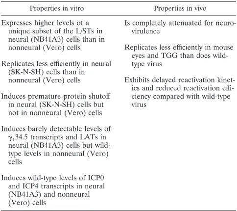
The herpes simplex virus type 1 L/S junction-spanning transcripts (L/STs) are a family of multisized tran-scripts expressed at high levels in cells infected with mutant viruses that (i) do not express ICP4, (ii) specify forms of ICP4 unable to bind to the consensus ICP4 binding site, or (iii) contain mutations in the ICP4 binding site located at the transcriptional start site of the L/STs. By extension, the failure to detect the L/STs in wild-type virus-infected cells is due to the repressive effect of ICP4 bound to its cognate binding site upstream of the L/ST transcription initiation site. ORF-P, the first and largest open reading frame (ORF) encoded by the L/STs, overlaps >90% of the ORF encoding ORF-34.5, a putative neurovirulence factor, which is transcribed from the opposite DNA strand. Viruses with mutations in the overlapping region of ORF-P and ICP34.5 exhibit premature shutoff of infected-cell protein synthesis and are highly attenuated following intracranial inocula-tion of juvenile mice. To determine whether the premature protein shutoff and neuroattenuated phenotypes of ORF-P ORF-34.5 double mutants are a consequence of alterations in ORF-P, ORF-34.5, or both, viruses containing mutations only in ORF-P or only in the ICP4 binding site in the L/ST promoter were isolated and charac-terized. Mutant virus L/ST-n38 contains a single-base-pair transition mutation in ORF-P codon 38, resulting in translational termination of the ORF-P protein (OPP). This mutation does not alter the amino acid se-quence of ICP34.5. Expression of a truncated form of OPP by mutant virus L/ST-n38 did not result in pre-mature shutoff of infected-cell protein synthesis and produced no other observable phenotype relative to wild-type virus in in vitro tests. Moreover, the 50% lethal dose (LD50) of L/ST-n38 was comparable to that of
wild-type virus following intracranial inoculation of 3-week-old mice, as were the latency and reactivation phenotypes of the virus. These properties of L/ST-n38 indicate that the attenuated phenotype of P ORF-34.5 double mutants is a consequence of mutations that affect the function of ICPORF-34.5 and not the function of OPP. Mutant virus L/ST-4BS contains four single-base-pair substitutions in the ICP4 binding site in the L/ST promoter that abrogate the binding of ICP4 to this site, leading to high-level expression of the L/STs and OPP. L/ST-4BS induced premature shutoff of viral and cellular protein synthesis and was slightly growth restricted in cells of neural lineage (SK-N-SH human neuroblastoma cells) but was wild type for these two parameters in cells of nonneural lineage (immortalized primate Vero cells). Of particular interest was the observation that L/ST-4BS exhibited cell-type-specific expression of both theg134.5 transcripts and the latency-associated
tran-scripts (LATs). Thus, expression of these trantran-scripts was barely detectable in cells of neural lineage (NB41A3 mouse neuroblastoma cells) but was wild type in Vero cells. In vivo, L/ST-4BS was reactivated from mouse trigeminal ganglia with reduced efficiency and delayed kinetics relative to wild-type virus. L/ST-4BS was com-pletely attenuated for neurovirulence (LD50> 106PFU) relative to wild-type virus (LD50< 900 PFU), although
the four single-base-pair substitutions lie outside the coding region for the neurovirulence factor, ICP34.5. Collectively, the complex in vitro and in vivo phenotypes of L/ST-4BS can be attributed to (i) disruptions of the ICP4 binding site in the L/ST promoter and subsequent overexpression of the L/STs and OPP; (ii) alterations in ORF-O, which is also mutated in L/ST-4BS; or (iii) alterations in other cryptic genes or cis-acting elements.
Herpes simplex virus type 1 (HSV-1) is a neurotropic virus that causes a variety of clinically significant diseases ranging from superficial cutaneous lesions to life-threatening enceph-alitis (49). The designation of HSV-1 as a neurotropic virus is
based on its ability to produce both lytic and latent infections of neurons of the peripheral and central nervous systems. The extent to which a given strain of HSV-1 can gain access to and travel within neurons (neuroinvasiveness) and the relative ef-ficiency with which a strain produces neuronal damage and death (neurovirulence) are major determinants of the disease-producing capabilities of that strain. Identification of the viral genes that control neuroinvasiveness and neurovirulence and elucidation of the functions of the protein products of these genes are necessary to understand the mechanism by which HSV-1 causes disease.
* Corresponding author. Mailing address: Department of Microbi-ology, University of Pennsylvania School of Medicine, 225A Johnson Pavilion, 3610 Hamilton Walk, Philadelphia, PA 19104-6067. Phone: (215) 573-9863. Fax: (215) 573-5344. E-mail: [email protected] .edu.
4250
on November 9, 2019 by guest
http://jvi.asm.org/
By definition, any viral gene which, when mutated, results in the failure of the virus to enter or spread in neurons or to replicate in epithelial or neuronal cells can be said to affect neuroinvasiveness or neurovirulence, respectively. Some viral genes, however, have unique properties which appear to func-tion exclusively or predominantly in neurons. Notably, a num-ber of viral genes which affect neurovirulence and which, col-lectively, determine whether infection will be productive or latent are located within the abb9a9c9ca repeat regions flanking the unique long and short regions of the HSV-1 genome (Fig. 1) (reviewed in reference 50).
The genes specifying the L/S junction-spanning transcripts (L/STs) and theg134.5 gene, are located within these repeat
regions and constitute the subject of this paper. The L/STs are a series of 59-coterminal polyadenylated RNAs transcribed from the same DNA strand as the latency-associated tran-scripts (LATs) (Fig. 1) (50). The L/STs range in size from 2.3 to .9.0 kb and are expressed at detectable levels only by mutant viruses that express no functional ICP4 or that lack an intact ICP4 binding site at the transcription initiation site of the L/ST gene (50). These properties suggest that ICP4 bound to its cognate binding site in the L/ST promoter blocks the expression of the L/STs. The L/STs overlap the 39terminal 2.3 kb of the 8-kb minor LAT on the same strand, and, critical to the subject of this paper, the L/STs overlap and are antisense to theg134.5 gene (Fig. 1).
The first and largest open reading frame (ORF) in the L/STs is ORF-P, which specifies the ORF-P protein (OPP) (Fig. 1C and Fig. 10). ORF-P is conserved in all HSV-1 strains whose genomes have been sequenced in this region. OPP is expressed at detectable levels only under conditions which allow expres-sion of the L/STs (26, 50). Overlapping the 59half of ORF-P, and encoded by the same DNA strand but in another reading frame, is ORF-O, which encodes the ORF-O protein (OOP) (Fig. 1C). In strain 17syn1, ORF-O is approximately 300 codons in length. In strains KOS, F, MGH10, and CVG2, a single-base-pair substitution terminates ORF-O after approx-imately 160 codons. Randall et al. have recently provided ev-idence suggesting that, like ORF-P, ORF-O is downregulated by ICP4 (34). The translational start codon of ORF-O lies immediately downstream of the TATA box in the L/ST pro-moter; thus, ORF-O is probably encoded by the LATs rather than the L/STs. Recently, however, Randall et al. have sug-gested that in strain F, ORF-O and ORF-P initiate from the same methionine codon and that the initiation of ORF-O at this codon results from frameshift or editing of its RNA (34). The L/STs overlap and are antisense to theg134.5 gene. The
g134.5 gene is located between the genes specifying two major
immediate-early (IE) transcriptional regulatory proteins, ICP0 and ICP4 (Fig. 1). The;8-kb LAT, specified by the opposite strand, overlaps and is antisense to the entire ICP0 andg134.5
genes. The g134.5 gene encodes ICP34.5, a protein with an
apparent molecular mass of 44 kDa (2, 11, 15). The g134.5
ORF (ORF-34.5) is conserved in all HSV-1 strains that have been sequenced to date (14, 18). Viruses with deletions in ORF-34.5 are replication competent in vitro; however, they are highly attenuated for neurovirulence in vivo following intra-cranial inoculation of mice (6, 14, 35, 46–48). Other properties ofg134.5 mutant viruses include (i) more efficient replication
at some primary sites of infection (e.g., the footpad and vagina) than at others (e.g., the eye) (49), (ii) defective replication in the sensory ganglia of mice, guinea pigs, and rabbits and in the central nervous system of mice (6, 14, 30–32, 35, 45, 48); (iii) wild-type growth in most cell types but restricted growth in SK-N-SH (human neuroblastoma) (13), CV-1 (African green monkey kidney) (32), murine 10T1/2 (stationary-phase primary
mouse embryo), and 3T6 cells (6, 7); (iv) premature shutoff of all protein synthesis (cellular and viral) following infection of SK-N-SH cells (12, 13); and (v) reduced establishment of la-tency compared to wild-type virus (31, 45).
During the course of the studies of ICP34.5 just described, a number of mutant viruses were constructed and characterized. By virtue of the complete overlap of theg134.5 gene with the
L/ST and the 8-kb LATs, mutations introduced into theg134.5
gene were also introduced into sequences specifying the L/STs and the LATs. Consequently, “g134.5 mutant viruses” are
ei-ther ORF-34.5 ORF-P double mutants or ORF-34.5 ORF-P ORF-O triple mutants (Fig. 1).
Because ORF-O is probably contained in the LATs rather than the L/STs, as noted above, we focused the present inves-tigation on the contribution of ORF-P to the phenotype of ORF-34.5 ORF-P double-mutant viruses. We approached this problem by using two genetic strategies. In the first, we mu-tated ORF-P without altering ORF-34.5, and in the second, we altered the transcriptional regulation of the L/STs by greatly increasing the levels of L/ST and ORF-P expression. We report here the construction and characterization of two mutant vi-ruses: one, L/ST-n38, containing a nonsense mutation that terminates ORF-P translation at amino acid 38 but does not alter the sequence of ICP34.5 encoded by the complementary DNA strand (Fig. 1G; also see Fig. 10), and another, L/ST-4BS, containing a mutation in the L/ST promoter that dere-presses L/ST expression by abrogating ICP4 binding to its cognate binding site near the transcription initiation site of the L/STs (Fig. 1G; also see Fig. 10). The properties of L/ST-n38 demonstrate that expression of a truncated OPP produces no observable phenotype relative to wild-type virus in vitro or in vivo. By contrast, L/ST-4BS, which is mutated in the ICP4 binding site in the L/ST promoter and overexpresses the L/STs and OPP, is highly attenuated following intracranial inocula-tion of juvenile mice, yet replicates efficiently in the mouse eye and trigeminal ganglia (TGG) without eliciting an adverse host response. Of special interest was the observation that L/ST-4BS induced cell-type-specific expression of bothg134.5
tran-scripts and LATs since both types of transcript were abundant in Vero cells but barely detectable in mouse neuroblastoma cells (NB41A3). The significance of these findings to the as-signment of specific functions to OPP and ICP34.5 can be summarized as follows: (i) ICP34.5 and not ORF-P is the neurovirulence factor responsible for attenuation of ORF-P ORF-34.5 double mutants and (ii) a factor or element affected by the 4BS mutation mediates cell-type-specific expression of theg134.5 gene and the LATs.
MATERIALS AND METHODS
Cells and viruses.African green monkey kidney cells (Vero; ATCC CCL 81) and human embryonic lung cells (HEL cells) were grown and maintained in Dulbecco’s modified Eagle’s medium as previously described (38). Mouse neu-roblastoma cells (NB41A3; ATCC CCL147) were propagated in Ham’s F10 medium supplemented with 2.5% fetal calf serum and 15% horse serum. Human neuroblastoma cells (SK-N-SH) were kindly provided by Patrick Vaughn (Uni-versity of California, Irvine, Calif.) and were propagated as described above for Vero and HEL cells. Rat pheochromocytoma cells (PC12) were the gift of John Wagner (Cornell University Medical College, New York, N.Y.) and were prop-agated and maintained as described previously (21).
The KOS strain of HSV-1 (41) and mutants derived from KOS were used in these studies. The ICP4 nonsense mutant, n12, and the ICP0 null mutant, 7134, were grown and assayed as previously described (9, 17). Mutant virus L/ST-n38 contains a nonsense mutation in ORF-P, and mutant virus L/ST-4BS contains four single-base-pair mutations in the ICP4 binding site in the L/ST promoter. L/ST-n38R is a rescuant of L/ST-n38, and L/ST-4BSR is a rescuant of L/ST-4BS. The 17syn1wild-type strain of HSV-1 and the ICP34.5 nonsense mutant, 1771, derived from 17syn1, were generous gifts of Alasdair MacLean (MRC Virology Unit, Glasgow, United Kingdom) and have been described previously (30).
on November 9, 2019 by guest
http://jvi.asm.org/
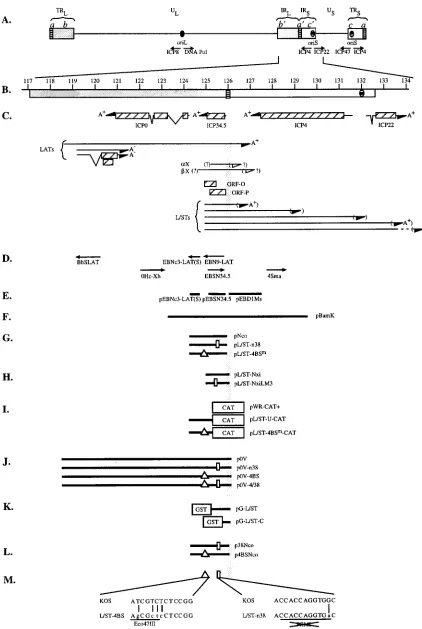
FIG. 1. Physical map of the internal repeat region of HSV-1 DNA. (A) Diagram of the HSV-1 genome. TRL, UL, and IRL, terminal repeat, unique, and internal
repeat sequences, respectively, of the long segment; IRS, US, and TRS, internal repeat, unique, and terminal repeat sequences, respectively, of the short segment; b9,
internal inverted repeat sequence bracketing UL; c9, internal inverted repeat sequence bracketing US; a9, inverted sequence located between the b9and c9sequences
and, in reverse orientation, at the genomic termini. (B) Expanded map of the internal repeat sequence b9-a9-c9lying between kbp 117 and 134 on the HSV-1 genome.
on November 9, 2019 by guest
http://jvi.asm.org/
Plasmids.For subcloning purposes, pBamK (Fig. 1F), containing the KOS BamHI K fragment from pSG28 (40), and pNco (Fig. 1G), containing the 1.75-kbp NcoI subclone of pBamK, were constructed as described previously (50). Plasmid pL/ST-n38 (Fig. 1G) is identical to pNco, except that it contains a single-base-pair transition in ORF-P. Mutagenesis was accomplished by in vitro phagemid mutagenesis with pNco as the parental plasmid and the mutant oligonucleotide 59-ACCAGGTGACGCACCCGG-39. The transition mutation from G to A (underlined) eliminates the cleavage site for the restriction enzyme PflMI. Plasmid pL/ST-4BSm(Fig. 1G) is identical to pNco except for four single-base-pair changes in the ICP4 binding site near the transcriptional start site of the L/STs. The mutations were introduced by in vitro phagemid mutagenesis with pNco as the parental plasmid and the mutant oligonucleotide 59-GCGGCTCC TGCCAGCGCTCCTCCGGAGAGC-39. The mutation of ATCGTCTCT to AG CGCTCCT generates a new restriction site for the endonuclease Eco47III.
For in vitro transcription and translation of L/ST ORF-P, a 2.6-kb SacI-NcoI fragment derived from pBamK was cloned into pGEM5Zf(1) (Promega Corp., Madison, Wis.) to generate plasmid Nxi (Fig. 1H). To construct pL/ST-NxiLM3 (Fig. 1H), the 0.7-kb SacI-BstEII fragment of pL/ST-Nxi was replaced by the corresponding fragment from pL/ST-n38. Plasmid pL/ST-NxiLM3 was used to test the efficacy of the stop codon.
The plasmids used in transient-expression assays were as follows. Plasmid pWR-CAT, an expression vector carrying a promoterless chloramphenicol acetyltrans-ferase (CAT) gene, was described previously (33). A modified vector, pWR-CAT1 (Fig. 1I), was used as the parent plasmid for all transient-expression plasmids constructed in this study. Plasmid pWR-CAT1was constructed by cloning the EcoRI (blunt ended with Klenow)-HindIII linker from pGEM3ZF(1) into the Ecl136II-HindIII sites of pWR-CAT. Plasmid pL/ST-U-CAT (Fig. 1I), contain-ing the L/ST promoter region, was constructed by cloncontain-ing a fragment spanncontain-ing the NcoI site at2935 through the entire 59untranslated region of the L/STs to position1122 in pWR-CAT1. In pL/ST-4BSm-CAT (Fig. 1I), the L/ST promoter (2935 to11) in pL/ST-U-CAT was replaced by the corresponding fragment (NcoI-BspEI) from pL/ST-4BSm, producing a plasmid in which the reporter CAT gene is driven by the L/ST promoter carrying the mutated ICP4 binding site. Effector plasmids, pSH, containing the wild-type ICP0 gene, and pn11, contain-ing the wild-type ICP4 gene, have been described previously (9, 16).
To generate L/ST mutant viruses, the following plasmids were constructed. p0V (Fig. J) contains the 7.1-kb PstI-DraI fragment from pSG28 cloned into the PstI-Ecl136II sites in pGEM3Zf(1) (Promega). p0V-n38 contains the 0.7-kb Tsp509I-DraIII fragment from pLST-n38 cloned into the EcoRI-PflMI sites of p0V. p0V-4BS contains the 0.3-kb MscI-PflMI fragment from pLST-4BSmcloned into the same sites in p0V. p0V-4/38 contains the 0.7-kb Tsp509I-DraIII fragment from pLST-n38 cloned into the EcoRI-PflMI sites of p0V-4BS.
Riboprobes EBN9-LAT, EBNc3-LAT(S), 4Sma, and BbSLAT have been de-scribed previously (Fig. 1D) (50). EBN9-LAT and EBNc3-LAT(S) were used to detect the L/STs; 4Sma was used to detect the ICP4 transcript; BbSLAT was used to detect the LATs. Riboprobe EBSN34.5, used to detect ICP34.5 transcripts, was derived from plasmid pEBSN34.5 (Fig. 1E) containing the SacI-NotI frag-ment of pNco (50).
Plasmids pEBNc3-LAT(S), pEBSN34.5, and pEBD1Ms (Fig. 1E) were also nick translated and used as probes in Southern blot analysis. pNco, pEBNc3-LAT(S), and pEBSN34.5 were described above. pEBD1Ms contains the 1.3-kb DraI-MscI fragment spanning the a sequences cloned into the HincII site of pGEM3Zf(1).
To express and purify OPP for the production of anti-OPP antibodies, a number of plasmids specifying glutathione S-transferase (GST) fusion proteins were constructed. In plasmid pG-L/ST (Fig. 1K), the ORF-P sequence, from its translational start site through the DraI restriction site in the a sequences, was cloned in frame to the 39end of the GST ORF in pGEX-2TK (Pharmacia Biotech Inc., Piscataway, N.J.) with BamHI linkers. To generate pG-L/ST-C (Fig. 1K), the plasmid expressing only the carboxyl-terminal 52 amino acids of ORF-P, a 278-bp NotI-EcoRI fragment from pG-L/ST, was blunt ended with Klenow enzyme and cloned in frame with GST in pGEX-2TK.
Plasmid mutagenesis.Competent Escherichia coli CJ236 (dut ung) was trans-formed with plasmid pNco, and single-stranded phagemid DNA was prepared as instructed by the manufacturer (Promega). Oligonucleotide-directed mutagene-sis was then performed by the Kunkel method (23). The mutations were
con-firmed by the absence (pL/ST-n38) or appearance (pL/ST-4BSm) of specific restriction enzyme sites and by DNA sequencing.
In vitro transcription and translation.Linearized template DNA (2mg) was transcribed in vitro with SP6 polymerase to generate capped RNA as specified by the manufacturer (Promega). Half of the RNA was used for in vitro translation; the other half was used for Northern blot analysis. In vitro translation was performed, as specified by the manufacturer (Promega), with rabbit reticulocyte lysate and either [35S]cysteine or [35S]methionine (New England Nuclear, Bos-ton, Mass.). Aliquots (2.5ml) of in vitro-translated proteins were analyzed by denaturing polyacrylamide gel electrophoresis.
Gel mobility shift assays. To generate probes for use in-gel mobility shift assays and competition experiments, single-stranded 30-mer oligonucleotides corresponding to the plus and minus strands of the wild-type and mutated ICP4 binding sites were synthesized by the Molecular Biology Core Facility at the Dana-Farber Cancer Institute. The nucleotide sequence of the wild-type binding site is 59-GCGGCTCCTGCCATCGTCTCTCCGGAGAGC-39, and that of the mutated binding site is 59 -GCGGCTCCTGCCAGCGCTCCTCCGGAGAGC-39. (The core ICP4 binding site is underlined.) Complementary single-stranded probes were annealed by heating to 98°C for 5 s followed by slow cooling to 68°C, incubation at 68°C for 1 h, and slow cooling to room temperature. Double-stranded probes were separated from unannealed single-Double-stranded oligonucleo-tides by polyacrylamide gel electrophoresis (PAGE), cut out of the gel, and electroeluted overnight at 35 V. Gel-purified double-stranded probes were ra-diolabeled with T4 polynucleotide kinase (New England Biolabs, Beverley, Mass.) and [g-32P]ATP (New England Nuclear).
As the source of proteins, Vero cells were mock infected or infected with KOS or n12, an ICP4 null mutant (17), at a multiplicity of infection of 10 PFU/cell. Extracts were prepared at 8 h postinfection (p.i.) by the method of Andrews and Faller (3). Individual binding-reaction mixtures contained 6 to 7mg of protein extract, 1 ng of32P-labeled probe, 20 mM HEPES, 50 mM NaCl, 1 mM EDTA, 10% glycerol, and 4mg of poly(dI-dC) (Boehringer Mannheim, Indianapolis, Ind.). The binding-reaction mixtures were incubated for 30 min at 25°C. For antibody supershift assays, 1ml of 58S (a monoclonal antibody to ICP4 [43]) was added after the 30-min binding reaction and the mixture was incubated for an additional 10 min at 25°C. Protein-DNA complexes were separated by electro-phoresis in 4% polyacrylamide gels (80:1 acrylamide/bisacrylamide) prepared and run in 0.53Tris-borate-EDTA buffer (TBE) (39). The gels were prerun at 170 V for 1 h. In competition experiments, radiolabeled probe and unlabeled competitor probe were premixed before the addition of buffer and extract. The gels were dried on filter paper and exposed to XAR-5 film (Kodak, Rochester, N.Y.).
Transient-expression assays. Vero cells (106cells) were plated in 60-mm-diameter petri dishes 24 h prior to transfection. The medium was replaced 3 h prior to transfection. Reporter and effector plasmids were transfected by the calcium phosphate–bis-ethane sulfonic acid method described by Ausubel et al. (4). Cell lysates for determination of CAT activity were prepared approximately 40 h posttransfection and assayed by the phase extraction method (42) as mod-ified by Frazier et al. (20). For transfection/superinfection assays, monolayers were infected with virus at an MOI of 10 PFU/cell 24 h after transfection and lysates were prepared at 3, 6, 12, and 24 h p.i.
Isolation of mutant and rescuant viruses.To isolate mutant viruses, L/ST-n38 and L/ST-4BS, Vero cells (53105cells) were seeded in 60-mm-diameter dishes and cotransfected 24 h later with 1mg of infectious 7134 DNA and 10mg of a DNA fragment containing the mutation (i.e., the 7,002-bp PstI-EcoRV fragment from p0V-n38 or the 7,129-bp PstI-EcoRI fragment from p0V-4BS [Fig. 1J]). Six days later, when cytopathic effects were generalized, the cultures were harvested, frozen and thawed, sonicated for 1 min, clarified by low-speed centrifugation, plated on Vero cell monolayers, and overlaid with methylcellulose. After incu-bation at 37°C for 4 days, 2 ml of 5-bromo-4-chloro-3-indolyl-b-D
-galactopyrano-side (X-Gal) (300mg/ml) in neutral red was added to each 35-mm petri dish. Parental 7134 virus would yield blue plaques because the ICP0 sequence has been replaced by lacZ. Recombinant plaques would be white if the wild-type ICP0 sequences had replaced lacZ sequences. In this way, the n38 and 4BS mutations were “piggybacked” simultaneously into the genome in a subset of recombinants. White plaques were picked and assayed by Southern blot analysis for the presence of the desired mutation in both copies of the b repeat sequences.
Beneath the scale of kilobase pairs are shown the locations of the b9-a9-c9sequence and oriS. (C) Beneath the b-a-c repeats are shown the locations of genes contained within kbp 117 to 134. Specifically, the map shows the locations of sequences specifying the transcripts encoding ICP0, ICP34.5, ICP4, and ICP22 and sequences specifying the LATs. ORFs, including ORF-O and ORF-P, are shown as hatched boxes beneath ORF34.5. The locations of sequences specifying the L/STs are shown beneath ORF34.5 and ORF-P. Beneath the L/STs are shown sequences specifying TR/UL RNA, theaX andbX transcripts, and oriSRNAs 1 and 2. Arrows indicate the direction of transcription, and the polyadenylation status is designated A1, A2or ? (D) DNA sequences specifying the riboprobes (arrows) used in this study. The arrows represent the orientation of these sequences in pGEM vectors as driven by the SP6 promoter. (E) Sequences specifying the DNA probes used for Southern blot analysis. (F to L) Physical map locations of the HSV-1 DNA sequences located in plasmids pBamK (F); pNco, pL/ST-n38, and pL/ST-4BSm(G); pL/ST-Nxi and pL/ST-NxiLM3 (H); pWR-CAT1, pL/ST-U-CAT, and pL/ST-4BSm-CAT (heterologous CAT sequences are shown as boxes) (I); p0V, p0V-n38, p0V-4BS, and p0V-4/38 (J); pG-L/ST and pG-L/ST-C (heterologous GST sequences are shown as boxes) (K); and p38Nco and p4BSNco (L). (M) Locations of the L/ST mutations constructed in this study. The position of the 4BS mutation is represented by the triangle. The position of the n38 mutation is represented by the rectangle. Below the physical map is the wild-type (KOS) sequence, beneath which are the mutated (L/ST-4BS and L/ST-n38) sequences. Each point mutation is designated by a vertical line. The Eco47III restriction site generated by the mutation in L/ST-4BS and the PflMI restriction site destroyed by the L/ST-n38 mutation are underlined.
on November 9, 2019 by guest
http://jvi.asm.org/
Mutants were subjected to two additional rounds of plaque purification. One viral isolate obtained from cotransfection of infectious 7134 and linearized p0V-n38 DNAs was designated L/ST-p0V-n38, whereas an isolate from the cotransfection of 7134 and p0V-4BS DNAs was designated L/ST-4BS.
To isolate rescuant viruses, L/ST-n38R and L/ST-4BSR, cotransfections were performed as described above with 1mg of infectious L/ST-n38 or L/ST-4BS DNA and 0.5mg of the wild-type 1.75-kB NcoI fragment from p0V. Rescuants generated from cotransfections were identified by Southern blot analysis, plaque purified two additional times, and designated L/ST-n38R and L/ST-4BSR, re-spectively.
Southern blot analysis was used to confirm the genotype of L/ST-n38, 4ST-4BS, and rescuants thereof. For this purpose, 53105Vero cells were infected with 0.01 PFU of the designated viruses per cell. When cytopathic effects were generalized, the cells were scraped into medium, frozen at270°C, thawed, sonicated for 1 min, pelleted, and resuspended in 400ml of lysis buffer (10 mM Tris-HCl [pH 8], 10 mM EDTA [pH 8], 2% sodium dodecyl sulfate [SDS], 400
mg of proteinase K per ml.). Lysis was carried out overnight at 37°C. DNA was extracted once with phenol and twice with phenol-chloroform-isoamyl alcohol (25:24:1), ethanol precipitated, and resuspended in deionized, distilled H2O. One-third of the DNA yield was digested by restriction enzymes, separated in an 0.8% agarose gel, and transferred onto a nitrocellulose membrane by the method of Southern (39, 44). The membranes were probed with nick-translated pEBSN34.5, pEBNc3-LAT(S), or pEBD1Ms. Briefly, the nick translation reac-tion mixture contained 0.5mg of plasmid DNA, 40 nM each dATP and dTTP (Pharmacia), about 625 nM each [a-32P]dCTP and [a-32P]dGTP (New England Nuclear), 2mU of DNase I (Promega), and 10 U of E. coli DNA polymerase I (New England Biolabs) in 13polymerase buffer (10 mM Tris-HCl [pH 7.5], 5 mM MgCl2, 7.5 mM dithiothreitol). The reaction mixture was incubated at 16°C for 1 h. Free nucleotides were removed with Sephadex G-25 Quick-Spin (TE) columns (Boehringer-Mannheim). To confirm the presence of the mutation in each of the L/ST mutant viruses, L/ST-n38 and L/ST-4BS, a 1.75-kb NcoI frag-ment containing the mutation was cloned as p38Nco and p4BSNco, respectively, and sequenced (Fig. 1L). The 1.75-kb fragment corresponds to the fragment present in pNco.
Western blot analysis.Cells were infected at a multiplicity of 10 PFU/cell and harvested at the indicated times p.i. Lysates were prepared as follows. Infected monolayers were washed twice with cold phosphate-buffered saline (PBS) (39), and scraped into PBS in the presence of protease inhibitors (10mg of TPCK [N-tosyl-L-phenylalanine chloromethyl ketone] per ml and 2 mM
phenylmethyl-sulfonyl fluoride [PMSF]). The cells were pelleted and resuspended in a small volume of PBS-TPCK-PMSF, aliquoted, and quick-frozen in liquid nitrogen. Aliquots were thawed only once for use. The protein concentration was deter-mined with the Bio-Rad protein assay reagent, as specified by the manufacturer. Cell lysis was completed by the addition of SDS-PAGE loading buffer (50 mM Tris, 2% SDS, 0.7 Mb-mercaptoethanol, 2.75% sucrose). Proteins were sepa-rated by denaturing polyacrylamide gel electrophoresis (10) and transferred to Immobilon-P membranes (Millipore Corp., Bedford, Mass.).
Western blot analysis was performed as follows. Blots were blocked for 2 h with 3% bovine serum albumin (BSA) in PBST (PBS containing 0.05% Tween 20). The primary antibody was diluted in 3% BSA–PBST and incubated with the blot for 1 h. The blot was then washed with two quick rinses, two 5-min washes, and three 10-min washes with PBST; it was then blocked again with 3% BSA– PBST for 20 min. The same procedure was repeated with the secondary anti-body, rabbit anti-mouse immunoglobulin G (Cappel Laboratories, Durham, N.C.), when applicable, and with horseradish peroxidase-conjugated goat anti-rabbit immunoglobulin G (Bio-Rad). Immunodetection was performed with the ECL chemiluminescence reagent from Amersham Life Science (Arlington Heights, Ill.) and visualized with BioMax-MR film (Kodak).
Mouse monoclonal antibodies H1112 and H1113 are specific for ICP0 and ICP27, respectively (1) (Goodwin Institute for Cancer Research, Plantation, Fla.). Monoclonal antibody 58S, specific for ICP4, was generated from a hybrid-oma cell line obtained from the American Type Culture Collection (43). Rabbit polyclonal antibody R45, which is directed against gD, was kindly provided by R. Courtney (Pennsylvania State University Health Science Center, Hershey, Pa.). Rabbit polyclonal antibody D111, directed against OPP, was generated in this laboratory as described below.
G-L/ST-C fusion protein purification.To prepare the G-L/ST-C fusion pro-tein, an overnight culture of bacteria transformed with pG-L/ST-C was diluted 1:50 and allowed to grow until its optical density at 595 nm reached 1.0. Fusion protein expression was induced by the addition of isopropyl-b-D -thiogalactopy-ranoside (IPTG) to a final concentration of 1 mM. At 2 h after induction, bacteria were pelleted and resuspended in Tris-buffered saline (TBS) (39) con-taining 10 mM EDTA, 75mg of TPCK per ml, 2 mM PMSF, and 2 mM leupeptin. The bacteria were lysed by sonication, incubation with Triton X-100 (final concentration, 1%), and rocking for 30 min at 4°C. The lysate was cleared by centrifugation for 15 min at 26,8903g in an SS34 rotor. Glycerol was added to the supernatant fluid to a final concentration of 10%. The G-L/ST-C fusion protein was purified on glutathione-Sepharose 4B beads (Pharmacia) as specified by the manufacturer instructions and eluted from beads with 20 mM glutathione in 100 mM Tris [pH 8]–120 mM NaCl.
Preparation of antibodies to OPP.Rabbit polyclonal antibodies were raised against the purified G-L/ST-C fusion protein by HRP, Inc (Denver, Pa.). Two
New Zealand White rabbits were inoculated intradermally with 250mg of G-L/ ST-C each and given booster doses three times at 2-week intervals by subcuta-neous dorsal injections of 125mg of G-L/ST-C prior to obtaining antisera. Antisera from both rabbits reacted with G-L/ST-C in Western blot analysis. Anti-G-L/ST-C antibodies were purified from one of the antisera, DF111, by the blot purification method of Harlow and Lane (22).
Analysis of infected-cell polypeptides.Vero cells (53105cells) or SK-N-SH cells (13107cells) were seeded in 35-mm-diameter petri dishes 24 h prior to infection. The cells were infected with the indicated viruses at a multiplicity of 10 PFU/cell. At the designated times p.i., the culture medium was replaced with 1 ml of methionine-minus medium (ICN Biomedicals, Inc., Costa Mesa, Calif.) supplemented with 50mCi of [35S]methionine (New England Nuclear). Mono-layers were labeled for 30 min at 37°C and then processed as described above to obtain protein lysates. Individual proteins in lysates (5mg of Vero and 10mg of SK-N-SH cell lysate) were separated by SDS-PAGE and analyzed by autora-diography as described previously (24).
PAGE.SDS-PAGE of infected cell lysates was performed by the method of Laemmli (24). Protein samples were boiled for 5 min in sample buffer (50 mM Tris, 2% SDS, 0.7 Mb-mercaptoethanol, 2.75% sucrose, Coomassie blue). In vitro-translated proteins were also analyzed on Tricine gels. The running gel contained 20% acrylamide (acrylamide/bisacrylamide ratio, 30:1), 1 M Tris (pH 8.45), 0.1% SDS, and 16.7% glycerol. The stacking gel contained 4% acrylamide, 0.75 M Tris (pH 8.45), and 0.075% SDS. Samples were boiled in Tricine gel sample buffer (0.125 M Tris [pH 6.8], 4% SDS, 0.7 Mb-mercaptoethanol, 10% glycerol, Coomassie blue). The anode buffer contained 200 mM Tris (pH 8.9). The cathode buffer contained 100 mM Tris, 100 mM Tricine, and 0.1% SDS. The pH of the cathode buffer was approximately 8.25 unadjusted. All the polyacryl-amide gels were cross-linked with ammonium persulfate and N,N,N9,N9 -tetra-methylethylenediamine (TEMED).
Northern blot analysis.Total-cell RNA was isolated and Northern blot anal-ysis was performed as previously described (50) with two exceptions. First, for Northern blot analysis, 7.5mg of RNA was used per lane, and second, after overnight hybridization of the32P-labeled probe, the final wash in 0.13SSC– 0.1% SDS was performed twice for approximately 50 min per wash. (0.13SSC is 15 mM NaCl plus 1.5 mM sodium citrate [pH 7].) Riboprobes were transcribed from the SP6 promoter in the vector as instructed by the manufacturer (Pro-mega).
Animal procedures.For neurovirulence assays, 3-week-old female BALB/C mice (Charles River Breeding Laboratories, Inc., Kingston, N.Y.) were anesthe-tized with sodium pentobarbital and inoculated intracranially with 10-fold dilu-tions of virus stock (n56 to 10 per dilution) in a volume of 30ml. The mice were monitored for 21 days, and the 50% lethal dose was calculated by the method of Reed and Muench (36).
For tests of latency and reactivation, 7-week-old randomly bred CD-1 mice (Charles River Breeding Laboratories) were anesthetized with sodium pento-barbital, both corneas were scarified, and 106PFU of virus in 10ml was added to each eye as described previously (28). At the indicated times p.i., both eyes of four mice were swabbed with cotton swabs dampened in cell culture medium. The material on the swab was suspended in 1 ml of cell culture medium. Two mice were also sacrificed at each time point, and their TGG were excised and frozen. Ganglion homogenates were prepared by thawing and homogenizing with 10 strokes of a Kontes pellet pestle (Fisher Scientific, Pittsburgh, Pa.). Eye swab material and ganglion homogenates were assessed for infectious virus by standard plaque assays in Vero cell monolayers.
For reactivation assays, mice were sacrificed 30 days after infection. Each TGG was removed, cut into eight pieces, and explanted into one well of a 24-well dish containing 23105Vero cells in suspension. On each day postexplant, for 15 or 21 days, 150ml of culture medium was removed and assayed for infectious virus. Fresh medium (150ml) was added back to the cocultivation to maintain the medium volume. To assay for infectious virus, 105Vero cells were infected in suspension with the 150ml of the cocultivation medium; cytopathic effects were scored (1or2) on day 5 after infection.
RESULTS
Sequence analysis of the 2.3-kb L/ST region. The DNA sequences used throughout these studies were derived from HSV-1 strain KOS. Since only the sequence of the L/ST pro-moter region of strain KOS had been published prior to these studies (5), we subcloned and sequenced the region corre-sponding to the 2.3-kb L/ST. Comparison of the KOS DNA sequence with that of HSV-1 strains 17syn1, F, MGH-10, and CVG-2 revealed a nearly identical sequence, confirming that this region of the genome is highly conserved among various strains of HSV-1 (data not shown). The L/ST promoter region, as well as sequences containing the 59 end of the L/STs, are also highly homologous to the corresponding region in HSV-2 strain HG52 (26, 29). As shown by the alignment of amino acid
on November 9, 2019 by guest
http://jvi.asm.org/
sequences, OPP of strain KOS is nearly identical to OPP of other HSV-1 strains (Fig. 2). The only major strain-specific difference lies in the variable number of AGV repeats near the amino terminus of the protein. These repeats correspond to the previously reported ATP repeats in the carboxy terminus of ICP 34.5 (14, 18, 26, 29).
The nonsense mutation (n38) terminates the translation of ORF-P at amino acid 38.To elucidate the functions of the L/STs and OPP, we introduced (i) nonsense mutations into both copies of ORF-P to yield a truncated protein consisting of only the amino terminus of OPP (the n38 mutation does not affect the amino acid sequence of ICP34.5 or the regulation of L/ST expression), and (ii) mutations into both copies of the ICP4 binding sites near the transcriptional start site of the L/STs that significantly enhance L/ST expression (and hence OPP expression) by eliminating ICP4-mediated repression.
To create the n38 nonsense mutation, we introduced a single G-to-A nucleotide transition in ORF-P, creating a termination codon at position 38 of this 225-codon ORF (Fig. 1M). This mutation disrupts a PflMI restriction enzyme cleavage site, thereby facilitating the identification of the mutation in the plasmid and in mutant viral constructs. Unlike previously pub-lished mutations in this region of the genome, the n38 muta-tion is a single-phase nonsense mutamuta-tion. Therefore, other proteins that may be encoded by this sequence on the same or opposite DNA strands would not be prematurely terminated. The n38 mutation does not alter ORF-34.5 coding specificity because the single-base-pair transition occurs at the wobble position of codon 180. The mutation does alter ORF-O, how-ever, changing amino acid 93 from Arg to Thr.
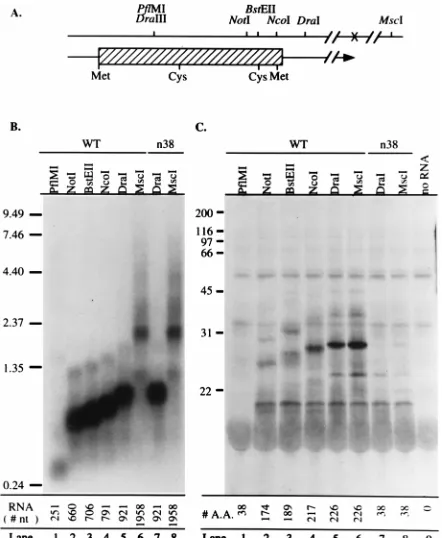
Prior to introduction of the mutation into the viral genome, the effect of the substitution on OPP synthesis was examined by in vitro transcription-translation. In the uncoupled in vitro transcription-translation system, the lengths of the transcript and the translated protein are dictated by the location of the restriction enzyme cleavage site used to linearize the expression plasmid. pLST-Nxi and pLST-NxiLM3, containing
wild-type and mutant ORF-P sequences, respectively, were lin-earized at the restriction sites shown in Fig. 3A. In vitro-tran-scribed RNAs generated from the linearized plasmids were divided into aliquots for use in Northern analysis and in vitro translation. As shown by the blot in Fig. 3B, which was probed with riboprobe EBN9-LAT for L/ST sense RNA (Fig. 1D), lin-earization of the plasmid containing wild-type ORF-P at con-secutive restriction sites downstream of the L/ST cap site re-sulted in the synthesis of progressively longer RNAs (Fig. 3B, lanes 1 to 6). As one would expect of a nonsense mutation that does not affect transcription, in vitro transcription from the mu-tant plasmid yielded RNAs (lanes 7 and 8) that were the same size as the corresponding wild-type RNAs (lanes 5 and 6). Be-cause the polyadenylation site of the 2.3-kb L/ST lies between the DraI and MscI sites (Fig. 3A), RNAs synthesized from the
MscI-linearized plasmid (MscI-RNAs) represent the entire
coding capacity of the 2.3-kb L/ST (Fig. 3B, lanes 6 and 8). An aliquot of each in vitro-transcribed RNA was used for in vitro translation (Fig. 3C). No protein was detected when
PflMI-RNA was translated because the 37-amino-acid product
does not contain [35S]Cys and therefore was not labeled (lane
[image:6.612.58.285.69.273.2]1). Progressively larger protein products were detected when progressively longer wild-type RNAs were translated (lanes 2
FIG. 2. Alignment of the ORF-P amino acid sequence in five strains of HSV-1. The amino acid sequences of HSV-1 strain KOS ORF-P are aligned with ORF-P amino acid sequences from strains 17syn1(18), F, MGH10, and CVG2 (26). Consensus amino acids are denoted by asterisks beneath the sequences. Periods represent gaps. The codon number of the last amino acid on each line is shown on the right. The numbers at the lower right represent the length of ORF-P in each viral strain.
FIG. 3. In vitro transcription/translation of wild-type (WT) and mutant ORF-P. (A) Restriction map of the region of the 2.3-kb L/ST. Beneath the restriction map is shown the location of ORF-P. The plasmid template was linearized at the restriction sites indicated before in vitro transcription (B) and translation (C). The positions of the methionine (Met) and cysteine (Cys) resi-dues used to radiolabel in vitro-translated proteins are shown. (B) Capped RNA from wild-type and n38 mutant plasmids was transcribed in vitro, separated electrophoretically, and transferred to Magnagraph paper. The resulting tran-scripts were detected by Northern blot analysis with riboprobe EBN9-LAT (Fig. 1D). The positions of RNA size markers (in kilobases) are shown on the left; the expected nucleotide (nt) sizes of the in vitro-transcribed RNA are indicated beneath each lane. (C) In vitro-transcribed wild-type and n38 mutant RNA from panel B were translated in the presence of [35S]Cys, separated electrophoreti-cally, and transferred to a polyvinylidene difluoride membrane. Proteins were visualized by autoradiography. The positions of molecular mass markers (in kilodaltons) are shown on the left. A.A., amino acids.
on November 9, 2019 by guest
http://jvi.asm.org/
[image:6.612.318.539.324.593.2]to 6). Translation of both DraI-RNA and the much longer
MscI-RNA yielded the same sized protein of approximately 26
kDa, since both restriction sites lie downstream of the ORF-P translation termination site (lanes 5 and 6). The molecular mass of this protein is similar to the theoretical molecular mass of OPP (23 kDa), suggesting that the observed protein band was derived from ORF-P.
Unlike in vitro translation of wild-type RNA, translation of
n38 RNA yielded no 26-kDa band, demonstrating that the n38
mutation terminated translation (Fig. 3C, lanes 7 and 8). In this in vitro system, translation of n38 should have produced a short (37-amino-acid) peptide. To determine if this was the case, protein translated from n38 DraI-RNA and n38 MscI-RNA in the presence of [35S]Met was analyzed on a 20%
denaturing Tricine polyacrylamide gel. A band corresponding to the 37-amino-acid peptide product of n38 RNA was de-tected in this test (data not shown). Because the RNAs con-taining the n38 mutation encode an otherwise full-length ORF-P, these results confirm that the single G-to-A transition terminated the translation of ORF-P.
The ICP4 binding-site mutation (4BS) abrogates ICP4 bind-ing at the L/ST transcriptional start site and significantly en-hances expression from the L/ST promoter.To disrupt ICP4-mediated repression of L/ST expression, we introduced four single-base-pair substitution mutations into the ICP4 binding site in the L/ST promoter (Fig. 1M). These mutations created a new Eco47III restriction enzyme site, facilitating the identi-fication of this mutation in plasmid and recombinant viral constructs. The effects of these mutations were first evaluated in vitro by gel mobility shift and transient-expression assays before they were introduced into the viral genome.
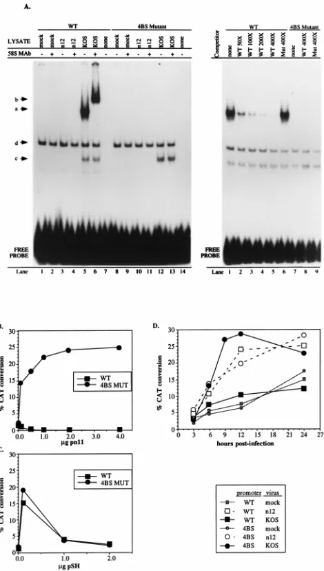
In gel mobility shift assays, proteins from infected-cell ly-sates formed a supershiftable complex with a labeled probe containing the wild-type ICP4 binding site but not with a probe containing the four single-base-pair substitution mutations (Fig. 4A). This complex was supershifted by an ICP4-specific anti-body. These findings were confirmed in a series of transient-expression and transfection/superinfection assays designed to determine the effect of the 4BS mutation on transcription from the L/ST promoter. In the first series of experiments, Vero cells were cotransfected with reporter constructs containing the L/ST promoter with the wild-type (pL/ST-U-CAT) or mu-tated (4BS; pL/ST-4BSm-CAT) ICP4 binding site and effector
plasmids, pn11 and pSH, expressing ICP4 and ICP0, respec-tively. At 40 h posttransfection, the cells were harvested and processed for CAT activity. Whereas expression from the wild-type promoter was repressed in the presence of ICP4, expres-sion from the promoter containing the 4BS mutation was strongly activated, demonstrating that ICP4 is able to activate the L/ST promoter but through a mechanism not involving the ICP4 consensus binding site in the L/ST promoter (Fig. 4B). To ascertain whether the 4BS mutation specifically affected regulation by ICP4 and not all regulators of gene expression, we cotransfected the wild-type and 4BS reporter plasmids with the ICP0-expressing plasmid, pSH. The responses of the wild-type and 4BS constructs to ICP0 were essentially identical (Fig. 4C) but were distinct from the responses of these plasmids to ICP4 (Fig. 4B). Thus, as reported previously, expression from both plasmids was high following cotransfection with low levels of ICP0 whereas higher levels of ICP0 resulted in lower levels of CAT activity (5). These results suggest that the 4BS muta-tion does not deregulate the L/ST promoter indiscriminately and that the ICP4 binding site is not a target of ICP0 or ICP4 activation. Together with the findings from gel mobility shift assays, the results of transient-expression assays with a 4BS
re-porter construct indicate that ICP4 cannot bind to the mutated binding site to repress L/ST expression.
[image:7.612.312.547.69.483.2]The cotransfection experiments examined the effects of spe-cific viral regulatory proteins on the L/ST promoter containing the 4BS mutation. Because L/ST promoter activation following superinfection may better reflect promoter function during
FIG. 4. Effects of the mutant ICP4 binding site on expression from the L/ST promoter. (A) Gel shift analysis. Radiolabeled 30-bp probe DNAs containing ei-ther the wild-type (WT) or mutated (4BS Mutant) ICP4 binding site were incu-bated with mock-, n12-, or KOS-infected Vero cell lysates prepared at 8 h p.i. and enriched for nuclear proteins. Anti-ICP4 monoclonal antibody (58S MAb) was added to the incubation mixture where indicated (lanes 2, 4, 6, 9, 11, and 13). The four major complexes detected are labeled a through d. (B to D) Transient-ex-pression assays. Vero cells were transfected with pL/ST-U-CAT (WT), contain-ing the wild-type ICP4 bindcontain-ing site, or pL/ST-4BSm-CAT (4BS MUT), contain-ing the mutated ICP4 bindcontain-ing site. Reporter plasmids were cotransfected with increasing amounts of the ICP4-expressing plasmid, pn11 (16) (B) or the ICP0-ex-pressing plasmid, pSH (9) (C). Cells were harvested at 40 h posttransfection or at the times indicated, and CAT enzyme activity was assayed. Data points rep-resent the average activities in two separately transfected plates. Transfection of pWR-CAT1, the promoter-less reporter plasmid, in the presence or absence of the effector plasmids, yielded background levels of CAT activity (data not shown). At 24 h after transfection of reporter plasmids alone, Vero cells were mock superinfected or superinfected with n12 or KOS at an MOI of 10 PFU/cell (D). The cells were harvested at 3, 6, 12, and 24 h p.i. A 9 h p.i., an additional data point was determined for 4BS reporter-transfected and KOS-superinfected Vero cells.
on November 9, 2019 by guest
http://jvi.asm.org/
natural viral infection, we performed a transient-expression/ superinfection assay in which transfected Vero cells were mock infected or infected with KOS or n12 virus 24 h after transfec-tion. At 3, 6, 12, and 24 h p.i., the cells were processed for CAT activity. As shown in Fig. 4D, expression from the wild-type promoter construct was repressed by superinfection with KOS (ICP41) but not n12 (ICP42). Expression from the mutated
4BS construct was also high following superinfection with ei-ther KOS or n12, demonstrating that introduction of the 4BS mutation into the L/ST promoter derepressed expression in the context of wild-type viral superinfection. The mutated 4BS construct was more active following superinfection with KOS than with n12, consistent with the data presented in Fig. 4B and demonstrating that ICP4 and ICP0 (KOS) or only ICP0 (n12) can activate the mutated L/ST promoter in the context of viral infection (10).
The introduction of the L/ST mutations n38 and 4BS into the viral genome was facilitated by the availability of 7134, an null mutant virus in which both copies of the ICP0-coding region have been replaced by the E. coli lacZ ICP0-coding sequence (9). Thus, the L/ST mutations were “piggybacked” into both copies of the L/ST gene simultaneously with the rescue of both copies of ICP0, producing the white-plaque phenotype. The mutations in the resulting viruses (L/ST-n38 and L/ST-4BS) were also rescued by homologous recombina-tion.
L/ST mutant viruses replicate efficiently in cells of neural and nonneural origin.ORF-34.5/ORF-P double mutants rep-licate to wild-type levels in permissive cells (e.g., Vero cells) but are growth restricted in certain neural cell lines (e.g., SK-N-SH cells) (12, 13). To assess the ability of mutant viruses L/ST-n38 and L/ST-4BS to replicate in Vero and SK-N-SH cells, single-cycle growth experiments were performed with the wild-type virus, the two mutants, and the rescuant viruses. In Vero cells, L/ST-n38, L/ST-4BS, and the rescuants replicated as efficiently as wild-type virus, indicating that no spurious mu-tations in genes that affect replication efficiency were present in any of the viruses (Fig. 5A). In SK-N-SH cells, however, L/ST-n38 replicated to wild-type levels whereas L/ST-4BS rep-licated to somewhat lower levels (Fig. 5B). This experiment was repeated twice with identical results. The modest reduc-tion in replicareduc-tion efficiency of L/ST-4BS in SK-N-SH cells was not observed with another ICP4 binding-site mutant virus, R7530 (25).
To determine if the modest reduction in the growth of L/ST-4BS in SK-N-SH cells was specific for cells of neural origin, single-cycle growth experiments were also performed in undif-ferentiated and nerve growth factor-difundif-ferentiated PC12 cells, mouse neuroblastoma cells (NB41A3), and dividing and rest-ing HEL cells as a control nonneural cell type. No major differences were observed in the growth patterns of L/ST-4BS, L/ST-n38, or KOS in any of the cell lines tested (data not shown), suggesting that the modest reduction in replication efficiency of L/ST-4BS in SK-N-SH is specific for this cell type. To further examine the general properties of the L/ST mu-tant viruses, patterns of protein synthesis in infected Vero cell extracts were analyzed at 3, 7, or 12 h p.i. by SDS-PAGE and by Western blot analysis with antibodies specific for ICP0, ICP4, ICP27, and glycoprotein D. Consistent with the similar-ities in the growth patterns of the L/ST mutants in Vero cells, the protein profiles of the mutants and their respective rescu-ants were indistinguishable from that of wild-type virus (data not shown).
Transcription of genes in the abb*a*c*ca repeats in cells of nonneural and neural lineage by L/ST mutant viruses.L/ST expression by the mutant viruses, KOS, and the rescuant
vi-ruses was examined by Northern blot hybridization of total RNA harvested from infected Vero and NB41A3 cells (Fig. 6A). The ICP4 nonsense mutant n12 was included as an L/ST-expressing control (50).
(i) L/STs.The L/STs were not detected in RNA from
L/ST-n38-infected Vero or NB41A3 cells (Fig. 6A, lanes 4 and 11);
[image:8.612.335.516.67.395.2]this was expected since the L/ST promoter in L/ST-n38 con-tains a wild-type ICP4 binding site and is therefore subject to repression by ICP4. Similarly, the L/STs were not detected in KOS-infected (lanes 2 and 9) or L/ST-n38R-infected (lanes 5 and 12) cells for the same reason. As expected from the results of transient-expression assays, the L/STs were expressed in 4BS-infected cells (lanes 6 and 13). In Vero cells, L/ST-4BS expressed lower levels of the 2.3-kb L/ST than did n12, whereas the opposite was true in NB41A3 cells. L/ST-4BS also expressed higher levels of L/STs than did n12 in differentiated and undifferentiated PC12 cells and in SK-N-SH cells (data not shown). In addition to the abundant 2.3-kb transcript, two smaller transcripts (,1.35 kb) were detected in L/ST-4BS-infected Vero and NB41A3 cells (lanes 6 and 13, respectively). The presence of these smaller transcripts in both infected Vero and NB41A3 cells argues that they represent independent transcripts and not degradation products of the larger L/STs. Finally, L/ST expression was not detected in L/ST-4BSR in-fected cells, confirming that the expression of the L/STs in
FIG. 5. Growth curves of L/ST mutant viruses in cell culture. Monolayers of Vero cells (A) or SK-N-SH cells (B) were infected at an MOI of 2.5 PFU/cell with KOS, L/ST-n38, L/ST-n38R, L/ST-4BS, or L/ST-4BSR as described in Materials and Methods. Total virus was harvested at the indicated times p.i. and quantified by a standard plaque assay on Vero cell monolayers. Titers are the average obtained in two tests.
on November 9, 2019 by guest
http://jvi.asm.org/
on November 9, 2019 by guest
http://jvi.asm.org/
L/ST-4BS was due to the 4-bp substitution in the ICP4 binding site (lanes 7 and 14).
(ii)g134.5 transcripts.Using a DNA probe specific forg134.5
transcripts, Chou and Roizman (15) detected two infected-cell-specific RNAs of 1.3 and 5.2 kb and one noninfected-cell-specific 3.5-kb RNA in wild-type virus (strain F)-infected Vero cells. As shown in Fig. 6B, the abundant 1.3-kbg134.5 RNA and two less
prom-inent 2.5- and 4-kb transcripts were detected in infected Vero cells with the EBSN34.5 riboprobe (lanes 2 and 4 through 7). Further mapping experiments are needed to determine wheth-er the 2.5- and 4-kb transcripts areg134.5 specific or
nonspe-cific. Levels of the 1.3-kbg134.5 RNA detected in cells infected
with wild-type virus and L/ST-n38 were similar (lanes 2 and 4) but somewhat higher in L/ST-4BS-infected cells, indicating that the n38 mutation had no major effect and that the 4BS mutation had, if anything, an enhancing effect on the levels of g134.5 RNA in Vero cells. As anticipated, transcripts were not
detected in n12-infected cells with theg134.5 probe (lane 3),
since ICP4-null viruses do not express late genes.
Three transcripts (1.3, 4, and 5.2 kb) were readily detected in KOS-infected (lane 9), L/ST-n38-infected (lane 11), and L/ST rescuant virus-infected (lanes 12 and 14) NB41A3 cells with the EBSN34.5 riboprobe. In contrast, only barely detectable levels ofg134.5 transcripts were observed in L/ST-4BS-infected
NB41A3 cells (lane 13). Similarly, only very low levels ofg134.5
transcripts were observed in L/ST-4BS-infected PC12 cells compared with those in KOS-infected PC12 cells (data not shown). In contrast, nearly equal levels of g134.5 transcripts
were detected in Vero cells infected with KOS (lane 2) or L/ST-4BS (lane 6). These findings demonstrate that g134.5
transcript accumulation in L/ST-4BS-infected cells is cell type specific. Figure 6B also shows that the levels ofg134.5-specific
transcripts detected were uniformly greater in NB41A3 cells than in Vero cells and that the sizes of the transcripts detected in the two cell types differed in that the 5.2-kb transcript was evident in NB41A3 cells but not in Vero cells.
(iii) ICP0 and ICP4 transcripts.Northern blot analysis of total RNA harvested from infected Vero cells at 6 h p.i. dem-onstrated that ICP0 and ICP4 RNAs were expressed by both L/ST-n38 and L/ST-4BS at levels comparable to those of wild-type virus (Fig. 6C, lanes 2, 4, and 6). Because these transcripts were present at very low levels in NB41A3 cells at 6 h p.i., a considerably longer autoradiographic exposure was necessary to visualize the bands in lanes 8 to 14. As in Vero cells, levels of ICP0 and ICP4 transcripts were similar to those seen in KOS-infected cells. Although abundant ICP4 transcripts were present in n12-infected Vero cells, no functional ICP4 protein was made because of the nonsense mutation in the ICP4 gene; hence, ICP0 and ICP4 transcription is not repressed and these transcripts are made at higher-than-wild-type levels. Notably, overexpression of ICP0 and ICP4 transcripts by n12 relative to KOS was evident in Vero cells but not in NB41A3 cells (or PC12 cells [data not shown]). Thus, overexpression of these two transcripts is cell type specific.
(iv) LATs. Because the L/STs may function during HSV latency (50), it was of interest to determine if the n38 or 4BS
mutations have an effect on LAT expression in vitro. We there-fore performed Northern blot analysis with a LAT-specific riboprobe, BbSLAT, with total RNA from Vero and NB41A3 cells harvested at 18 h p.i. with the L/ST mutant and rescuant viruses (Fig. 6D). RNAs from KOS-infected cells (lanes 2 and 9) and n12-infected cells (lanes 3 and 10) were included as LAT-positive and LAT-negative controls, respectively.
In Vero cells, L/ST-n38 and L/ST-4BS and their respective rescuant viruses (lanes 4 to 7) induced the synthesis of wild-type or near-wild-wild-type levels of the LATs, although we have consistently noted a slightly lower level of LAT expression during infection of these cells with L/ST-4BS (lane 6).
In NB41A3 cells, the LATs were reproducibly detected dur-ing infection with KOS, L/ST-n38, and the L/ST rescuant vi-ruses (lanes 9, 11, 12, and 14); however, only a faint LAT signal was detected in repeat experiments in RNA from 4BS-infected cells (lane 13). (The low LAT signal seen for
L/ST-n38R in lane 12 was not reproducible; near-wild-type levels of
LAT expression in NB41A3 cells are more characteristic of this virus.) Since the overall intensities of the LAT signals in NB41A3 cells were considerably lower than those in Vero cells, the low signal intensity in L/ST-4BS-infected NB41A3 cells (lane 13) may reflect the slight reduction observed in Vero cells noted above (lane 6). Whatever the basis for this reduc-tion, the 4BS mutation had a major effect on LAT expression
[image:10.612.310.548.442.673.2]FIG. 7. Expression of ORF-P protein by the L/ST mutant viruses. Vero cells were mock infected or infected with KOS, n12, L/ST-n38, L/ST-4BS, L/ST-4BSR, or L/ST-4/38 at a multiplicity of 10 PFU/cell. Infected-cell proteins were pre-pared at the indicated times p.i., separated by SDS-PAGE, and transferred to a polyvinylidene difluoride membrane. ORF-P protein was detected by Western blot analysis with rabbit polyclonal antibody, DF111, diluted 1:1,000. FIG. 6. Transcription of viral genes in the b9-a9-c9sequence in L/ST mutant virus-infected Vero and NB41A3 cells. Vero and NB41A3 cells were mock infected or infected with 10 PFU of KOS, n12 (an ICP4 nonsense mutant virus), L/ST mutant viruses L/ST-n38 or L/ST-4BS, or L/ST rescued viruses L/ST-n38R or L/ST-4BSR per cell. Total-cell RNA (Vero [lanes 1 to 7] or NB41A3 [lanes 8 to 14]) was isolated at 18 h p.i. (A, B, and D) or 6 h p.i. (C), separated electrophoretically, and transferred to Magnagraph paper. The positions of RNA size markers (in kilobases) are shown on the left. (A) L/STs were detected with riboprobe EBN9-LAT (Fig. 1D). (B)g134.5 RNAs were detected with riboprobe EBSN34.5 (Fig. 1D). (C) ICP0 and ICP4 RNAs were detected concurrently with riboprobes 0Hc-Xh and 4Sma (Fig. 1D), respectively. The specificity of each riboprobe was determined independently (data not shown). ICP0 and ICP4 RNAs from NB41A3 cells (lanes 8 to 14) were detected after a twofold-longer autoradiographic exposure than RNAs from Vero cells (lanes 1 to 7). The positions of ICP4 RNA and ICP0 are marked by black and white arrowheads, respectively. (D) LATs were detected with riboprobe BbSLAT (Fig. 1D).
on November 9, 2019 by guest
http://jvi.asm.org/
in NB41A3 cells relative to wild-type virus and L/ST-n38. Sim-ilar results were observed in PC12 cells. We conclude from these tests that LAT expression, like the expression ofg134.5
transcripts, is cell type specific, being significantly higher in nonneural cells (Vero) than in cells of neural lineage (NB41A3 and PC12).
OPP expression by L/ST mutant viruses. Having demon-strated high-level expression of the L/STs by L/ST-4BS but not by L/ST-n38 in both Vero and NB41A3 cells, we attempted to assess the kinetics and levels of expression of OPP in Vero cells. To facilitate these studies, rabbit antibody (DF111) was raised against the C-terminal 52 amino acids of ORF-P fused to GST. Using this antibody, we first examined the ability of the L/ST mutant viruses and their respective rescuants to ex-press OPP. Extracts of KOS- and n12-infected Vero cells were included as OPP-negative and -positive controls, respectively. As shown in Fig. 7, OPP was not detected in Vero cells infected with KOS, L/ST-n38, and L/ST-4BSR, viruses that do not ex-press the L/STs as demonstrated by Northern blot analysis (Fig. 6A). In cells infected with L/ST-4BS, three proteins of approximately 24, 29, and 33 kDa were detected (Fig. 7, lanes 10 to 12). The size of the 24-kDa band is consistent with the molecular mass of OPP (23 kDa) and with the size of the OPP band observed by in vitro transcription-translation (26 kDa) (Fig. 3). The size and relative abundance of each protein spe-cies at 6, 12, and 18 h p.i. suggest that the 24-kDa protein is modified posttranslationally during the course of lytic infec-tion. As anticipated, the 24-kDa protein was also detected in
n12-infected Vero cells, although the appearance of the larger
(29-kDa) species was delayed and these proteins were present in lower abundance relative to that in L/ST-4BS-infected cells. OPP expression by n12 and L/ST-4BS was also detected in NB41A3 cells, nerve growth factor-differentiated and undiffer-entiated PC12 cells, and SK-N-SH cells (data not shown), demonstrating that like expression of the L/STs, OPP expres-sion is not cell type specific.
L/ST-4BS but not L/ST-n38 induces premature shutoff of protein synthesis in SK-N-SH cells. Gradual shutoff of host cell protein synthesis is observed after HSV infection of cul-tured cells (19). In contrast to wild-type virus infection, ORF-34.5/ORF-P double-mutant viruses induce the shutoff of all protein synthesis (viral and host cell) at early times (3 to 6 h p.i.) in SK-N-SH cells relative to other cell types (13). To examine protein shutoff in L/ST-n38- and L/ST-4BS-infected Vero and SK-N-SH cells, we performed metabolic labeling experiments. An ICP34.5-null mutant, 1771, derived from strain 17syn1was included as a positive control, and wild-type strains KOS and 17syn1were included as negative controls. In Vero cells, infection with the L/ST mutant viruses, both rescu-ants and the positive and negative control viruses, produced wild-type (KOS or 17syn1) protein profiles, characterized by a gradual shift from host to viral protein synthesis over the 12-h period (Fig. 8A). In SK-N-SH cells, L/ST-n38 and both rescu-ant viruses produced KOS wild-type-like protein profiles (Fig. 8B). In contrast, L/ST-4BS and 1771 infection resulted in rapid and extensive shutoff of both viral and cellular protein synthe-sis relative to their respective wild-type viruses. Rescue of the 4BS mutation to produce the wild-type genotype (L/ST-4BSR) returned the protein synthesis profile to that seen with wild-type virus. The results obtained with L/ST-n38 suggest that the premature protein shutoff phenotype of the ORF-P ORF-34.5 double-mutant viruses in SK-N-SH cells is not likely to be a consequence of mutations in ORF-P but, rather, is a conse-quence of mutations in ORF-34.5. The molecular basis for the premature protein shutoff phenotype of L/ST-4BS is not clear and will be discussed below.
L/ST-4BS but not L/ST-n38 is attenuated for neurovirulence in mice. Having shown that the protein shutoff phenotype is characteristic of L/ST-4BS but not L/ST-n38, we tested these mutants in vivo for their neurovirulence phenotypes and their ability to establish and reactivate from latency in mice.
To determine if the n38 and 4BS mutations result in the attenuated phenotype characteristic of ORF-P ORF-34.5 dou-ble-mutant viruses, we assessed the neurovirulence of the L/ST mutant viruses by intracranial inoculation of BALB/c mice. The results of two independent experiments are shown in Ta-ble 1. Consistent with published data, wild-type strain 17syn1 was more neurovirulent than KOS and the 17syn1 mutant, 1771, was highly attenuated in these tests. L/ST-n38, although somewhat less virulent than KOS in experiment 1 and some-what more virulent than KOS in experiment 2, is clearly a neu-rovirulent virus, demonstrating that the mutation of ORF-P in ORF-P ORF-34.5 double mutants is not responsible for the attenuated phenotypes of these viruses.
The reduced levels ofg134.5 transcripts (Fig. 6B) and the
premature shutoff of protein synthesis in SK-N-SH cells ob-served in in vitro tests of L/ST-4BS (Fig. 8B) predict that L/ ST-4BS might behave in vivo like ORF-P ORF-34.5 double-mutant viruses. As predicted, L/ST-4BS was completely at-tenuated in both experiments (Table 1), causing no
detect-FIG. 8. Kinetics of the shutoff of viral and cellular protein synthesis in Vero and SK-N-SH cells infected with L/ST mutant viruses. Cells were mock infected (M) or infected with 10 PFU of the indicated viruses per cell, labeled with [35S]methionine for 30 min at 3, 7, or 12 h p.i., and processed for PAGE as described in Materials and Methods. (A) Vero cells, 5mg of protein per lane. (B) SK-N-SH cells, 10mg of protein per lane.
on November 9, 2019 by guest
http://jvi.asm.org/
[image:11.612.337.518.69.402.2]able morbidity or mortality in mice. Rescue of the mutation in L/ST-4BS restored the wild-type neurovirulence phenotype, confirming that the attenuated phenotype is due to the 4BS mutation.
L/ST-4BS replicates in the mouse eye and in TGG during acute infection following corneal inoculation.Because the at-tenuation of L/ST-4BS in mice could, in theory, be due to a general defect in the ability of this virus to replicate in mice, we examined the ability of KOS, L/ST-4BS, and L/ST-4BSR to replicate in the mouse eye and in TGG following corneal inoculation. Tear film from four mice and TGG from two mice were collected daily during acute infection (the first 7 days p.i.) and assayed for infectious virus by a standard plaque assay on Vero cell monolayers. The arithmetic mean titers of multiple samples at each time point are shown in Fig. 9. In tear film, KOS and L/ST-4BSR reached peak titers of approximately 105
PFU/ml on day 1 p.i., and these titers were maintained at high levels through day 6 p.i., dropping slightly on day 7 p.i. (Fig. 9A). Replication of these viruses in TGG was evident by day 2 and peaked between days 3 and 4 p.i. (Fig. 9B). In both tear film and TGG, L/ST-4BS replicated to near-wild-type levels during the first few days p.i. but exhibited an earlier and more rapid decline in titer thereafter. Rescue of the mutation in L/ST-4BS restored wild-type levels of replication in TGG as in eyes. Notably, these results demonstrate that L/ST-4BS is ca-pable of significant replication (albeit less efficiently than the wild type) in the peripheral nervous system.
L/ST-4BS exhibits delayed reactivation from latency in the mouse ocular model.Because the L/STs and/or OPP may be involved in some way in the establishment or reactivation of latency, we examined the ability of the L/ST mutant viruses to reactivate from latently infected mouse TGG. In two indepen-dent experiments, mice were inoculated with 23106PFU/eye.
On day 30 p.i., TGG were removed and cocultivated with Vero cells. The cocultivation medium was then assayed daily for the presence of infectious, reactivated virus. The cumulative per-centage of cocultivated ganglia which scored positive for infec-tious virus in both experiments is shown in Fig. 9. Consistent with its wild-type phenotype in vitro, L/ST-n38 reactivated with wild-type kinetics and efficiency in both experiments. L/ST-4BS also reactivated but with delayed kinetics and reduced effi-ciency relative to wild-type virus in both experiments. In ex-periment 1, reactivation of L/ST-4BS was delayed somewhat and was less efficient than that of the wild type (71% TGG [17 of 24]) by day 15 postexplant versus 100% of TGG [24 of 24] by day 6). In this experiment, it was not determined whether the TGG remaining on day 15 harbored latent L/ST-4BS virus; therefore, in the second experiment, explant cocultures were monitored for an extended period (21 days postexplant). In
this experiment, L/ST-4BS ultimately reactivated from 89% of TGG (16 of 18), a level comparable to KOS (100% [18 of 18 TGG]) and L/ST-n38 (100% [19 of 19 TGG]) in the 21-day experiment. The delayed kinetics of L/ST-4BS reactivation noted in the first experiment were also evident in the second experiment. Rescue of the 4BS mutation restored the wild-type reactivation profile (L/ST-4BSR, experiment 2). Viral DNA iso-lated from half of the reactivated cultures in experiment 1 and all of the cultures in experiment 2 was analyzed by Southern blot analysis; the L/ST mutations were present in reactivated mutant viruses in all cases (data not shown).
DISCUSSION
The abb9c9a9ca repeats of the HSV-1 genome contain genes
[image:12.612.49.290.81.180.2]and cis-acting elements that play central roles in establishing the programs of gene expression characteristic of productive infection and latency. The large number of these genes and elements and the extent to which they overlap have compli-cated the genetic and functional analysis of this region of the genome in that mutations introduced into one gene or element invariably alter the nucleotide sequence of other overlapping genes and elements. Such has been the case in studies of the g134.5 gene, which is encoded on the DNA strand opposite
TABLE 1. Neurovirulence of L/ST mutant viruses in mice
Virus LD50
aafter i.c. inoculation in:
Expt 1 Expt 2
KOS 72 898
L/ST-n38 537 273
L/ST-n38R NDb 626
L/ST-4BS .2.43106 .2.63106
L/ST-4BSR ND 997
17syn1 1.1 ND
1771 .7.43105 ND
aMice were infected as described in the text. The 50% lethal doses (LD
50) of the indicated viruses were calculated by the method of Reed and Muench (36). i.c., intracranical.
bND, not determined.
FIG. 9. Production of infectious virus in mouse eyes and TGG during acute infection and in explanted ganglia during reactivation. Mice were inoculated with 23106PFU of KOS, L/ST-n38, L/ST-4BS, or L/ST-4BSR per eye. (A) Eye
swabs were taken 1 to 3 h p.i. and then daily from 1 to 7 days p.i. Each point represents the arithmetic mean titer of eight eye swabs (four mice). (B) TGG were removed and assayed directly for the presence of infectious virus on days 1 through 7 p.i. Each point represents the arithmetic mean titer for TGG (two mice). The titers were determined by a standard plaque assay on Vero cells. Data points below the asterisk represent undetectable levels of virus. (C) At 30 days p.i., TGG were removed and cocultivated with Vero cells. Aliquots of the cocul-tivation medium were then assayed for infectious virus on the indicated days postexplant. Each point represents the cumulative percentage of cocultivated ganglia scoring positive for infectious virus. In experiment 1, the total numbers of TGG tested were as follows: KOS, 18; L/ST-n38, 18; L/ST-4BS, 24. In exper-iment 2, the total numbers of TGG used were as follows: KOS, 18; L/ST-n38, 19; L/ST-4BS, 18; L/ST-4BSR, 14.

![FIG. 6. Transcription of viral genes in the binfected with 10 PFU of KOS,per cell. Total-cell RNA (Vero [lanes 1 to 7] or NB41A3 [lanes 8 to 14]) was isolated at 18 h p.i](https://thumb-us.123doks.com/thumbv2/123dok_us/1238719.76393/10.612.310.548.442.673/transcription-viral-genes-binfected-total-lanes-lanes-isolated.webp)