Int. J. Electrochem. Sci., 11 (2016) 1568 - 1580
International Journal of
ELECTROCHEMICAL
SCIENCE
www.electrochemsci.org
Effect of Some Rare Earth Oxides Doping on the Morphology,
Crystallite Size, Electrical Conductivity and N
2O Decomposition
Activity of CuO Catalyst
Bahaa M. Abu-Zied1,*, Salem M. Bawaked2, Samia A. Kosa2, Wilhelm Schwieger3 1
Center of Excellence for Advanced Materials Research (CEAMR), King Abdulaziz University, P.O. Box 80203, Jeddah 21589, Saudi Arabia
2
Chemistry Department, Faculty of Science, King Abdulaziz University, P.O. Box 80203, Jeddah 21589, Saudi Arabia
3
Institut Für Chemische Reaktionstechnik, Friedrich-Alexander-Universita¨t Erlangen-Nu¨rnberg, Egerlandstraße 3, 91058 Erlangen, Germany
*
E-mail: [email protected], [email protected]
Received: 28 September 2015 / Accepted: 19 November 2015 / Published: 1 January 2016
The present work presents the results of direct catalytic decomposition of nitrous oxide over CuO and its Pr-, Sm- and Tb-promoted catalysts. These catalysts were prepared by the microwave assisted precipitation method, using oxalic acid as precipitant, and subsequent calcination at 500 °C. X-ray diffraction (XRD) was used for identification of the composition of the dried oxalate precursors and their calcination products. Thermogravimetric analysis (TGA) was used to follow up the thermal stability of the prepared precursors. The obtained catalysts were characterized by Fourier transform infrared spectroscopy (FT-IR), field emission scanning electron microscopy (FESEM), transmission electron microscopy, electrical conductivity and (TEM) H2 temperature programmed reduction (H2 -TPR). An enhancement effect of the added rare earth elements was observed during N2O decomposition over the prepared catalysts Assessment of the de-N2O activity was discussed based on the electrical conductivity as well as the H2-TPR results.
Keywords: Greenhouse gases, N2O decomposition, nitrous oxide, CuO catalyst, RE promotedCuO
1. INTRODUCTION
nanocrystalline CuO in different geometrical morphologies. For instance, Xia eta al. [7] have prepared chain-like hierarchically nanostructured CuO by precipitation-reflux method.
Jiang et al. [8] successfully prepared CuO nanosheets via the template-free hydrothermal method. Bozkurt et al. [9] have used the surfactants-assisted precipitation method to prepared nanocrystalline CuO with different morphologies (plate-, rod-, leaf-, particle-, caterpillar-, and corncoblike and nanowires structures). Their results revealed that the surfactant type (including zwitterionic SB12, cationic CTAB and anionic SDS) as well as its addition order to the synthesis environment also affects the morphology of the CuO structures [9]. Liu et al. [10] demonstrated that by changing the volume of PEG200 and the alkalinity during the preparation of CuO nanostructures would tune its morphology from 1D (nanoseeds, nanoribbons) to 3D (shuttle-like, shrimp-like, and nanoflowers).
Nitrous oxide (N2O) is known to be a greenhouse gas, that has a global warming potential (GWP) of about 310 times of carbon dioxide (CO2) [11]. N2O has a very long atmospheric lifetime
(~120 years). Since the citation of N2O as the second non-CO2 greenhouse gas, according to the Kyoto Protocol of the United Nations Convention on Climate Change (December 1997), the studies focusing on its abatement has gained more attention. Industrially, N2O arise as a co-product from some chemical processes, which includes nitric acid production and the production of large amounts of adipic acid, which is produced from the HNO3 oxidation of cyclohexanol–cyclohexanone mixtures, for Nylon 6,6 and 6,12 [11]. Direct catalytic decomposition of nitrous oxide to its elements (N2 and O2) is considered as one of the potential solutions to minimize the N2O emissions. Various catalysts exhibited promising activity for this reaction such as metals, mixed oxides, supported oxides, spinels, perovskites, and zeolites based catalysts [12–15].
In this contribution, we report effect of doping with some rare earth (RE) elements (Pr, Sm and Tb) on the morphology and crystallite size of CuO nanorparticles, which were prepared via the microwave assisted method of copper oxalate and subsequent calcination at 500 °C. The decomposition pathways of the three RE/copper oxalate mixtures were followed using thermogravimetric analysis (TGA). It was found that the oxalate precursors dehydrate till 150 C and decompose at the temperature range of 200-400 0C. Based on thermal analysis results, Pr, Sm, and Tb doped CuO samples were obtained via the calcination, in static air, at 500 C for 1h for their oxalate precursors. XRD, FTIR, SEM, TEM and XPS analyses were used to characterize the morphology and crystalline quality RE doped copper oxide samples.
2. EXPERIMENTAL
2.1. Catalysts preparation
employing oxalic acid as precipitant at microwave power (MWP) of 280 W. It is to be mentioned that the selection of the microwave power of 280 W during the precursor preparation was based on our recent results, which indicated that lower MWP yield material with smaller particles size [16]. CuO was obtained by the subsequent calcination of the copper oxalate precursor for 1 h at 500 °C in air. The three rare earths promoted CuO catalysts were prepared by the co-precipitation method. In such method the amounts of a rare earth cation (Pr3+, Sm3+ and Tb3+), required to achieve a rare earth (RE)/Cu ratio of 0.05, were added during the precipitation of oxalate using the same working procedure. After drying of the co-precipitated oxalates precursors, they were calcined for 1 h at 500 °C in air to yield the corresponding earths promoted CuO catalysts.
2.2. Characterization techniques
The structure and composition of the as-prepared oxalate precursors as well as their calcination products powders were investigated at ambient temperature using Thermo-Scientific ARL X'TRA Powder Diffractometer. TGA measurements were carried out at a heating rate of 10 °C min-1
in nitrogen flow (40 ml min–1) with a TA instrument apparatus (model TGA-Q500). The weight of the sample taken in each experiment was around 5 mg. FT-IR spectra (4000–400 cm-1) were recorded using Attenuated Total Reflectance (ATR) sampling accessory on the Nicolet iS50 FT-IR spectrometer. The size and morphology of the prepared catalysts were investigated by using field emission scanning electron microscope (FE-SEM) on a JEOL model JSM-7600F microscope and transmission electron microscopy (TEM) using JEOL (model JEM1011) microscopy. The electrical conductivity of the prepared catalysts was measured using a Pyrex glass cell. In each experiment a 0.5 g of the catalyst powder was placed between two silver electrodes having 1.0 cm diameter. The resistance measurements were carried out using Keithley 6517A electrometer. Hydrogen temperature-programmed reduction (H2-TPR) experiments were performed on Quantachrome CHEMBET-3000 instrument operated with a TCD detector. In each experiment, the catalyst was pre-treated in helium at 300 °C for 30 min followed by cooling to 25 °C. Then, the flow of the gas was changed to 5% H2+ 95% Ar and the sample temperature increased to 600◦C at the rate of 5 °C/min.
2.3. Activity measurements
concentrations in the inlet and outlet streams were measured using non-dispersive infrared analyzer (Hartmann and Braun, Uras 10E).
3. RESULTS AND DISCUSSION
3.1. Catalysts Characterization
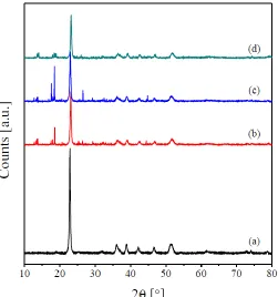
[image:4.596.172.423.350.618.2]Fig. 1 shows the XRD patterns of the obtained oxalates precursors. The obtained patterns reveal the crystalline nature of the prepared precursors. All the diffraction peaks of the copper oxalate (Fig. 1(a)) match well with those of orthorhombic phase of copper oxalate (JCPDS 21– 0297, space group Pnnm) and are similar to the XRD patterns reported by other research groups [1,4,17–19]. No other peaks, due to the presence of impurities, were identified. This, in turn, suggests the formation of purely crystalline CuC2O4.H2O. The diffractograms of the RE-promoted copper oxalate precursors (Fig. 1(b–d)) reveals the presence of the reflections due to copper oxalate together with small reflections at 2θ = 10–20° and 24-29°. These reflections could be assigned to the presence of the RE-oxalates.
Figure 1. X-ray powder diffraction patterns of Cu oxalate (a), Pr/Cu oxalates (b), Sm/Cu oxalates (c), and Tb/Cu oxalates (d).
formation of Cu2O (41.3 %). Similar results were reported by Lamprecht et al. for the decomposition of copper oxalate in argon and nitrogen flows [17]. In this context, it was shown that the decomposition of copper oxalate in air flow proceeds with the formation of CuO [19,20].
[image:5.596.65.538.273.699.2]The obtained TGA-DTG thermograms for the RE-containing precursors (Fig. 2(b–d)) reveal the presence of three WL steps. These steps are maximized at the temperature ranges of 60–89 °C, 268–275 °C and 379–413 °C. By comparison with the TGA-DTG curves of copper oxalate, the obtained curves (Fig. 2(b–d)) could reasonably related to the precursors-dehydration, copper oxalate decomposition and RE-oxalates decomposition, respectively. Based on the TGA-DTA results the various parents were calcined at 500 °C for 1h in order to obtain the relevant CuO as well as the RE-promotes CuO catalysts.
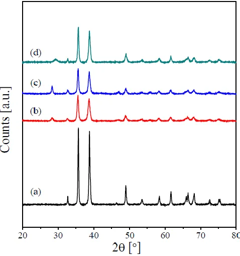
Typical XRD patterns of the calcination products of copper and its RE-containing oxalates are shown in Fig. 3. Fig. 3(a) reveals that calcining the parent copper oxalate at 500 °C is accompanied by a complete disappearance of all reflections due to the oxalate phase and the emergence of new reflections at 2 θ = 32.65°, 35.57°, 38.79°, 46.38°, 48.91°, 53.53°, 58.38°, 61.65°, 65.85°, 66.38°, 68.08°, 72.44°, 75.06° and 75.36°. These reflections agree well with those reported for the monoclinic phase of CuO (JCPDS 80-1917, space group Cc). No peaks due to the presence of impurities were observed in the XRD pattern. This indicates the complete decomposition of copper oxalate in air leading to the formation of pure CuO as the decomposition product. The XRD pattern of the Pr-containing CuO (Fig. 3(b)) reveals the presence of all reflections due to the monoclinic CuO together with very-small intensity ones at 2θ = 28.27°, and 46.432°. These reflections could be assigned to Pr6O11 phase (JCPDS 42-1121), which suggests that this catalyst is composed of CuO as a major phase together with trace amount of Pr6O11. In addition to the peaks characterizing the monoclinic CuO phase, the diffractogram of the calcined Sm/Cu (Fig. 3(c)) reveals the existence of a weak reflection at 2θ = 28.24°. This reflection could be assigned to the Sm2O3 phase (JCPDS 76-0153). This, in turn, suggests the co-existence of CuO (major) and Sm2O3 (trace) for this sample. Finally, the composition of the calcined Tb/Cu can be suggested to be CuO (major) and Tb4O7 (trace). This can be judged from the existence of all reflections due to monoclinic CuO together with the emergence a new weak reflection at 2θ = 28.31°, which could be due to the presence of Tb4O7 (JCPDS card No. 13-0387). The broadening of the XRD peaks of the catalysts presented in Fig. 4 indicates nanometer size of these calcination products. The crystallite sizes of these catalysts were determined from the XRD line broadening applying the Scherrer formula [12,21]. The obtained values are listed in Table 1. The obtained values indicate that doping CuO with the various RE-oxides leads to a noticeable decrease in its crystallite sizes.
[image:6.596.177.416.467.721.2]
Table 1. Crystallite sizes, electrical conductivities, T25 and T50 values obtained for bare CuO ant its REpromoted catalysts.
Catalyst Crystallite size [nm] σ10-5 [Ω-1 cm-1] T25 [°C] T50 [°C]
CuO 53.5 2.0 446 497
Pr/CuO 15.8 25.9 385 458
Sm/CuO 18.9 46.5 390 451
Tb/CuO 23.7 45.5 408 463
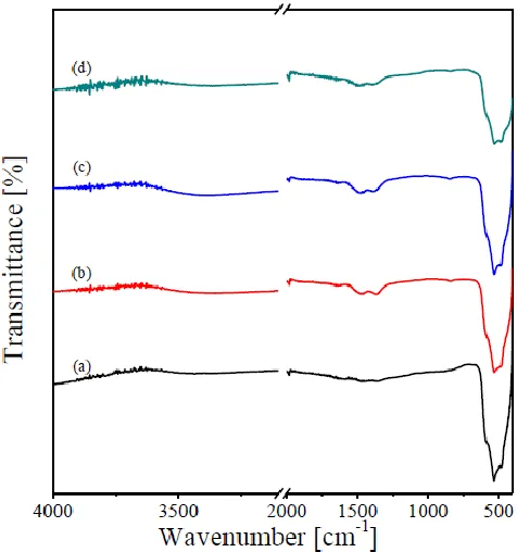
The FT-IR spectra of as prepared CuO and its RE-containing catalysts are shown in Fig. 4. All the spectra revealed the presence of strong absorptions located at 500–600 cm-1 assignable CuO vibration in CuO [1,22–24]. The RE oxide containing catalysts reveals the presence of other weak absorptions at the wavenumber range of 1700-1200 cm-1, which can be assigned to the presence of surface carbonate species. This suggests the ability of the added RE oxides to form weak surface carbonates upon contact with atmospheric air [21,25].
Figure 4. FT-IR spectra taken for CuO (a), Pr/CuO (b), Sm/CuO (c), and Tb/CuO (d) catalysts.
[image:7.596.179.417.367.621.2]
of sphere-like and rice-like morphologies of CuO nanoparticles via the heating of CuC2O4.H2O alone and CuC2O4.H2O and NaOH mixture, respectively. Similar spherelike morphology, with diameters in the range 13.3–31.2 nm, was reported by Jayaprakash et al. [24], for their CuO prepared by the sol–gel method and subsequent annealing at 400 °C. Jia et al. [19] demonstrated that morphology of CuO prepared by the decomposition of CuC2O4.H2O depends also on the copper ions source; sphere-like particles were obtained using CuSO4 and CuCl2 precursors where a biscuit-like morphology was obtained using Cu(CH3COO)2 precursor. Regarding the effect of the added RE-oxides, our results (Fig. 5(b–d)) indicated that the REoxides/CuO solids maintain the sphere-like morphology of CuO. Moreover the Pr- and Sm containing catalysts are more porous and have smaller agglomerates.
Figure 5. FE-SEM nano-graphs obtained for CuO (a), Pr/CuO (b), Sm/CuO (c), and Tb/CuO (d) catalysts.
[image:8.596.104.497.242.584.2]
Figure 6. TEM images obtained for CuO (a), Pr/CuO (b), Sm/CuO (c), and Tb/CuO (d) catalysts.
3.2. N2O decomposition activity
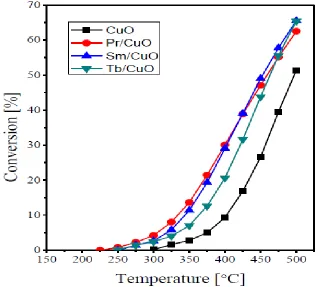
Figure 7. Dependence of N2O conversion % on the reaction temperature over the CuO, Pr6O11/CuO, Sm2O3/CuO and Tb4O7/CuO catalysts.
The promotion effect via doping of various transition-metal based catalysts with different dopants is well documented in the literature. For instant an activity increase was reported for Co3O4 based catalysts upon doping with alkali and alkali earth cations [26–28]. An activity promotion was also reported upon doping NiO catalyst with Li, Na, K, and Cs ions during N2O direct decomposition [29,30]. Regarding N2O decomposition over CuO catalysts, Pasha et al. [31] reported a superior activity of Cs-promoted catalysts, particularly the molar ratio of Cs/Cu at 0.1, compared to the bare CuO. More recently, better catalytic performance during N2O decomposition over Ce–Cu mixed oxides, prepared by co-precipitation, compared to the bare CuO or CeO2 [32].
The earlier work of Dell et al. [33] revealed the correlation of the N2O decomposition activity of a series of metal oxides with their electrical conductivity. The highest activity was exhibited by the p-type semiconductors, intermediate activity was exhibited by insulators whereas the n-type oxided showed the lowest activity. Based on the in situ electrical conductivity measurements, the N2O abetment activity of a series of transition metal exchanged ZSM-5 zeolites was classified into two series [15]. The first one includes Fe-, Co-, Cu-, Pd-, La-, Ce- and Ag-ZSM-5 catalysts, which showed higher magnitude of electrical conductivity decrease upon the admission of N2O. On the other hand, the second group contains Ni-, Mn-, Cd-, Zn-, and Y-ZSM-5 catalysts, which exhibited only a mild conductivity decrease during the N2O admission. Concurrently, the N2O decomposition activity of Cu-X zeolite catalysts was correlated with their electrical conductivity properties [34]. The reported mechanism for N2O direct decomposition over various transition-metal based catalysts involves three steps [15,28,32]:
N2O− (ads.) ... Mn+1 → N2(g) + O−(ads.) ... Mn+1 ………. (2) O−(ads.) ... Mn+1 → ½O2(g) + Mn+ ………. (3) The first step in this mechanism includes an electron donation from the catalyst surface to an N2O molecule leading to its adsorption, i.e. formation of N2O− (ads.), and an increase in oxidation state of the active cations at the catalyst surface. It is believed that the added promoters enhances the electron donation ability of the surface transition metal active centers [13,14,26–32]. This means that the reaction is controlled by the electrical properties of the catalyst, where the ability of the electrons to move from the catalyst bulk to its surface is of great importance in enhancing the N2O adsorption and, thus, decomposition. The conductivity values of bare CuO and its RE-promoted catalysts are listed in Table 1. From the inspection of the obtained values, it appears that doping the bare CuO with the various RE-oxides is accompanied by a noticeable increase of its conductivity. In other words, the electron movement from the catalyst bulk will become more significant when the CuO is doped with the various RE oxides.
Fig. 8 shows the TPR profiles obtained for bare CuO and its Pr-, Sm- and Tb-containing catalysts. The obtained TPR curves of all the catalysts are very similar, which are characterized by the presence of one broad band. This peak could be assigned to the reduction processes Cu2+ to Cu+ and Cu+ to Cu0 according to [32,35]:
CuO + H2 Cu2O + H2O ………. (4)
Cu2O + H2 2 Cu + H2O ………. (5) In this respect, Zhang [35] reported that the sequential reduction of copper oxide (Cu2+ → Cu+ →Cu0
[image:11.596.144.474.499.734.2]) is difficult to be resolve using the H2-TPR experiments. Inspection of Fig. 8 reveals that all the added RE-oxides shift the H2-TPR peak towards lower temperatures. This indicates that the incorporation of the RE-oxides enhanced the reducibility of CuO. This finding is in agreement with the results of Zhang et al. [35], who reported a lowering the CuO H2-TPR peak a result of CeO2-ZrO2 incorporation. Similar findings were reported by Konsolakis et al. [32] for Cu-Ce.
It is agreed in the open literature that the added promotors interacts with the active transition metal ions forming an electron-rich species. These species have the electron-donation ability to the adsorbed nitrous oxide molecules, which leads to the weakening of the N-O bond in nitrous oxide and, thus, its decomposition into N2 molecule and an adsorbed O-species (equations 1 and 2). From the observed increase in the measured electrical conductivity upon incorporating the RE-oxides to CuO, it is plausible to suggest that this will increase the rate of equations 1 and 2 forming Cu2+ ions. The recoverability of the copper ions with lower oxidation states, i.e. Cu+ ions, is essential for the completion of the reaction mechanism and the regeneration of the active sites. From the observed enhancement of the CuO reducibility (Fig. 8) via oxides addition, we can suggest that these RE-oxides enhance the regeneration of the catalysts active centers, and thus enhancing the N2O direct decomposition.
4. CONCLUSIONS
In this paper, a series of catalysts comprises of bare CuO and RE-doped CuO was prepared by the microwave assisted precipitation method and subsequent calcination at 500 °C. Structural characterization of the prepared solids indicates that these catalysts are composed of nanocrystalline monoclinic CuO and a mixture of monoclinic CuO together with the RE-oxides, respectively. All the prepared solids showed a sphere-like morphology, where the RE-promoted catalysts exhibit smaller particles size. Higher N2O decomposition activity RE-promoted catalysts compared to bare CuO. This enhanced activity of the RE-containing catalysts was ascribed to their role in enhancing both the electrical conductance and the reduction of CuO during the catalytic experiments.
ACKNOWLEDGEMENTS
This project was supported by the National Science, Technology and Innovation Plan (MAARIFAH) strategic technologies programs, number (12-ENV2756-03) of the Kingdom of Saudi Arabia. The authors thankfully acknowledge Science and Technology Unit, Deanship of Scientific Research at King Abdulaziz University for their technical support.
References
1. F. Behnoudnia and H. Dehghani, Polyhedron, 56 (2013) 102. 2. L. Zhang, R. Liu and H. Yang, Physica E, 44 (2012) 1592.
3. A. A. Bush, V. Ya. Shkuratov, A. B. Kuz’menko and E. A. Tishchenko, Cryst. Rep., 47 (2002) 335.
4. X. Zhang, D. Zhang, X. Ni and H. Zheng, Solid-State Electron., 52 (2008) 245. 5. T. Zhang, W. Li and J.-P. Croué, Appl. Catal. B, 121–122 (2012) 88.
6. L. Winnubst, P. J. de Veen, S. Ran and D. H. A. Blank, Ceram. Int., 36 (2010) 847. 7. C. Xia, C. Xiaolan, W. Ning and G. Lin, Anal. Chim. Acta, 691 (2011) 43.
10. J. Liu, J. Jin, Z. Deng, S.-Z. Huang, Z.-Y. Hu, L. Wang, C. Wang, L.-H. Chen, Y. Li, G. Van Tendeloo and B.-L. Su, J. Colloid Interface Sci., 384 (2012) 1.
11. J. Pérez-Ramírez, Appl. Catal. B, 70 (2007) 31. 12. B.M. Abu-Zied, Appl. Catal. A, 334 (2008) 234.
13. B.M. Abu-Zied, S.A. Soliman and S.E. Abdellah, J. Ind. Eng. Chem., 21 (2015) 814. 14. F. Kapteijn, J. Rodriguez-Mirasol and J. A. Moulijn, Appl. Catal. B, 9 (1996) 25. 15. B.M. Abu-Zied, W. Schwieger and A. Unger, Appl. Catal. B, 84 (2008) 277.
16. B. M. Abu-Zied, S. M. Bawaked, S. A. Kosa and W. Schwieger, Appl. Surf. Sci., 351 (2015) 600. 17. E. Lamprecht, G. M. Watkins and M. E. Brown, Thermochim. Acta, 446 (2006) 91.
18. D. X. Zhang, H. Xu, Y. Z. Liao, H. S. Li and X. J. Yang, Powder Technol., 189 (2009) 404. 19. Z. Jia, L. Yue, Y. Zheng and Z. Xu, Mater. Res. Bull., 43 (2008) 2434.
20. A. Aimable, A. T. Puentes and P. Bowen, Powder Technol., 208 (2011) 467. 21. B.M. Abu-Zied and S.A. Soliman, Thermochim. Acta, 470 (2008) 91.
22. A. B. Devi, D. S. Moirangthem, N. C. Talukdar, M. D. Devi, N. R. Singh and M. N. Luwang, Chin. Chem. Lett., 25 (2014) 1615.
23. J. K. Sharma, M. S. Akhtar, S. Ameen, P. Srivastava and G. Singh, J. Alloys Compd., 632 (2015) 321.
24. J. Jayaprakash, N. Srinivasan, P. Chandrasekaran and E. K. Girija, Spectrochim. Acta, Part A, 136 (2015) 1803.
25. S.A. Soliman and B.M. Abu-Zied, Thermochim. Acta, 491 (2009) 84.
26. G. Maniak, P. Stelmachowski, A. Kotarba, Z. Sojka, V. Rico-Pérez and A. Bueno-López, Appl. Catal. B, 136–137 (2013) 302.
27. N. Pasha, N. Lingaiah, N. S. Babu, P. S. S. Reddy and P. S. S. Prasad, Catal. Commun., 10 (2008) 132.
28. B. M. Abu-Zied and S. A. Soliman, Catal. Lett., 132 (2009) 299.
29. N. Pasha, N. Lingaiah, P. S. S. Reddy and P. S. S. Prasad, Catal. Lett., 118 (2007) 64.
30. B.M. Abu-Zied and A.M. Asiri, Chin. J. Catal., 36 (2015), proof (doi: 10.1016/S18722067(15)60963-9).
31. N. Pasha, N. Lingaiah, P. S. S. Reddy and P. S. S. Prasad, Catal. Lett., 127 (2009) 101.
32. M. Konsolakis, S. A. C. Carabineiro, E. Papista, G. E. Marnellos, P. B. Tavares, J. Agostinho Moreira, Y. Romaguera-Barcelay and J. L. Figueiredo, Catal. Sci. Technol., 5 (2015) 3714.
33. R. M. Dell, F. S. Stone and P. F. Tiley, Trans. Faraday Soc. 49 (1953) 201. 34. B. M. Abu-Zied, Microporous Mesoporous Mater., 139 (2011) 59.
35. Q. Zhang, L. Xu, P. Ning, J. Gu and Q. Guan, Appl. Surf. Sci., 317 (2014) 955.