16 Using Cap Analysis Gene Expression Technology
Ayumi Taguchi,aKazunori Nagasaka,aKei Kawana,aKosuke Hashimoto,bRika Kusumoto-Matsuo,cCharles Plessy,bMiranda Thomas,d Hiroe Nakamura,aAlessandro Bonetti,bKatsutoshi Oda,aIwao Kukimoto,cPiero Carninci,bLawrence Banks,dYutaka Osuga,a Tomoyuki Fujiia
Department of Obstetrics and Gynecology, Graduate School of Medicine, The University of Tokyo, Tokyo, Japana
; RIKEN Center for Life Science Technologies, Division of Genomic Technologies, Yokohama, Japanb
; Pathogen Genomic Center, National Institute of Infectious Diseases, Tokyo, Japanc
; International Centre for Genetic Engineering and Biotechnology, Trieste, Italyd
We have performed cap-analysis gene expression (CAGE) sequencing to identify the regulatory networks that orchestrate
ge-nome-wide transcription in human papillomavirus type 16 (HPV16)-positive cervical cell lines of different grades: W12E, SiHa,
and CaSki. Additionally, a cervical intraepithelial neoplasia grade 1 (CIN1) lesion was assessed for identifying the transcriptome
expression profile. Here we have precisely identified a novel antisense noncoding viral transcript in HPV16. In conclusion,
CAGE sequencing should pave the way for understanding a diversity of viral transcript expression.
C
ervical cancer is the third most common female cancer
world-wide, with 530,000 new cases and 275,000 deaths annually (
1
),
and infection by one of a subset of high-risk human
papillomavi-ruses (HPVs) is a prerequisite for its development (
2
).
A number of elegant studies on the HPV type 16 (HPV16)
transcriptome (
3–10
) have shown that transcription in all HPVs
occurs unidirectionally from at least two promoters and involves
multiple splice sites. The major promoters in HPV16 are the early
promoter (p97), located in the locus control region (LCR), and
the differentiation-inducible promoter (p670), with additional
promoters in the LCR and within E4 and E5, potentially
generat-ing late transcripts. HPV transcription is polycistronic,
undergo-ing differential splicundergo-ing to produce numerous viral mRNAs (at
least 13 transcripts from 8 HPV16 genes in W12E cells [
11
]).
Pre-vious analyses considered only coding RNA, and it is possible that
new transcription start sites (TSSs) are obscured by prominent
promoters. Therefore, we have used cap analysis gene expression
(CAGE) for high-throughput transcriptome analysis (
12–16
)
CAGE specifically determines transcriptional start sites (TSS)
ge-nome wide, by capping 5
=
ends of RNA transcripts, reverse
tran-scribing them, and mapping cDNAs to the reference genome to
identify TSSs (
17–20
).
To assess all potential TSSs in HPV16-infected cell lines, we
performed CAGE analysis using cervical carcinoma-derived
CaSki and SiHa cells (
21
,
22
), cervical intraepithelial neoplasia
grade 1 (CIN1)-derived W12E (20863) cells, containing episomal
copies of HPV16 (
3
,
23
) (3T3 feeder cells removed by
trypsiniza-tion), and a biopsy specimen from a CIN1 cervical lesion
(ap-proved by the Institutional Review Board, University of Tokyo
Hospital). From each sample, 5
g RNA was extracted using a
miRNeasy kit (Qiagen). RNA quality was assessed by the use of a
Bioanalyzer (Agilent) and standardized at an RNA integrity
num-ber (RIN) of
⬎
7.0. Quantitation by NanoDrop analysis confirmed
that the
A
260/
A
290and
A
260/
A
230ratios were
⬎
1.7.
First-strand cDNAs were transcribed to the 5
=
end of capped
RNAs and attached to CAGE “bar code” tags (
Table 1
), and the
sequenced CAGE tags were mapped to the HPV16 genome and
the human hg19 genomes using BWA software (v0.5.9),
discard-ing ribosomal or non-A/C/G/T base-containdiscard-ing RNAs (
24
).
CAGE tags were normalized by the total number of tags per
sam-ple mapped to the human genome and are indicated as tags per
million (TPM) (
25
). For HPV16 genes, CAGE tag 5
=
coordinates
were input for Paraclu clustering, with these parameters: (i) a
minimum of 5 tags/cluster, (ii) (maximum density/baseline
den-sity)
⭌
2, and (iii) 100-bp maximal cluster length (
18
).
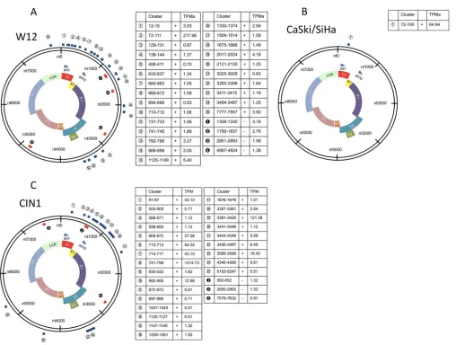
HPV16 transcriptomes differ between carcinoma- and
CIN1-derived cells.
Viral gene expression was high in W12E cells
and CIN1 clinical samples (
Fig. 1C
) but was low in CaSki and SiHa
cells (
Table 1
). To quantitatively visualize CAGE-tagged HPV16
genes from each cell line, we compared the data to the HPV16
reference genome (NCBI GI no.
333031
). We found 25
positive-strand and 4 negative-positive-strand tag clusters (TCs [regions with
⬎
0.5
TPM/sample]) in W12E cells (
Fig. 1A
), and only 1 positive-strand
TC in CaSki and SiHa cells (
Fig. 1B
), corresponding to the p97
promoters. In W12E cells and a CIN1 clinical sample, numerous
additional TSSs span the HPV16 genome, suggesting that
precan-cerous cells show diversity and abundance of viral gene expression
that are lost upon viral genome integration and disease
progres-sion, although investigation of W12 derivatives with integrated
HPV16 and of additional clinical samples is needed to confirm
this.
The six most highly expressed TCs in each cell line are
com-pared in
Fig. 2
. The six TCs most frequently found in W12E cells
originate from nucleotide (nt) 90 to 97, nt 1125 to 1149, nt 2017 to
Received1 December 2014 Accepted2 December 2014
Accepted manuscript posted online10 December 2014
CitationTaguchi A, Nagasaka K, Kawana K, Hashimoto K, Kusumoto-Matsuo R, Plessy C, Thomas M, Nakamura H, Bonetti A, Oda K, Kukimoto I, Carninci P, Banks L, Osuga Y, Fujii T. 2015. Characterization of novel transcripts of human
papillomavirus type 16 using cap analysis gene expression technology. J Virol 89:2448 –2452.doi:10.1128/JVI.03433-14.
Editor:M. J. Imperiale
Address correspondence to Kazunori Nagasaka, [email protected], or Kei Kawana, [email protected].
Copyright © 2015, American Society for Microbiology. All Rights Reserved.
doi:10.1128/JVI.03433-14
on November 7, 2019 by guest
http://jvi.asm.org/
2024, nt 12 to 15, nt 1330 to 1322, and nt 741 to 745, some of which
were previously identified (
26
). The TC at nt 90 to 97 (
Fig. 2A
)
corresponds to the p97 promoter, while those at nt 129 to 131 and
nt 138 to 144 had probably leaked from the P97 promoter (
21
).
The TC at nt 1125 to 1149, detected only in W12 cells (
Fig. 2B
), is
probably a TSS of E8-E2 (
27
,
28
), and the TC at nt 12 to 15
corre-sponds to a promoter upstream of p97, indicating limited E1
tran-scription (
Fig. 2D
) (
27
). The TCs at nt 741 to 745 would be a TSS
of E1Ê4, corresponding to the p670 promoter (
Fig. 2F
) (
3
). Most
interesting, however, was the previously unreported TC at nt 1330
to 1322, which is a potential antisense cluster, highly expressed in
W12E cells (
Fig. 2E
). In addition, we identified another new TC at
nt 2017 to 2024 (
Fig. 2C
).
[image:2.585.41.288.78.216.2]To verify these novel transcripts, W12 RNA with or without
poly(A) was reverse transcribed using a ReverTra Ace qPCR RT kit
(Toyobo), and 3
=
rapid amplification of cDNA ends (3
=
RACE)
TABLE 1Number of tags mapped to HPV16 genomeCell line Bar code
No. of tags mapped to HPV16 genome
No. of tags mapped to hg19 genome
Tags per million (TPM)
CaSki-1 GTA 548 9,632,333 56.89 CaSki-2 ACC 664 13,139,414 50.53 CaSki-3 CAC 494 11,535,883 42.79 SiHa-1 AGT 1,504 12,064,850 124.66 SiHa-2 GCG 991 9,050,850 109.49 W12-1 TAC 5,750 15,955,328 350.95 W12-2 GCT 4,019 11,517,132 348.96 W12-3 ATG 3,928 12,018,142 326.84 CIN1 ATG 20,181 9,861,351 2,046.47
FIG 1Clustered TSSs of the HPV16 genomes of cervical cell lines of different grades. CAGE tags of HPV16 genes from each cell lines are quantitatively visualized. The data are representative of the results determined with the respective cell lines. The CaSki and SiHa cultured cells, representing a transcriptional program characteristic of the conditions in a tumor, were cultured in Dulbecco’s modified Eagle’s medium supplemented with 10% heat-inactivated fetal bovine serum (FBS) and 1% penicillin-streptomycin. W12E (20863) is an HPV16-positive cell line derived from a CIN1 lesion (23). W12E (20863) cells were cocultured with mitomycin C-treated 3T3 fibroblast feeder cells. (A) Circular map depicting viral transcripts identified by Paraclu clustering. CAGE tags were clustered for HPV16 genes from W12E cells. Regions containing over 0.5 tags per million (TPM) in all samples were selected as tag clusters (TCs) and mapped to the circular HPV16 genome. (B) We defined regions containing over 0.5 TPM in the sample as TCs. Then, CAGE tags were clustered for HPV16 genes from the CaSki and SiHa cells. (C) CAGE tags were clustered for HPV16 genes from a CIN1 clinical sample. Notably, we found 25 positive-strand TCs and 4 negative-strand TCs in W12E cells (A), whereas only 1 positive-strand TC was found in CaSki or SiHa cells (B). TPMa, the average number of TPMs.
on November 7, 2019 by guest
http://jvi.asm.org/
[image:2.585.42.550.257.641.2]was carried out using
Ex Taq
(TaKaRa) on the detected clusters
found in clustering analysis of HPV16 genome expression, using
the nt-97 cluster as a positive control.
To verify RNA without poly(A), a 3
=
preadenylated DNA
adap-tor sequence was ligated at the 3
=
end of RNA. To increase
speci-ficity, we carried out first RACE and second RACE experiments.
Primer pairs for each peak [oligo(dT)] adaptor are shown in
Fig. 3
.
The PCR conditions for the first RACE and second RACE
exper-FIG 2Expression levels of the top 6 tag clusters (TCs) in W12E, CaSki, and SiHa cells. The top 6 expressed TCs were selected, and expression levels are indicated as tags per million (TPM) (25). Six TSS clusters were frequently found to be prominent in W12E cells, originating from nt 90 to 97 (90_97_⫹), nt 1125 to 1149 (1125_1149_⫹), nt 2017 to 2024 (2017_2024_⫹), nt 12 to 15 (12_15_⫹), nt 1322 to 1330 (1330_1322_⫺), and nt 741 to 745 (741_745_⫹).FIG 3Validation of novel transcripts from 3=RACE analysis. Agarose gel electrophoresis of transcripts from W12 cell eluted RNA. The results of the experiment indicate that the transcript originating at nt 1330 to 1322 could correspond to a newly identified viral antisense noncoding RNA without poly(A). Further, we could encode a newly identified transcript of nt 2017 to 2024 starting in the middle of E1 coding gene. ddC, dideoxycytosine; rApp, adenylation-5=; RT, reverse transcriptase.
on November 7, 2019 by guest
http://jvi.asm.org/
[image:3.585.139.451.65.330.2] [image:3.585.101.486.456.680.2]iments were as follows: for the first RACE experiment, 35 cycles at
94°C for 30 s, 57°C for 30 s, and 72°C for 30 s; for second RACE
experiment, 35 cycles at 94°C for 30 s, 57°C for 30 s, and 72°C for
30 s. The results are shown in
Fig. 3
. PCR products were extracted
(Qiagen) and sequenced by Applied Biosystems 3130xl (Fasmac).
Importantly, we identified, for the first time, the full coordinates
of a novel antisense transcript starting from nt 1330 as a
noncod-ing RNA (ncRNA) without poly(A) and also a novel transcript, nt
2017 to 2024, starting in the middle of the E1 gene. Although most
viral RNAs are considered to be polyadenylated at the 3
=
end, our
finding confirms that the noncoding viral RNA starting from nt
1330 lacks poly(A) signals.
In this study, we performed CAGE analysis and investigated
TSSs from the perspective of both the HPV16 and human
ge-nomes, using cell lines containing episomal and integrated HPV
sequences. All the TSSs used in the HPV16 transcriptome, in the
HPV16-containing cervical keratinocyte W12E cell line, and in
the cervical cancer-derived SiHa and CaSki cell lines were
investi-gated. Intriguingly, we also show that a diversity of viral
tran-scripts, seen in W12E cells, shared some identity with the CIN1
biopsy specimen subjected to a precise colposcopic examination.
Recently, as shown previously in the mammalian transcriptome
(
29
), abundant virally encoded noncoding RNAs (ncRNAs) were
identified (
30
), but this is the first full confirmation of a
prelimi-nary observation of antisense transcripts in HPV.
Using the CAGE method, we have successfully established
un-biased analyses of reproducible transcriptional start sites across
the HPV16 genome, potentially identifying novel transcripts,
in-cluding ncRNAs, for future RNA therapies (
31
). Further
compre-hensive studies will aim to identify possible markers to predict the
outcome of infections with HPV.
Nucleotide sequence accession number.
The sequence data
reported are available in the DDBJ BioProject under the accession
number PRJDB3385.
ACKNOWLEDGMENTS
We are grateful to Ri-ichiroh Manabe and Michihira Tagami (Division of Genomic Technologies, RIKEN) for the excellent advice and technical guidance on the preparation of CAGE analysis. All sequencing was per-formed by the Genome Analysis Support Facility (Division of Genomic Technologies, RIKEN). We are also very grateful to Paul Lambert (Uni-versity of Wisconsin School of Medicine and Public Health, Madison, Wisconsin) for his kind gift of W12E (20863) cell lines.
This work was supported by a Grant-in-Aid for Scientific Research (K.N., K.K.) from the Ministry of Education, Science and Culture, Japan.
REFERENCES
1.Arbyn M, Castellsagué X, de Sanjosé S, Bruni L, Saraiya M, Bray F, Ferlay J.2011. Worldwide burden of cervical cancer in 2008. Ann Oncol 22:2675–2686.http://dx.doi.org/10.1093/annonc/mdr015.
2.zur Hausen H.2002. Papillomaviruses and cancer: from basic studies to clinical application. Nat Rev Cancer2:342–350.http://dx.doi.org/10.1038 /nrc798.
3.Doorbar J, Parton A, Hartley K, Banks L, Crook T, Stanley M, Craw-ford L.1990. Detection of novel splicing patterns in a HPV16-containing keratinocyte cell line. Virology178:254 –262.
4.Grassmann K, Rapp B, Maschek H, Petry KU.1996. Identification of a differentiation-inducible promoter in the E7 open reading frame of hu-man papillomavirus type 16 (HPV-16) in raft cultures of a new cell line containing high copy numbers of episomal HPV-16 DNA. J Virol70: 2339 –2349.
5.Braunstein TH, Madsen BS, Gavnholt B, Rosenstierne MW, Johnsen CK, Norrild B.1999. Identification of a new promoter in the early region
of the human papillomavirus type 16 genome. J Gen Virol80(Pt 12):3241– 3250.
6.Rosenstierne MW, Vinther J, Hansen CN, Prydsoe M, Norrild B.2003. Identification and characterization of a cluster of transcription start sites located in the E6 ORF of human papillomavirus type 16. J Gen Virol 84:2909 –2920.http://dx.doi.org/10.1099/vir.0.19332-0.
7.Zheng Z, Tao M, Yamanegi K, Bodaghi S, Xiao W.2004. Splicing of a cap-proximal human papillomavirus 16 E6E7 intron promotes E7 expres-sion, but can be restrained by distance of the intron from its RNA. J Mol Biol337:1091–1108.http://dx.doi.org/10.1016/j.jmb.2004.02.023. 8.Schmitt M, Pawlita M.2011. The HPV transcriptome in HPV16 positive
cell lines. Mol Cell Probes25:108 –113.http://dx.doi.org/10.1016/j.mcp .2011.03.003.
9.Schmitt M, Dalstein V, Waterboer T, Clavel C, Gissmann L, Pawlita M. 2011. The HPV16 transcriptome in cervical lesions of different grades. Mol Cell Probes 25:260 –265. http://dx.doi.org/10.1016/j.mcp.2011.05 .003.
10. Schwartz S.2013. Papillomavirus transcripts and posttranscriptional reg-ulation. Virology445:187–196.http://dx.doi.org/10.1016/j.virol.2013.04 .034.
11. Milligan SG, Veerapraditsin T, Ahamet B, Mole S, Graham SV.2007. Analysis of novel human papillomavirus type 16 late mRNAs in differen-tiated W12 cervical epithelial cells. Virology360:172–181.http://dx.doi .org/10.1016/j.virol.2006.10.012.
12. Shiraki T, Kondo S, Katayama S, Waki K, Kasukawa T, Kawaji H, Kodzius R, Watahiki A, Nakamura M, Arakawa T, Fukuda S, Sasaki D, Podhajska A, Harbers M, Kawai J, Carninci P, Hayashizaki Y.2003. Cap analysis gene expression for high-throughput analysis of transcriptional starting point and identification of promoter usage. Proc Natl Acad Sci U S A100:15776 –15781.http://dx.doi.org/10.1073/pnas.2136655100. 13. Kodzius R, Kojima M, Nishiyori H, Nakamura M, Fukuda S, Tagami
M, Sasaki D, Imamura K, Kai C, Harbers M, Hayashizaki Y, Carninci P.2006. CAGE: cap analysis of gene expression. Nat Methods3:211–222.
http://dx.doi.org/10.1038/nmeth0306-211.
14. Plessy C, Bertin N, Takahashi H, Simone R, Salimullah M, Lassmann T, Vitezic M, Severin J, Olivarius S, Lazarevic D, Hornig N, Orlando V, Bell I, Gao H, Dumais J, Kapranov P, Wang H, Davis CA, Gingeras TR, Kawai J, Daub CO, Hayashizaki Y, Gustincich S, Carninci P. 2010. Linking promoters to functional transcripts in small samples with nano-CAGE and nano-CAGEscan. Nat Methods 7:528 –534. http://dx.doi.org/10 .1038/nchembio.586.
15. Kanamori-Katayama M, Itoh M, Kawaji H, Lassmann T, Katayama S, Kojima M, Bertin N, Kaiho A, Ninomiya N, Daub CO, Carninci P, Forrest ARR, Hayashizaki Y.2011. Unamplified cap analysis of gene expression on a single-molecule sequencer. Genome Res21:1150 –1159.
http://dx.doi.org/10.1101/gr.115469.110.
16. Takahashi H, Lassmann T, Murata M, Carninci P.2012. 5=end-centered expression profiling using cap-analysis gene expression and next-generation sequencing. Nat Protoc7:542–561.http://dx.doi.org/10.1038 /nprot.2012.005.
17. Carninci P, Kasukawa T, Katayama S, Gough J, Frith MC, Maeda N, Oyama R, Ravasi T, Lenhard B, Wells C, Kodzius R, Shimokawa K, Bajic VB, Brenner SE, Batalov S, Forrest AR, Zavolan M, Davis MJ, Wilming LG, Aidinis V, Allen JE, Ambesi-Impiombato A, Apweiler R, Aturaliya RN, Bailey TL, Bansal M, Baxter L, Beisel KW, Bersano T, Bono H, Chalk AM, Chiu KP, Choudhary V, Christoffels A, Clutter-buck DR, Crowe ML, Dalla E, Dalrymple BP, de Bono B, Della Gatta G, di Bernardo D, Down T, Engstrom P, Fagiolini M, Faulkner G, Fletcher CF, Fukushima T, Furuno M, Futaki S, et al.2005. The transcriptional landscape of the mammalian genome. Science309:1559 –1563.http://dx .doi.org/10.1126/science.1112014.
18. Frith MC, Valen E, Krogh A, Hayashizaki Y, Carninci P, Sandelin A. 2008. A code for transcription initiation in mammalian genomes. Genome Res18:1–12.http://dx.doi.org/10.1101/gr.6831208.
19. Djebali S, Davis CA, Merkel A, Dobin A, Lassmann T, Mortazavi A, Tanzer A, Lagarde J, Lin W, Schlesinger F, Xue C, Marinov GK, Khatun J, Williams BA, Zaleski C, Rozowsky J, Röder M, Kokocinski F, Abdel-hamid RF, Alioto T, Antoshechkin I, Baer MT, Bar NS, Batut P, Bell K, Bell I, Chakrabortty S, Chen X, Chrast J, Curado J, Derrien T, Drenkow J, Dumais E, Dumais J, Duttagupta R, Falconnet E, Fastuca M, Fejes-Toth K, Ferreira P, Foissac S, Fullwood MJ, Gao H, Gonzalez D, Gordon A, Gunawardena H, Howald C, Jha S, Johnson R, Kapranov P,
on November 7, 2019 by guest
http://jvi.asm.org/
King B, et al.2012. Landscape of transcription in human cells. Nature 489:101–108.
20. FANTOM Consortium and the RIKEN PMI and CLST (DGT), Forrest AR, Kawaji H, Rehli M, Baillie JK, de Hoon MJ, Lassmann T, Itoh M, Summers KM, Suzuki H, Daub CO, Kawai J, Heutink P, Hide W, Freeman TC, Lenhard B, Bajic VB, Taylor MS, Makeev VJ, Sandelin A, Hume DA, Carninci P, Hayashizaki Y.2014. A promoter-level mamma-lian expression atlas. Nature 507:462– 470. http://dx.doi.org/10.1038 /nature13182.
21. Smotkin D, Wettsteintt F.1986. Transcription of human papillomavirus type 16 early genes in a cervical cancer and a cancer-derived cell line and identification of the E7 protein. Proc Natl Acad Sci U S A83:4680 – 4684.
http://dx.doi.org/10.1073/pnas.83.13.4680.
22. Yee C, Krishnan-Hewlett I, Baker CC, Schlegel R, Howley PM.1985. Presence and expression of human papillomavirus sequences in human cervical carcinoma cell lines. Am J Pathol119:361–366.
23. Stanley MA, Browne HM, Minson AC. 1989. Properties of a non-tumorigenic human cervical keratinocyte cell line. Int J Cancer43:672– 676.
24. Li H, Durbin R.2010. Fast and accurate long-read alignment with Bur-rows-Wheeler transform. Bioinformatics26:589 –595.http://dx.doi.org /10.1093/bioinformatics/btp698.
25. Valen E, Pascarella G, Chalk A, Maeda N, Kojima M, Kawazu C, Murata M, Nishiyori H, Lazarevic D, Motti D, Marstrand TT, Tang MH, Zhao X, Krogh A, Winther O, Arakawa T, Kawai J, Wells C, Daub C, Harbers M, Hayashizaki Y, Gustincich S, Sandelin A, Carninci P. 2009. Genome-wide detection and analysis of hippocampus core
promot-ers using DeepCAGE. Genome Res19:255–265.http://dx.doi.org/10.1101 /gr.084541.108.
26. Graham SV.2010. Human papillomavirus: gene expression, regulation and prospects for novel diagnostic methods and antiviral therapies. Future Microbiol5:1493–1506.http://dx.doi.org/10.2217/fmb.10.107. 27. Lace MJ, Anson JR, Turek LP, Haugen TH.2008. Functional mapping of
the human papillomavirus type 16 E1 cistron. J Virol82:10724 –10734.
http://dx.doi.org/10.1128/JVI.00921-08.
28. Lace MJ, Anson JR, Thomas GS, Turek LP, Haugen TH.2008. The E8âE2 gene product of human papillomavirus type 16 represses early transcription and replication but is dispensable for viral plasmid persis-tence in keratinocytes. J Virol82:10841–10853.http://dx.doi.org/10.1128 /JVI.01481-08.
29. Derrien T, Johnson R, Bussotti G, Tanzer A, Djebali S, Tilgner H, Guernec G, Martin D, Merkel A, Knowles DG, Lagarde J, Veeravalli L, Ruan X, Ruan Y, Lassmann T, Carninci P, Brown JB, Lipovich L, Gonzalez JM, Thomas M, Davis CA, Shiekhattar R, Gingeras TR, Hubbard TJ, Notredame C, Harrow J, Guigó R.2012. The GENCODE v7 catalog of human long noncoding RNAs: analysis of their gene struc-ture, evolution, and expression. Genome Res22:1775–1789.http://dx.doi .org/10.1101/gr.132159.111.
30. Sullivan CS.2008. New roles for large and small viral RNAs in evading host defences. Nat Rev Genet9:503–507.http://dx.doi.org/10.1038/nrg 2349.
31. Takahashi H, Carninci P.1 September 2014, posting date. Widespread genome transcription: new possibilities for RNA therapies. Biochem Bio-phys Res Communhttp://dx.doi.org/10.1016/j.bbrc.2014.08.139.
on November 7, 2019 by guest
http://jvi.asm.org/